R E S E A R C H A R T I C L E
Open Access
Premenopausal serum androgens and breast
cancer risk: a nested case-control study
Anne Zeleniuch-Jacquotte
1,2*, Yelena Afanasyeva
1, Rudolf Kaaks
3, Sabina Rinaldi
4, Stephanie Scarmo
1,
Mengling Liu
1,2, Alan A Arslan
1,2,5, Paolo Toniolo
1,2,5,6, Roy E Shore
1,7and Karen L Koenig
1,2Abstract
Introduction:Prospective epidemiologic studies have consistently shown that levels of circulating androgens in postmenopausal women are positively associated with breast cancer risk. However, data in premenopausal women are limited.
Methods:A case-control study nested within the New York University Women’s Health Study was conducted. A total of 356 cases (276 invasive and 80in situ) and 683 individually-matched controls were included. Matching variables included age and date, phase, and day of menstrual cycle at blood donation. Testosterone,
androstenedione, dehydroandrosterone sulfate (DHEAS) and sex hormone-binding globulin (SHBG) were measured using direct immunoassays. Free testosterone was calculated.
Results:Premenopausal serum testosterone and free testosterone concentrations were positively associated with breast cancer risk. In models adjusted for known risk factors of breast cancer, the odds ratios for increasing quintiles of testosterone were 1.0 (reference), 1.5 (95% confidence interval (CI), 0.9 to 2.3), 1.2 (95% CI, 0.7 to 1.9), 1.4 (95% CI, 0.9 to 2.3) and 1.8 (95% CI, 1.1 to 2.9;Ptrend= 0.04), and for free testosterone were 1.0 (reference), 1.2 (95% CI, 0.7 to 1.8), 1.5 (95% CI, 0.9 to 2.3), 1.5 (95% CI, 0.9 to 2.3), and 1.8 (95% CI, 1.1 to 2.8,Ptrend= 0.01). A marginally significant positive association was observed with androstenedione (P= 0.07), but no association with DHEAS or SHBG. Results were consistent in analyses stratified by tumor type (invasive,in situ), estrogen receptor status, age at blood donation, and menopausal status at diagnosis. Intra-class correlation coefficients for samples collected from 0.8 to 5.3 years apart (median 2 years) in 138 cases and 268 controls were greater than 0.7 for all biomarkers except for androstenedione (0.57 in controls).
Conclusions:Premenopausal concentrations of testosterone and free testosterone are associated with breast cancer risk. Testosterone and free testosterone measurements are also highly reliable (that is, a single measurement is reflective of a woman’s average level over time). Results from other prospective studies are consistent with our results. The impact of including testosterone or free testosterone in breast cancer risk prediction models for women between the ages of 40 and 50 years should be assessed. Improving risk prediction models for this age group could help decision making regarding both screening and chemoprevention of breast cancer.
Introduction
Prospective epidemiologic studies have consistently shown that circulating androgens in postmenopausal women are positively associated with breast cancer risk [1-8], an association which is thought to be largely due to their role as precursors of estrogens. Positive associa-tions have also been reported for androgens in
premenopausal women but data are limited to date, with the majority of studies having small numbers of cases [9-17]. If results in premenopausal women are confirmed, androgens could be considered for inclusion in breast cancer risk prediction models, such as the Gail model [18]. Improving breast cancer risk prediction models could be particularly valuable for women between the ages of 40 and 50, as they could help with decisions on screening, since guidelines for this age group are not consistent [19,20]. In addition, improved models might help younger women with an elevated
* Correspondence: anne.jacquotte@nyumc.org
1
Department of Environmental Medicine, New York University School of Medicine, 650 First Avenue, New York, NY 10016, USA
Full list of author information is available at the end of the article
risk of breast cancer decide whether to take tamoxifen for chemoprevention, as is recommended [21,22]. Tamoxifen has been approved by the US Food and Drug Administration for chemoprevention in women age 35 or older with a 5-year Gail-model risk greater than 1.66%, but is not often used in practice for this purpose [23,24]. A better understanding of the associa-tion between premenopausal concentraassocia-tions of andro-gens and breast cancer risk is also important because some experimental studies [25,26], although not all [27], have suggested that androgens may protect against breast cell proliferation in an estrogen-rich environment, such as the time period before menopause. Finally, it is also important to assess the association between andro-gens and breast cancer risk because androandro-gens have been advocated for relief of sexual symptoms such as low libido [28], including in older premenopausal women [29].
We report here the results of a case-control study nested within the New York University (NYU) Women’s Health Study cohort. Prediagnostic concentrations of testosterone, androstenedione, dehydroepiandrosterone sulfate (DHEAS) and sex hormone-binding globulin (SHBG) were measured in serum samples from 356 inci-dent cases and 683 controls who were premenopausal at enrollment (time of initial blood donation), and the association of these biomarkers with subsequent risk of breast cancer was assessed.
Materials and methods
Study Population
The NYU Women’s Health Study (NYUWHS) enrolled 14,274 women 34 to 65 years old at a breast cancer screening center in New York City between 1985 and 1991 [30]. Women who had been pregnant or taken hor-monal medications in the six months preceding their visit were excluded. After written informed consent was obtained, demographic, medical, anthropometric, repro-ductive, and dietary data were collected through self-administered questionnaires. Non-fasting peripheral venous blood was drawn prior to breast examination and serum samples were stored at -80°C for subsequent bio-chemical analyses. Up until 1991, women who returned to the clinic for annual breast cancer screening were asked to donate blood at each of their visits. For 52% of the women, two or more blood samples were collected.
Women were classified as premenopausal at enroll-ment/first blood donation if they reported at least one menstrual cycle in the six months prior to their visit. To determine the phase of cycle at blood donation, the start date of the menstrual period prior to the visit was recorded, and women were asked to return a postcard indicating the start date of their next menstrual cycle. Seventy-two percent of the women returned the
postcard. For women with length of cycle 20 to 41 days, phase of cycle was calculated based on the number of days between the date of blood donation and the start date of the next menses: < 12 days: luteal; 12 to 16 days: peri-ovulatory; 17 to 19 days: late follicular; ≥20 days: early follicular. For the 15% of women who did not return the postcard but reported regular menstrual cycles, the date of menstrual period prior to the visit and the usual length of cycle were used to compute the phase of cycle at blood donation. Women who had had a hysterectomy without total oophorectomy prior to natural menopause and were less than 52 years of age at enrollment were also classified as premenopausal, with subsequent verification of menopausal status by follicle-stimulating hormone (FSH) measurements on a nested case-control basis. Women with a concentration < 12.75 mIU/mL were confirmed as premenopausal. For these women (7%), the phase of cycle at blood donation was coded as unknown. Phase of cycle was also coded as unknown for women who did not return the postcard and reported having irregular cycles and for women who returned the postcard but had a length of cycle less than 20 or more than 41 days (13%). The study was approved by the Institutional Review Board at the New York University School of Medicine.
Nested case-control study of breast cancer
Breast cancer cases were identified through active fol-low-up of the cohort by mailed questionnaires approxi-mately every two to four years and telephone interviews for non-respondents, as well as record linkages with state cancer registries in New York, New Jersey, and Florida, and with the US National Death Index. A cap-ture-recapture analysis estimated the breast cancer case ascertainment rate in our cohort to be 95% [31]. Only incident cases (that is, diagnosed at least six months after blood donation) were included. Medical and pathology reports were reviewed to confirm the diagnosis.
For each case, two controls were selected at random from the appropriate risk set. The risk set for a case consisted of all women premenopausal at enrollment who were alive and free of cancer at the time of diagno-sis of the case (index date) and who matched the case on age at enrollment/first blood donation (± 6 months), date of enrollment (± 3 months), number (0, 1, 2+) and dates (± 6 months) of subsequent blood donations, and phase (early follicular, late follicular, peri-ovulatory, luteal, unknown) and day of menstrual cycle at the first blood donation.
Laboratory analyses
Cancer in Lyon, France. Assays were selected based on the results of a validity study [32]. Testosterone and DHEAS were measured by direct radioimmunoassays from Immunotech (Marseille, France), androstenedione and FSH by direct double-antibody radioimmunoassays from DSL (Diagnostic System Laboratories, Webster, Texas), and SHBG by a direct ‘sandwich’ immunoradio-metric assay (Cis-Bio, Gif-sur-Yvette, France). Mean intra-batch and inter-batch coefficients of variation were 8.7% and 15.8% for testosterone, 7.8% and 13.5% for androstenedione, 5.4% and 14.7% for DHEAS and 5.6% and 13.5% for SHBG. Free testosterone was calculated using mass action equations and the concentrations of testosterone and SHBG [33].
Statistical methods
Case and control characteristics were compared using the conditional logistic regression model, to take into account the individual matching. Median, 10thand 90th percentiles were calculated for the hormonal measure-ments and a mixed-effects regression model accounting for the matching was used to compare concentrations in cases and controls. Odds ratios were estimated using conditional logistic regression. Biomarkers were ana-lyzed as quintiles based on the distribution of cases and controls combined, and trend tests were carried out using ordered categorical variables. We also conducted an analysis using the mean hormone level for women who had two samples, and the single available measure-ment for the remaining women. Finally, each biomarker was also analyzed after log2-transformation to estimate
odds ratios corresponding to a doubling in concentra-tion and to compute a trend test on the continuous scale. Adjusted models included known risk factors for breast cancer, that is, age at menarche, family history of breast cancer, parity, age at first birth, history of breast biopsy, and body mass index. Analyses were also done stratifying by tumor type (invasive andin situ), estrogen receptor status, age at enrollment, menopausal status at index date, lag time between blood donation and diag-nosis of the case, and in the subgroup of women who had reported a history of regular menstrual cycles as well as five to seven cycles in the six months prior to blood donation. Analyses stratified by body mass index (BMI) and menopausal status at diagnosis and in the subgroup of women with five to seven regular menstrual cycles in the six months prior to blood donation were carried out using unconditional logistic regression, con-trolling for the matching factors, to avoid exclusion from the analysis of matched sets whose case and con-trols would be in different strata. All stratified analyses were carried out on log2-transformed hormones because
of the smaller sample sizes in subgroups. The P-value obtained when adding a cross-product term to the
model containing main effects was used to assess inter-action. All statistical tests were two-sided.
The intra-class correlation coefficient (ICC) [34] was used to assess the temporal reliability of the biomarker measurements using two samples collected at different time points in cases and controls who donated blood more than once.
Results
Among the 7,220 (50.6%) women who were premeno-pausal at the time of initial blood donation, 366 breast cancer cases (285 invasive and 81 in situ) were diag-nosed by 1 January 2000. Ten cases were excluded because of FSH concentrations compatible with postme-nopausal status. As a result, 356 cases (276 invasive and 80in situ) are included in this analysis. Among the initi-ally selected individuiniti-ally-matched controls, 29 were excluded because of their FSH concentrations, resulting in the inclusion of 683 controls in this analysis. For 138 cases and 268 controls, serum samples collected at two separate visits were analyzed.
Table 1 presents descriptive statistics of the study participants. Fifty-five percent of the cases were between the ages of 34 and 44 at blood donation. Fourteen percent of the cases were diagnosed before age 45 and 70% before age 55. As expected, there was a higher proportion of nulliparous women among cases than controls (40% versus 34%, P = 0.04) and cases tended to have a later age at first full-term preg-nancy than controls (P = 0.03). There was also a higher proportion of women with a family history of breast cancer among cases than controls (P = 0.004). There was no difference between cases and controls in the proportions of overweight (25 to 29.9 kg/m2
; 21% and 22%, respectively) and obese (≥30 kg/m2; 10% in both groups) women. Seventy-six percent of the cases and 78% of the controls had a history of regular cycles and also reported five to seven cycles in the six months prior to enrollment.
Table 1 Case and control subject characteristics
Characteristic Case subjects
(n= 356)
Control subjects (n= 683) P
-valuea
Age at enrollment, years matched
34 to 40 77 (22%) 152 (22%)
40 to 44 118 (33%) 235 (35%)
45 to 49 110 (31%) 205 (30%)
≥50 51 (14%) 91 (13%)
Age at diagnosis, years
< 45 48 (14%)
45 to 49 99 (28%)
50 to 54 101 (28%)
≥55 108 (30%)
Menopausal status at index date 0.06b
Premenopausal 152 (49%) 316 (52%)
Postmenopausal 161 (51%) 293(48%)
Missing 43 74
Age at menarche, years 0.49c
< 12 87 (25%) 157 (23%)
12 97 (27%) 167 (25%)
13 108 (30%) 210 (31%)
> 13 64 (18%) 144 (21%)
Missing 0 5
Nulliparous (%) 142 (40%) 230 (34%) 0.04b
Age at first full-term pregnancy, years 0.03c
< 25 91 (44%) 238 (54%)
25 to 30 54 (26%) 122 (27%)
30 to 34 41 (20%) 60 (14%)
≥35 21 (10%) 24 (5%)
Missing 7 9
First-degree family history of breast cancer (%) 0.004b
No 264 (74%) 550 (81%)
Yes, one relative≥45 years-old 64 (18%) 106 (15%)
Yes, one relative < 45 years-old or more than one relative
28 (8%) 27 (4%)
History of breast biopsy (%) 79 (22%) 127 (19%) 0.11b
Ever use of oral contraceptives (%) 175 (56%) 362 (59%) 0.37b
Missing 42 67
Body mass index (kg/m2) 0.19c
< 20 55 (16%) 70 (10%)
20 to 22.4 111 (31%) 233 (34%)
22.5 to 24.9 79 (22%) 164 (24%)
25 to 29.9 76 (21%) 149 (22%)
≥30 34 (10%) 66 (10%)
Missing 1 1
Menstrual cycle regularity and number of periods in six months prior to blood donation 0.64b
Irregular cycles 78 (23%) 139 (21%)
Regular cycles
< 5 cycles in 6 months 3 (1%) 8 (1%)
5 to7 cycles in 6 months 259 (76%) 516 (78%)
> 7 cycles in 6 months 2 2
Missing 14 18
a
Based on conditional logistic regression.b
P-value for unordered categorical variable, except for family history of breast cancer (ordered categorical variable).c
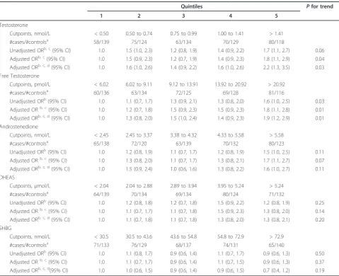
Table 3 reports odds ratios for breast cancer by quin-tiles of hormone concentrations. In multivariate-adjusted models, risk of breast cancer increased signifi-cantly with concentrations of testosterone (P = 0.04) and free testosterone (P = 0.01), with odds ratio (95% CI) of 1.8 (1.1, 2.9) and 1.8 (1.1, 2.8), respectively, for women in the highest versus lowest quintile. The increase in risk appeared more linear for free testoster-one (odds ratios for quintiles were 1.0, 1.2, 1.5, 1.5 and 1.8) than testosterone (odds ratios for quintiles were 1.0, 1.5, 1.2, 1.4 and 1.8). A marginally significant trend (P= 0.07) of increasing risk with increasing concentration of androstenedione was also observed, while no significant association was observed between concentrations of
Table 2 Median (10thand 90thpercentiles) of hormone concentrations in cases and controls
Case subjects (n= 356)
Control subjects (n= 683)
P -value
Testosterone, nmol/L 0.90 (0.41, 2.07) 0.83 (0.31, 1.87) 0.01 Free testosterone, pmol/
L
11.91 (4.92, 32.01)
11.02 (3.70, 27.81)
0.01
Androstenedione, nmol/ L
3.90 (1.97, 6.80) 3.74 (1.80, 6.53) 0.08
DHEAS,μmol/L 3.58 (1.57, 7.22) 3.33 (1.54, 6.75) 0.50 SHBG, nmol/L 48.3 (22.3, 87.2) 49.4 (21.7, 89.5) 0.64
[image:5.595.58.538.281.671.2]DHEAS, dehydroandrosterone sulfate; SHBG, sex hormone-binding globulin.
Table 3 Odds ratios (ORs) and 95% confidence intervals (CIs) for breast cancer by hormone concentration
Quintiles Pfor trend
1 2 3 4 5
Testosterone
Cutpoints, nmol/L < 0.50 0.50 to 0.74 0.75 to 0.99 1.00 to 1.41 > 1.41
#cases/#controlsa 58/139 75/124 63/134 70/129 80/118
Unadjusted ORb, c(95% CI) 1.0 1.5 (1.0, 2.3) 1.2 (0.8, 1.9) 1.4 (0.9, 2.2) 1.7 (1.1, 2.7) 0.06 Adjusted ORb, c(95% CI) 1.0 1.5 (0.9, 2.3) 1.2 (0.7, 1.9) 1.4 (0.9, 2.3) 1.8 (1.1, 2.9) 0.04 Adjusted ORb, c, d(95% CI) 1.0 1.6 (1.0, 2.6) 1.4 (0.9, 2.2) 1.6 (1.0, 2.6) 2.2 (1.3, 3.5) 0.03 Free Testosterone
Cutpoints, pmol/L < 6.02 6.02 to 9.11 9.12 to 13.91 13.92 to 20.92 > 20.92
#cases/#controlsa 60/136 63/134 72/125 69/128 81/116
Unadjusted ORb(95% CI) 1.0 1.1 (0.7, 1.7) 1.3 (0.9, 2.1) 1.3 (0.8, 2.0) 1.6 (1.0, 2.5) 0.03
Adjusted ORb, c(95% CI) 1.0 1.2 (0.7, 1.8) 1.5 (0.9, 2.3) 1.5 (0.9, 2.3) 1.8 (1.1, 2.8) 0.01
Adjusted ORb, c, d(95% CI) 1.0 1.3 (0.8, 2.0) 1.5 (1.0, 2.4) 1.4 (0.9, 2.3) 1.9 (1.2, 2.9) 0.01
Androstenedione
Cutpoints, nmol/L < 2.45 2.45 to 3.37 3.38 to 4.32 4.33 to 5.58 > 5.58
#cases/#controlsa 65/138 72/120 63/139 70/132 80/123
Unadjusted ORb(95% CI) 1.0 1.2 (0.8, 1.9) 1.1 (0.7, 1.7) 1.2 (0.8, 1.9) 1.5 (1.0, 2.5) 0.11 Adjusted ORb, c(95% CI) 1.0 1.3 (0.8, 2.0) 1.1 (0.7, 1.7) 1.3 (0.8, 2.1) 1.7 (1.1, 2.7) 0.07 Adjusted ORb, c, d(95% CI) 1.0 1.5 (0.9, 2.4) 1.0 (0.6, 1.6) 1.3 (0.8, 2.2) 1.6 (1.0, 2.7) 0.11 DHEAS
Cutpoints,μmol/L < 2.04 2.04 to 2.88 2.89 to 3.94 3.95 to 5.24 > 5.24
#cases/#controlsa 64/139 70/134 69/134 80/124 71/132
Unadjusted ORb(95% CI) 1.0 1.2 (0.8, 1.8) 1.2 (0.7, 1.8) 1.5 (0.9, 2.2) 1.2 (0.8, 1.9) 0.25 Adjusted ORb, c(95% CI) 1.0 1.1 (0.7, 1.7) 1.1 (0.7, 1.8) 1.5 (0.9, 2.3) 1.3 (0.8, 2.0) 0.14 Adjusted ORb, c, d(95% CI) 1.0 1.1 (0.7, 1.8) 1.1 (0.7, 1.8) 1.3 (0.8, 2.0) 1.3 (0.8, 2.1) 0.20
SHBG
Cutpoints, nmol/L < 30.5 30.5 to 43.6 43.6 to 54.8 54.8 to 72.9 > 72.9
#cases/#controlsa 71/133 76/129 68/137 74/131 65/140
Unadjusted ORb(95% CI) 1.0 1.1 (0.8, 1.7) 0.9 (0.6, 1.4) 1.1 (0.7, 1.7) 0.9 (0.6, 1.3) 0.50
Adjusted ORb, c(95% CI) 1.0 1.1 (0.7, 1.7) 0.9 (0.6, 1.4) 1.1 (0.7, 1.5) 0.9 (0.6, 1.3) 0.37
Adjusted ORb, c, d(95% CI) 1.0 1.0 (0.6, 1.5) 0.9 (0.6, 1.4) 0.9 (0.6, 1.5) 0.7 (0.4, 1.2) 0.19 a
The number of subjects varies for each hormone depending on the number of the values below the detection limit.bControlling for age, date, and phase and day of cycle at blood donation through matching and use of conditional logistic regression.c
Adjusted for age at menarche (< 12, 12, 13, > 13, missing), family history of breast cancer (no, one affected first-degree relative > 45 yrs old, one affected first degree relative < 45 yrs old or more than one affected first-degree relative), parity/age at first birth (≤20 years at first full-term pregnancy, 21-25 years at first full-term pregnancy, 26-30 years at first full-term pregnancy, > 30 years at first full-term pregnancy, nulliparous, missing), history of breast biopsy, and body mass index (< 20, 20-22.5, 22.6-24.9, 25-29.9, 30+, missing).d
Using the average of two measurements for women for whom two blood samples were available and one measurement for all other women and adjusting for all factors listed inc
DHEAS and SHBG with breast cancer risk. Associations were similar in analyses using the mean hormone level for women who had two samples and the single avail-able measurement for the remaining women, except for testosterone for which higher odds ratios were observed when the average was used if available.
Table 4 reports odds ratios associated with a doubling of biomarker concentrations for all women, as well as by various subject characteristics. Although the odds ratios varied in magnitude according to subgroups and were not always consistently statistically significant, the associations between testosterone and free testosterone and breast cancer risk were usually in the same direc-tion, and none of the tests for interaction was signifi-cant. In particular, odds ratios associated with a doubling in testosterone or free testosterone were ele-vated for invasive and in situ tumors, as well as for tumors diagnosed before and after menopause. Odds ratios greater than one were also observed for estrogen receptor-negative tumors, although the associations were weaker than for estrogen receptor-positive tumors and not statistically significant.
Table 5 shows the ICCs for androgens and SHBG measured at two visits a median of two years (range 0.8 to 5.3 years) apart. ICCs were very similar in cases and controls. The lowest ICC was observed for androstene-dione (0.57, 95% CI: 0.49, 0.65) with all other ICCs greater than 0.7.
Discussion
We observed positive associations between premenopau-sal concentrations of total and free testosterone and breast cancer risk, with women in the highest quintile having a risk approximately 80% greater than women in the lowest quintile. We also observed a marginally sig-nificant positive association with androstenedione but no association with DHEAS or SHBG. The observation of similar associations (except for testosterone for which the odds ratios increased slightly) in an analysis using the mean hormone level for women who had two sam-ples and the single available measurement for the remaining women strengthened our conclusions. There was no evidence of heterogeneity in the associations of total and free testosterone with breast cancer risk in subgroups according to tumor type, estrogen receptor status, age and BMI at enrollment, menopausal status at diagnosis and lag time between blood donation and diagnosis.
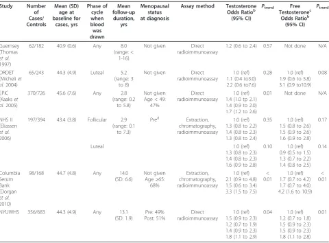
Two prospective studies also reported statistically sig-nificant positive associations between premenopausal concentrations of testosterone and free testosterone and breast cancer risk [14,17], three reported non-significant positive associations [10,12,16], and the smallest (17 cases) reported no association [9]. Overall, results are
consistent across studies, despite variations in the phase of menstrual cycle when blood was drawn, follow-up duration, menopausal status at diagnosis and assay used (table 6). In particular, the two largest studies (the Eur-opean Prospective Investigation into Cancer and Nutri-tion (EPIC) study and this one) both reported statistically significant positive trends for testosterone level and breast cancer risk; free testosterone was not evaluated in the EPIC study. We also observed a high temporal reliability of total testosterone and free testos-terone over a median time of two years, as was also reported by others [36,37].
It has been suggested that including circulating con-centrations of sex hormones could improve risk predic-tion models [38,39]. In fact, imputed postmenopausal concentrations of estradiol improved the discriminatory accuracy of the log-incidence model for breast cancer risk prediction developed by Rosner and Colditz [40], although only modestly. Estrogens, though, are not good candidates for inclusion in risk prediction models of breast cancer in premenopausal women because no con-sistent association has been demonstrated with breast cancer risk in these women [9-11,14,16,17,41], which may be due to the large variations in estrogen concen-trations over the menstrual cycle. The positive
associa-tion of premenopausal testosterone and free
Table 4 Odds ratios (ORs) and 95% confidence intervals (CIs) for a doubling in hormone concentration for all women and according to subject characteristicsa
Characteristic Hormone
(number of cases) Testosterone Free
Testosterone
Androstenedione DHEAS SHBG
All women (n= 354)
OR (95% CI) 1.6 (1.2, 2.3) 1.3 (1.1, 1.5) 1.3 (1.0, 1.7) 1.1 (0.9, 1.4) 0.9 (0.8, 1.1)
P-value 0.01 0.003 0.07 0.31 0.47
Tumor type
Invasive (n= 274)
OR (95% CI) 1.5 (1.1, 2.2) 1.2 (1.0, 1.4) 1.3 (0.9, 1.8) 1.1 (0.8, 1.4) 1.0 (0.8, 1.2)
P-value 0.03 0.02 0.11 0.60 0.84
In situ (n= 80)
OR (95% CI) 2.3 (0.9, 5.9) 1.4 (1.0, 2.1) 2.0 (0.9, 4.5) 1.6 (0.9, 2.8) 0.8 (0.5, 1.3)
P-value 0.09 0.07 0.10 0.14 0.37
Estrogen receptor statusb Positive (n= 104)
OR (95% CI) 2.4 (1.2, 4.6) 1.6 (1.2, 2.2) 2.2 (1.3, 3.8) 1.2 (0.8, 1.8) 0.9 (0.6, 1.3)
P-value 0.01 0.003 0.01 0.42 0.56
Negative (n= 60)
OR (95% CI) 1.7 (0.7, 4.3) 1.2 (0.9, 1.8) 0.7 (0.3, 1.6) 1.4 (0.7, 2.6) 0.8 (0.5, 1.4)
P-value 0.25 0.26 0.37 0.34 0.41
Age at enrollmentc < 40 yrs (n= 77)
OR (95% CI) 1.7 (0.7, 4.2) 1.3 (0.9, 2.0) 1.3 (0.6, 2.7) 0.9 (0.5, 1.5) 0.7 (0.4, 1.2)
P-value 0.27 0.19 0.45 0.61 0.17
40- to yrs (n= 117)
OR (95% CI) 1.4 (0.7, 2.6) 1.1 (0.8, 1.4) 1.1 (0.7, 1.8) 1.1 (0.8, 1.6) 1.2 (0.8, 1.8)
P-value 0.33 0.50 0.72 0.53 0.36
≥45 yrs (n= 160)
OR (95% CI) 1.9 (1.2, 3.2) 1.4 (1.1, 1.7) 1.5 (1.0, 2.3) 1.2 (0.9, 1.7) 0.9 (0.7, 1.2)
P-value 0.01 0.004 0.08 0.21 0.57
Menopausal status at diagnosisd, e
Pre (n= 152)
OR (95% CI) 1.4 (0.8, 2.3) 1.1 (0.9, 1.4) 1.0 (0.7, 1.5) 1.1 (0.8, 2.5) 1.0 (0.8, 1.4)
P-value 0.23 0.36 0.98 0.56 0.80
Post (n= 161)
OR (95% CI) 1.7 (1.1, 2.6) 1.3 (1.1, 1.6) 1.3 (0.9, 1.9) 1.2 (0.8, 1.6) 0.9 (0.7, 1.1)
P-value 0.02 0.006 0.18 0.39 0.26
BMId
< 25 kg/m2(n= 239)
OR (95% CI) 1.8 (1.2, 2.6) 1.3 (1.1, 1.5) 1.2 (0.9, 1.6) 1.3 (1.0, 1.6) 0.9 (0.7, 1.1)
P-value 0.003 0.003 0.30 0.08 0.41
25 to 29.9 kg/m2(n= 74)
OR (95% CI) 2.4 (1.1, 5.0) 1.5 (1.1, 2.1) 1.9 (1.0, 3.5) 1.3 (0.8, 2.1) 1.1 (0.8, 1.7)
P-value 0.02 0.01 0.05 0.29 0.58
≥30 kg/m2(n= 108)
OR (95% CI) 1.5 (0.8, 2.7) 1.2 (0.9, 1.6) 1.6 (1.0, 2.5) 1.0 (0.7, 1.5) 1.1 (0.8, 1.5)
P-value 0.20 0.11 0.08 0.95 0.64
Lag time between blood donation and diagnosisf
< 7 yrs (n= 154)
OR (95% CI) 2.0 (1.1, 3.5) 1.3 (1.0, 1.7) 1.3 (0.9, 2.1) 1.4 (1.0, 1.9) 1.0 (0.8, 1.5)
P-value 0.02 0.04 0.20 0.06 0.78
approved for chemoprevention in such women. Use of tamoxifen for prevention of breast cancer, though, has been limited [23,24]. It has been shown that the higher the risk of breast cancer relative to the risk of adverse events, the more likely a woman is to accept tamoxifen chemoprevention [45]. Factors helping to predict more accurately the absolute risk of breast cancer might thus lead to increased acceptance of chemoprevention by the women most likely to benefit and, therefore, result in a larger number of prevented breast cancers. A challenge that needs to be addressed prior to incorporation of cir-culating hormone levels in risk prediction models, though, is standardization of assay methods [38,46,47].
Positive associations between circulating androgens in postmenopausal women and risk of breast cancer have been observed consistently [1], although the association may vary according to the estrogen receptor (ER) status of the tumor. Whereas most studies found a positive association with ER-positive tumors [4,8,48,49], no sig-nificant association was observed in three studies [4,6,48], and the largest study to date found a significant inverse, rather than positive, association with ER-nega-tive tumors [49]. Notwithstanding these differences, the main mechanism proposed to explain the increase in risk observed among postmenopausal women is the aro-matization of androgens into estrogens in peripheral adipose tissue [50]. Because of the reduced production of estrogens by the ovaries, peripheral production is an important contributor to circulating concentrations of
estrogens after menopause. This mechanism is thought to contribute to the well documented positive associa-tion between BMI and breast cancer risk observed after menopause [51]. It is unlikely, though, that this mechan-ism explains the association observed between andro-gens and breast cancer in premenopausal women because estrogens are mostly produced by the ovaries prior to menopause. Further, under this mechanism, one would expect a positive association between BMI and breast cancer risk in premenopausal women, as is seen in post-menopausal women, since more aromatiza-tion of androgens in adipose tissue is expected with increased BMI. However, although we did not observe an association of BMI with breast cancer risk in our study, an inverse, rather than positive, association between BMI and breast cancer risk in premenopausal women has been found in most studies [52,53]. Addi-tional evidence against an important role of this mechanism is the observation made by Eliassen et al. that adjustment for concentrations of estradiol in pre-menopausal women did not affect risk estimates asso-ciated with testosterone concentrations [16]. It is of interest that we observed an association in women with regular cycles, as well as women of normal weight, sug-gesting that androgen concentrations increase breast cancer risk even in women with no evidence of hyperandrogenism.
[image:8.595.58.539.113.185.2]In addition to their role as estrogen precursors, it has been proposed that androgens directly impact cell pro-liferation [54,55], possibly through binding to androgen receptors which are present in both normal breast tissue and most breast cancers [56]. Results from experimental studies, though, have been inconsistent, with some stu-dies [25,26] reporting an inhibitory effect of androgens on estrogen-induced breast cell proliferation, while others did not [27]. The only human study that exam-ined the effect of testosterone on breast cell prolifera-tion found that postmenopausal women who received testosterone (300μg/day patch) in addition to hormone replacement therapy (2 mg estradiol and 1 mg
Table 5 ICCs (95% CI) for hormonal biomarkers
Cases N= 138
Controls N= 268
Testosterone 0.74 (0.65 - 0.81) 0.78 (0.73 - 0.82) Androstenedione 0.58 (0.46 - 0.68) 0.57 (0.49 - 0.65) DHEAS 0.82 (0.76 - 0.87) 0.76 (0.70 - 0.81) SHBG 0.86 (0.81 - 0.90) 0.78 (0.73 - 0.82) Free testosterone 0.86 (0.81 - 0.90) 0.82 (0.78 - 0.86)
[image:8.595.57.289.633.717.2]DHEAS, dehydroandrosterone sulfate; SHBG, sex hormone-binding globulin.
Table 4 Odds ratios (ORs) and 95% confidence intervals (CIs) for a doubling in hormone concentration for all women and according to subject characteristicsa(Continued)
OR (95% CI) 1.5 (1.0, 2.3) 1.3 (1.0, 1.5) 1.3 (0.9, 1.9) 1.0 (0.7, 1.3) 0.8 (0.6, 1.1)
P-value 0.07 0.02 0.17 0.80 0.22
5 to7 cycles in 6 months prior to enrollment and regular cyclesd(n= 253)
OR (95% CI) 1.6 (1.1, 2.3) 1.2 (1.1, 1.5) 1.3 (1.0, 1.8) 1.1 (0.9, 1.5) 0.9 (0.8, 1.2)
P-value 0.02 0.01 0.07 0.29 0.59
a
Adjusted for age at menarche (< 12, 12, 13, > 13, missing), family history of breast cancer (no, one affected first-degree relative > 45 yrs old, one affected first degree relative < 45 yrs old or more than one affected first-degree relative), parity/age at first birth (≤20 years at first full-term pregnancy, 21 to 25 years at first full-term pregnancy, 26 to 30 years at first full-term pregnancy, > 30 years at first full-term pregnancy, nulliparous, missing), history of breast biopsy, and body mass index (< 20, 20-22.5, 22.6-24.9, 25-29.9, 30+, missing).b
0.05 <Pinteraction< 0.15 for androstenedione.c0.05 <Pinteraction< 0.15 for free testosterone.dUsing
unconditional logistic regression, adjusting for matching factors in addition to factors listed ina.e0.05 <Pinteraction< 0.15 for free testosterone. f
0.05 <Pinteraction<
norethisterone acetate) did not have an increase in cell proliferation, while a more than five-fold increase in cell proliferation was observed in women who received only the estrogen + progestin therapy [57]. These studies, though, were conducted in primates or in women in the postmenopausal stage who received estrogens orally, the effect of which may differ from that of endogenous hor-mones. For instance, oral estrogens are known to increase production of SHBG and reduce the concentra-tions of free testosterone [58]. It is, therefore, not clear whether results of these studies apply to endogenous androgens in premenopausal women. Additional research is needed to explain why circulating concentra-tions of androgens in premenopausal women are asso-ciated with an increase in risk of breast cancer.
Androgens, in particular testosterone, have been pro-posed for relief of menopausal symptoms, in particular sexual desire deficit [28]. As symptoms may start well before menopause, androgen therapies may be pre-scribed beginning in the late premenopausal years
[29,59]. In light of the increased risk of breast cancer associated with higher concentrations of circulating androgens both pre- and post-menopause, and the results of two prospective studies that reported an increased risk of breast cancer in women receiving estrogen + testosterone therapy [60,61], although this association was significant only in one of the two studies [60], caution should be exercised regarding long-term prescription of androgens.
[image:9.595.58.539.98.452.2]The NYUWHS was designed primarily to examine the association of endogenous sex hormones with risk of breast cancer. We therefore excluded women taking exogenous estrogens and collected data on date of next menstrual period which allowed us to calculate the phase of cycle more precisely than some other studies. Other strengths of our study include the large number of cases, which allowed us to examine various subgroups and the availability of two serum samples in a fairly large number of both cases and controls which allowed us to show that a single androgen concentration
Table 6 Prospective studies of testosterone and breast cancer risk in premenopausal womena
Study Number of Cases/ Controls Mean (SD) age at baseline for cases, yrs Phase of cycle when blood was drawn Mean follow-up duration, yrs Menopausal status at diagnosis
Assay method Testosterone Odds Ratiob
(95% CI)
Ptrend Free
Testosteronec Odds Ratiob
(95% CI) Ptrend Guernsey (Thomas et al. 1997)
62/182 40.9 (0.6) Any 8.0 (range: <
1-16)
Not given Direct radioimmunoassay
1.2 (0.6 to 2.4) 0.57 Not done N/A
ORDET (Micheliet al.2004)
65/243 44.3 (4.9) Luteal 5.2 (range: 3
to 8)
Not given Direct radioimmunoassay
1.0 (ref) 1.1 (0.4 to3.0) 2.2 (0.6 to7.6)
0.28 1.0 (ref) 1.9 (0.6 to 5.8) 3.1 (0.9 to10.9)
0.08
EPIC (Kaakset al.2005)
370/726 45.6 (7.6) Any 2.8 (range: 0.2
to 5.8)
Not given Age < 49:
47%
Direct radioimmunoassay
1.0 (ref) 1.4 (1.0 tp 2.1) 1.4 (0.9 to 2.0) 1.7 (1.2 to 2.6)
0.01 Not done N/A
NHS II (Eliassen
et al.
2006)
197/394 43.4 (3.8) Follicular 2.9 (range: 0.1
to 7.3)
Pred Extraction,
chromatography, radioimmunoassay
1.0 (ref) 1.3 (0.8 to 2.2) 1.4 (0.8 to 2.3) 1.3 (0.8 to 2.4)
0.35 1.0 (ref) 1.5 (0.8 to 2.6) 1.5 (0.9 to 2.6) 1.6 (0.9 to 2.8)
0.17
Luteal 1.0 (ref)
1.3 (0.8 to 2.3) 1.4 (0.8 to 2.3) 1.6 (0.9 to 2.8)
0.10 1.0 (ref) 0.9 (0.5 to 1.5) 1.3 (0.7 to 2.2) 1.4 (0.8 to 2.5)
0.14 Columbia Serum Bank (Dorgan et al. 2010)
98/168 44.7 (4.8) Any 14.0 (SD: 6.6)
Not given Age≥65:
68%
Extraction, chromatography, radioimmunoassay
1.0 (ref) 2.1 (0.9 to 4.8) 1.5 (0.6 to 3.4) 3.3 (1.5 to 7.5)
< 0.01
1.0 (ref) 1.7 (0.7 to 4.2) 1.7 (0.7 to 4.0) 4.2 (1.6 to 10.9)
< 0.01
NYUWHS 356/683 44.3 (4.9) Any 13.1 (SD: 1.9) Pre: 49% Post: 51% Direct radioimmunoassay 1.0 (ref) 1.5 (0.9 to 2.3) 1.2 (0.7 to 1.9) 1.4 (0.9 to 2.3) 1.8 (1.1 to 2.9)
0.04 1.0 (ref) 1.2 (0.7 to 1.8) 1.5 (0.9 to 2.3) 1.5 (0.9 to 2.3) 1.8 (1.1 to 2.8)
0.01
a
Wysowskiet al.[9] not included in table because no odds ratio was provided (no statistical difference was observed in testosterone concentrations in 17 women).b
Odds Ratios (95% CIs) based on a 1 unit increase in the natural log of hormone concentration, tertiles, quartiles, or quintiles of serum concentration.
c
Bioavailable testosterone (free plus albumin bound) calculated in Dorganet al.2010 study.d
measurement is quite representative of a woman’s con-centration over several years. This is despite the fact that we did not control for time of day of blood dona-tion, and, therefore, for possible circadian variations of androgen production. A weakness of our study is that we used radioimmunassays without an extraction step, and the sensitivity and specificity of such assays have been questioned [46,62]. It should be noted, though, that results from other studies did not appear to vary according to whether or not a purification step was used (table 6). A similar observation was made for sex hormone concentrations in postmenopausal women [1].
Conclusions
In conclusion, and in agreement with other cohorts, we observed associations between pre-diagnostic concentra-tions of total and free testosterone in premenopausal women and risk of breast cancer. These results suggest that androgen concentrations should be considered for inclusion in risk prediction models for women between the ages of 40 and 50, which could help in decision making regarding both screening and chemoprevention of breast cancer.
Abbreviations
BMI: body mass index; CI: confidence interval; DHEAS: dehydroandrosterone sulfate; EPIC: European Prospective Investigation into Cancer and Nutrition; ER: estrogen receptor; FSH: follicle-stimulating hormone; ICC: intraclass correlation coefficient; NYUWHS: New York University Women’s Health Study; SHBG: sex hormone-binding globulin.
Acknowledgements
We thank all of the participants of the NYUWHS and Elizabeth Clancy, Denise Heimowitz and Lynne Quinones for data collection and technical assistance. This study was supported by the National Institutes of Health R01 CA098661, center grant CA16087 and National Institute of Environmental Health Sciences center grant ES00260.
Author details
1Department of Environmental Medicine, New York University School of
Medicine, 650 First Avenue, New York, NY 10016, USA.2New York University Cancer Institute, New York University School of Medicine, 530 First Avenue, New York, NY 10016, USA.3Division of Cancer Epidemiology, German Cancer Research Centre, Im Neuenheimer Feld 280, D-69120 Heidelberg, Germany.
4
International Agency for Research on Cancer, 150, Cours Albert Thomas, 69372 Lyon Cedex 08, France.5Department of Obstetrics and Gynecology,
New York University School of Medicine, 550 First Avenue, New York, NY 10016, USA.6Unit of Cancer Epidemiology, Institute of Social and Preventive
Medicine, Centre Hospitalier Universitaire Vaudois, Biopôle 1, 2 Route de la Corniche, CH-1066 Epalinges, Switzerland.7Radiation Effects Research Foundation, 5-2 Hijiyama Park, Minami-ku, Hiroshima, 732-0815, Japan.
Authors’contributions
AZJ participated in the design of the study, statistical analysis, and manuscript preparation. YA performed the statistical analysis and contributed to manuscript preparation. RK participated in the conception of the study and manuscript preparation. SR performed serum hormone analyses and contributed to manuscript preparation. SS contributed to manuscript preparation. ML participated in the statistical analysis and manuscript preparation. AAA participated in data acquisition and manuscript preparation. PT and RES participated in the conception and design of the study and manuscript preparation. KLK participated in the design of the
study, the statistical analysis and manuscript preparation. All authors read and approved the final manuscript.
Competing interests
The authors declare that they have no competing interests.
Received: 19 August 2011 Revised: 20 January 2012 Accepted: 16 February 2012 Published: 16 February 2012
References
1. The Endogenous Hormones Breast Cancer Collaborative Group:
Endogenous sex hormones and breast cancer in postmenopausal women: reanalysis of nine prospective studies.J Natl Cancer Inst2002,
94:606-616.
2. Kaaks R, Rinaldi S, Key TJ, Berrino F, Peeters PHM, Biessy C, Dossus L, Lukanova A, Bingham S, Khaw KT, Allen NE, Bueno-de-Mesquita HB, van Gils CH, Grobbee D, Boeing H, Lahmann PH, Nagel G, Chang Claude J, Clavel Chapelon F, Fournier A, Thibaut A, Gonzlez CA, Quirs JR, Tormo MJ, Ardanaz E, Amiano P, Krogh V, Palli D, Panico S, Tumino R,et al:
Postmenopausal serum androgens, oestrogens and breast cancer risk: the European prospective investigation into cancer and nutrition.Endocr Relat Cancer2005,12:1071-1082.
3. Woolcott C, Shvetsov Y, Stanczyk F, Wilkens L, White K, Caberto C, Henderson B, Le-Marchand L, Kolonel L, Goodman M:Plasma sex hormone concentrations and breast cancer risk in an ethnically diverse population of postmenopausal women: the Multiethnic Cohort Study.Endocr Relat Cancer2010,17:125-134.
4. Missmer S, Eliassen AH, Barbieri R, Hankinson S:Endogenous estrogen, androgen, and progesterone concentrations and breast cancer risk among postmenopausal women.J Natl Cancer Inst2004,96:1856-1865. 5. Zeleniuch-Jacquotte A, Shore RE, Koenig KL, Akhmedkhanov A,
Afanasyeva Y, Kato I, Kim MY, Rinaldi S, Kaaks R, Toniolo P:Postmenopausal levels of estrogen, androgen, and SHBG and breast cancer risk: long-term results of a prospective study.Br J Cancer2004,90:153-159. 6. Baglietto L, Severi G, English D, Krishnan K, Hopper J, McLean C, Morris H,
Tilley W, Giles G:Circulating steroid hormone levels and risk of breast cancer for postmenopausal women.Cancer Epidemiol Biomarkers Prev
2010,19:492-502.
7. Manjer J, Johansson R, Berglund G, Janzon L, Kaaks R, Agren A, Lenner P:
Postmenopausal breast cancer risk in relation to sex steroid hormones, prolactin and SHBG (Sweden).Cancer Causes Control2003,14:599-607. 8. Cummings S, Lee J, Lui L-Y, Stone K, Ljung B, Cauleys J:Sex hormones, risk
factors, and risk of estrogen receptor-positive breast cancer in older women: a long-term prospective study.Cancer Epidemiol Biomarkers Prev
2005,14:1047-1051.
9. Wysowski DK, Comstock GW, Helsing KJ, Lau HL:Sex hormone levels in serum in relation to the development of breast cancer.Am J Epidemiol
1987,125:791-799.
10. Thomas H, Key T, Allen D, Moore J, Dowsett M, Fentiman I, Wang D:A prospective study of endogenous serum hormone concentrations and breast cancer risk in premenopausal women on the island of Guernsey.
Br J Cancer1997,75:1075-1079.
11. Kabuto M, Akiba S, Stevens R, Neriishi K, Land C:A prospective study of estradiol and breast cancer in Japanese women.Cancer Epidemiol Biomarkers Prev2000,9:575-579.
12. Micheli A, Muti P, Secreto G, Krogh V, Meneghini E, Venturelli E, Sieri S, Pala V, Berrino F:Endogenous sex hormones and subsequent breast cancer in premenopausal women.Int J Cancer2004,112:312-318. 13. Page J, Colditz G, Rifai N, Barbieri R, Willett W, Hankinson S:Plasma adrenal
androgens and risk of breast cancer in premenopausal women.Cancer Epidemiol Biomarkers Prev2004,13:1032-1036.
14. Kaaks R, Berrino F, Key T, Rinaldi S, Dossus L, Biessy C, Secreto G, Amiano P, Bingham S, Boeing H, Bueno de Mesquita H, Chang-Claude J, Clavel-Chapelon F, Fournier A, van Gils C, Gonzalez C, Gurrea A, Critselis E, Khaw K, Krogh V, Lahmann P, Nagel G, Olsen A, Onland-Moret N, Overvad K, Palli D, Panico S, Peeters P, Quirós J, Roddam A,et al:Serum sex steroids in premenopausal women and breast cancer risk within the European Prospective Investigation into Cancer and Nutrition (EPIC).J Natl Cancer Inst2005,97:755-765.
sulfate with breast cancer risk in predominantly premenopausal women.
Cancer Epidemiol Biomarkers Prev2006,15:967-971.
16. Eliassen A, Missmer S, Tworoger S, Spiegelman D, Barbieri R, Dowsett M, Hankinson S:Endogenous steroid hormone concentrations and risk of breast cancer among premenopausal women.J Natl Cancer Inst2006,
98:1406-1415.
17. Dorgan J, Stanczyk F, Kahle L, Brinton L:Prospective case-control study of premenopausal serum estradiol and testosterone levels and breast cancer risk.Breast Cancer Res2010,12:R98.
18. Gail MH, Brinton LA, Byar DP, Corle DK, Green SB, Schairer C, Mulvihill JJ:
Projecting individualized probabilities of developing breast cancer for white females who are being examined annually.J Natl Cancer Inst1989,
81:1879-1886.
19. U.S. Preventive Services Task Force:Screening for breast cancer: U.S. Preventive Services Task Force recommendation statement.Ann Intern Med2009,151:716-726, W-236.
20. American Cancer Society Guidelines for the Early Detection of Cancer.
[http://www.cancer.org/Healthy/FindCancerEarly/
CancerScreeningGuidelines/american-cancer-society-guidelines-for-the-early-detection-of-cancer].
21. U.S. Preventive Services Task Force:Chemoprevention of breast cancer: recommendations and rationale.Ann Intern Med2002,137:56-58. 22. Chlebowski R, Col N, Winer E, Collyar D, Cummings S, Vogel V, Burstein H,
Eisen A, Lipkus I, Pfister D:American Society of Clinical Oncology technology assessment of pharmacologic interventions for breast cancer risk reduction including tamoxifen, raloxifene, and aromatase inhibition.
J Clin Oncol2002,20:3328-3343.
23. Armstrong K, Quistberg DA, Micco E, Domchek S, Guerra C, Armstrong K, Quistberg DA, Micco E, Domchek S, Guerra C:Prescription of tamoxifen for breast cancer prevention by primary care physicians.Arch Intern Med
2006,166:2260-2265.
24. Waters EA, Cronin KA, Graubard BI, Han PK, Freedman AN, Waters EA, Cronin KA, Graubard BI, Han PK, Freedman AN:Prevalence of tamoxifen use for breast cancer chemoprevention among U.S. women.Cancer Epidemiol Biomarkers Prev2010,19:443-446.
25. Zhou J, Ng S, Adesanya Famuiya O, Anderson K, Bondy CA:Testosterone inhibits estrogen-induced mammary epithelial proliferation and suppresses estrogen receptor expression.FASEB J2000,14:1725-1730. 26. Dimitrakakis C, Zhou J, Wang J, Belanger A, LaBrie F, Cheng C, Powell D,
Bondy C:A physiologic role for testosterone in limiting estrogenic stimulation of the breast.Menopause2003,10:292-298.
27. Wood C, Lees C, Cline JM:Mammary gland and endometrial effects of testosterone in combination with oral estradiol and progesterone.
Menopause2009,16:466-476.
28. Davis S, Moreau M, Kroll R, Bouchard C, Panay N, Gass M, Braunstein G, Hirschberg A, Rodenberg C, Pack S, Koch H, Moufarege A, Studd J:
Testosterone for low libido in postmenopausal women not taking estrogen.N Engl J Med2008,359:2005-2017.
29. Glaser R, York A, Dimitrakakis C:Beneficial effects of testosterone therapy in women measured by the validated Menopause Rating Scale (MRS).
Maturitas2011,68:355-361.
30. Toniolo PG, Pasternack BS, Shore RE, Sonnenschein EG, Koenig KL, Rosenberg C, Strax P, Strax S:Endogenous hormones and breast cancer: a prospective cohort study.Breast Cancer Res Treat1991,18:S23-S26. 31. Kato I, Toniolo P, Koenig K, Kahn A, Schymura M, Zeleniuch-Jacquotte A:
Comparison of active and cancer registry-based follow-up for breast cancer in a prospective cohort study.Am J Epidemiol1999,149:372-378. 32. Rinaldi S, Dechaud H, Biessy C, Morin-Raverot V, Toniolo P,
Zeleniuch-Jacquotte A, Akhmedkhanov A, Shore RE, Secreto G, Ciampi A, Riboli E, Kaaks R:Reliability and validity of commercially available, direct radioimmunoassays for measurement of blood androgens and estrogens in postmenopausal women.Cancer Epidemiol Biomark Prev
2001,10:757-765.
33. Rinaldi S, Geay A, Dechaud H, Biessy C, Zeleniuch-Jacquotte A, Akhmedkhanov A, Shore RE, Riboli E, Toniolo P, Kaaks R:Validity of free testosterone and free estradiol determinations in serum samples from postmenopausal women by theoretical calculations.Cancer Epidemiol Biomarkers Prev2002,11:1065-1071.
34. Donner A:A review of inference procedures for the intraclass correlation coefficient in the one-way random effects model.Int Stat Rev1986,
54:67-82.
35. Rothman M, Carlson N, Xu M, Wang C, Swerdloff R, Lee P, Goh VHH, Ridgway EC, Wierman M:Reexamination of testosterone,
dihydrotestosterone, estradiol and estrone levels across the menstrual cycle and in postmenopausal women measured by liquid
chromatography-tandem mass spectrometry.Steroids2011,76:177-182. 36. Missmer S, Spiegelman D, Bertone Johnson E, Barbieri R, Pollak M,
Hankinson S:Reproducibility of plasma steroid hormones, prolactin, and insulin-like growth factor levels among premenopausal women over a 2- to 3-year period.Cancer Epidemiol Biomarkers Prev2006,15:972-978. 37. Burger HG, Dudley EC, Cui J, Dennerstein L, Hopper JL:A prospective
longitudinal study of serum testosterone, dehydroepiandrosterone sulfate, and sex hormone-binding globulin levels through the menopause transition.J Clin Endocrinol Metab2000,85:2832-2838. 38. Santen RJ, Boyd NF, Chlebowski RT, Cummings S, Cuzick J, Dowsett M,
Easton D, Forbes JF, Key T, Hankinson SE, Howell A, Ingle J, Breast Cancer Prevention Collaborative G:Critical assessment of new risk factors for breast cancer: considerations for development of an improved risk prediction model.Endocr Relat Cancer2007,14:169-187.
39. Rockhill B, Spiegelman D, Byrne C, Hunter DJ, Colditz GA:Validation of the Gail et al. model of breast cancer risk prediction and implications for chemoprevention.J Natl Cancer Inst2001,93:358-366.
40. Rosner B, Colditz GA, Iglehart JD, Hankinson SE:Risk prediction models with incomplete data with application to prediction of estrogen receptor-positive breast cancer: prospective data from the Nurses’ Health Study.Breast Cancer Res2008,10:R55.
41. Helzlsouer K, Alberg AJ, Bush TL, Longcope C, Gordon GB, Comstock GW:A prospective study of endogenous hormones and breast cancer.Cancer Detect Prev1994,18:79-85.
42. Eliassen AH, Missmer S, Tworoger S, Hankinson S:Endogenous steroid hormone concentrations and risk of breast cancer: does the association vary by a woman’s predicted breast cancer risk?J Clin Oncol2006,
24:1823-1830.
43. Smith R, Cokkinides V, Brooks D, Saslow D, Brawley O:Cancer screening in the United States, 2010: a review of current American Cancer Society guidelines and issues in cancer screening.CA Cancer J Clin2010,
60:99-119.
44. Gail MH, Costantino JP, Bryant J, Croyle R, Freedman L, Helzlsouer K, Vogel V:Weighing the risks and benefits of tamoxifen treatment for preventing breast cancer.J Natl Cancer Inst1999,91:1829-1846. 45. Ozanne E, Wittenberg E, Garber J, Weeks J:Breast cancer prevention:
patient decision making and risk communication in the high risk setting.Breast J2010,16:38-47.
46. Stanczyk F:Measurement of androgens in women.Semin Reprod Med
2006,24:78-85.
47. Blair I:Analysis of estrogens in serum and plasma from postmenopausal women: past present, and future.Steroids2010,75:297-306.
48. Sieri S, Krogh V, Bolelli G, Abagnato CA, Grioni S, Pala V, Evangelista A, Allemani C, Micheli A, Tagliabue G, Schunemann HJ, Menard S, Berrino F, Muti P, Sieri S, Krogh V, Bolelli G, Abagnato CA, Grioni S, Pala V, Evangelista A, Allemani C, Micheli A, Tagliabue G, Schunemann HJ, Menard S, Berrino F, Muti P:Sex hormone levels, breast cancer risk, and cancer receptor status in postmenopausal women: the ORDET cohort.
Cancer Epidemiol Biomarkers Prev2009,18:169-176.
49. Farhat G, Cummings S, Chlebowski R, Parimi N, Cauley J, Rohan T, Huang A, Vitolins M, Hubbell FA, Manson J, Cochrane B, Lane D, Lee J:Sex hormone levels and risks of estrogen negative and estrogen receptor-positive breast cancers.J Natl Cancer Inst2011,103:562-570.
50. Key T:Endogenous oestrogens and breast cancer risk in premenopausal and postmenopausal women.Steroids2011,76:812-815.
51. The Endogenous Hormones Breast Cancer Collaborative Group:Body mass index, serum sex hormones, and breast cancer risk in postmonopausal women.J Natl Cancer Inst2003,95:1218-1226.
52. van den Brandt PA, Spiegelman D, Yaun SS, Adami HO, Beeson L, Folsom AR, Fraser G, Goldbohm RA, Graham S, Kushi L, Marshall JR, Miller AB, Rohan T, Smith-Warner SA, Speizer FE, Willett WC, Wolk A, Hunter DJ:Pooled analysis of prospective cohort studies on height, weight, and breast cancer risk.Am J Epidemiol2000,152:514-527. 53. Tehard B, Clavel Chapelon F:Several anthropometric measurements and
breast cancer risk: results of the E3N cohort study.Int J Obes2006,
54. Somboonporn W, Davis S:Testosterone effects on the breast: implications for testosterone therapy for women.Endocr Rev2004,
25:374-388.
55. Liao D, Dickson R:Roles of androgens in the development, growth, and carcinogenesis of the mammary gland.J Steroid Biochem Mol Biol2002,
80:175-189.
56. Tiefenbacher K, Daxenbichler G:The role of androgens in normal and malignant breast tissue.Breast Care2008,3:325-331.
57. Hofling M, Hirschberg A, Skoog L, Tani E, Hagerstrom T, von Schoultz B:
Testosterone inhibits estrogen/progestogen-induced breast cell proliferation in postmenopausal women.Menopause2007,14:183-190. 58. Campagnoli C, Colombo P, De Aloysio D, Gambacciani M, Grazioli I,
Nappi C, Serra G, Genazzani A:Positive effects on cardiovascular and breast metabolic markers of oral estradiol and dydrogesterone in comparison with transdermal estradiol and norethisterone acetate.
Maturitas2002,41:299-311.
59. Davis S, Papalia M-A, Norman R, O’Neill S, Redelman M, Williamson M, Stuckey BGA, Wlodarczyk J, Gard’ner K, Humberstone A:Safety and efficacy of a testosterone metered-dose transdermal spray for treating decreased sexual satisfaction in premenopausal women: a randomized trial.Ann Int Med2008,148:569-577.
60. Tamimi R, Hankinson S, Chen W, Rosner B, Colditz G:Combined estrogen and testosterone use and risk of breast cancer in postmenopausal women.Arch Int Med2006,166:1483-1489.
61. Ness R, Albano J, McTiernan A, Cauley J:Influence of estrogen plus testosterone supplementation on breast cancer.Arch Int Med2009,
169:41-46.
62. Rosner W, Auchus R, Azziz R, Sluss P, Raff H:Position statement: Utility, limitations, and pitfalls in measuring testosterone: an Endocrine Society position statement.J Clin Endocrinol Metab2007,92:405-413.
doi:10.1186/bcr3117
Cite this article as:Zeleniuch-Jacquotteet al.:Premenopausal serum
androgens and breast cancer risk: a nested case-control study.Breast Cancer Research201214:R32.
Submit your next manuscript to BioMed Central and take full advantage of:
• Convenient online submission
• Thorough peer review
• No space constraints or color figure charges
• Immediate publication on acceptance
• Inclusion in PubMed, CAS, Scopus and Google Scholar
• Research which is freely available for redistribution