Open Access
Short Report
Effects of chronic estradiol treatment on the thyroid gland
structure and function of ovariectomized rats
Menna M Abdel-Dayem
†1and Mohamed S Elgendy*
†2Address: 1Histology Department, Faculty of Medicine, Cairo University, Cairo, Egypt and 2Histology Department, Faculty of Medicine, Fayoum University, Fayoum, Egypt
Email: Menna M Abdel-Dayem - [email protected]; Mohamed S Elgendy* - [email protected] * Corresponding author †Equal contributors
Abstract
Background: Estrogen therapy is widely used nowadays in women to treat many postmenopausal symptoms but it may have some undesirable effects due to multiple organs affection. So, the aim of this study was to determine the effects of chronic estradiol treatment on the structure and function of the thyroid gland in ovarictomized rats as a model simulating menopause.
Findings: Thirty adult female Wistar rats divided into three groups were used in this study; the first group was sham-operated, while the second and third groups were ovariectomized. The first and second groups were injected with olive oil while the third group was injected with estradiol dipropionate daily for three months, after that; hormonal assay for T3, T4, TSH and specimens of the thyroid were taken and processed to be examined by light and electron microscopy. The results of this study revealed that serum levels of T3 and T4 decreased in ovariectomized animals and significantly increased after estradiol treatment, while TSH increased in ovariectomized animals and decreased with estradiol treatment. Histological and morphometric study in ovariectomized group revealed marked accumulation of colloid in follicular lumens with decreased epithelial height in addition to increased connective tissue amount. After estradiol treatment the follicles became smaller in size, having small amount of colloid with increased epithelial height in addition to decreased connective tissue content. Ultrastructural study supported these results in addition to the presence of large amount of intracytoplasmic colloid vesicles after estradiol treatment.
Conclusion: Low estrogen level may lead to mild thyroidal hypofunction while estradiol treatment may lead to hyperactivity so it should be used very cautiously in the treatment of postmenopausal symptoms to avoid its undesirable stimulatory effect on the thyroid.
Published: 30 August 2009
BMC Research Notes 2009, 2:173 doi:10.1186/1756-0500-2-173
Received: 5 August 2009 Accepted: 30 August 2009
This article is available from: http://www.biomedcentral.com/1756-0500/2/173
© 2009 Elgendy et al; licensee BioMed Central Ltd.
Background
There is a great wealth of published data on the value of estrogen therapy in alleviating menopausal symptoms such as hot flushes and insomnia and in preventing uro-genital atrophy and osteoporosis [1].
During hormonal therapy both beneficial and undesira-ble side effects appear due to multiple target organs for estrogen [2]. Epidemiological studies suggest that the use of estrogen may contribute to the pathogenesis of thyroid tumors [3] additionally; thyroid diseases are more com-mon in women [4].
Although some studies have demonstrated influences of estrogen on the development, physiology and histology of the thyroid gland, questions about the effects of estrogen replacement therapy on the thyroid gland remained unan-swered [5].
As regard the effect of estradiol on thyroid activity; numer-ous studies have demonstrated contradictory results either stimulatory effect through activation of pituitary-thyroid axis following estradiol treatment [6,7] or no alterations of TSH although decreased T3 in ovariectomized rats which became normal after estradiol treatment [8].
In women treated with conjugated estrogen 0.625-2.5 mg/kg/day, there was no change in serum TSH concentra-tions [9]. In other studies; perimenopausal and postmen-opausal women had "elevated" serum TSH concentrations after 1 month [10] or 6 weeks [11] of estrogen therapy.
Having in mind discrepancies in literature, this study aimed to examine the effects of chronic estradiol treat-ment on structure and function of thyroid gland in ovaric-tomized rats as an animal model for menopause.
Methods
Animals
Thirty adult female Wistar albino rats of 200 - 220 g body weight were used in this study; they were obtained and housed in the animal house of Kasr-El-Aini Faculty of Medicine, Cairo University, Egypt. The animals received a standard diet for rodents and allowed free access to water. The animals were treated humanely and care was taken to ease suffering.
Surgical procedure
The animals were divided randomly into three groups of ten animals each. The first group (Group I) was sham-operated. The other two groups were ovariectomized. All animals were anaesthetized with intraperitoneal injection of Phenobarbital sodium (15 mg/kg body weight). The lower abdomen of the rats was shaved and incised.
Fallo-pian tubes and ovaries were identified. Absorbable catgut sutures were used to tie the Fallopian tubes below the ova-ries. Then, the ovaries were removed. Sham-operated group underwent a similar surgical incision exposing the ovaries and replacing them in the same position [12].
Treatment protocol
One month after surgical procedures, rats of the first and second groups were injected intra peritoneal (i.p) with sterile olive oil once daily for three months. The third group was injected (i.p) with 0.625 mg/kg bw. estradiol dipropionate (EDP) (Sigma Chemical Co.), dissolved in sterile olive oil once daily for the same duration. This dose is equivalent to a commonly used dose for estrogen replacement in the clinical practice in postmenopausal women and used in previous experimental designs [13].
This study has been approved by the ethics committee on animal research in the animal house of Kasr-El-Aini Fac-ulty of Medicine, Cairo University, Egypt following inter-national ethics and regulations for animal research in laboratory applications [14].
Evaluation methods
Hormonal assay
At the end of experimental period, blood samples were collected from the tail vein of all animals and centrifuged at 3000 rpm for ten minutes. Sera were separated and stored at -20°C until hormonal assay.
Total T3 and T4 levels were measured by radioimmu-noassay (RIA) using commercial kits (Coat-A-Coat), while Serum TSH was measured by RIA using a specific rat TSH kit (supplied by Diagnostic Products Corporation DPC, Los Angeles, USA). Radioactivity was determined by the gamma-counter [15].
The data were presented as mean ± SD.
Histological study
Twenty four hours after the last injection, all animals were anaesthetized with Phenobarbital sodium, and after int-racardiac perfusion with the fixative solution (2% gluter-aldehyde in phosphate buffer), the thyroid glands were removed together with a portion of the adjoining trachea. One lobe of the thyroid gland of the animals were dis-sected, cut into small cubes (about 1 mm3) and
immedi-ately fixed in 2% gluteraldehyde. for electron microscopic study:
Light microscopic study
Morphometry
The data were obtained by using "Leica Qwin 500" image analyzer computer system (England). The measurements were done using an objective lens of magnification 40, examining the central regions of the glands. (peripheral and isthmic follicles were excluded).
Epithelial height and follicular area were measured in sec-tions stained with Hx&E. colloidal area percent was assessed in PAS stained sections and connective tissue area percent in sections stained with Masson's trichrome stain. For each parameter, ten readings per animal in each group were done.
Electron microscopic study
Small fragments from the thyroid glands were rinsed in phosphate buffer (PH 7.4) fixed in 2% gluteraldehyde, postfixed in 1% osmium tetroxide and dehydrated. After embedding in ultrathin sections were cut and stained with lead citrate and uranyl acetate. The grids were examined and photographed with electron microscope.
Statistical analysis
The data obtained from hormonal assay and morphome-try were presented as mean ± SD. Data analysis was per-formed using "SPSS" Statistical Analysis System Software. The obtained data were analyzed through the use of the analysis of variance (ANOVA). Comparison among each two groups was performed using the t-test. The significant differences were defined as P < 0.05.
Results
Hormonal assay
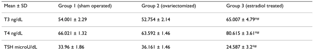
Animals underwent ovariectomy demonstrated decreased serum levels of T3 and T4 with increase of TSH levels when compared to the control and estradiol treated groups. Animals treated with estradiol demonstrated highly statistically significant increased levels of T3, T4 and decrease in TSH levels when compared to both con-trol and ovariectomized groups (table 1).
Light microscopic results
In the sham-operated group, the thyroid gland was found to be composed of thyroid follicles which appeared
gen-erally oval or rounded lined with a single layer of cells. The central follicles appeared smaller in size than periph-eral ones. The central follicles were lined with cuboidal cells with rounded nuclei, while the epithelial lining of the peripheral follicles was formed of flattened or low cuboidal cells. The amount of colloid substance varied from one follicular lumen to another. It showed different staining affinity with PAS. Some follicles contained con-spicuous peripheral vacuoles. The connective tissue cap-sule was thin and the connective tissue septa were hardly identified dividing the gland into numerous small ill-defined lobules (fig. 1a-d).
In the ovariectomized rats, preserved secretory activity of the thyroid gland was noticed, it was mainly characterized by central follicles of a flattened, cuboidal or low pris-matic epithelium surrounding the colloid. Occasionally, enlarged distended follicles with very flat epithelium were noticed. The peripheral follicles were large and lined with flattened epithelium. The colloid appeared filling up the entire lumen in most of the follicles with no or little peripheral vacuolization. The connective tissue around the follicles was more prominent than in sham-operated group (fig. 2a-d).
The thyroid gland of estradiol treated group demonstrated very small follicles in the central region with high colum-nar or pyramidal epithelial lining surrounding colum-narrow lumen. There were areas of disorganized follicles with complete obstruction of their lumina. The peripheral fol-licles were larger. The colloid was faintly stained and dem-onstrated extensive vacuolization. Connective tissue was minimal between the follicles (fig. 3a-d).
Morphometric analysis of light microscopic results
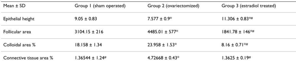
In ovariectomized group, epithelial height was signifi-cantly decreased while the follicular area, colloidal area percent and connective tissue content were significantly increased when compared to sham-operated group.
[image:3.612.52.561.628.714.2]In estradiol treated group, there was marked increase in epithelial height and decreased follicular area and colloi-dal area percent when compared to the other two groups. Connective tissue content was more or less similar to the
Table 1: changes in the mean serum levels of thyroid hormones T3, T4 and TSH in the different groups
Mean ± SD Group 1 (sham operated) Group 2 (ovariectomized) Group 3 (estradiol treated)
T3 ng/dL 54.001 ± 2.29 52.754 ± 2.14 65.007 ± 4.79*#
T4 ng/dL 66.021 ± 1.32 63.592 ± 1.46 80.615 ± 3.61*#
TSH microU/dL 33.96 ± 1.86 36.161 ± 1.46 24.587 ± 3.2*#
sham-operated group. These results were summarized in (table 2).
Electron microscopic results
Electron microscopic study of the thyroid follicles in con-trol sham operated rats showed rounded follicles formed of cuboidal cells with rounded nuclei. The follicles are bounded with a basement membrane and surrounded with capillaries. The cells are rich in rough endoplasmic reticulum and mitochondria in addition to a prominent Golgi apparatus and intracytoplasmic colloid vesicles. The apex of the cells adjacent to the follicular lumen showed apical cytoplasmic vacuolization with the presence of many lysosomes (fig. 4a).
Electron microscopic study of the thyroid follicles in ova-riectomized rats showed thyroid follicles with cuboidal cells with rounded nuclei. Intracytoplasmic colloid vesi-cles were more prominent with well developed Golgi apparatus. In addition to that the apical cytoplasmic vac-uolization and lysosomes could be demonstrated (fig 4b).
Electron microscopic study of the thyroid follicles in ova-riectomized rats treated with estradiol showed increased
activity of thyroid follicular columnar cells with oval nuclei and intracytoplasmic colloid vesicles which were much more prominent with clear apical cytoplasmic vac-uolization (fig. 4c).
Discussion
The end of the reproductive lifespan, in females of many species, is associated with hypoestrogenic state that lead to other physiological changes with increased risk of oste-oporosis and cardiovascular diseases. Estrogen replace-ment therapy was accepted for prevention and treatreplace-ment of human menopausal problems [16]. Unfortunately, recent evidence has connected hormonal treatment with undesirable side effects especially as a carcinogenic stimu-lator of cell proliferation and inducing genetic damage [17].
The main goal of the present study was to elucidate the effects of chronic estradiol treatment on thyroid gland structure and function in ovariectomized rat model, in doses comparable with those used in human therapy.
In the present study, ovariectomized rats demonstrated a decrease in the levels of T3 and T4 with an increase in the level of TSH when compared to animals with intact ova-ries. A recent study by De Araujo et al. [7], demonstrated
[image:4.612.54.296.86.255.2]light microscopic pictures of thyroid gland from sham oper-ated rats
Figure 1
light microscopic pictures of thyroid gland from sham operated rats. (a) Large peripheral thyroid follicles (thick arrows) under the capsule (caps) lined with flattened and cuboidal epithelial lining (thin arrows) filled with colloid (C). The central follicles appear smaller in size (Hx&E × 200). (b) Higher magnification of the central zone from the previ-ous section. The epithelial lining of the follicles is cuboidal (thin arrows) with rounded central or basal nuclei. The folli-cles are variable in size. The colloid contains peripheral vacu-olization (arrow heads) (Hx&E × 400). (c) The central zone of thyroid of sham-operated rats showing variable amount of colloid filling the lumina of the follicles (C) (PAS × 400). (d) There is minimal amount of connective tissue in between the follicles (thin arrows) (Masson trichrome × 400).
[image:4.612.313.553.86.256.2]light microscopic pictures of thyroid gland of group II (ovaric-tomized rat)
Figure 2
similar findings in ovariectomized rats. Also high levels of TSH were recorded in postmenopausal women in several studies [2,18]. While Sosic-Jurjevic et al. [19] reported increased levels of T3 and T4 this difference may be due to a shorter duration of estrogen administration.
In the present study, serum thyroid hormone levels seem to reflect the histological and morphometric changes in thyroid glands. In ovariectomized rats; the follicles were lined with low prismatic, cuboidal or flattened epithelium with significant decrease in epithelial height. The follicu-lar area was increased as well as colloidal area percent.
These findings indicated hypoactivity of the thyroid gland [20].
Estrogen has antifibrotic effects through the inhibition of key profibrotic factors, and through attenuation of fibrob-last differentiation [21]. So the increased connective tissue content shown in this study in ovariectomized rats could be due to decreased estrogen antifibrotic effects as a result of estrogen withdrawal.
In our study electron microscopic examination of the thy-roid gland from ovariectomized rats showed the cuboidal follicular cells with increased amount of intracytoplasmic colloid vesicles. While with estradiol treatment in ovariec-tomized rats, the follicular cells appeared columnar with oval nuclei with much more prominent intracytoplasmic colloid vesicles and clear apical vacuolization.
Zayed et al. [22] also reported decreased follicular colloid with increased intracytoplasmic colloid vesicles due to decreased activity of thyroid gland after ovariectomy with increased intracytoplasmic vesicles stored later as colloid in the follicular lumen which became wider.
Meanwhile, some reports have shown that the effects of estrogens on the thyroid gland may be indirect through the pituitary-thyroid axis [18] or as a consequence of increased serum levels of thyroxin binding globulin and the associated changes in thyroid function [23].
Taken together, the present results demonstrate that the thyroid glands of rats remain susceptible to the influence of ovariectomy and estrogen. The growth stimulatory effect of estrogen on benign and malignant human thy-roid cells support the epidemiological data showing higher prevalence of thyroid carcinomas in females [24].
Although the extrapolation of these results to human should be interpreted with caution, it is highly conceiva-ble that monitoring of thyroid function may be important for patients who are under or going to be under estrogen therapy.
light microscopic pictures of thyroid gland of group III (ovarictomized rat treated with estradiol)
Figure 3
[image:5.612.53.554.601.709.2]light microscopic pictures of thyroid gland of group III (ovarictomized rat treated with estradiol). (a) The peripheral follicles (thick arrows) under the capsule (caps) are relatively larger than the central follicles (Hx&E × 200). (b) Higher magnification of the central zone showing very small follicles. Some follicles have no apparent lumina (thick arrows), while others contain absent or highly vacuolated colloid material (arrow heads) (Hx&E × 400). (c) There is lit-tle amount of the colloid in some follicles (C) while others demonstrate absent colloid (thin arrows) (PAS × 400). (d) There is minimal connective tissue (thin arrows) surrounding the thyroid follicles (Masson trichrome × 400).
Table 2: the statistical analysis of the morphometric data obtained by the image analysis
Mean ± SD Group 1 (sham operated) Group 2 (ovariectomized) Group 3 (estradiol treated)
Epithelial height 9.05 ± 0.83 7.577 ± 0.9* 11.306 ± 0.83*#
Follicular area 3104.15 ± 216 4485.01 ± 577* 1841.78 ± 146*#
Colloidal area % 18.158 ± 1.34 23.958 ± 1.53* 8.16 ± 0.71*#
Connective tissue area % 1.36544 ± 1.24# 4.72668 ± 0.43* 1.3625 ± 0.19#
Conclusion
Decreased amount of estrogen may lead to thyroid hypo-function while estradiol treatment may lead to hyperactiv-ity so it should be used very cautiously in the treatment of postmenopausal symptoms to avoid its undesirable stim-ulatory effect on the thyroid.
Competing interests
The authors declare that they have no competing interests.
Authors' contributions
All authors have made substantial contributions to design of the work, in addition to analysis and interpretation of data; and have been involved in drafting the article and revising it critically for important intellectual content; and have given final approval of the version to be published.
Acknowledgements
Thanks for Prof. Hanaa abdelkader Professor and Chair of histology depart-ment Kasr-El-Aini Faculty of Medicine Cairo University Egypt for her
encouragements and support during preparation of this manuscript Thanks for Prof. Hend Shafik Professor of histology in Kasr-El-Aini Faculty of Med-icine Cairo University Egypt for her great effort in critically revising this manuscript and making valuable notes to improve it
Thanks for researchers and technicians in the animal house of Kasr-El-Aini Faculty of Medicine, Cairo University, Egypt who helped us greatly in mak-ing the laboratory work
As regard funding it was from faculties of medicine in both Cairo and Fay-oum universities to get the materials and to use labs facilities
References
1. Hendrix SL: Long-term use of hormone therapy for urogenital complaints: is there a role? Med Clin North Am 2003, 87:1029-37. 2. Schindler AE: Thyroid function and postmenopause. Gynecol
Endocrinol 2003, 17:79-85.
3. Ron E, Kleinerman RA, Boice JD, LiVolsi VA, Flannery JT, Fraumeni JF:
A population-based case-control study of thyroid cancer. J Natl Cancer Ins 1987, 79(1):1-12.
4. Hollowell JG, Staehling NW, Flanders WD, Hannon WH, Gunter EW, Spencer CA, et al.: Serum TSH, T (4), and thyroid antibod-ies in the United States population (1988 to 1994): National electron micrograph of thyroid follicular cells in (a) sham operated rats, (b) ovariectomized rats and (c) ovariectomized rats treated with estradiol
Figure 4
Publish with BioMed Central and every scientist can read your work free of charge "BioMed Central will be the most significant development for disseminating the results of biomedical researc h in our lifetime."
Sir Paul Nurse, Cancer Research UK
Your research papers will be:
available free of charge to the entire biomedical community
peer reviewed and published immediately upon acceptance
cited in PubMed and archived on PubMed Central
yours — you keep the copyright
Submit your manuscript here:
http://www.biomedcentral.com/info/publishing_adv.asp
BioMedcentral
Health and Nutrition Examination Survey (NHANES III). J Clin Endocrinol Metab 2002, 87:489-99.
5. Banu SK, Arosh JA, Govindarajulu P, Aruldhas MM: Testosterone and estradiol differentially regulate thyroid growth in Wistar rats from immature to adult age. Endocr Res 2001, 27:447-63. 6. Banu SK, Govindarajulu P, Aruldhas MM: Developmental profiles
of TSH, sex steroids, and their receptors in the thyroid and their relevance to thyroid growth in immature rats. Steroids 2002, 67:137-44.
7. De Araujo LF, Soares JM, Simoes RS, Calio PL, Oliveira-Filho RM, Simoes J, Haidar MA, Baracat EC: Effect of conjugated equine estrogens and tamoxifen administration on thyroid gland histomorphology of the rat. Clinics 2006, 61(4):321-6.
8. Lima LP, Barros IA, Lisboa PC, Arajo RL, Silva AC, Rosenthal D, Fer-reira AC, Carvalho DP: Estrogen effects on thyroid iodide uptake and thyroperoxidase activity in normal and ovariect-omized rats. Steroids 2006, 71(8):653-9.
9. Abdalla HI, Beastall G, Fletcher D, Hawthorn JS, Smith J, Mc Hart KD:
Sex steroid replacement in post-menopausal women: effects on thyroid hormone status. Maturitas 1987, 9:49-54.
10. Contreras I, Parra D: Estrogen replacement therapy and the prevention of coronary heart disease in postmenopausal women. Am J Health Syst Pharm 2000, 57:1963-1968.
11. Marqusee E, Braverman LE, Lawrence JE, Carroll JS, Seely EW: The Effect of Droloxifene and Estrogen on Thyroid Function in Postmenopausal Women. J Clin Endocrinol Metab 2000,
85:4407-4410.
12. Seko K, Kagami H, Senga K, Ozeki K, Mizutani H, Ueda M: Effects of ovariectomy and estrogen replacement on rat oral mucosa.
Maturitas 2005, 50(1):44-51.
13. Sosic-Jurjevic B, Filipovic B, Milosevic V, Nestorovic N, Manojlovic-Stojanoski M, Brkic B, Sekulic M: Chronic estradiol exposure modulates thyroid structure and decreases T4 and T3 serum levels in middle-aged female rats. Horm Res 2005, 63(1):48-54. 14. Russell WMS, Burch RL: The Principles of Humane Experimen-tal Technique. London: Methuen & Co. Ltd 1959. (Reissued: 1992, Universities Federation for Animal Welfare, Herts, England) .
15. Chopra IJ, Solomon DH, Gnho RS: A radioimmunoassay of thy-roxin. J Clin Endocrinol 1971, 33:865-7.
16. Davidson MR: Pharmacotherapeutics for osteoporosis pre-vention and treatment. J Midwifery Womens Health 2003,
48(1):39-52.
17. Liehr J: Is Estradiol a Genotoxic Mutagenic Carcinogen?
Endocr Rev 2000, 21:40.
18. Abech DD, Moratelli HB, Leite SC, Oliveira MC: Effects of estro-gen replacement therapy on pituitary size, prolactin and thy-roid-stimulating hormone concentrations in menopausal women. Gynecol Endocrinol 2005, 21(4):223-6.
19. Sosic-Jurjevic B, Filipovic B, Milosevic V, Nestorovic N, Negic N, Sekulic M: Effects of ovariectomy and chronic estradiol admin-istration on pituitary-thyroid axis in adult rats. Life Sci 2006,
79(9):890-7.
20. Rao-Rupanagudi S, Heywood R, Gopinath C: Age-related changes in thyroid structure and function in Sprague-Dawley rats. Vet Pathol 1992, 29:278.
21. Lekgabe ED, Royce SG, Hewitson TD, Tang ML, Zhao C, Moore XL, Tregear GW, Bathgate RA, Du XJ, Samuel CS: The Effects of Relaxin and Estrogen Deficiency on Collagen Deposition and Hypertrophy of Nonreproductive Organs. Endocrinology 2006,
147(12):5575-5583.
22. Zayed I, Esch E, Mcconnell RF: Systemic and histopathologic changes in Beagle dogs after chronic daily oral administra-tion of synthetic (ethinylestradiol) or natural (estradiol) estrogens, with special reference to the kidney and thyroid.
Toxicol Pathol 1998, 26:730-41.
23. Ain KB, Mori Y, Refetoff S: Reduced clearance rate of thyrox-inebinding globulin (TBG) with increased sialylation: a mech-anism for estrogen-induced elevation of serum TBG concentration. J Clin Endocrinol 1987, 65:689-96.
24. Henderson BE, Ross RK, Pike MC, Casagrande JT: Endogenous hor-mones as a major factor in human cancer. Cancer Res 1982,