R E S E A R C H A R T I C L E
Open Access
erbB3 recruitment of insulin receptor substrate
1 modulates insulin-like growth factor receptor
signalling in oestrogen receptor-positive breast
cancer cell lines
Janice M Knowlden
1, Julia MW Gee
1, Denise Barrow
1, John F Robertson
2, Ian O Ellis
3, Robert I Nicholson
1and
Iain R Hutcheson
4*Abstract
Introduction:Recently we reported that insulin receptor substrate 1 (IRS-1), classically an adaptor protein for the insulin-like growth factor type I receptor (IGF-IR), associates with the epidermal growth factor receptor in oestrogen receptor (ER)-positive (ER+) tamoxifen-resistant breast cancer cells. In this study, we examined whether IRS-1 also associates with another erbB receptor family member, erbB3, and what impact this might have on IGF-IR signalling in three ER+ breast cancer cell lines.
Methods:Immunoprecipitation and Western blot analysis were utilised to examine the potential association between erbB3 and IRS-1 in MCF-7, T47D and BT-474 cells in the absence and presence of the erbB3/4 ligand heregulinb1 (HRGb1). Subsequently, the impact of a selective IGF-IR/IR inhibitor 4-anilino-5-bromo-2-[4-(2-hydroxy-3-(N, N-dimethylamino)propoxy)anilino]pyrimidine on this association and HRGb1 signalling was assessed in these
cell lines. Immunohistochemical analysis of a small cohort of ER+ breast cancer patient samples was also performed to determine the potential clinical relevance of this novel interaction.
Results:Immunoprecipitation and Western blot analysis revealed an interaction between erbB3 and IRS-1 in MCF-7, T47D and BT-474 cells, with HRGb1 significantly enhancing this recruitment and promoting IRS-1
phosphorylation at Y612. IRS-1 participates in erbB3 signalling in MCF-7 and T47D cells as IRS-1 knockdown impaired HRGb1 signalling. Importantly, recruitment of IRS-1 by erbB3 reduced IRS-1 association with IGF-IR in MCF-7 and T47D cells, whilst blockade of IGF-IR-enhanced erbB3-IRS-1 interaction and sensitised both cell lines to HRGb1, allowing HRGb1 to override IGF-IR blockade. Consequently, suppression of IRS-1 signalling enhanced the effects of IGF-IR inhibition in these cells. This novel interaction may have clinical relevance, as
immunohistochemical analysis of a small ER+ breast tumour series revealed significant positive correlations between phosphorylated IRS-1 Y612 expression and total erbB3, phosphorylated Akt and Ki-67 expression. Conclusions:IRS-1 can be recruited to IGF-IR and erbB3 in ER+ breast cancer cells, and this provides an adaptive resistance mechanism when these receptors are targeted individually. Consequently, cotargeting IGF-IR and either erbB3 or IRS-1 should prove to be a more effective strategy for the treatment of ER+ breast cancer.
Keywords:breast cancer, erbB3, IRS-1, IGF-IR, resistance
* Correspondence: hutchesonir@cf.ac.uk 4
Department of Pharmacology, Radiology & Oncology, Cardiff University, School of Medicine, Heath Park, Cardiff, CF14 4XN, UK
Full list of author information is available at the end of the article
Introduction
There is strong experimental and clinical evidence impli-cating the insulin-like growth factor type I receptor (IGF-IR) in breast cancer development and growth [1-3]. The IGF-IR, which belongs to a family of receptor tyrosine kinases that includes the insulin receptor (IR), has been found to be expressed in a high percentage of breast tumours, where its expression is positively correlated with oestrogen receptor (ER) status and is usually coex-pressed with markers of a better overall prognosis [2,4-6]. Expression of the IGF-IR has also been demon-strated in the majority of ER+ breast cancer cell lines [7,8]. Indeed, in MCF-7 cells, IGF-IR has been shown not only to be a key receptor in mediating hormone-sensitive growth but also to engage in significant cross-talk with ER [9,10]. Importantly, this leads to synergistic interac-tions between ER and IGF-IR signalling to promote effi-cient growth responses [2].
However, conversely, increased expression and activa-tion of IGF-IR and its associated downstream signalling components have also been reported in some clinical breast cancers and have been linked to disease progres-sion and recurrence [11,12]. On the basis of these data, IGF-IR has been identified as a potential therapeutic tar-get for the treatment of breast cancer [13]. Activation of the IGF-IR promotes binding of insulin receptor sub-strate (IRS) members, a family of structurally related adaptor molecules which have classically been identified as key signalling intermediates of the IR and IGF-IR [14]. Binding results in phosphorylation of their carboxyl ter-mini at multiple tyrosine residues, and these phosphotyr-osine residues provide docking sites for the recruitment of key signalling pathways, such as the mitogen-activated protein kinase (MAPK)/extracellular signal-regulated kinase 1/2 (ERK1/2) and phosphatidylinositol 3-kinase (PI3K) pathways [15]. These signalling cascades can med-iate mechanisms underlying tumour growth and progres-sion, implicating a potential role for IRS members in oncogenesis [1,15-18]. Indeed, IRS-1 has been reported to be overexpressed and constitutively phosphorylated in breast tumours [18,19], and high expression of this adap-tor protein has been associated with lymph node metas-tases and poor patient prognosis [11,20,21]. Furthermore, IRS-1 and IRS-2 have been implicated in the regulation of proliferation, survival and metastatic potential in a range of breast cancer cell lines [17].
However, there is now increasing evidence that IRS-1 is not restricted to binding to IR/IGF-IR but is also capable of associating with a variety of other signalling-related pro-teins [17]. One such protein is the epidermal growth factor receptor (EGFR), a member of the erbB receptor tyrosine kinase family also comprising erbB2, erbB3 and erbB4 and which has been shown to play a central role in driving
bothde novoand acquired anti-hormone-resistant growth and invasion in breast cancer [22-25]. Evidence of an EGFR-IRS-1 interaction arises from reports by Fujioka and colleagues [26,27], who reported that the phosphorylated NPXY motifs in activated IR and IGF-IR to which IRS molecules bind are also present in the C-terminus region of activated EGFR and were indispensable for EGF-induced IRS-1 tyrosine phosphorylation in EGFR-trans-fected CHO cells [27]. Furthermore, a potential interaction between EGFR and IRS-1 has been predicted on the basis of the binding of peptides, representing the physical sites of EGFR tyrosine phosphorylation, to protein microarrays comprising all Src homology 2 and phosphotyrosine bind-ing domains encoded in the human genome [28]. Recently, we provided strong evidence that IRS-1 can function as a key signalling intermediate for EGFR, a receptor that drives the growth of a tamoxifen-resistant MCF-7 breast cancer cell line [29]. In these cells, we showed that IRS-1 physically complexes with EGFR and is preferentially phosphorylated on Y896, a Grb2-binding/MAPK recruit-ment site [15]. Moreover, EGFR was the dominant recrui-ter of IRS-1, which thus served to limit the availability of IRS-1 to associate with IGF-IR in these cells and, as a result, suppressed IGF-IR signalling via this receptor.
cells and demonstrate that this novel interaction can serve to reduce the association between IRS-1 and IGF-IR and inhibits signalling via this receptor. We show in turn that suppression of IGF-IR by the use of a tyrosine kinase inhi-bitor and siRNA technology can promote erbB3 down-stream signalling by reinforcement of erbB3 interplay with IRS-1. This provides a potential novel resistance signal, which, when targeted, may generate more effective inhibi-tion of cell growth compared to IGF-IR treatment alone.
Materials and methods Cell culture
MCF-7 cells (a gift from AstraZeneca Pharmaceuticals, Cheshire, UK) and T47D cells (American Type Culture Collection (ATCC), Manassas, VA, USA), which are both nonamplified erbB2 breast cancer cell lines, were grown in RPMI medium containing 5% FCS and glutamine (4 mM). Both cell lines were maintained at 37°C in a humidified 5% CO2atmosphere. BT-474 over amplified erbB2 breast
can-cer cells (ATCC) were grown in RPMI medium containing 10% FCS and glutamine (4 mM).
Experimental procedures
The cell lines were grown to 70% confluence prior to transfer into phenol red/steroid-and serum growth fac-tor-free dendritic cell conditioned medium (Biosynergy Europe, Cambridge, UK) for 24 hours followed by expo-sure for up to 20 minutes to either 0.1 to 10 ng/ml HRGb1 or 10 ng/ml IGF-I in 10 mM acetic acid/0.1% BSA or appropriate vehicle control. To examine the effects of pharmacological blockade of IGF-IR, cells were incubated in phenol red-free (white) RPMI medium supplemented with 5% FCS and either the IGF-IR/IR tyrosine kinase inhibitor 4-anilino-5-bromo-2-[4-(2-hydroxy-3-(N,N -dimethylamino)propoxy)anilino]pyrimi-dine (ABDP) (1μM in dimethyl sulphoxide, AstraZeneca, Macclesfield, UK) [40] or appropriate vehicle control for 1 to 2 days. All experiments were performed at least three times. Cells were then lysed to measure protein expression.
siRNA studies
Dharmacon SMARTpool siRNA Design specific for IRS-1 (IRS si), erbB3 (3 si) or IGF-IR (IGF si; all 20 mM, Dhar-macon RNAi Technologies, Lafayette, CO, USA) were mixed with DharmaFECT 1 transfection reagent (lipid; Dharmacon RNAi Technologies) at a ratio of 10μl of siRNA to 1μl of lipid and incubated at room temperature for 20 minutes. The mix was added to the cells, which were maintained in white RPMI medium containing 5% FCS to give a final siRNA concentration of 100 nM per dish. Control experiments consisted of transfection with the ON-TARGETplus Nontargeting siRNA control pool (100 nM; Dharmacon RNAi Technologies), medium only
(nontransfected cells) or lipid. All experimental arms were set up in duplicate. Cells were incubated in growth medium containing either IRS si, 3 si, IGF si or control (C si) (100 nM for each) for 4 days prior to treatment with either 10 ng/ml HRGb1 or vehicle alone for 5 min-utes. To examine the effect of IRS-1 knockdown and IGF-IR blockade, cells were incubated in medium con-taining either 100 nM C si, 100 nM IRS si, 1μM ABDP or a combination of these treatments prior to a 5-minute incubation with HRGb1 (10 ng/ml). The cells were then lysed, and protein extracts were examined by Western blot analysis.
Immunoprecipitation and Western blot analysis
Fresh cell lysates containing 500μg of protein were immu-noprecipitated using 1μg of specific antibody as described previously [24]. Protein samples from either immunopreci-pitation or total cell lysates (20 to 50μg) were separated on a 7.5% polyacrylamide gel and then transblotted onto nitrocellulose membrane as described previously [24]. The antibodies used were directed against total EGFR (SC-03), total erbB2 (SC-284), total erbB3 (SC-285), total IGF-IR (SC-712), total IRS-1 (SC-7200; Insight Biotechnology Ltd, Wembley, UK), total and phosphorylated Akt YS473, ERK1/2 and phosphorylated c-erbB3 Y1289 (Cell Signaling Technology, Hitchin, UK), phosphorylated EGFR (Y1068), phosphorylated IRS-1 (Y612 and Y896; BioSource Interna-tional, Camarillo, CA, USA), b-actin (Sigma-Aldrich, Dorset, UK) and specific phosphorylated IGF-IR Y1316 (a kind gift from AstraZeneca, Macclesfield, UK). The Wes-tern blots were then scanned by densitometry to provide data for semiquantification. Each experiment was per-formed at least three times with representative gels shown in figures.
Cell proliferation studies
Cells were seeded at 40, 000 cells per well overnight in phenol red-free RPMI medium supplemented with 5% FCS and then incubated in fresh medium containing 0.1 to 1 μM ABDP, 10 ng/ml HRGb1, vehicle control or a combination of these agents for 4 days. Cell population growth was evaluated by means of trypsin dispersion of the cell monolayers, and cell number was measured using a COULTER COUNTER (Beckman Coulter (UK) Ltd, High Wycombe, UK). All experiments were per-formed in triplicate.
Clinical series
paraffin. No patient had previously received any form of adjuvant endocrinological or cytotoxic therapy. The use of these samples for research purposes, without the requirement of further patient informed consent, was approved by Nottingham Research Ethics Committee 2 under the title‘Development of a molecular genetics clas-sification of breast cancer’(C2020313).
Immunocytochemical assays
Immunocytochemical assays for phosphorylated Akt, Ki-67 and total erbB3 and specific phosphorylated IGF-IR Y1316 were performed on 50 primary ER+ breast tumours as previously described [6,30,41] and the clini-copathological parameters for the clinical set of these tumours are as shown (Additional file 1, Table S1). For the detection of phosphorylated IRS-1 Y612, paraffin wax sections from each tumour sample were dewaxed using xylene treatment and then rehydrated through graded ethanols to PBS. Endogenous peroxidases were destroyed by immersing the sections in 3% hydrogen peroxide pre-pared in methanol for 5 minutes, followed by rinsing with distilled water for 5 minutes. Antigen retrieval was achieved by pressure cooking the slides in 0.01 M sodium citrate buffer, pH 6.0, for 4 minutes. Slides were then immersed in slowly running tap water for 10 minutes before being transferred to PBS for 5 minutes. Sections were blocked in 1% l BSA for 5 minutes prior to incuba-tion overnight at 37°C in anti-phosphorylated IRS-1 Y612 rabbit primary antibody diluted 1:50 in PBS. Sections were washed for 3 minutes in PBS, washed twice for 5 minutes in 0.02% PBS-Tween 20 and then incubated for 2 hours at room temperature in a peroxidase-labelled polymer secondary antibody EnVision Kit (Dako Ltd, Ely, UK). Slides were then washed for 3 minutes in PBS, washed twice for 5 minutes in 0.02% PBS-Tween 20 and incubated for 10 minutes at room temperature in EnVi-sion DAB chromogen solution (diaminobenzidine; Dako Ltd). Slides were then rinsed twice for 2 minutes in distilled water, incubated in 0.5% methyl green for 25 seconds as a counterstain, rinsed in distilled water and allowed to air-dry before DPX mountant (a mixture of distyrene, a plasticizer and xylene) was applied. Com-bined cytoplasmic and plasma membrane staining inten-sity and percentage positivity were assessed by HScore analysis as described previously [41]. Expression of phos-phorylated membrane and cytoplasmic IRS-1 Y612 has previously been verified by immunocytochemistry in ER+ breast cancer cell lines [29].
Statistics
For immunocytochemical analysis of clinical material, Hscores were compared using Spearman’s rank-correlation and Mann-WhitneyUtests for nonparametric data. For experimental growth studies, overall differences between
the control and treatment groups were determined by ana-lysis of variance withpost hoc t-tests with the Bonferroni adjustment factor. A two-sidedt-test was performed on the densitometry values obtained following Western blot analysis. Differences were considered significant at the P≤ 0.05 level.
Results
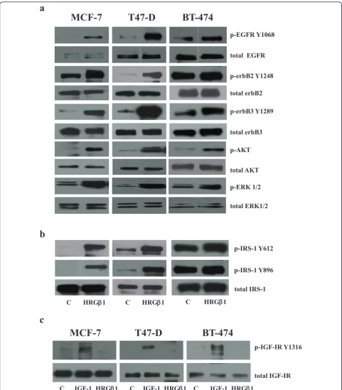
Heregulinb1 promotes phosphorylation of insulin receptor substrate 1 in MCF-7, T47D and BT-474 oestrogen receptor-positive breast cancer cells
Western blot analysis demonstrated that 5-minute HRGb1 stimulation promoted EGFR, erbB3, erbB2 and EGFR phosphorylation and activation of the downstream signal-ling components Akt and ERK1/2 in MCF-7, T47D and BT-474 breast cancer cells, as shown in Figure 1a. Inter-estingly, HRGb1 also promoted phosphorylation of IRS-1 at Y612 and at Y896 in all cell lines, but less dramatically in the BT-474 cells (Figure 1b). Treatment of HRGb1 had no effect on either specific IGF-IR Y1316 phosphorylation or total IGF-IR protein expression levels in these cell lines, whilst, as expected, 10 ng/ml IGF-I stimulation promoted IGF-IR phosphorylation (Figure 1c).
Insulin receptor substrate 1 associates with erbB receptors in MCF-7, T47D and BT-474 cells
T47-D
BT-474
MCF-7
a
b
p-AKT
total AKT p-erbB2 Y1248
total erbB2
p-erbB3 Y1289
total erbB3
p-ERK 1/2
total ERK1/2
p-IRS-1 Y612
total IRS-1 p-IRS-1 Y896 p-EGFR Y1068
total EGFR
c
C HRGEE1 C HRGE1 C HRGE1
C IGF-1 HRGE1
p-IGF-IR Y1316
C IGF-1 HRGE1 C IGF-1 HRGE1
total IGF-IR
T47-D
BT-474
[image:5.595.57.539.87.637.2]MCF-7
IRS-1 C HRGEE1
IRS-1
erbB3 C HRGE1 IP: erbB3 erbB3
T47D
IRS-1 erbB3 IP: erbB3
IP: IRS-1
BT474
IRS-1 erbB3
C HRGE1
C HRGE1 EGFR erbB2
EGFR erbB2 IP: IRS-1
b
MCF-7
a
erbB3 IRS-1
C HRGE1 C HRGE1 IRS-1 erbB3
C HRGE1 C HRGE
IP: IgG IP: IRS-1
IP: erbB3
0 1 2 0 20 mins (HRGE1) IP: IgG IP: erbB3
IRS-1 erbB2
C HRGE1 EGFR IP: total IRS-1
IRS-1 erbB3
C HRGE1
total IGF-IR total IRS-1 IP-total IRS-1
IP-total IRS-1
MCF-7
T47D
C HRGE1
IP-total IRS-1
C HRGE1
BT474
c
C HRGβ1
MCF-7
T47D
*
*
d
C HRGβ1
0 20 40 60 80 100 120
0 20 40 60 80 100 120
IG
F
-1R/IRS-1 association
IG
F
[image:6.595.56.544.87.639.2]-1R/IRS-1 association
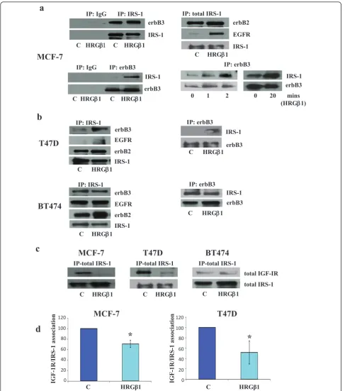
HRGb1 treatment (Figure 2b). In addition, there was no decrease in association between IGF-IR and IRS-1 follow-ing HRGb1 stimulation in these cells (Figure 2c) and, as a consequence, BT-474 cells were excluded from further studies.
erbB3 siRNA knockdown reduces heregulinb1-induced insulin receptor substrate 1 phosphorylation in MCF-7 and T47D cells
Western blot analysis demonstrated that both basal and HRGb1-primed IRS-1 Y612 and Y896 phosphorylation levels were markedly reduced following incubation of MCF-7 and T47D breast cancer cells with siRNA-targeting erbB3 protein expression. The reduction in IRS-1 Y612 and Y896 phosphorylation was found to be statistically sig-nificant following densitometric analysis (P≤0.01 (n= 3) andP≤0.01 (n= 3), respectively, for MCF-7;P≤ 0.01 (n= 3) andP≤0.001 (n= 3), respectively, for T47D cells) (Figures 3b and 3c). Basal and HRGb1-primed Akt and ERK1/2 phosphorylation levels were similarly inhibited by the erbB3 siRNA treatment (Figure 3a), whilst total Akt, ERK1/2 andb-actin protein levels remained constant fol-lowing total erbB3 downregulation in both cell lines.
Insulin receptor substrate 1 siRNA knockdown reduces heregulinb1-primed erbB3 signalling
Interestingly, the ability of HRGb1 to prime Akt phos-phorylation was reduced substantially following incubation of MCF-7 and T47D cells with siRNA-targeting IRS-1 pro-tein expression as demonstrated by Western blot analysis (Figure 4a). Moreover, this decrease in Akt phosphoryla-tion was statistically significant following densitometric analysis (P≤0.01 (n= 3) andP≤0.001 (n= 3) for MCF-7 and T47D cells, respectively) (Figures 4b and 4c). How-ever, there was no obvious reduction in HRGb1-induced ERK1/2 phosphorylation in these cells following IRS-1 protein downregulation. Total Akt, ERK1/2 andb-actin protein levels remained constant.
Insulin-like growth factor type I receptor inhibition facilitates insulin receptor substrate 1 association with erbB3 and promotes heregulinb1-induced
phosphorylation of insulin receptor substrate 1 Y612 in MCF-7 and T47D cells
We next examined whether recruitment of IRS-1 by erbB3 can provide a resistance mechanism to IGF-IR-targeted therapy. A 24-hour treatment with the IGF-IR inhibitor ABDP enhanced the sensitivity of MCF-7 and T47D cells to HRGb1, with increased phosphorylation of IRS-1 Y612, IRS-1 Y896, Akt and ERK1/2 apparent at lower concentra-tions of this ligand (Figure 5a). These increases were not due to increased IGF-IR activity, as specific phosphory-lated IGF-IR levels were completely inhibited following ABDP treatment. Densitometric analysis showed that the
increase in Akt phosphorylation was statistically significant for both MCF-7 cells (P≤0.05 (n= 3)) and T47D cells (P≤0.05 (n= 3)) at the 1 ng/ml HRGb1 concentration (Figures 5b and 5c). The increased ERK1/2 phosphoryla-tion observed in MCF-7 cells failed to reach statistical sig-nificance (P = 0.06 (n = 3)) (Figure 5b), although significance was reached for the T47D cells (P≤0.05 (n= 3)) (Figure 5c). Total protein levels remained constant fol-lowing IGF-IR inhibition in both cell lines. These results were verified further in both cell lines using siRNA to spe-cifically target IGF-IR (Additional file 2, Figure S1). Immu-noprecipitation and Western blot analysis also revealed that, following treatment of MCF-7 and T47D cells with ABDP, there was a reduced association between IRS-1 and IGF-IR and an enhanced association between IRS-1 and erbB3, as shown in Figure 5d.
siRNA knockdown of insulin receptor substrate 1 reverses the increased sensitivity to heregulinb1 observed following insulin-like growth factor type I receptor blockade in MCF-7 and T47D cells
The increase in HRGb1-induced Akt phosphorylation observed following treatment of MCF-7 and T47D breast cancer cells with the IGF-IR/IR inhibitor ABDP for 24 hours was effectively blocked using siRNA speci-fic to IRS-1 (Figure 6a). However, IRS-1 knockdown had only a small effect on HRGb1-induced ERK1/2 phos-phorylation, and it had no effect on total IGF-IR, Akt, ERK1/2 and b-actin protein levels in both these cell lines.
Growth inhibition by ABDP can be overcome by heregulinb1 in MCF-7 and T47D cells
The growth of MCF-7 and T47D breast cancer cells was significantly increased in the presence of HRGb1 (P ≤
0.001 (n= 3) andP≤0.01 (n= 3), respectively) and signifi-cantly reduced by approximately 50% in the presence of either 0.1μM ABDP in MCF-7 cells (P≤0.001 (n= 3)) or 0.75μM ABDP in T47D cells (P≤0.001 (n= 3)) (Figures 6b and 6c). This inhibition in cell growth observed with ABDP was potently and significantly overridden by treat-ment of MCF-7 and T47D cells with 10 ng/ml HRGb1 (P≤0.001 (n= 3) andP≤0.01 (n= 3), respectively) as shown in Figures 6b and 6c.
Phosphorylated insulin receptor substrate 1 Y612 expression positively correlates with erbB3 and insulin-like growth factor type I receptor expression in oestrogen receptor-positive clinical breast cancer material
MCF-7
T47-D
p-AKT
EE- actin
a
C si C si 3 si 3 si + + HRGβ1 HRGβ1 C si C si 3 si 3 si
+ + HRGβ1 HRGβ1
MCF-7
T47D
IRS-1 Y612
IRS-1 Y896
C si 3 si C si 3 si
C si 3 si C si 3 si
*
*
b
c
*
*
IRS-1 Y612
IRS-1 Y896
arbitrary optical densitom
et
ry
units
arbitrary optical densitom
et
ry
units
total erbB3 p-erbB3 Y1289
total IRS-1 p-IRS-1 Y612
p-IRS-1 Y896
total AKT
p-ERK1/2
[image:8.595.58.538.88.635.2]total ERK1/2
MCF-7
p-IRS-1 Y612
p-IRS-1 Y896
T47-D
C si C si IRS si IRS si + + HRGβ1 HRGβ1
total IRS-1
p-AKT
total AKT
p-ERK1/2
total ERK1/2
EE- actin
a
b
C si C si IRS si IRS si + + HRGβ1 HRGβ1
C si IRSsi Csi C si IRS si C si IRS si
MCF-7
C si IRS si C si IRS si
T47D
p-AKT
p-AKT
p-ERK1/2
p-ERK1/2
*
*
c
ar
bitrar
y optical
de
nsitom
et
ry
units
ar
bitrar
y optical
de
nsitom
et
ry
[image:9.595.58.545.87.640.2]units
T47D
*
*
p-AKT p-ERK1/2
*
*
C ABDP IP: IRS-1
IGF-IR erbB3 IRS-1 IP: IRS-1
C ABDP MCF-7 T47-D
d
c
MCF-7
b
p-ERK1/2 p-AKT
*
HRGβ1 ABDP HRGβ1 ABDP + HRGβ1 + HRGβ1
a
arbitrary optical densitom
et
ry
units
arbitrary optical densitom
et
ry
units
HRGβ1 ABDP HRGβ1 ABDP + HRGβ1 + HRGβ1
MCF-7
T47-D
total IRS-1 p-IRS-1 Y612
p-AKT
total AKT p-ERK1/2 total ERK1/2 p-erbB3 Y1289 total erbB3
HRGEE1 (ng/ml) p-IGF-IR Y1316 total IGF-IR
0 0.1 1 10 0 0.1 1 10 -ABDP +ABDP
0 0.1 1 10 0 0.1 1 10 -ABDP +ABDP
[image:10.595.59.539.87.641.2]p-IRS-1 Y896
Figure 5Western blot analysis.(a)Total and phosphorylated insulin-like growth factor type I receptor (IGF-IR), insulin receptor substrate 1 (IRS-1), erbB3, Akt and extracellular-signal regulated kinase 1/2 (ERK1/2) protein expression following incubation of MCF-7 and T47D cells in medium containing either the specific IGF-IR/IR tyrosine kinase inhibitor 4-anilino-5-bromo-2-[4-(2-hydroxy-3-(N,N-dimethylamino)propoxy)anilino] pyrimidine (ABDP) (1μM) or appropriate vehicle control for 24 hours and subsequently challenged with increasing concentrations of heregulin
b1 (HRGb1) (0.1 to 10 ng/ml) or vehicle control for 5 minutes. Densitometric analysis of phosphorylated Akt and ERK1/2 protein levels in(b)
C si + HRGβ1
C si + ABDP
+ HRGβ1
IRS si + ABDP
+ HRGβ1
total IGF-IR
p-IRS-1 Y612
p-AKT
total AKT
p-ERK1/2
total ERK1/2 total IRS-1 p-IGF-IR Y1316
MCF-7
IRS si + HRGβ1
β-actin
T47-D
C si + HRG
C si + ABDP
+ HRGβ1
IRS si + ABDP
+ HRGβ1 IRS si
+ HRG
a
b
%
gr
ow
th at
day 4
*
*
†
%
gr
ow
th
at
day
4
*
*
†
c
0 20 40 60 80 100 120 140 160
control HRGβ1 ABDP + HRGβ1 ABDP
control HRGβ1 ABDP + HRGβ1 ABDP
[image:11.595.57.539.88.640.2]0 50 100 150 200 250
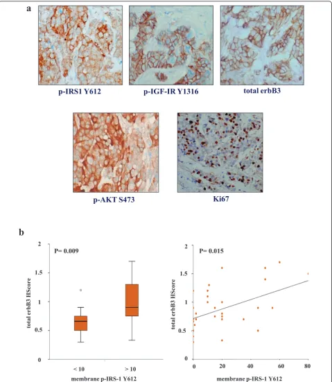
cytoplasmic staining was also observed (Figure 7a). Immunocytochemical assays for phosphorylated Akt, specific phosphorylated IGF-IR (Y1316), nuclear Ki-67 and total erbB3 were also performed as previously described on the 50 primary ER+ breast tumours [6,30,41], and the clinicopathological parameters for the clinical set of these tumours are given in Additional file 1, Table S1. A Mann-WhitneyU test was applied to these samples to determine the relationships between phosphorylated membrane IRS-1 Y612 immunostaining (using a median HScore of 10 as a cutoff for positivity) and total membrane and cytoplasmic erbB3, membrane phosphorylated IGF-IR Y1316, membrane and cytoplas-mic phosphorylated Akt and percentage of nuclear Ki-67 immunostaining HScore values. Interestingly, total mem-brane and cytoplasmic erbB3 expression was significantly higher in phosphorylated membrane IRS-1 Y612-positive tumours than in phosphorylated membrane IRS-1 Y612-negative tumours (P = 0.009 (n = 33)) (Figure 7b). Further analysis also revealed that phosphorylated mem-brane and cytoplasmic IRS-1 Y612-positive tumours (median HScore cutoff of 61) expressed higher levels of phosphorylated membrane IGF-IR Y1316 (P= 0.011 (n= 50)), phosphorylated membrane and cytoplasmic Akt (P≤0.001 (n= 50)) and nuclear Ki-67 (P = 0.022 (n= 40)) immunostaining than did phosphorylated membrane and cytoplasmic IRS-1 Y612-negative tumours (not shown). In addition, Spearman’s rank-correlation test was applied to this group of ER+ patients. This analysis confirmed the Mann-WhitneyUtest findings revealing that phosphorylated membrane IRS-1 Y612 immunos-taining positively correlated with immunosimmunos-taining for total membrane and cytoplasmic erbB3 (P= 0.015 (n= 33)) (Figure 7b). Significant positive correlations between phosphorylated membrane and cytoplasmic IRS-1 Y612 and phosphorylated membrane IGF-IR (P= 0.025 (n= 50)), phosphorylated membrane and cytoplasmic Akt (P= 0.001 (n= 50)) and nuclear Ki-67 (P= 0.007 (n= 40)) were also observed (not shown).
Discussion
IRS-1 is not restricted to binding to IR/IGF-IR but also has the capacity to interact with a variety of other proteins [21]. Recently, we reported that IRS-1 can interact with EGFR, resulting in loss of recruitment of IRS-1 by IGF-IR and reducing signalling via this receptor in an ER+, tamox-ifen-resistant MCF-7 breast cancer cell line [29]. In the present study, we examined whether IRS-1 can associate with other erbB family members, notably erbB3, and whether this has a direct impact on IGF-IR signalling in three ER+ breast cancer cell lines (MCF-7, T47D and BT-474) previously shown to express IRS-1 protein [42-44].
Initial characterisation of these cell lines showed that EGFR, erbB2, erbB3 and associated downstream signalling
elements MAPK and Akt were activated following HRGb1 treatment, with this ligand having a more potent effect on phosphorylation levels in MCF-7 and T47D cells that on BT-474 cells. Interestingly, HRGb1 treatment also increased levels of IRS-1 phosphorylation at both the Y612 and Y896 residues, with this effect being greater in MCF-7 and T47D cells than in the BT-474 cell line. The more modest effect of HRGb1 priming of such activity in BT-474 cells most likely reflects the fact that these cells constitutively overexpress erbB2 and consequently have higher basal phosphorylation levels of all these signalling elements. As such, any increase in activity is harder to dis-tinguish compared to the erbB2 low-expressing MCF-7 and T47D cell lines [45]. Using immunoprecipitation and Western blot analysis, we confirmed that HRGb1-induced phosphorylation of IRS-1 was a result of IRS-1’s complex-ing with erbB3/EGFR and erbB3/erbB2 heterodimers in both MCF-7 and T47D cells. The ability of erbB3 to het-erodimerise with both EGFR and erbB2 in response to HRGb1 stimulation explains the increased phosphoryla-tion of IRS-1 at Y896 in these two cell lines. We have pre-viously described the recruitment and phosphorylation of IRS-1 at this tyrosine residue by EGFR/erbB2 heterodi-mers in a tamoxifen-resistant MCF-7 breast cancer cell line [29]. We have previously reported that phosphoryla-tion of IRS-1 Y612 results from recruitment and activaphosphoryla-tion by IGF-IR. In the present study, however, HRGb1-induced IRS-1 Y612 phosphorylation appeared to be IGF-IR-inde-pendent. There was no effect of this ligand on IGF-IR phosphorylation, as verified by the use of a specific pY1316 IGF-IR antibody in these cell lines [40]. Indeed, HRGb1 treatment reduced the association of IRS-1 with IGF-IR in both cell lines. This leaves association of IRS-1 with erbB3 as the likely mediator of HRGb1-induced IRS-1 Y6IRS-12 phosphorylation in these cells.
p-AKT S473
p-IRS1 Y612
p-IGF-IR Y1316
total erbB3
Ki67
a
2
0.5
membrane p-IRS-1 Y612
20 40 60 80
00 1.5
1
P= 0.015
b
0 0
0.5 1 1.5
2
total erbB3
HScore
< 10 > 10
P= 0.009
membrane p-IRS-1 Y612
total erbB3
[image:13.595.59.539.88.640.2]HScore
suggest that an IRS-1-independent mechanism underlying HRGb1-induced ERK1/2 activity was at work in our cell lines. Consequently, the remainder of our study focused primarily on IRS-1 Y612/Akt phosphorylation, as this appeared to be the IRS-1-dependent pathway in response to HRGb1 in our cell models. In BT-474 cells, there was a strong basal association between IRS-1 and erbB3, as observed in immunoprecipitation studies, which could not be enhanced further by exogenous ligand stimulation. Again, this could be due to the high constitutive erbB2 activity present within these cells masking the exogenous stimulatory effects of HRGb1 treatment. Moreover, IRS-1 itself may potentially be more freely available to interact with erbB3 in these cells, as they have somewhat less IGF-IR protein with which to associate compared to MCF-7 and T47D cells, as shown in this study and as shown else-where previously [46]. As this HRGb1-induced association between IRS-1 and erbB3 was not evident in the BT-474 cells, these cells were omitted from further study.
As mentioned previously, another interesting phenom-enon noted in these studies is the finding that whilst HRGb1 treatment enhanced erbB3-IRS-1 interactions, it also promoted a decrease in the association between IRS-1 and IGF-IR, an effect that was clearly apparent in the MCF-7 and T47D cell lines. This finding suggests that the enhanced physical interaction between erbB3 and IRS-1 following HRGb1 treatment may serve to limit the availability of IRS-1 to associate with IGF-IR, potentially resulting in inhibition of signalling via this receptor. Indeed, as mentioned above, we have previously reported that EGFR can similarly suppress IGF-IR signalling through such a mechanism in a tamoxifen-resistant MCF-7 cell line [29]. A potential consequence of the abil-ity of HRGb1/erbB3 signalling to suppress IGF-IR signal-ling activity is that such a mechanism could severely affect the efficacy of IGF-IR-targeted agents in these breast cancer cells. Indeed, there is now evidence emer-ging from experimental breast cancer cell models impli-cating a role for erbB receptors in resistance to IGF-IR blockade, with Haluska and colleagues [47] showing that EGFR/erbB2 signalling can confer resistance to the IGF-IR tyrosine kinase inhibitor BMS-536924 in MCF-7 cells. In our present study, a role for erbB3 signalling in resis-tance to IGF-IR blockade is also clearly implicated, as HRGb1 readily overcame the growth-inhibitory effects of the IGF-IR/IR tyrosine kinase inhibitor ABDP in the MCF-7 and T47D cell lines. ABDP is a novel dual IGF-IR/IR tyrosine kinase inhibitor that has previously been reported to potently inhibit IGF-IR signalling in breast and prostate cancer cell lines [40]. Blockade of IGF-IR signalling in these cells using ABDP also enhanced responses to HRGb1 in both cell lines, with phosphoryla-tion of IRS-1 Y612, Akt and ERK1/2 apparent at lower concentrations of this ligand and with a greater
magnitude of phosphorylation also observed at the high-est concentrations of HRGb1 in ABDP-treated compared to untreated cells. Similar results were observed when IGF-IR signalling was blocked using an IGF-IR siRNA. This rapid enhancement of HRGb1 signalling by IGF-IR inhibition is likely a consequence of two mechanisms. The first is an IRS-1-mediated mechanism, which immu-noprecipitation and Western blot analysis revealed a loss of IRS-1 association with IGF-IR and an increased asso-ciation of this adaptor protein with erbB3 in MCF-7 and T47D cells following treatment with ABDP, mirroring results observed for HRGb1 treatment alone in these cell types. Thus, HRGb1 signalling was enhanced as a result of increased availability and association of IRS-1 with erbB3. This is further supported by the finding that knockdown of IRS-1 protein levels by siRNA not only reduced HRGb1-primed Akt phosphorylation but also prevented the ABDP-induced sensitisation of the cells to this ligand, greatly reducing signalling via Akt in particu-lar. The second is that an erbB3-dependent mechanism appears to play a role, as IGF-IR inhibition by either ABDP or IGF-IR siRNA knockdown also enhanced HRGb1-induced erbB3 phosphorylation, with this effect being most apparent in the MCF-7 cells. The reasons behind this effect remain unclear, although similar find-ings have been reported in hepatocellular carcinoma cells treated with the novel IGF-IR monoclonal antibody AVE1642 [39]. One possible mechanism was recently identified by Gijsen and colleagues [48], who reported that blockade of Akt can activate ADAM17 (ADAM metallopeptidase domain 17) in erbB2-overexpressing breast cancer cells, leading to release of heregulins, which can act in an autocrine manner to activate erbB3. As IGF-IR blockade can acutely inhibit Akt activity in our cell lines, such a mechanism may explain the subsequent phosphorylation of erbB3; however, further studies are required to confirm this hypothesis. Interestingly, IRS-1 knockdown was not as effective in reducing HRGb 1-induced ERK1/2 activity compared to Akt activity in ABDP-treated MCF-7 and T47D cell lines. One possible explanation for this is that the increased erbB3 phosphor-ylation observed in response to ABDP may provide the input maintaining ERK1/2 phosphorylation in these cells; however, further investigation into this mechanism is required and is currently ongoing.
these ER+ primary breast tumours. As the majority of these ER+ tumours were found to express low and/or negative erbB2 levels, these findings directly support our cell line work and suggest that an association between erbB3 and IRS-1 may well occur within ER+ breast tumours. The link between IRS-1 Y612 phosphorylation levels and Akt activity identified in the cell lines was also observed in the clinical samples, with significant correla-tions between phosphorylated levels of IRS-1 Y612 and Akt in ER+ patients. Moreover, there was a significant correlation between IRS-1 Y612 and the proliferation marker Ki-67 in these ER+ tumours, suggesting that the potential interplay between erbB3, IRS-1 and Akt in these tumours may culminate in driving cell proliferation. However, for such signalling to arise, heregulins must be synthesized and accessible within the cancer milieu. Importantly, our previous findings based on the same clinical breast cancer series used in this study, as well as others [30,49], clearly demonstrate that neuregulins such as HRGb1 are ubiquitously expressed in clinical breast tissue, thus making such interplay a distinct possibility and warranting a more extensive study to be carried out in a larger breast cancer series. In light of these findings, the recent suggestion that IRS-1 should be considered as a biomarker for IGF-IR activity in cancers susceptible to IGF-IR targeting [50] should be viewed with a degree of caution, especially in cancer types that also express erbB receptors and their ligands.
Conclusions
These and previous findings identify IRS-1 as a key signal-ling component for both IGF-IR and erbB receptor tyro-sine kinases in ER+ breast cancer cells and as an important convergence point for cross-talk between these two receptor tyrosine kinase families. These studies pro-vide further epro-vidence that this versatile adaptor molecule may provide an adaptive resistance mechanism when either of these receptor families is targeted individually. Consequently, targeting IRS-1 alongside such agents may prove to be a more effective strategy for the treatment of ER+ breast cancer, particularly when heregulins are abun-dant. Although direct targeting of IRS-1 may prove to be problematic, it may be achievable in ER+ breast cancer with the use of antihormones, as IRS-1 is an oestrogen-regulated gene [8]. Indeed, recent reports have provided evidence that such a therapeutic strategy may prove highly effective with the IGF-IR inhibitor NVP-AEW541 when used in combination with an aromatase inhibitor by syner-gistically inducing apoptosis in aromatase-expressing MCF-7 and T47D cellsin vitro[51] and with a novel anti-IGF-IR antibody when combined with tamoxifen-suppres-sing breast tumour cell growthin vivo[52]. However, it should be noted that it remains to be determined whether reduced IRS-1 expression is a major contributing factor to
the improved response of the combination treatments uti-lised in these studies.
Additional material
Additional file 1: Clinicopathological parameters for oestrogen receptor-positive breast tumour set. Table S1 gives the
clinicopathological parameters of a small historical series of 50 primary tumours excised from oestrogen receptor-positive (ER+) patients with histologically proven breast cancer who presented for surgery at the Nottingham City Hospital. No patient had previously received any form of adjuvant endocrinological or cytotoxic therapy. EGFR = epidermal growth factor receptor.
Additional file 2: Effect of insulin-like growth factor receptor knockdown on heregulinb1 signalling in MCF-7 and T47D cells. Figure S1 shows the results of Western blot analysis of total insulin-like growth factor type I receptor (IGF-IR), phosphorylated and total insulin receptor substrate 1 (IRS-1), erbB3, Akt, ERK1/2 andb-actin protein expression in MCF-7 and T47D cells following incubation with either lipid and C si mix (100 nM) or lipid and IGF-IR siRNA (IGF si) mix (100 nM) for 4 days and subsequently challenged with either heregulinb1 (HRGb1) (10 ng/ml) or vehicle control alone for 5 minutes. Data are representative of three separate experiments. p-AKT = phosphorylated Akt; pERK1/2 = phosphorylated extracellular signal-regulated kinase 1/2; c Si = siRNA control pool.
Abbreviations
ABDP: 4-anilino-5-bromo-2-[4-(2-hydroxy-3-(N,N-dimethylamino)propoxy) anilino]pyrimidine; BSA: bovine serum albumin; EGFR: epidermal growth factor receptor; ER: oestrogen receptor; ERK1/2: extracellular signal-regulated kinase 1/2; FCS: foetal calf serum; HRGβ1: heregulinβ1; IGF-IR: insulin-like growth factor type I receptor; IRS-1: insulin receptor substrate 1; MAPK: mitogen-activated protein kinase; PBS: phosphate-buffered saline; PI3K: phosphatidylinositol 3-kinase; RPMI: phenol red-free Roswell Park Memorial Institute; siRNA: small interfering RNA; TBS: Tris-buffered saline; Y: tyrosine.
Acknowledgements
Written consent for publication was obtained from all patients or their relatives. The authors thank Carol Dutkowski and Pauline Finlay for expert technical assistance and Lynne Farrow for performing the statistical analyses. This research was generously supported by the Breast Cancer Campaign. We also thank AstraZeneca for kindly providing us with their IGF-IR/IR inhibitor and specific IGF-IR Y1316 antibody.
Author details
1Welsh School of Pharmacy, Cardiff University, Redwood Building, King
Edward VII Avenue, Cardiff, CF10 3NB, UK.2Professorial Unit of Surgery, Nottingham City Hospital, Hucknall Road, Nottingham, NG5 1PB, UK. 3
Department of Histopathology, Nottingham City Hospital, Hucknall Road, Nottingham, NG5 1PB, UK.4Department of Pharmacology, Radiology & Oncology, Cardiff University, School of Medicine, Heath Park, Cardiff, CF14 4XN, UK.
Authors’contributions
IRH conceived of the study, participated in its design and execution and helped draft the manuscript. JMK drafted the manuscript, carried out all the Western blot analyses and conducted all siRNA and cell culture studies, including growth studies with the help and support of DB. JMWG carried out the immunocytochemistry on a small series of 50 primary breast tumours excised from ER+ patients recruited by JFR and IOE. RIN
participated in the design and coordination of the study and helped to draft the manuscript. All authors read and approved the final manuscript.
Competing interests
Received: 28 March 2011 Revised: 15 July 2011
Accepted: 22 September 2011 Published: 22 September 2011
References
1. Sachdev D, Yee D:The IGF system and breast cancer.Endocr Rel Cancer 2001,8:197-209.
2. Surmacz E:Function of the IGF-I receptor in breast cancer.J Mammary Gland Biol Neoplasia2000,5:95-105.
3. Fagan DH, Yee D:Crosstalk between IGF1R and estrogen receptor
signaling in breast cancer.J Mammary Gland Biol Neoplasia2008,
13:423-429.
4. Surmacz E, Guvakova MA, Nolan MK, Nicosia RF, Sciacca L:Type I
insulin-like growth factor receptor function in breast cancer.Breast Cancer Res
Treat1998,47:255-267.
5. Happerfield LC, Miles DW, Barnes DM, Thomsen LL, Smith P, Hanby A:The localization of the insulin-like growth factor receptor 1 (IGFR-1) in
benign and malignant breast tissue.J Pathol1997,183:412-417.
6. Gee JM, Robertson JF, Gutteridge E, Ellis IO, Pinder SE, Rubini M, Nicholson RI:Epidermal growth factor receptor/HER2/insulin-like growth factor receptor signalling and oestrogen receptor activity in clinical
breast cancer.Endocr Relat Cancer2005,12(Suppl 1):S99-S111.
7. Stewart AJ, Johnson MD, May FE, Westley BR:Role of insulin-like growth factors and the type I insulin-like growth factor receptor in the
estrogen-stimulated proliferation of human breast cancer cells.J Biol
Chem1990,265:21172-21178.
8. Lee AV, Jackson JG, Gooch JL, Hilsenbeck SG, Coronado-Heinsohn E, Osborne CK, Yee D:Enhancement of insulin-like growth factor signaling in human breast cancer: estrogen regulation of insulin receptor
substrate-1 expressionin vitroandin vivo.Mol Endocrinol1999,
13:787-796.
9. Hamelers IH, Steenbergh PH:Interactions between estrogen and insulin-like growth factor signaling pathways in human breast tumor cells.
Endocr Relat Cancer2003,10:331-345.
10. Yee D, Lee AV:Crosstalk between the insulin-like growth factors and
estrogens in breast cancer.J Mammary Gland Biol Neoplasia2000,
5:107-115.
11. Rocha RL, Hilsenbeck SG, Jackson JG, VanDenBerg CL, Weng C, Lee AV, Yee D:Insulin-like growth factor binding protein-3 and insulin receptor substrate-1 in breast cancer: correlation with clinical parameters and
disease-free survival.Clin Cancer Res1997,3:103-109.
12. Turner BC, Haffty BG, Narayanan L, Yuan J, Havre PA, Gumbs AA, Kaplan L, Burgaud JL, Carter D, Baserga R, Glazer PM:Insulin-like growth factor-I receptor overexpression mediates cellular radioresistance and local
breast cancer recurrence after lumpectomy and radiation.Cancer Res
1997,57:3079-3083.
13. Gualberto A, Pollak M:Emerging role of insulin-like growth factor receptor inhibitors in oncology: early clinical trial results and future
directions.Oncogene2009,28:3009-3021.
14. Lee YH, White MF:Insulin receptor substrate proteins and diabetes.Arch Pharm Res2004,27:361-370.
15. White MF:The insulin signalling system and the IRS proteins.
Diabetologia1997,40(Suppl 2):S2-S17.
16. Lawlor MA, Alessi DR:PKB/Akt: a key mediator of cell proliferation,
survival and insulin responses?J Cell Sci2001,114:2903-2910.
17. Nicholson KM, Anderson NG:The protein kinase B/Akt signalling pathway
in human malignancy.Cell Signal2002,14:381-395.
18. Chang Q, Li Y, White MF, Fletcher JA, Xiao S:Constitutive activation of insulin receptor substrate 1 is a frequent event in human tumors:
therapeutic implications.Cancer Res2002,62:6035-6038.
19. Dearth RK, Cui X, Kim HJ, Kuiatse I, Lawrence NA, Zhang X, Divisova J, Britton OL, Mohsin S, Allred DC, Hadsell DL, Lee AV:Mammary tumorigenesis and metastasis caused by overexpression of insulin
receptor substrate (IRS)-1 or IRS-2.Mol Cell Biol2006,26:9302-9314.
20. Koda M, Sulkowska M, Kanczuga-Koda L, Golaszewska J, Kisielewski W, Baltaziak M, Wincewicz A, Sulkowski S:Expression of the insulin receptor substrate 1 in primary tumors and lymph node metastases in breast
cancer: correlations with Bcl-xL and Bax proteins.Neoplasma2005,
52:361-363.
21. Dearth RK, Cui X, Kim HJ, Hadsell DL, Lee AV:Oncogenic transformation by the signaling adaptor proteins insulin receptor substrate (IRS)-1 and
IRS-2.Cell Cycle2007,6:705-713.
22. Nicholson RI, McClelland RA, Gee JMW, Manning DL, Cannon P, Robertson JF, Ellis IO, Blamey RW:Epidermal growth factor receptor expression in breast cancer: association with response to endocrine
therapy.Breast Cancer Res Treat1994,29:117-125.
23. Knowlden JM, Hutcheson IR, Jones HE, Madden T, Gee JM, Harper ME, Barrow D, Wakeling AE, Nicholson RI:Elevated levels of epidermal growth factor receptor/c-erbB2 heterodimers mediate an autocrine growth
regulatory pathway in tamoxifen-resistant MCF-7 cells.Endocrinology
2003,144:1032-1044.
24. Hiscox S, Morgan L, Barrow D, Dutkowskil C, Wakeling A, Nicholson RI: Tamoxifen resistance in breast cancer cells is accompanied by an
enhanced motile and invasive phenotype: inhibition by gefitinib (’Iressa’,
ZD1839).Clin Exp Metastasis2004,21:201-212.
25. Nicholson RI, Hutcheson IR, Hiscox SE, Knowlden JM, Giles M, Barrow D, Gee JM:Growth factor signalling and resistance to selective oestrogen receptor modulators and pure anti-oestrogens: the use of anti-growth factor therapies to treat or delay endocrine resistance in breast cancer.
Endocr Relat Cancer2005,12(Suppl 1):S29-S36.
26. Fujioka T, Kim JH, Adachi H, Saito K, Tsujimoto M, Yokoyama S, Ui M: Further evidence for the involvement of insulin receptor substrates in epidermal growth factor-induced activation of phosphatidylinositol
3-kinase.Eur J Biochem2001,268:4158-4168.
27. Fujioka T, Ui M:Involvement of insulin receptor substrates in epidermal growth factor induced activation of phosphatidylinositol 3-kinase in rat
hepatocyte primary culture.Eur J Biochem2001,268:25-34.
28. Jones RB, Gordus A, Krall JA, MacBeath G:A quantitative protein interaction network for the ErbB receptors using protein microarrays.
Nature2006,439:168-174.
29. Knowlden JM, Jones HE, Barrow D, Gee JM, Nicholson RI, Hutcheson IR: Insulin receptor substrate-1 involvement in epidermal growth factor receptor and insulin-like growth factor receptor signalling: implication
for gefitinib (’Iressa’) response and resistance.Breast Cancer Res Treat
2007,111:79-91.
30. Hutcheson IR, Knowlden JM, Hiscox SE, Barrow D, Gee JM, Robertson JF, Ellis IO, Nicholson RI:Heregulinβ1 drives gefitinib-resistant growth and
invasion in tamoxifen-resistant MCF-7 breast cancer cells.Breast Cancer
Res2007,9:R50.
31. Prigent SA, Gullick WJ:Identification of c-erbB-3 binding sites for
phosphatidylinositol 3’-kinase and SHC using an EGF receptor/c-erbB-3
chimera.EMBO J1994,13:2831-2841.
32. Baselga J, Swain SM:Novel anticancer targets: revisiting ERBB2 and
discovering ERBB3.Nat Rev Cancer2009,9:463-475.
33. Hamburger AW:The role of ErbB3 and its binding partners in breast cancer progression and resistance to hormone and tyrosine kinase
directed therapies.J Mammary Gland Biol Neoplasia2008,13:225-233.
34. Lemoine NR, Barnes DM, Hollywood DP, Hughes CM, Smith P, Dublin E, Prigent SA, Gullick WJ, Hurst HC:Expression of the ERBB3 gene product in
breast cancer.Br J Cancer1992,66:1116-1121.
35. Naidu R, Yadav M, Nair S, Kutty MK:Expression of c-erbB3 protein in
primary breast carcinomas.Br J Cancer1998,78:1385-1390.
36. Travis A, Pinder SE, Robertson JF, Bell JA, Wencyk P, Gullick WJ,
Nicholson RI, Poller DN, Blamey RW, Elston CW, Ellis IO:C-erbB3 in human breast carcinoma: expression and relation to prognosis and established
prognostic indicators.Cancer1996,74:229-233.
37. Pinkas-Kramarski R, Soussan L, Waterman H, Levkowitz G, Alroy I, Klapper L, Lavi S, Seger R, Ratzkin BJ, Sela M, Yarden Y:Diversification of Neu differentiation factor and epidermal growth factor signaling by
combinatorial receptor interactions.EMBO J1996,15:2452-2467.
38. Wiseman SM, Makretsov N, Nielsen TO, Gilks B, Yorida E, Cheang M, Turbin D, Gelmon K, Huntsman DG:Coexpression of the type 1 growth factor receptor family members HER-1, HER-2, and HER-3 has a synergistic negative prognostic effect on breast carcinoma survival.
Cancer2005,103:1770-1777.
39. Desbois-Mouthon C, Baron A, Blivet-Van Eggelpoël MJ, Fartoux L, Venot C, Bladt F, Housset C, Rosmorduc O:Insulin-like growth factor-1 receptor inhibition induces a resistance mechanism via the epidermal growth factor receptor/HER3/AKT signaling pathway: rational basis for cotargeting insulin-like growth factor-1 receptor and epidermal growth
factor receptor in hepatocellular carcinoma.Clin Cancer Res2009,
40. Jones HE, Gee JMW, Barrow D, Tonge D, Holloway B, Nicholson RI: Inhibition of insulin receptor isoform-A signalling restores sensitivity to
gefitinib in previouslyde novoresistant colon cancer cells.Br J Cancer
2006,95:172-180.
41. Knowlden JM, Gee JM, Seery LT, Farrow L, Gullick WJ, Ellis IO, Blamey RW, Robertson JF, Nicholson R:c-erbB3 and c-erbB4 expression is a feature of
the endocrine responsive phenotype in clinical breast cancer.Oncogene
1998,17:1949-1957.
42. Byron SA, Horwitz KB, Richer JK, Lange CA, Zhang X, Yee D:Insulin receptor substrates mediate distinct biological responses to insulin-like
growth factor receptor activation in breast cancer cells.Br J Cancer2006,
95:1220-1228.
43. Mukohara T, Shimada H, Ogasawara N, Wanikawa R, Shimomura M, Nakatsura T, Ishii G, Park JO, Jänne PA, Saijo N, Minami H:Sensitivity of breast cancer cell lines to the novel insulin-like growth factor-1 receptor (IGF-IR) inhibitor NVP-AEW541 is dependent on the level of IRS-1
expression.Cancer Lett2009,282:14-24.
44. Jackson JG, White MF, Yee D:Insulin receptor substrate-1 is the predominant signaling molecule activated by insulin-like growth factor-I, insulin, and interleukin-4 in estrogen receptor-positive human breast
cancer cells.J Biol Chem1998,273:9994-10003.
45. Alimandi M, Romano A, Curia MC, Muraro R, Fedi P, Aaronson SA, Di Fiore PP, Kraus MH:Cooperative signaling of ErbB3 and ErbB2 in
neoplastic transformation and human mammary carcinomas.Oncogene
1995,10:1813-1821.
46. Chakraborty AK, Liang K, DiGiovanna MP:Co-targeting insulin-like growth factor I receptor and HER2: dramatic effects of HER2 inhibitors on
nonoverexpressing breast cancer.Cancer Res2008,68:1538-1545.
47. Haluska P, Carboni JM, Ten Eyck C, Attar RM, Hou X, Yu C, Sagar M, Wong TW, Gottardis MM, Erlichman CM:HER receptor signaling confers resistance to the insulin-like growth factor-I receptor inhibitor,
BMS-536924.Mol Cancer Ther2008,7:2589-2598.
48. Gijsen M, King P, Perera T, Parker PJ, Harris AL, Larijani B, Kong A:HER2 phosphorylation is maintained by a PKB negative feedback loop in
response to anti-HER2 herceptin in breast cancer.PLoS Biol2010,8:
e1000563.
49. Dunn M, Sinha P, Campbell R, Blackburn E, Levinson N, Rampaul R, Bates T, Humphreys S, Gullick WJ:Co-expression of neuregulins 1, 2, 3 and 4 in
human breast cancer.J Pathol2004,203:672-680.
50. Baserga R:The insulin receptor substrate-1: a biomarker for cancer?Exp Cell Res2009,315:727-732.
51. Lisztwan J, Pornon A, Chen B, Chen S, Evans DB:The aromatase inhibitor letrozole and inhibitors of insulin-like growth factor I receptor
synergistically induce apoptosis inin vitromodels of
estrogen-dependent breast cancer.Breast Cancer Res2008,10:R56.
52. Ye JJ, Liang SJ, Guo N, Li SL, Wu AM, Giannini S, Sachdev D, Yee D, Brünner N, Ikle D, Fujita-Yamaguchi Y:Combined effects of tamoxifen and a chimeric humanized single chain antibody against the type I IGF
receptor on breast tumor growthin vivo.Horm Metab Res2003,
35:836-842.
doi:10.1186/bcr3018
Cite this article as:Knowldenet al.:erbB3 recruitment of insulin receptor substrate 1 modulates insulin-like growth factor receptor signalling in oestrogen receptor-positive breast cancer cell lines.Breast Cancer Research201113:R93.
Submit your next manuscript to BioMed Central and take full advantage of:
• Convenient online submission
• Thorough peer review
• No space constraints or color figure charges
• Immediate publication on acceptance
• Inclusion in PubMed, CAS, Scopus and Google Scholar
• Research which is freely available for redistribution