C A S E R E P O R T
Open Access
Reconstruction of a ruptured patellar tendon
using ipsilateral semitendinosus and gracilis
tendons with preserved distal insertions: two case
reports
Yuji Takazawa
*, Hiroshi Ikeda, Muneaki Ishijima, Mitsuaki Kubota, Yoshitomo Saita, Haruka Kaneko, Yohei Kobayashi,
Ryo Sadatsuki, Shinnosuke Hada and Kazuo Kaneko
Abstract
Background:Acute patellar tendon ruptures with poor tissue quality. Ruptures that have been neglected are difficult to repair. Several surgical techniques for the repair of the patellar tendon have been reported, however, these techniques remain difficult because of contractures, adhesions, and atrophy of the quadriceps muscle after surgery. Case presentation:We report the cases of 2 Japanese patients (Case 1: a 16-year-old male and Case 2: a 43-year-old male) with patellar tendon ruptures who were treated by reconstruction using semitendinosus-gracilis (STG) tendons with preserved distal insertions. Retaining the original insertion of the STG appears to preserve its viability and provide the revascularization necessary to accelerate healing. Both tendons were placed in front of the patella, in a figure-of-eight fashion, providing stability to the patella.
Conclusion:Both patients recovered near normal strength and stability of the patellar tendon as well as restoration of function after the operation.
Background
Patellar tendon ruptures are uncommon injuries that may become chronic or neglected if not correctly diag-nosed [1]. The management goal of this type of injury is the restoration of the extensor mechanism that fa-cilitates active knee extension. This can be accom-plished by end-to-end sutures or reinsertion, usually combined with an additional reconstruction to protect the injured tendon [2]. Several reconstruction tech-niques have been proposed, including the use of syn-thetic material; autografts using the semitendinosus alone, or together with the gracilis tendon; the contra-lateral patellar tendon; turndown of the quadriceps tendon; or an allograft using the Achilles tendon [3-6]. However, the technique remains difficult to perform because of contractures, adhesions, and atrophy of the quadriceps muscle after surgery [7].
Here, we describe the cases of 2 patients with patellar tendon ruptures who were treated by reconstruction and restoration using semitendinosus–gracilis (STG) tendons with preserved distal insertions.
Surgery was performed using an anterior midline ap-proach from the proximal pole of the patella to a pos-ition approximately 8 cm distal to the tibial tuberosity. After raising the flaps, scar tissues were excised and distal mobilization of the patella was performed. The remnants were then repaired with primary sutures, as far as possible.
The ends of the semitendinosus and gracilis tendons were exposed through another 4-cm incision over the surface of the pes anserinus. An open-ended tendon stripper was used to harvest the semitendinosus and graci-lis tendons, with preservation of the tibial insertions. Both tendons were sutured along their longitudinal axes, after cleaning away any remaining muscle and fatty tissue. A horizontal tunnel, with a 4.5-mm diameter, was drilled in a medial to lateral direction at a level 1 cm posterior to the tibial tuberosity. The free end of the semitendinosus
* Correspondence:takayuuji@aol.com
Department of Orthopaedics, Juntendo University School of Medicine, 2-1-1 Hongo, Bunkyou-ku, Tokyo 113-8421, Japan
tendon was passed through this tunnel in a medial to lateral direction. Both tendons were then placed in front of the patella in a figure-of-eight fashion. The gracilis tendon was passed through the medial side of tibia, towards the inferolateral part of the patella, whereas the semitendinosus tendon was passed through the lateral side of tibia, towards the inferomedial part of the patella. Each tendon was then introduced, transversely, through the distal end of the quadriceps tendon along the proximal margin of the patella. The 2 tendons were pulled distally and placed in a figure-of-eight fashion, again. Finally, the tendons were sutured with interrupted sutures where they overlapped (Figure 1). The tension was set in such a man-ner that the lengths of the reconstructed tendons were ap-proximately equal to the height of the patella. This distance conforms to the Insall–Salvati ratio [8] and standard post-operative radiographs were obtained to confirm the patellar height. The residual tissue between the patella and the tubercle tibia was sutured with overlapping sutures.
After the surgery, the knee was not splinted, but continu-ous passive motion training and isometric quadriceps exer-cise were gradually initiated on Day 1. Non-weight-bearing ambulation, using 2 crutches for balance, was initiated within 2 days after the operation. Partial weight-bearing walking was permitted, beginning 2 weeks after the surgery, with use of an extension brace. The brace was removed and closed kinetic chain exercises were performed, starting at 4 weeks after the operation. Jogging was encouraged after 3 months, and open kinetic chain exercises were allowed 4 months after the surgery. Sprinting and various competitive exercises were permitted 6 months after the operation and
the patient was allowed to return to full sports activities, in a step-by-step manner, starting 8 months after the surgery.
Case presentation
Case 1
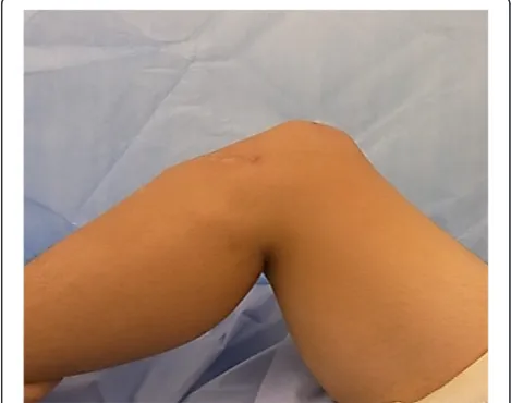
A 16-year-old male Japanese patient presented to our clinic with an inability to extend his left knee. He had injured his left knee during a jump while playing handball and sustained an avulsion fracture of the tibial tubercle Open reduction and cannulated screw fixation for the tib-ial plateau fracture had been performed at another institu-tion 10 months before he visited our hospital. However, the rupture of the patellar tendon had been missed during the initial emergency treatment. He complained of patellar dislocation, an inability to run, and difficulty in walking up and down stairs. An examination indicated that his left patella had migrated proximally and was higher than the right (Figure 2). There was evidence of quadriceps wast-ing, and a palpable gap below the patella was noted. The range of movement was from 0 to 130°; however, there was no active knee extension. The preoperative Lysholm knee-score [9] was 65 points. Both plain radiography and computed tomography with three-dimensional recon-struction confirmed that there was massive ectopic calcifi-cation along the patellar tendon and severe patella alta (Insall-Salvati index, 1.6) (Figure 3). Magnetic resonance imaging indicated the absence of continuity in the patellar tendon and an irregularity in the tibial tuberosity.
[image:2.595.56.291.472.627.2]During surgery, two-thirds of the patellar tendon mass was found to have been replaced by bone. The bony mass, which was 5.5 × 3.0 × 2.0 cm in size, was re-moved. Rupture of the patellar tendon from the inferior pole of the patella was identified during the operation. The semitendinosus and gracilis tendons were used to re-construct the patellar tendon, as described. The ruptured
Figure 1Reconstruction of the patellar tendon using semitendinosus and gracilis tendons with preserved distal insertions.The free end of the semitendinosus tendon was passed through a tunnel in the tibia tubercle in a medial to lateral direction. The free ends of both tendons were brought up proximally to cross in front of the patella as a figure-of-eight, towards the inferolateral side of the patella. Each tendon was passed transversely through the distal end of the quadriceps tendon and pulled distally. Both tendons were treated with interrupted sutures where they overlapped. ST: semitendinosus tendon, G: gracilis tendon.
[image:2.595.304.539.519.704.2]patellar tendon remnant was sutured to strengthen the reconstruction, and the course of rehabilitation proceeded, as described. His knee functions gradually returned to normal, and no pain was reported. He was able to resume his sports activities 6 months after surgery and returned to full training 8 months after surgery. The patient was last examined 1 year after his operation. At that time, there was no extensor lag and full flexion could be achieved. Isokinetic peak torque measurements of quadriceps strength at 90°/s was only 5% less than that of the contralateral extremity, as confirmed by Biodex (Biodex Medical Systems Inc., New York, USA) testing. The Lysholm knee-score was 100 points.
Case 2
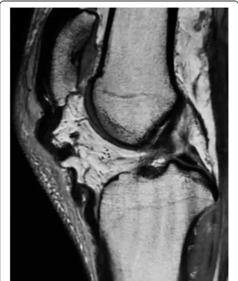
A 43-year-old male Japanese patient had injured his left knee during a jump while playing basketball. He had a history of rupturing both Achilles tendons and his body weight was 95 kg. A clinical examination revealed promin-ent pain in the patellar retinaculum, and lateral radiographs indicated the presence of patella alta (Insall index = 1.5). A diagnosis of patellar instability was made and surgical intervention was recommended. Proton magnetic res-onance imaging revealed a rupture of the left patellar tendon (Figure 4).
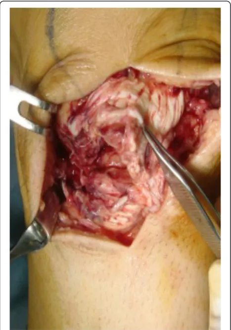
During the surgery, which was performed 10 days after the injury, the remnants of the patellar tendons were observed to be thin and fragile. Those were repaired with primary sutures, as far as possible; however, the tissue quality was poor and augmentation was required (Figure 5). Reconstruction using semitendinosus and gracilis tendons with preserved distal insertion was performed and rehabilitation proceeded, as previously
[image:3.595.58.539.89.283.2]described. The patient returned to work as a technician 2 months after surgery. At the 1-year follow-up, he did not experience any pain or difficulty during his daily activities. Full flexion could be achieved and was comparable with the contralateral side; his quadriceps strength was marked
Figure 3Lateral radiography (a) and computed tomography with three-dimensional reconstruction (b) of the right knee shows the presence of patella alta (Insall-Salvati index (Length of the patella/Length of the patella tendon) = 1.6) and ectopic calcification along the patellar tendon in case 1.
[image:3.595.305.538.418.693.2]as 5/5 and his thigh girth, 10 cm above the patella, was comparable to that of the right leg. The Lysholm knee-score was 95 points.
Discussion
Fresh ruptures of the patellar tendon are relatively un-common and require immediate surgical restoration of the extensor mechanism for optimal return to preinjury functional status. Ruptures are associated with tendinopathy caused by repetitive microtrauma, diabetes mellitus, rheumatoid arthritis, chronic renal failure, or steroid medication [10]. We reported 2 cases of patellar tendon disruption. One case involved a neglected disruption that had been missed during an initial emergency treatment. The second case involved an obese man who experienced a sports-related injury; poor quality of the ruptured tendon was suspected. Both cases required additional augmentation.
There are several surgical procedures for the repair of fresh and neglected ruptures of the patellar tendon; however, none of the methods are widely accepted. The
treatment goals for ruptured patellar tendons include restoration of the quadriceps mechanism, restoration of the anatomic congruity of the patellofemoral joint to avoid chondral lesions, improved range of motion, and splinting of the patellar tendon to allow early mobilization. Early surgical treatment can yield optimal clinical results, and end-to-end repair or reinsertion is used, with or without cerclage reinforcement [2]. In cases involving poor tissue quality due to an underlying disease process or the severity of the injury, primary repair, combined with autogenous graft augmentation, has been considered.
[image:4.595.57.291.89.424.2]Neglected rupture of the patellar tendon is a rare oc-currence, the treatment of which is a difficult challenge for orthopedic surgeons. Use of a contralateral patellar tendon [5] may result in additional damage to the un-injured leg, whereas the use of synthetic materials [3] and allografts [6] increases the risk of bacterial or viral infection and neoplasia. We performed reconstruction and restoration of ruptured patellar tendons using STG tendons with preserved distal insertions, without the use of any synthetic materials. We suggest that this modified technique has several advantages. STG tendons are rich in tendon fibers and can be used to yield a strong graft; the ultimate tensile load of doubled semitendinosus tendon grafts have been reported to reach 2330 N [11] and they are often used in anterior cruciate ligament [ACL] re-constructions. The strength and stability of the extensor mechanism can also be restored with minimal donor site morbidity [4,12]. Preservation of the original distal insertion of the STG appears to preserve its viability and provide sufficient blood supply to accelerate healing. Chen et al. [13] reported the reconstruction of a ruptured patel-lar tendon using STG tendons with preserved distal inser-tions. However, in their technique, a combined fixation with wire was required. We did not use wire cerclage, and this may have reduced the risk of infection and obviated the need for a second intervention to remove the implanted material. Nyemb et al. [14] also described a reconstruction technique with a semitendinosus auto-graft that maintained its original insertion. However, their technique required the formation of a horizontal tunnel in the patella; such a hole in the small patella promotes a risk of fracture [15]. The presently described technique does not require the formation of a hole in the patella as both tendons were placed in front of the pa-tella in a figure-of-eight fashion for additional stabilization. This technique is able to transmit the tension load from the patella directly to the tubercle tibia. When the knee bends, the patella is pulled downward and prevented from floating anteriorly by both tendons. In addition, this technique helps to stabilize the patella and maintain appropriate tracking. Most importantly, this technique avoids postoperative cast-ing and facilitates immediate postoperative mobilization.
Conclusion
In the two cases of patellar tendon ruptures that we de-scribe here, we have tried to depict the usefulness of a technique that uses reconstruction using semitendinosus-gracilis (STG) tendons with preserved distal insertions. This procedure could be used for similar cases with acute patellar tendon ruptures involving poor tissue quality as well as for neglected ruptures because it provides good functional results, appropriate patellar alignment, and full active range of motion for the repaired knee. In addition, this technique may allow optimal healing of the repaired site and early knee mobilization.
Consent
Written informed consent was obtained from the patient’s mother, in case of Case 1 and the patient himself for case 2 for publication of this case report and accompanying images. A copy of the written consent is available for review by the Editor-in-Chief of this journal.
Competing interests
The authors declare that they have no competing interests.
Authors’contributions
YT (corresponding author) mainly performed medical examinations and surgeries for all patients. As assistant of operative procedure, YS and HK supported for case1 and HI and SH supported for case2. MI, MK, YK, RS and KK discussed and advised about the method of the operation. All authors read and approved the final manuscript.
Received: 30 June 2013 Accepted: 5 September 2013 Published: 8 September 2013
References
1. Kannus P, Natri A:Etiology and pathophysiology of tendon ruptures in sports.Scand J Med Sci Sports1997,7:107–112.
2. Greis PE, Holmstrom MC, Lahav A:Surgical treatment options for patella tendon rupture. Part I.Acute Orthop2005,28:672–679.
3. Kim JR, Park H, Roh SG, Shin SJ:Concurrent bilateral patellar tendon rupture in a preadolescent athlete: a case report and review of the literature.J Pediatr Orthop B2010,19:511–514.
4. Van der Bracht H, Verdonk R, Stuyts B:Augmentation of a patellar tendon repair with an autologous graft.Acta Orthop Belg2009,75:417–419. 5. Milankov MZ, Miljkovic N, Stankovic M:Reconstruction of chronic patellar
tendon rupture with contralateral BTB autograft: a case report.Knee Surg Sports Traumatol Arthrosc2007,15:1445–1448.
6. Labib SA, Wilczynski MC, Sweitzer BA:Two-layer repair of a chronique patellar tendon rupture: a novel technique and literature review.
Am J Orthop (Belle Mead NJ)2010,39:277–282.
7. Cadambi A, Engh GA:Use of a semitendinosus tendon autogenous graft for rupture of the patellar ligament after total knee arthroplasty. A report of seven cases.J Bone Joint Surg Am1992,74:974–979. 8. Insall J, Salvati E:Patella position in the normal knee joint.Radiol1971,
101:101–104.
9. Bengtsson J, Mollborg J, Werner S:A study for testing the sensitivity and reliability of the Lysholm knee scoring scale.Knee Surg Sports Traumatol Arthrosc1996,4:27–31.
10. Amlang MH, Zwipp H:Damage to large tendons: Achilles, patellar and quadriceps tendons.Chir2006,77:637–650.
11. Hamner DL, Brown CH Jr, Steiner ME, Hecker AT, Hayes WC:Hamstring tendon grafts for reconstruction of the anterior cruciate ligament: biomechanical evaluation of the use of multiple strands and tensioning techniques.J Bone Joint Surg Am1999,81:549–557.
12. Tashiro T, Kurosawa H, Kawakami A, Hikita A, Fukui N:Influence of medial hamstring tendon harvest on knee flexor strength after anterior cruciate
ligament reconstruction. A detailed evaluation with comparison of single- and double-tendon harvest.Am J Sports Med2003,31:522–529. 13. Chen B, Li R, Zhang S:Reconstruction and restoration of neglected
ruptured patellar tendon using semitendinosus and gracilis tendons with preserved distal insertions: two case reports.Knee2012,19:508–512. doi:10.1016/j.knee.2011.07.007.
14. Nguene-Nyemb AG, Huten D, Ropars M:Chronic patellar tendon rupture reconstruction with a semitendinosus autograft.Orthop Traumatol Surg Res2011,97:447–450.
15. Shah JN, Howard JS, Flanigan DC, Brophy RH, Carey JL, Lattermann C:
A systematic review of complications and failures associated with medial patellofemoral ligament reconstruction for recurrent patellar dislocation.
Am J Sports Med2012,40:1916–1923. doi:10.1177/0363546512442330.
doi:10.1186/1756-0500-6-361
Cite this article as:Takazawaet al.:Reconstruction of a ruptured patellar tendon using ipsilateral semitendinosus and gracilis tendons with preserved distal insertions: two case reports.BMC Research Notes
20136:361.
Submit your next manuscript to BioMed Central and take full advantage of:
• Convenient online submission
• Thorough peer review
• No space constraints or color figure charges
• Immediate publication on acceptance
• Inclusion in PubMed, CAS, Scopus and Google Scholar
• Research which is freely available for redistribution