..
..
..
..
..
..
..
..
..
..
..
..
..
..
..
..
..
..
..
.
Angiopoietin-1 promotes atherosclerosis
by increasing the proportion of circulating
Gr1
1
monocytes
Takeshi Fujisawa
1,2†, Keqing Wang
1,2*
†, Xi-Lin Niu
3, Stuart Egginton
4, Shakil Ahmad
1,2,
Peter Hewett
5, Christopher D. Kontos
3, and Asif Ahmed
1,2*
1
Aston Medical Research Institute, Aston Medical School, Aston University, Birmingham B4 7ET, U.K;2
Gustav Born Centre for Vascular Biology and BHF Centre for Cardiovascular
Sciences, University of Edinburgh, Edinburgh EH16 4TJ, UK;3Department of Medicine, Division of Cardiology, Duke University Medical Centre, Durham, NC 27710, USA;
4
Multidisciplinary Cardiovascular Research Centre, School of Biological Sciences, University of Leeds, Leeds, UK;5
Institute of Cardiovascular Sciences, College of Medical and Dental Sciences, University of Birmingham, Birmingham, UK
Received 5 April 2016; revised 11 October 2016; editorial decision 17 October 2016; accepted 17 October 2016
Time of primary review: 41 days
Aims Atherosclerosis is a chronic inflammatory disease occurring within the artery wall. A crucial step in atherogenesis is the infiltra-tion and reteninfiltra-tion of monocytes into the subendothelial space of large arteries induced by chemokines and growth factors. Angiopoietin-1 (Ang-1) regulates angiogenesis and reduces vascular permeability and has also been reported to promote mono-cyte migrationin vitro. We investigated the role of Ang-1 in atherosclerosis-prone apolipoprotein-E (Apo-E) knockout mouse. ... Methods
and results
Apo-E knockout (Apo-E-/-) mice fed a western or normal chow diet received a single iv injection of adenovirus encoding Ang-1 or control vector. Adenovirus-mediated systemic expression of Ang-1 induced a significant increase in early atherosclerotic lesion size and monocyte/macrophage accumulation compared with control animals receiv-ing empty vector. Ang-1 significantly increased plasma MCP-1 and VEGF levels as measured by ELISA. FACS analysis showed that Ang-1 selectively increased inflammatory Gr1þmonocytes in the circulation, while the cell-surface expression of CD11b, which mediates monocyte emigration, was significantly reduced.
...
Conclusions Ang-1 specifically increases circulating Gr1þinflammatory monocytes and increases monocyte/macrophage
reten-tion in atherosclerotic plaques, thereby contributing to development of atherosclerosis.
䊏 䊏 䊏 䊏 䊏 䊏 䊏 䊏 䊏 䊏 䊏 䊏 䊏 䊏 䊏 䊏 䊏 䊏 䊏 䊏 䊏 䊏 䊏 䊏 䊏 䊏 䊏 䊏 䊏 䊏 䊏 䊏 䊏 䊏 䊏 䊏 䊏 䊏 䊏 䊏 䊏 䊏 䊏 䊏 䊏 䊏 䊏 䊏 䊏 䊏 䊏 䊏 䊏 䊏 䊏 䊏 䊏 䊏 䊏 䊏 䊏 䊏 䊏 䊏 䊏 䊏 䊏 䊏 䊏 䊏 䊏 䊏 䊏 䊏 䊏 䊏 䊏 䊏 䊏 䊏 䊏 䊏 䊏 䊏 䊏 䊏 䊏 䊏 䊏 䊏 䊏 䊏 䊏 䊏 䊏 䊏 䊏 䊏 䊏 䊏 䊏 䊏 䊏 䊏 䊏 䊏 䊏 䊏 䊏 䊏 䊏 䊏 䊏 䊏 䊏 䊏 䊏 䊏 䊏 䊏 䊏 䊏 䊏 䊏 䊏 䊏 䊏 䊏 䊏 䊏 䊏 䊏 䊏 䊏 䊏 䊏 䊏 䊏 䊏 䊏 䊏 䊏 䊏 䊏 䊏 䊏 䊏 䊏 䊏 䊏 䊏 䊏 䊏 䊏 䊏 䊏 䊏 䊏 䊏 䊏 䊏 䊏 䊏 䊏 䊏 䊏 䊏 䊏 䊏 䊏 䊏 䊏 䊏 䊏 䊏 䊏 䊏 䊏 䊏 䊏 䊏 䊏 䊏 䊏 䊏 䊏 䊏 䊏 䊏 䊏 䊏 䊏 䊏 䊏 䊏 䊏 䊏 䊏 䊏 䊏 䊏 䊏 䊏 䊏 䊏 䊏 䊏 䊏 䊏 䊏 䊏 䊏
Keywords Angiopoietin-1
•
Atherosclerosis•
Monocytes1. Introduction
Atherosclerosis is a chronic inflammatory disease of the artery wall.1–3 Monocyte-derived macrophages participate in a maladaptive, non-resolving inflammatory response that expands the subendothelial layer due to the accumulation of cells, lipid and matrix.4Signals such as mono-cyte chemoattractant protein-1 (MCP-1) increase monomono-cyte recruit-ment into atherogenic foci,5,6in particular, Ly6Chigh(Gr1þ) inflammatory monocytes that gives rise to macrophages in atheromata.7
Angiopoietin-1 (Ang-1) and its cognate receptor Tie2 are well-established regulators of vascular development and angiogenesis.8–10 Ang-1 plays a crucial role in endothelial cell survival, vessel wall remodel-ling and mural cell recruitment.11Overexpression of Ang-1 dramatically
blocks increases in vascular permeability induced by VEGF,12,13 suggest-ing that it has protective properties in the microvasculature.14However, long-term Ang-1 expression using adeno-associated virus failed to pro-tect against the development of rat cardiac allograft arteriosclerosis.15 Jeanssonet al.8recently reported that Ang-1 is not necessary for normal steady-state physiological processes in the adult, being expendable in the blood vasculature from E13.5 onwards,10 but when combined with injury, Ang-1 deficiency results in accelerated angiogenesis and fibrosis.10 Longet al.14have reported that Ang-1 therapy accompanied pro-fibrotic and inflammatory effects in folic acid-induced tubular necrosis and in a murine model of acute renal injury.16 More importantly, Ang-1 is expressed at a higher level in atherosclerotic lesions obtained from endarterectomy of the carotid artery compared to healthy controls.17
*Corresponding authors. Tel:þ44 121 204 4967; fax: +44 121 204 5142, E-mail: asif.ahmed@aston.ac.uk; k.wang@aston.ac.uk
†
The first two authors contributed equally to the study.
VCThe Author 2016. Published by Oxford University Press on behalf of the European Society of Cardiology.
This is an Open Access article distributed under the terms of the Creative Commons Attribution License (http://creativecommons.org/licenses/by/4.0/), which permits unrestricted reuse, distribution, and reproduction in any medium, provided the original work is properly cited.
by guest on January 17, 2017
..
..
..
..
..
..
..
..
..
..
..
..
..
..
..
..
..
..
..
..
..
..
..
..
..
..
..
..
..
..
..
..
..
..
..
..
..
..
..
..
..
..
..
..
..
..
..
..
..
..
..
..
..
..
..
..
..
..
..
..
..
..
..
..
..
..
..
..
..
..
..
..
..
..
..
..
..
..
..
..
..
..
..
..
..
..
..
..
..
.
Furthermore, Ang-1 stimulates TNF-a, a key cytokine that modulates the inflammatory process of atherosclerosis.18expression in peripheral blood mononuclear cells,19Indeed, we and others have demonstrated that Ang-1 stimulates monocyte20and neutrophil21migration, both of which are the critical players in atherosclerosis,22,23implicating Ang-1 as a potential player in monocyte recruitment and retention mechanisms especially in a high-lipid environment.Based on these observations, we hypothesized that Ang-1 plays a role in the progression of atherosclerosis in a hypercholesterolaemic environ-ment through its effects on inflammatory monocytes. In this study, we demonstrate that high circulating Ang-1 levels promote a pro-atherogenic phenotype by specifically increasing Gr1þinflammatory monocytes and elevating the circulating levels of pro-remodelling cytokines, VEGF and MCP-1 in ApoE-/-mice. Furthermore, Ang-1-induced monocyte/macro-phage retention in atherosclerotic plaques was accompanied by decreased cell-surface expression of CD11b on circulating monocytes.
2. Methods
2.1 Adenoviruses
Recombinant adenovirus encoding human Ang-1* (AdAng-1) was provided by Regeneron Pharmaceuticals (Tarrytown, New York, USA), propagated in Human embryonic kidney cells 293 (HEK293), purified on CsCl gradients, titered, and stored at80C in 4% sucrose buffer. Control, empty adenovi-rus (AdEV) was generated as described previously.22
2.2 Adenovirus-mediated expression of
Ang-1 in ApoE
-/-mice
All procedures conformed to the recommendations of the Guide for the Care and Use of Laboratory Animals published by the US National Institutes of Health (NIH Publication, 8th Edition, 2011) and also in accordance with Directive 2010/63/EU of the European Parliament and with the UK Home Office Animal (Scientific Procedures) Act 1986. All procedures passed local ethical review. Eight- to nine-week-old male ApoE-/- mice on a C57BL/6J background (B6.129P2-apoEtm1Unc, SN: 002052; Jackson Labs, Maine, USA) were maintained with a 12-hour light/ dark cycle and had free access to food and water. To investigate the effects of Ang-1 on atherosclerosis under high-fat environment, the mice were fed a western-style diet (TD 88137; Harlan Teklad, South Easton, MA, USA) for 1 week and then divided into two groups (n¼12 per group) and injected via tail vein with 5109pfu of AdAng-1 or control empty virus (AdEV) diluted into 100lL PBS. These mice were maintained on a western diet for another 4 weeks. In another sets of experiments, mice fed a normal chow diet (n¼5 in each group in each experiment). These mice were injected with 5109pfu of AdAng-1 or control empty virus (AdEV) diluted into 100lL PBS through tail vain. The mice were euthanized 4 weeks after virus delivery. Briefly, cardiac puncture was per-formed with 2.3% isoflurane inhalation, and blood was collected in EDTA tube. Mice were injected with Pentabarbitone (Euthatal, 270 mg/kg, ip injection) to euthanize and then perfuse-fixed with 1% paraformaldehyde. Heart and proximal aortae were harvested. Serial 5mm frozen sections of the aortae were prepared as described previously.24For the evaluation of atherosclerotic lesions, nine sections were taken at 40 mm intervals, stained with oil red O, counterstained with Mayer’s hematoxylin, and the lesions were quantified using NIH Image software (v. 1.62). The mean area in nine sections was determined for each animal. Up to three sec-tions from each animal were immunostained for monocytes/macro-phages using a monoclonal rat anti-mouse monocyte/macrophage
antibody (MOMA-2, BD PharMingen) and quantification of MOMA-2-positive area was performed. The mean MOMA-2-MOMA-2-positive area was determined for each animal. The aortic arches were available from some animals and Sudan IVen facestaining was performed for analysis of lipid accumulation, as described elsewhere.25Blood was taken at 3 and 10 days after virus injection by tail bleed and at 28 days by cardiac puncture.
2.3 Aortic ring culture
Male ApoE-/- mice (8–10 weeks old) were euthanized and bled out. Thoracic aortas were removed into a Petri dish filled with cold sterile PBS and aortas were mechanically cleaned of surrounding fat tissue. Using a surgical blade, aortas were evenly cut into 1 mm rings, which were transferred to fresh DMEM medium supplemented with 1% fetal calf serum (FCS), penicillin and streptomycin. Aortic rings were stimu-lated with Ang-1 (400 ng/mL; R&D Systems, Abingdon, UK) for 24 hours at 37C. Some of rings were pre-incubated with Tie2 blocking peptide26 (NLLMAAS, 100mM; Peptide Protein Research Ltd, Hampshire, UK) for 30 min prior to stimulation with Ang-1. Each condition was performed in duplicates. Commercial ELISA as described below measured the levels of MCP-1 and VEGF in the conditioned medium.
2.4 ELISA assay
Whole blood was collected into EDTA-containing tubes. ELISA was used to measure plasma levels of Ang-1, MCP-1 and VEGF (R&D Systems).
2.5 Immunohistochemistry
Immunohistochemistry was performed on serial frozen sections. Primary rat anti-mouse antibodies to monocyte/macrophage (MOMA-2), and CD31 were from BD Pharmingen. Isotype-matched non-binding immunoglobulin was used as a negative control. Binding of secondary antibodies (Vector Laboratories, Peterborough, UK) was detected with Vectastain ABC reagent (Vector Laboratories, Peterborough, UK) and DAB substrate kits (Dako, Cheadle, UK). Cells were counterstained with Mayer’s hematoxylin.
2.6 Flow cytometry
Whole-blood samples (15lL) were washed with cold PBS before incu-bating for 20 min on ice with directly conjugated antibodies: anti-Ly6C-APC (eBioscience, Cheshire, UK), anti-CD11b (eBioscience, Cheshire, UK) and anti-Ly6G (Gr1)-FITC (eBioscience, Cheshire, UK). Monocytes were gated according to CD11b expression and side scatter and then further separated by Gr1 and Ly6C staining. Two monocyte subsets were identified as SSClowCD11bþGr1þLy6Chigh and SSClowCD11bþGr1-Ly6Clow monocytes, whereas neutrophils were identified as SSChighCD11bþGr1highLy6Cinte (Figure 3A and B) using FACSCalibur. Combination of anti-CD11b, anti-Gr1 and anti-Tie2-PE (eBioscience, Cheshire, UK) were used to stain cells to analyse Tie2-positive monocyte population.
2.7 Measurement of plasma Ang-1
Blood was collected from mice at days 0, 3, 7, 14, 21, and 28 after injec-tion of AdEV (n¼3) or AdAng-1 (n¼3), mixed with EDTA. Plasma lev-els of Ang-2 were determined a using specific ELISA following the manufacturer’s instructions (R&D Systems, MN, USA).
2.8 Confocal immunofluorescence staining
Tissue sections were stained with rat monoclonal anti-Ly6C (IgG2c; eBioscience, Paisley, UK) followed by Alexa Fluor 568-conjugatedanti-by guest on January 17, 2017
..
..
..
..
..
..
..
..
..
..
..
..
..
..
..
..
..
..
..
..
..
..
..
..
..
..
..
..
..
..
..
..
..
..
..
..
..
..
..
..
..
.
rat IgG (Invitrogen, Paisley, UK). Then counter stained with anti-CD11b-FITC (eBioscience, Cheshire, UK) followed by goat anti-anti-CD11b-FITC- anti-FITC-conjugated with Alexa Fluor488 (Invitrogen, Paisley, UK). Sections were washed with PBS, coverslip mounted and imaged on a Leica SP5C inverted confocal laser scanning microscope.2.9 Statistical analysis
Results are expressed as the mean6SEM. Comparisons between two groups were performed using unpaired Student’st-tests, and compari-sons among multiple groups were performed using One-way Analysis of variance (ANOVA). Statistical analyses were performed using Prism 7.0 (GraphPad Software, Inc., La Jolla, CA, USA).Pvalues<0.05 were con-sidered statistically significant.
3. Results
3.1 Ang-1 enhances atherosclerotic plaque
formation in ApoE
-/-mice fed a western
diet
To investigate the effect of Ang-1 on atherosclerosis development, ApoE-/-mice fed a western diet for 1 week were given AdAng-1 or con-trol AdEV injection and maintained on a western diet for a further 4 weeks. It has been demonstrated that iv delivery of adenovirus leads to a widespread distribution of vector with the highest level of expression in the liver.27The level of Ang-1 in plasma peaked 3 days after injection and remained elevated for >10 days (Supplementary material online,
Figure S1). No significant differences in body weight or circulating lipid profiles were detected between these groups (Supplementary material
online,Table S1). Four weeks after systemic adenovirus administration,
atherosclerotic lesions were analysed in aortas sections using oil red O staining (Figure 1A). Systemic overexpression of Ang-1 significantly increased the mean atherosclerotic lesion size compared with AdEV-infected ApoE-/-mice (0.68760.071 mm2vs. 0.43760.045 mm2;P<0. 01;Figure1B). Uninfected ApoE-/-mice had lesions similar in size to the AdEV-infected group (0.54760.05 mm2, data not shown). In addition, with macrophages being a primary cell type contributing to development of the atherosclerotic lesion, we examined the effect Ang-1 on mono-cyte/macrophage accumulation in atherosclerotic lesion using monocyte/ macrophage marker MOMA-2. As expected, Ang-1 induced a significantly increased monocyte/macrophage accumulation in lipid lesions compared with control animals receiving empty vector (0.1560.015 mm2 vs. 0. 0960.021 mm2;P<0.05;Figure1C). Endothelium staining using CD31 antibody showed no significant vascularization in aortas (Figure1A)
3.2 Ang-1 promotes atherosclerotic
plaque formation in ApoE
-/-mice fed a
normal chow diet
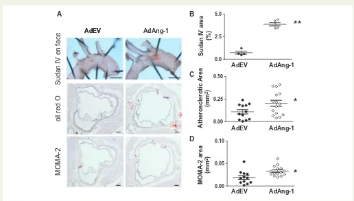
In order to further confirm the effects of Ang1 in atherosclerosis devel-opment without the confounding effects of western diet on inflammation in apoE-/-mice, AdAng-1 or control AdEV were injected in apoE-/-mice fed a normal chow diet anden faceanalyses of Sudan IV-stained areas in thoracic aortas were quantified 4 weeks after treatment. Ang-1 signifi-cantly increased plaque area compared with AdEV-treated ApoE-/-mice
A B
C
AdAng-1 AdEV
MOMA-2
oil red O
CD31
0.0 0.5 1.0 1.5
Atherosclerotic Area
(mm
2)
0.0 0.1 0.2 0.3
MOMA-2 area
(mm
2)
*
*
AdEV AdAng-1 [image:3.595.38.558.409.637.2]AdEV AdAng-1
Figure 1Ang-1 enhances atherosclerotic plaque formation in ApoE-/-mice fed a Western diet. ApoE-/-mice fed a Western diet for 4 weeks were infected
systemically with adenoviruses encoding Ang1 (AdAng-1) or a control virus (AdEV). The mice were euthanized 4 weeks after adenovirus treatment and effects on early to intermediate atherosclerotic lesions were investigated. (A) Representative sections stained with oil red O (top panel) and immunohisto-chemical staining of monocytes/macrophages and endothelium with the MOMA-2 monoclonal antibody (middle panel) and CD31 antibody (bottom panel) respectively in AdEV or AdAng1-treated ApoE-/-mice. Original magnification,40 (Oil red),100 (MOMA-2). Bars, 100mm. (B) Atherosclerotic lesion
area was quantified by oil red O staining of lesions from serial aortic sections using ImageJ image analysis software. Nine serial sections at 40mm intervals were used from each animal for analysis. Results show the mean from nine cross-sectional lesion size (mm2) for each animal and the line indicates median
value per treatment of mice.*P<0.01. (C) Similarly, five mice were randomly picked from each treatment group, and tissue sections from each animal were analysed for MOMA-2-positive lesion area.*P<0.05.
by guest on January 17, 2017
..
..
..
..
..
..
..
..
..
..
..
..
..
..
..
..
..
..
..
..
..
..
..
..
..
..
..
..
..
..
..
..
..
..
.
(3.9060.195 mm2vs. 0.7360.169 mm2;P<0.01;Figure2A and B). Oil red O-positive atherosclerotic area and MOMA-2-positive area were also analysed as before (Figure2C and D). As expected, mice fed a normal chow diet developed much smaller plaques compared with mice on a western diet. However, the plaques were again significantly larger in the AdAng-1-treated group compared to the AdEV-treated group (0.2060.034 mm2vs. 0.1160.024 mm2;P<0.05;Figure2C). Similarly, the accumulation of MOMA-2-positive cells, which co-localized with oil red O staining in the aortic root (Figure1A), was also increased in the AdAng-1-treated group (0.0360.003 mm2 vs. 0.0260.004 mm2;P<0.01;Figure2D)
3.3 Ang-1 increases proportion of
circulating Gr1
þ/Ly6C
highmonocytes
It is widely accepted that bone marrow-derived circulating monocytes play an important role in atherosclerosis28and that different subsets of monocytes commit for specific functions while still in the circulation.29 Particularly, Ly6Chigh(Gr1þ) inflammatory monocytes give rise to mac-rophages in atheromata.7 To investigate the role of Ang-1 on the dynamics of monocyte turnover/recruitment, peripheral monocytes in ApoE-/-mice fed a normal chow diet were evaluated at different time points after AdAng-1 treatment (3, 10 and 28 days). Monocytes were
gated according CD11bþand SSC (Figure3AR1) and two monocyte subsets were identified as SSClowCD11bþGr1þLy6Chigh(Figure3A
R3) and SSClowCD11bþGr1-Ly6Clow monocytes (Figure 3A R2), which correspond to inflammatory and residential subsets, respectively.30A third CD11bþcells population with high SSC, low Ly6C are neutrophils (Figure3AR4).7,30The proportion of circulating Gr1þmonocytes in AdAng-1-treated mice was significantly increased at day 10 compared with AdEV-treated mice (4.9560.503 vs. 2.9360.163; P<0.01), whereas Gr1- monocytes remained unchanged (Figure3B). Interestingly, neutrophil proportion was also increased (9.5363.92 vs. 13.7464.04;P<0.05). Significant increase in the ratio of Gr1þ/Gr1-monocytes (Figure3C) was positively corre-lated with plaque size (Figure3D), indicating the importance of mono-cyte subsets balance in disease progression. At day 3 and day 28, the proportions of Gr1- and Gr1þmonocytes showed no differences between AdEV and AdAng-1 treated groups (data not shown). Furthermore, immunostaining of aorta obtained from ApoE-/- mice received AdAng-1 treatment revealed that Ly6C-positive cells local-ized in the plaques (Figure4) in line with the current understanding that this group of cells is likely to account for the observed accumula-tion of monocytes/macrophages in the plaques.
It is believed that inflammatory monocytes derived from bone mar-row play a crucial role in atherogenesis. Recently, spleen has been
B A
C
D AdEV
MOMA-2
oil red O
AdEV AdAng-1
Sudan IV
en face
0.0 2.5 5.0
0.00 0.05 0.10 0.00 0.25 0.50
Sudan IV
area
(%)
Atherosclerotic Area
(mm
2)
MOMA-2 area
(mm
2)
*
*
**
AdEV AdAng-1
AdEV AdAng-1
[image:4.595.38.554.356.649.2]AdEV AdAng-1
Figure 2Ang-1 promotes early atherosclerotic plaque formation in ApoE-/-mice fed a normal chow diet. ApoE-/-mice fed a normal chow diet were
infected systemically with adenoviruses encoding Ang-1 (AdAng-1), or control virus (AdEV). The mice were euthanized after 4 weeks of adenovirus treat-ment and effects on early to intermediate atherosclerotic lesions was investigated. (A) Representative picture of Sudan IVen face(top panel) (n¼4 per group). (B) Sudan IVen facestaining was quantified from the beginning of aortic arch to the left common carotid artery using image analysis software and expressed as the percentage lesion area in each vessel. **P<0.001. (C) Oil red O-positive atherosclerotic areas were calculated by using ImageJ image analy-sis software (n¼12–15 per group). Atherosclerotic plaques were quantified as described in the legend toFigure1.*P<0.05. (D) Similarly, three sections from each animal were analysed for MOMA-2-positive lesion size (mm2) (n¼12–15 per group).*P<0.05. Original magnification2.5 (Sudan IVen face); bars, 1 mm (Sudan IV).
by guest on January 17, 2017
..
..
..
..
..
..
..
..
..
..
..
..
..
..
..
..
..
..
..
..
..
..
..
..
..
..
..
..
..
..
..
..
..
..
..
..
..
..
..
..
..
..
..
.
identified as a site for storage and rapid deployment of monocytes, which contribute to atherosclerosis development.31Interestingly, preliminary
study showed that proportion of bone marrow Ly6Chighmonocytes was
reduced following AdAng-1 treatment, whereas splenic Ly6Chigh mono-cytes remained unchanged (data not shown), suggesting that Ly6Chigh
inflammatory monocytes are likely mobilized from bone marrow in response to Ang-1.
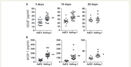
3.4 Ang-1 increases plasma levels of VEGF
and MCP-1 in ApoE
-/-mice fed a normal
chow diet
Chemokines such as MCP-1 is crucial for monocyte mobilization from bone marrow.32To test whether overexpression of Ang-1 alters
circu-lating cytokine/chemokine profiles, we analysed the plasma concentra-tions of VEGF and MCP-1 by ELISA. Ang-1 significantly increased both plasma VEGF (Figure5A) and MCP-1 (Figure5B) levels on day 3 and 10 after administration of viruses. Notably, the increased level of MCP-1 persisted even on day 28 when plasma Ang-1 concentrations had sub-sided (Supplementary material online, Figure S1). Furthermore, the plasma level of VEGF significantly correlated with expression of MCP-1 on day 10 in AdAng-1-treated but not in AdEV-treated animals
(Supplementary material online,Figure S2).
3.5 Ang-1 induces release of VEGF and
MCP-1 from aortic rings
Next, we performed aortic ring cultures to investigate the origin of VEGF and MCP-1 upon stimulation with Ang-1. Ang-1 significantly increased release of both VEGF and MCP-1 from aortic rings, and these effects were blocked by pre-incubation with the Tie2 peptide (Figure6), suggesting that Ang-1 induces vascular cell expression of VEGF and MCP-1, thereby creating a pro-remodelling environment in large vessels in which atherosclerotic plaques form. In addition, expression of VEGF and MCP-1 was significantly correlated with one another in these aortic rings (Supplementary material online,Figure S3).
CD11b
SSC
R1
A
B
D
C
R2 R3
Ly
6
C
Gr1 R4
Atherosclerotic Area
(mm
2)
Gr1 p/n ratio 0.0 1.5 3.0 4.5 0.00
0.02 0.04 0.06 0.08
0 1 2 3 4 5
AdEV AdAng-1
G
r1
p
/n
ra
ti
o
**
0 2 4 6 8 10
AdEV AdAng-1
Gr1+
G
r1
+%
o
f
a
ll
c
e
ll
s
**
0 2 4 6 8 10
G
r1
-%
o
f
a
ll
c
e
ll
s
AdEV AdAng-1
Gr1
-Figure 3Ang-1 increases circulating numbers of Gr1þmonocytes. Whole-blood samples were stained with CD11b, Gr1 and Ly6C antibodies and ana-lysed by flow cytometry. (A) CD11b-positive cells were gated (R1) according to CD11b expression and side scatter. R1 cells were further divided into SSClowCD11bþ
Gr1-Ly6Clowmonocytes (R2) and SSClowCD11bþ
Gr1þLy6Chighmonocytes (R3) and SSChighCD11bþ
Gr1highLy6Cinteneutrophils (R4). (B)
10 days after virus administration, the proportions of Gr1-and Gr1þmoncoytes in each treatment group was calculated as the percentage of total white blood cells. (C) To correct for variability among individual experiments, the Gr1þ/Gr1-ratio was calculated for each treatment group. (D) The correlation
between MOMA-2-positive areas and Gr1þand Gr1-monocytes on day 10 after AdAng-1 administration was analysed. Pooled data are presented from three separate independent experiments (n¼4–5 per experiment).**P<0.01.
[image:5.595.40.563.55.294.2]X 40
X 80
Figure 4Ang-1 induces Ly6CþCD11bþmonocytes accumulation in
ApoE-/-mice fed a normal chow diet. Representative immunofluorescence staining of Ly6C and CD11b monoclonal antibodies in AdAng1-treated ApoE-/-mice. Red: Ly6C, green: CD11b, blue: 4’,6-diamidino-2-phenylin-dole (DAPI). Original magnification,40 (left),80 (right). White arrow is the border line between arterial wall and plaque judging by out layer of muscle filament. Yellow arrow is double staining mononuclear cell.
by guest on January 17, 2017
[image:5.595.39.283.414.564.2]..
..
..
..
..
..
..
..
..
..
..
..
..
.
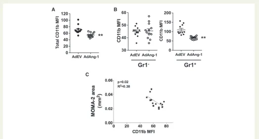
3.6 Ang-1 reduces cell-surface expression
of CD11b on Gr1
þmonocytes
Theb2-integrin heterodimer CD11b/CD18 (aMb2, also called Mac-1)
is one of the major adhesion molecules on monocytes that mediates firm adhesion to endothelial cellsviaintercellular adhesion molecule-1 (ICAM-1).33,34It is required for monocyte reverse migration and may play a role in atherosclerosis development.35,36 Immunostaining of
ICAM1 at the sinus did not show any difference between control virus-treated and AdAng-1-treated mice (Supplementary material
online, Figure S4). However, CC chemokine receptor 2 (CCR2)
expressions on circulating monocytes showed significant increase 10 days after AdAng-1 infection (data not shown), suggesting that Ang-1 may contribute to increased monocyte recruitment. To determine whether the pro-atherogenic effects of Ang-1 are associated with changes inb2-integrins on monocytes, we analysed the expression of
CD11b on circulating monocytes. Mice treated with AdAng-1 have a significantly lower level of monocyte CD11b expression 10 days after AdAng-1 treatment (Figure7A). In addition, this Ang-1-induced
A
B
3 days
10 days
28 days
0 10 20 30 40 50 60
V
E
G
F
(p
g
/m
l)
**
AdEV AdAng-1 0 10 20 30 40 50 60
**
AdEV AdAng-1 0 10 20 30 40 50 60
AdEV AdAng-1
0 100 200 300 400 500
M
C
P
-1
(p
g
/m
l)
**
AdEV AdAng-1 0 100 200 300 400 500
AdEV AdAng-1
**
0 50 100
AdEV AdAng-1
[image:6.595.37.560.57.327.2]*
Figure 5Ang-1 increases plasma levels of VEGF and MCP-1ApoE-/- mice fed a normal chow diet. Whole blood was collected at 3 and 10 and 28 days after AdEV or AdAng-1 administration and plasma was isolated. ELISA was used to measure the level of VEGF (A) and MCP-1 (B). Pooled data are presented from four independent experiments (n¼17–19 per group).**P<0.01.
Ang-1 Tie2 pep
- + - + DMSO
- - + +
Ang-1 Tie2 pep
- + - + DMSO
- - + +
0 5 10 15 20 25
VEGF (pg/ml)
p<0.001 p<0.001
0 100 200 300
MCP-1 (pg/ml)
p<0.05
p<0.05
A B
Figure 6Ang-1 induces the VEGF and MCP-1 release in conditioned medium from aortic ring. Thoratic artery from apoE-/-mouse was cut into 1 mm seg-ments and aortic rings were stimulated with Ang-1 (400 ng/mL) with or without pre-incubation with Tie2 blocking peptide (NLLMAAS, 100mM) for 30 min. Medium containing 0.01% DMSO was served as vehicle control. The levels of VEGF (A) and MCP-1 (B) in conditioned medium were measured by ELISA assay. Pooled data are presented from four independent experiments performed in duplicates.
by guest on January 17, 2017
[image:6.595.46.555.407.562.2]..
..
..
..
..
..
..
..
..
..
..
..
..
..
..
..
..
..
..
..
..
..
..
..
..
..
..
..
..
..
..
..
..
..
..
..
..
..
..
.
down-regulation of CD11b was Gr1þmonocyte specific (Figure7B), and the down-regulation of CD11b persisted up to 28 days after virus administration, after expression of Ang-1 had declined(Supplementary material online,Figure S5). Furthermore, there was a
significant negative correlation between CD11b and MOMA-2-posi-tive lesion area in ApoE-/-mice (Figure7C), suggesting a possible pro-tective function of CD11b, which is inhibited by Ang-1, thereby resulting in enhanced atherosclerosis in ApoE-/-mice.
To further investigate the mechanism by which down-regulation of CD11b may reduce monocyte transmigration, we examined the interac-tions of monocytes with human umbilical vein endothelial cells (HUVECs). We found that blocking CD11b function with the monoclo-nal antibody ICRF44 reduced monocytes reverse transmigration through HUVEC monolayers by more than two-fold (IgG control;n¼3, transmi-grated 15.70% vs. ICRF44; n¼3, transmigrated 32.43%, P¼0.068)
(Supplementary material online, Figure S6), suggesting that
down-regulation of CD11b expression on monocytes may lead to increased monocyte retention.
In addition, we explored Tie2 expression in Gr1þCD11bþand Gr1-CD11bþmonocytes and found that Tie2 expression was largely restricted to Gr1þmonocytes compared to Gr1-monocytes in both blood (Supplementary material online,Figure S7, 4.00060.1941 vs. 1. 97360.176;P<0.01) and spleen (Supplementary material online,Figure S7, 10.4461.376vs. 3.89260.3050;P<0.01). Immunostaining of aorta obtained from ApoE-/-mice received AdAng-1 treatment revealed that Tie2-positive macrophages were present in the plaques (Supplementary
material online,Figure S8), suggesting that Ang-1/Tie2 pathway on
mono-cytes, is in part, responsible for Ang-1 pro-atherogenic effects.
4. Discussion
The activation of endothelial cells and the recruitment of monocytes are key events in the early onset of atherosclerosis; however, these initial processes may be reversible and typically do not cause clinical conse-quences.1,37It is the subsequent prolonged retention of monocytes/mac-rophages in the intimal space and their uptake of oxidized LDL to form foam cells that constitutes the major cellular component in early athero-sclerotic lesion development.38The major finding of this study is that sys-temic overexpression of Ang-1 accelerates atherosclerosis development in ApoE-/-mice. It has been shown that levels of Ang-1, but not of Ang-2, are significantly increased in conditioned medium from cultured athero-sclerotic arteries compared to healthy arteries.17It has also been shown that Tie2 expression is increased in atherosclerotic arteries compared to healthy arteries. These findings support our results and suggest that the Ang-1/Tie2 system has a significant role in the development of athero-sclerosis. Although Ang-1 has been shown to have anti-inflammatory properties in endothelial cells, our present study clearly demonstrates pro-remodelling effects of Ang-1 on monocytes that translate into increased atherosclerosis in the context of elevated cholesterol levels in ApoE-/-mice. The mechanisms by which Ang-1 promotes atherosclero-sis appear to involve (i) Ang-1-induced pro-remodelling cytokine release;
A B
C
Gr1
-Gr1
+0 50 100 150 200
CD1
1
b M
F
I
AdEV AdAng-1
**
30 40 50 60
CD1
1
b
MF
I
AdEV AdAng-1
0 20 40 60 80 100 120
To
ta
l C
D
1
1
b
M
F
I
AdEV AdAng-1
[image:7.595.39.564.55.336.2]**
Figure 7Ang-1 reduces cell-surface CD11b expression on Gr1þmonocytes. Whole-blood samples were stained with anti-CD11b and anti-Gr1 antibod-ies, and analysed by flow cytometry as described in the legend toFigure3. (A) CD11b expressions [median fluorescence intensity (MFI)] on circulating mono-cytes in control and AdAng-1 groups were compared 10 days after virus administration. (B) CD11b expression on Gr1-and Gr1þmonocytes in control and AdAng-1 groups were analysed and representative histograms were depicted.(C)The correlation between MOMA-2-positive areas and CD11b expression on Gr1þmonocytes on day 10 after AdAng-1 administration was analysed. CD11b expression was represented by MFI. Pooled data are presented from three independent experiments (n¼5 per experiment).**P<0.01.
by guest on January 17, 2017
..
..
..
..
..
..
..
..
..
..
..
..
..
..
..
..
..
..
..
..
..
..
..
..
..
..
..
..
..
..
..
..
..
..
..
..
..
..
..
..
..
..
..
..
..
..
..
..
..
..
..
..
..
..
..
..
..
..
..
..
..
..
..
..
..
..
..
..
..
..
..
..
..
..
..
..
..
..
..
..
..
..
..
..
..
..
..
..
..
.
(ii) Ang-1 increases the proportion of circulating inflammatory cytes and (iii) Ang-1-mediated reduction in CD11b expression on mono-cytes, resulting in increased monocyte/macrophage retention in atherosclerotic plaques.In the present study, plasma levels of VEGF and MCP-1, both of which promote atherosclerosis in mice,39,40are increased following systemic overexpression of Ang-1. Ang-1 increased VEGF and MCP-1 release via Tie2 from aortic rings suggesting that the inflammatory effects of Ang-1 are evident in large arteries, in which atherosclerotic plaques form. Taken together our data suggest that Ang-1/Tie2 pathway participates in the inflammatory process under these conditions. VEGF is a potent regu-lator of vascular permeability.41It not only stimulates endothelial cell proliferation but also upregulates other pro-remodelling cytokines release from endothelial cells, such as MCP-1.42 Ang-1-induced increased levels of VEGF and MCP-1 are positively correlated with each other in bothin vivoandin vitrosystems, suggesting Ang-1 can create a positive pro-remodelling cytokine feedback loop in large arteries, which resulted in sustained inflammation even after systemic Ang-1 levels dropped to baseline 14 days after AdAng-1 injection (Supplementary
material online,Figure S1).
Hypercholesterolemia induces monocytosis and monocytes accumu-lation in the plaques.43Recently, the importance of neutrophil in early atherosclerosis development has been increasingly recognized.44Doring
et al.43 have demonstrated that the mechanism of neutrophil-driven atherosclerosis is mediated through neutrophil granule protein cathelicidin-induced inflammatory monocytes recruitment,45suggesting that bone marrow-derived monocytes are crucial in atherosclerosis development. Hypercholesterolemia is known to induce selective expansion of Gr1þ/Ly6Chighmonocytes in ApoE–/–mice, and these cells preferentially adhere to activated endothelium, infiltrate the arterial wall and develop into atherosclerotic macrophages.7Ang-1 overexpression leads to a reduction in the prevalence of Gr1þ/Ly6Chighinflammatory monocytes in bone marrow but an increase in circulation in AdAng-1-treated animals, suggesting that Ang-1 may trigger monocyte mobiliza-tion from bone marrow most likely through up-regulamobiliza-tion of MCP-1.32,40,46–49The imbalance of monocyte subsets in circulation induced by Ang-1 overexpression is associated with increased atheroma (Figure3D). In addition, we show for the first time that Gr1þ/Ly6Chigh not Gr1-/ Ly6Clowmonocytes express Tie-2 in ApoE-/-mice (Supplementary mate
rial online,Figure S7). Until recently, Tie-2 was thought to be restricted
to endothelial cells. However, De Palmaet al.48identified a subset of Tie-2-positive monocytes that promote angiogenesis in experimental tumour model.50Recently, studies have revealed that functions of Tie-2-expressing monocytes (TEMs) may not be restricted to angiogenesis and immunosuppression. TEMs are involved in inflammatory process.51,52 Our findings suggest that Ang-1/Tie2 is, in part, responsible for migra-tion/recruitment of Gr1þ/CCR2þmonocytes in addition to MCP-1. Furthermore, the presence of Ly6Cþcells within atherosclerotic pla-ques (Figure4) provides further evidence that this group of cells is likely to account for the observed accumulation of monocytes/macrophages, which contributed to Ang-1-induced atherosclerosis development.
Adhesion molecules participating in monocyte–endothelial cell inter-actions are known to play a critical role in atherogenesis.35In this study, we observed that overexpression of Ang-1 increased oil red-positive lesion size and it is associated with down-regulation of CD11b expres-sion on circulating Gr1þmonocytes. It has become increasingly clear that the dynamic trafficking of monocyte-derived cells within athero-sclerotic lesions is closely linked to disease progression.53Prolonged retention of monocytes/macrophages in the intimal space1,2or reduced
rate of mononuclear cell emigration (i.e. reverse migration) from lesions53is crucial for atherosclerosis development. Reverse migration is a physiological feature of human mononuclear phagocytes54and that this process is dependent on both ICAM-1 and CD18 (integrinb2).54Since
CD11b forms functional heterodimer complex with CD18, down-regulation of CD11b expression is likely to affect monocyte reverse migration and as a consequence would favour monocyte retention and progression of atherosclerosis. Our assumption is supported byin vitro
study showing that blockade of CD11b function leads to reduced mono-cyte reverse transmigration through endothelial cells and inverse corre-lation between CD11b expression and plaque monocyte/macrophage accumulation (Figure7C). Merchedet al.53showed thatb2 integrin defi-ciency accelerates early atherosclerosis in LDLR/mice, suggesting that CD11b may play a dynamic role in the development of atherosclerosis. Interestingly, CD11b/CD18 activation has been proven to be able to inhibit macrophage lipid uptake, and CD11b-deficient peritoneal macro-phages have up-regulated level of CD36 expression, and lipid accumula-tion compare to wild-type controls,54 suggesting that Ang-1-induced down-regulation of CD11b may contribute to lipid accumulation in the lesions.
In conclusion, Ang-1 overexpression induces MCP-1 and VEGF in cir-culation and subsequently caused inflammatory monocyte mobilization, and down-regulated CD11b expression on these cells, leading to mono-cytes/macrophages accumulation and atherosclerotic plaque formation. Our findings provide a novel mechanism by which Ang-1 may contribute to atherosclerosis development.
Supplementary material
Supplementary materialis available atCardiovascular Researchonline.
Acknowledgements
A special thanks goes to Professor Hiroyuki Kuramoto, Kitasato University, Kanagawa, Japan, for providing the early guidance and support to TF. We thank Clara Yates and Professor Gerard Nash from University of Birmingham for their assistance in data acquisition of mono-cyte migration assay. For their continuous and generous financial support to Aston Medical School research portfolio, a special thanks goes to Sir Doug Ellis and Tim Watts of the West Midlands.
Conflict of interest:None declared.
Funding
This study was funded by grants from the British Heart Foundation (PG/06/ 114) and Medical Research Council (G0601295 and G0700288) to A.A., and by grants from the NIH (R01 HL70165) and Mid-Atlantic Affiliate of the American Heart Association (0355792U) to C.D.K..
References
1. Libby P, Ridker PM, Hansson GK. Progress and challenges in translating the biology
of atherosclerosis.Nature2011;473:317–325.
2. Hansson GK, Hermansson A. The immune system in atherosclerosis.Nat Immunol
2011;12:204–212.
3. Ridker PM, Silvertown JD. Inflammation, C-reactive protein, and atherothrombosis.J
Periodontol2008;79:1544–1551.
4. Moore KJ, Tabas I. Macrophages in the pathogenesis of atherosclerosis. Cell
2011;145:341–355.
by guest on January 17, 2017
..
..
..
..
..
..
..
..
..
..
..
..
..
..
..
..
..
..
..
..
..
..
..
..
..
..
..
..
..
..
..
..
..
..
..
..
..
..
..
..
..
..
..
..
..
..
..
..
..
..
..
..
..
..
..
..
..
..
..
..
..
..
..
..
..
..
..
..
..
..
..
..
..
..
..
..
..
..
..
..
..
..
..
..
..
..
5. Swirski FK, Libby P, Aikawa E, Alcaide P, Luscinskas FW, Weissleder R, Pittet MJ. Ly-6Chi monocytes dominate hypercholesterolemia-associated monocytosis and give
rise to macrophages in atheromata.J Clin Invest2007;117:195–205.
6. Suri C, Jones PF, Patan S, Bartunkova S, Maisonpierre PC, Davis S, Sato TN, Yancopoulos GD. Requisite Role of Angiopoietin-1, a Ligand for the TIE2 Receptor,
during Embryonic Angiogenesis.Cell1996;87:1171–1180.
7. Fiedler U, Reiss Y, Scharpfenecker M, Grunow V, Koidl S, Thurston G, Gale NW, Witzenrath M, Rosseau S, Suttorp N, Sobke A, Herrmann M, Preissner KT, Vajkoczy P, Augustin HG. Angiopoietin-2 sensitizes endothelial cells to TNF-alpha and has a
crucial role in the induction of inflammation.Nat Med2006;12:235–239.
8. Jeansson M, Gawlik A, Anderson G, Li C, Kerjaschki D, Henkelman M, Quaggin SE. Angiopoietin-1 is essential in mouse vasculature during development and in response
to injury.J Clin Invest2011;121:2278–2289.
9. Witzenbichler B, Maisonpierre PC, Jones P, Yancopoulos GD, Isner JM. Chemotactic properties of angiopoietin-1 and -2, ligands for the endothelial-specific receptor
tyro-sine kinase Tie2.J Biol Chem1998;273:18514–18521.
10. Thurston G, Suri C, Smith K, McClain J, Sato TN, Yancopoulos GD, McDonald DM. Leakage-resistant blood vessels in mice transgenically overexpressing angiopoietin-1.
Science1999;286:2511–2514.
11. Thurston G, Rudge JS, Ioffe E, Zhou H, Ross L, Croll SD, Glazer N, Holash J, McDonald DM, Yancopoulos GD. Angiopoietin-1 protects the adult vasculature
against plasma leakage.Nat Med2000;6:460–463.
12. Witzenbichler B, Westermann D, Knueppel S, Schultheiss H-P, Tschope C.
Protective role of angiopoietin-1 in endotoxic shock.Circulation2005;111:97–105.
13. Nykanen AI, Pajusola K, Krebs R, Keranen MA, Raisky O, Koskinen PK, Alitalo K, Lemstrom KB. Common protective and diverse smooth muscle cell effects of
AAV-mediated angiopoietin-1 and -2 expression in rat cardiac allograft vasculopathy.Circ
Res2006;98:1373–1380.
14. Long DA, Price KL, Ioffe E, Gannon CM, Gnudi L, White KE, Yancopoulos GD, Rudge JS, Woolf AS. Angiopoietin-1 therapy enhances fibrosis and inflammation
fol-lowing folic acid-induced acute renal injury.Kidney Int2008;74:300–309.
15. Le Dall J, Ho-Tin-Noe´ B, Louedec L, Meilhac O, Roncal C, Carmeliet P, Germain S, Michel J-B, Houard X. Immaturity of microvessels in haemorrhagic plaques is associated
with proteolytic degradation of angiogenic factors.Cardiovasc Res2010;85:184–193.
16. McKellar GE, McCarey DW, Sattar N, McInnes IB. Role for TNF in atherosclerosis?
Lessons from autoimmune disease.Nat Rev Cardiol2009;6:410–417.
17. Seok SH, Heo JI, Hwang JH, Na YR, Yun JH, Lee EH, Park JW, Cho CH. Angiopoietin-1 elicits pro-inflammatory responses in monocytes and differentiating
macrophages.Mol Cells2013;35:550–556.
18. Ahmad S, Cudmore MJ, Wang K, Hewett P, Potluri R, Fujisawa T, Ahmed A. Angiopoietin-1 Induces Migration of Monocytes in a Tie-2 and Integrin-Independent
Manner.Hypertension2010;56:477–483.
19. Sturn DH, Feistritzer C, Mosheimer BA, Djanani A, Bijuklic K, Patsch JR,
Wiedermann CJ. Angiopoietin affects neutrophil migration. Microcirculation
2005;12:393–403.
20. Weber C, Noels H. Atherosclerosis: current pathogenesis and therapeutic options.
Nat Med2011;17:1410–1422.
21. Woollard KJ, Geissmann F. Monocytes in atherosclerosis: subsets and functions.Nat
Rev Cardiol2010;7:77–86.
22. Ahmed A, Fujisawa T, Niu XL, Ahmad S, Al-Ani B, Chudasama K, Abbas A, Potluri R, Bhandari V, Findley CM, Lam GK, Huang J, Hewett PW, Cudmore M, Kontos CD. Angiopoietin-2 confers Atheroprotection in apoE-/- mice by inhibiting LDL oxidation
via nitric oxide.Circ Res2009;104:1333–1336.
23. Holman RL, Mc GH, Jr., Strong JP, Geer JC. Technics for studying atherosclerotic
lesions.Lab Invest1958;7:42–47.
24. Tournaire R, Simon MP, le Noble F, Eichmann A, England P, Pouyssegur J. A short synthetic peptide inhibits signal transduction, migration and angiogenesis mediated by
Tie2 receptor.EMBO reports2004;5:262–267.
25. Hansson GK. Inflammation, Atherosclerosis, and Coronary Artery Disease.N Engl J
Med2005;352:1685–1695.
26. Tacke F, Alvarez D, Kaplan TJ, Jakubzick C, Spanbroek R, Llodra J, Garin A, Liu J, Mack M, van Rooijen N, Lira SA, Habenicht AJ, Randolph GJ. Monocyte subsets dif-ferentially employ CCR2, CCR5, and CX3CR1 to accumulate within atherosclerotic
plaques.J Clin Invest2007;117:185–194.
27. Wood M, Perrotte P, Onishi E, Harper ME, Dinney C, Pagliaro L, Wilson DR. Biodistribution of an adenoviral vector carrying the luciferase reporter gene following
intravesical or intravenous administration to a mouse. Cancer Gene Ther 1999;
6(4):367–372.
28. Gautier EL, Jakubzick C, Randolph GJ. Regulation of the migration and survival of monocyte subsets by chemokine receptors and its relevance to atherosclerosis.
Arterioscler Thromb Vasc Biol2009;29:1412–1418.
29. Robbins CS, Chudnovskiy A, Rauch PJ, Figueiredo JL, Iwamoto Y, Gorbatov R, Etzrodt M, Weber GF, Ueno T, van Rooijen N, Mulligan-Kehoe MJ, Libby P, Nahrendorf M, Pittet MJ, Weissleder R, Swirski FK. Extramedullary hematopoiesis
generates Ly-6C(high) monocytes that infiltrate atherosclerotic lesions.Circulation
2012;125:364–374.
30. Tsou CL, Peters W, Si Y, Slaymaker S, Aslanian AM, Weisberg SP, Mack M, Charo IF. Critical roles for CCR2 and MCP-3 in monocyte mobilization from
bone marrow and recruitment to inflammatory sites. J Clin Invest
2007;117:902–909.
31. Shang X-Z, Issekutz AC. Contribution of CD11a/CD18, CD11b/CD18, ICAM-1
(CD54) and2 (CD102) to human monocyte migration through endothelium and
connective tissue fibroblast barriers.Eur J Immunol1998;28:1970–1979.
32. von Andrian UH, Chambers JD, McEvoy LM, Bargatze RF, Arfors KE, Butcher EC. Two-step model of leukocyte-endothelial cell interaction in inflammation: distinct
roles for LECAM-1 and the leukocyte beta 2 integrins in vivo.Proc Natl Acad Sci USA
1991;88:7538–7542.
33. Nageh MF, Sandberg ET, Marotti KR, Lin AH, Melchior EP, Bullard DC, Beaudet AL.
Deficiency of Inflammatory Cell Adhesion Molecules Protects Against
Atherosclerosis in Mice.Arterioscler Thromb Vasc Biol1997;17:1517–1520.
34. Collins RG, Velji R, Guevara NV, Hicks MJ, Chan L, Beaudet AL. P-Selectin or inter-cellular adhesion molecule (ICAM)-1 deficiency substantially protects against
athero-sclerosis in apolipoprotein E-deficient mice.J Exp Med2000;191:189–194.
35. Libby P. Inflammation in atherosclerosis.Nature2002;420:868–874.
36. Glass CK, Witztum JL. Atherosclerosis. the road ahead.Cell2001;104:503–516.
37. Celletti FL, Waugh JM, Amabile PG, Brendolan A, Hilfiker PR, Dake MD. Vascular
endothelial growth factor enhances atherosclerotic plaque progression. Nat Med
2001;7:425–429.
38. Aiello RJ, Bourassa PA, Lindsey S, Weng W, Natoli E, Rollins BJ, Milos PM. Monocyte chemoattractant protein-1 accelerates atherosclerosis in apolipoprotein E-deficient
mice.Arterioscler Thromb Vasc Biol1999;19:1518–1525.
39. Yeo KT, Wang HH, Nagy JA, Sioussat TM, Ledbetter SR, Hoogewerf AJ, Zhou Y, Masse EM, Senger DR, Dvorak HF, et al. Vascular permeability factor (vascular endo-thelial growth factor) in guinea pig and human tumor and inflammatory effusions.
Cancer Res1993;53:2912–2918.
40. Marumo T, Schini-Kerth VB, Busse R. Vascular endothelial growth factor activates nuclear factor-kappaB and induces monocyte chemoattractant protein-1 in bovine
retinal endothelial cells.Diabetes1999;48:1131–1137.
41. Soehnlein O, Drechsler M, Hristov M, Weber C. Functional alterations of myeloid
cell subsets in hyperlipidaemia: relevance for atherosclerosis. J Cell Mol Med
2009;13:4293–4303.
42. Soehnlein O. Multiple roles for neutrophils in atherosclerosis. Circ Res
2012;110:875–888.
43. Doring Y, Drechsler M, Wantha S, Kemmerich K, Lievens D, Vijayan S, Gallo RL, Weber C, Soehnlein O. Lack of neutrophil-derived CRAMP reduces atherosclerosis
in mice.Circ Res2012;110:1052–1056.
44. Lu M, Perez VL, Ma N, Miyamoto K, Peng HB, Liao JK, Adamis AP. VEGF Increases
Retinal Vascular ICAM-1 Expression In Vivo. Invest Ophthalmol Vis Sci
1999;40:1808–1812.
45. Clauss M, Weich H, Breier G, Knies U, Rockl W, Waltenberger J, Risau W. The vas-cular endothelial growth factor receptor Flt-1 mediates biological activities. Implications for a functional role of placenta growth factor in monocyte activation
and chemotaxis.J Bio Chem1996;271:17629–17634.
46. Boring L, Gosling J, Cleary M, Charo IF. Decreased lesion formation in CCR2-/- mice
reveals a role for chemokines in the initiation of atherosclerosis. Nature
1998;394:894–897.
47. Gosling J, Slaymaker S, Gu L, Tseng S, Zlot CH, Young SG, Rollins BJ, Charo IF. MCP-1 deficiency reduces susceptibility to atherosclerosis in mice that overexpress
human apolipoprotein B.J Clin Invest1999;103:773–778.
48. De Palma M, Venneri MA, Galli R, Sergi Sergi L, Politi LS, Sampaolesi M, Naldini L. Tie2 identifies a hematopoietic lineage of proangiogenic monocytes required for tumor vessel formation and a mesenchymal population of pericyte progenitors.
Cancer Cell2005;8:211–226.
49. Hamilton JA, Tak PP. The dynamics of macrophage lineage populations in
inflamma-tory and autoimmune diseases.Arthritis Rheum2009;60:1210–1221.
50. Garcia S, Krausz S, Ambarus CA, Fernandez BM, Hartkamp LM, van Es IE, Hamann J, Baeten DL, Tak PP, Reedquist KA. Tie2 signaling cooperates with TNF to promote the pro-inflammatory activation of human macrophages independently of
macro-phage functional phenotype.PLoS One2014;9:e82088.
51. Llodra J, Angeli V, Liu J, Trogan E, Fisher EA, Randolph GJ. Emigration of monocyte-derived cells from atherosclerotic lesions characterizes regressive, but not
progres-sive, plaques.Proc Natl Acad Sci U S A2004;101:11779–11784.
52. Randolph GJ, Furie MB. Mononuclear phagocytes egress from an in vitro model of the vascular wall by migrating across endothelium in the basal to apical direction:
role of intercellular adhesion molecule 1 and the CD11/CD18 integrins.J Exp Med
1996;183:451–462.
53. Merched A, Tollefson K, Chan L.b2 integrins modulate the initiation and progression
of atherosclerosis in low-density lipoprotein receptor knockout mice.Cardiovasc Res
2010;85:853–863.
54. Yakubenko VP, Bhattacharjee A, Pluskota E, Cathcart MK. alphaMbeta(2) integrin activation prevents alternative activation of human and murine macrophages and
impedes foam cell formation.Circ Res2011;108:544–554.
by guest on January 17, 2017