RESEARCH
Protein inhibitor of activated STAT3
reduces peripheral arthritis and gut
inflammation and regulates the Th17/Treg cell
imbalance via STAT3 signaling in a mouse model
of spondyloarthritis
Hong‑Ki Min
1,2†, JeongWon Choi
2†, Seon‑Yeong Lee
2, Hyeon‑Beom Seo
2, KyungAh Jung
3, Hyun Sik Na
2,4,
Jun‑Geol Ryu
2, Seung‑Ki Kwok
1,2, Mi‑La Cho
2,4,5*‡and Sung‑Hwan Park
1,2,6*‡Abstract
Background: Spondyloarthritis (SpA) is chronic inflammatory arthritis, and interleukin (IL)‑17 is crucial in SpA pathogenesis. Type 17 helper T (Th17) cells are one of major IL‑17‑secreting cells. Signal transducer and activator of transcription (STAT)‑3 signaling induces Th17 differentiation. This study investigated the effects of protein inhibitor of activated STAT3 (PIAS3) on SpA pathogenesis. Curdlan was injected into SKG ZAP‑70W163C mice for SpA induction.
Methods: The PIAS3 or Mock vector was inserted into mice for 10 weeks. Clinical and histologic scores of the paw, spine, and gut were evaluated. The expression of IL‑17, tumor necrosis factor‑α (TNF‑α), STAT3, and bone morphogenic protein (BMP) was measured. Confocal microscopy and flow cytometry were used to assess Th cell differentiation.
Results: PIAS3 significantly diminished the histologic scores of the paw and gut. PIAS3‑treated mice displayed decreased expression of IL‑17, TNF‑α, and STAT3 in the paw, spine, and gut. BMP‑2/4 expression was lower in the spines of PIAS3‑treated mice. Th cell differentiation was polarized toward the upregulation of regulatory T cells (Tregs) and the downregulation of Th17 in PIAS3‑treated mice.
Conclusion: PIAS3 had beneficial effects in mice with SpA by reducing peripheral arthritis and gut inflammation. Pro‑inflammatory cytokines and Th17/Treg differentiation were controlled by PIAS3. In addition, BMPs were decreased in the spines of PIAS3‑treated mice. These findings suggest that PIAS3 could have therapeutic benefits in patients with SpA.
Keywords: Spondyloarthritis, PIAS3, Type 17 helper T cell, Regulatory T cell
© The Author(s) 2019. This article is distributed under the terms of the Creative Commons Attribution 4.0 International License (http://creat iveco mmons .org/licen ses/by/4.0/), which permits unrestricted use, distribution, and reproduction in any medium, provided you give appropriate credit to the original author(s) and the source, provide a link to the Creative Commons license, and indicate if changes were made. The Creative Commons Public Domain Dedication waiver (http://creat iveco mmons .org/ publi cdoma in/zero/1.0/) applies to the data made available in this article, unless otherwise stated.
Open Access
*Correspondence: iammila@catholic.ac.kr; rapark@catholic.ac.kr †Hong‑Ki Min and JeongWon Choi contributed equally to this work ‡Mi‑La Cho and Sung‑Hwan Park contributed equally to this work 5 Rheumatism Research Center, Catholic Institutes of Medical Science, The Catholic University of Korea, 222 Banpo‑Daero, Seocho‑gu, Seoul 137‑701, South Korea
6 Division of Rheumatology, Department of Internal Medicine, College of Medicine, Seoul St. Mary’s Hospital, The Catholic University of Korea, 222 Banpo‑Daero, Seocho‑gu, Seoul 137‑701, South Korea
Background
Sacroiliitis and spondylitis are the main symptoms of spondyloarthritis (SpA), which can be accompanied by inflammation in peripheral joints, enthesitis, gut inflam-mation, psoriasis, and uveitis. Various factors including environmental factors, genetic background, and immune dysregulation are involved in SpA pathogenesis. Interleu-kin (IL)-17 is a key pathologic cytoInterleu-kine in SpA pathogen-esis, and various immune cells such as type 17 helper T (Th17) cells, natural killer T (NKT) cells, and γδ T cells secrete IL-17 [1].
IL-23 and IL-17 play pivotal roles in the development and progression of SpA [2, 3]. IL-23 is secreted from anti-gen-presenting cells, such as dendritic cells, and affects immune cells of the innate and adaptive immune systems to differentiate into IL-17-secreting immune cells [1, 2]. Although IL-17-secreting cells secrete not only IL-17, but also various pathologic cytokines, including IL-22, tumor necrosis factor alpha (TNF-α), and interferon (IFN)-γ, it is obvious that IL-17 is a key pathologic cytokine [2]. The IL-17-blocking monoclonal antibody, secukinumab, has already been approved for use in SpA patients [4, 5]. Th17 cells are a major source of IL-17, and an increased number of circulating Th17 cells was discovered in SpA patients [6, 7]. In contrast to IL-17-secreting cells, regu-latory T cells (Tregs) maintain self-tolerance and play a suppressive role in autoimmune diseases [8]. Tregs in peripheral blood are decreased in ankylosing spondyli-tis (AS) patients relative to healthy controls [9, 10]. An imbalance in Th17 cells and Tregs is observed in SpA, and research studies have shown that potential therapeu-tic agents can ameliorate SpA symptoms by regulating the Th17/Treg cell imbalance [11, 12].
Naïve cluster of differentiation (CD) 4+ T cells can dif-ferentiate into various Th cells such as Th1, Th2, Th17, and Tregs. Signal transducer and activator of transcrip-tion (STAT) proteins act as essential transmitters of Th cell differentiation [13]. STAT3 is a regulatory factor that induces Th17 cell development from naïve CD4+ T cells [14]. Upon Th17 cell polarization, IL-23 plays a role in Th17 cell expansion and stabilization via the IL-23 recep-tor [15]. The expression of protein inhibitor of activated STAT3 (PIAS3) showed negative correlation with the level of Th17 in rheumatoid arthritis (RA) patients, and mice model of graft-versus-host disease revealed that PIAS3 upregulates Th17 population and downregulates Treg population [16, 17]. Blocking STAT3 signaling by PIAS3 may disturb SpA development by modifying Th17 cell and Treg cell differentiation.
The SKG ZAP-70W163C mutation of the BALB/c mouse
induces SpA features by curdlan injection [18]. A
pre-vious study showed that CD4+ cells extracted from
curdlan-induced SpA mice transferred SpA features to
mice with severe combined immunodeficiency (SCID) [18]. This finding supports the pathological role of the adaptive immune system in SpA pathogenesis. There-fore, curdlan-induced SpA mice are suitable models for investigating the mechanisms of therapeutic agents with regard to modulating the Th cell subtype.
This study determined the beneficial effects of a direct STAT3 inhibitor, protein inhibitor of activated STAT3 (PIAS3), in a mouse model of SpA. We measured the clinical and histologic scores, gut inflammation, and expression of cytokines in joints and gut tissues. We also determined the effects of PIAS3 on Th cell polarization by assessing Th17 and Treg cell populations to explain the mechanism of PIAS3 in SpA pathogenesis.
Materials and methods
Mice
SKG mice on a BALB/c background were kindly pro-vided by Professor Shimon Sakaguchi (Department of Experimental Immunology, World Premier International Immunology Frontier Research Center, Osaka Univer-sity). Mice were bred under specific-pathogen-free con-ditions and fed standard mouse chow (Ralston Purina, St. Louis, MO, USA) and water ad libitum. All of the experiments were assessed and approved by the Institu-tional Animal Care and Use Committee of the School of Medicine and the Animal Research Ethics Committee of the Catholic University of Korea, and were conducted in accordance with the Laboratory Animals Welfare Act, Guide for the Care and Use of Laboratory Animals.
SpA induction and PIAS3 treatment
Curdlan (3 mg/kg) was injected intraperitoneally (i.p) into SKG mice aged 8–10 weeks. PIAS3 cDNA was obtained from Korea Human Gene Bank, Medical Genomics ResearchCenter, KRIBB, Korea. PIAS3 cDNA was
sub-cloned to Kpn1 and Xho1 sties of pcDNA3.1 +
(Invitro-gen). The PIAS3 vector (100 µg) or Mock vector (100 µg) was inserted into SKG mice by electroporation and weekly hydrodynamic injection for 10 weeks according to previous research methods [16]. Clinical scores were measured weekly for the first 4 weeks and twice weekly for the following 6 weeks according to a previous study [18]. Scores of the affected joints were summed for each mouse.
Histopathological analysis
sections of the peripheral joints and spines were scored for inflammation. The histologic scores of the peripheral joints and spine were calculated according to a previous study [18]. The histologic scores of the colon and small intestine were measured using a previously described method [19, 20].
Immunohistochemistry
Immunohistochemical analyses were performed using the Vectastain ABC Kit (Vector Laboratories, Burl-ingame, CA, USA). Tissues were first incubated with pri-mary antibodies (Abs) against IL-17, TNF-α, total STAT3, phosphorylated STAT3 (pSTAT3) s727, pSTAT3 y705, bone morphogenetic protein (BMP)-2, or BMP-4 (Santa Cruz Biotechnology, Santa Cruz, CA, USA) overnight at 4 °C followed by incubation with biotinylated second-ary Abs against goat (Santa Cruz Biotechnology) and streptavidin–peroxidase complex (Vector Laboratories) for 1 h. The final colored product was developed using a chromogen (3,3-diaminobenzidine; Dako, Carpinteria, CA, USA). Two independent, blinded observers assessed all of the histologic scores. Images were taken using a DP71 digital camera (Olympus, Center Valley, PA, USA)
attached to a BX41 microscope (Olympus) at a 3400×
magnification. The positive cell was counted at high-power field (magnifications: 200×, 400×) with the aid of Adobe Photoshop software and averaged 3 randomly selected fields per tissue section.
Confocal microscopy
Spleens were obtained from mice in both groups at 10 weeks after curdlan injection. To assess the differen-tiation of Th17 cells and Tregs, the spleen tissues were stained with Abs against CD4–fluorescein isothiocyanate (FITC), IL-17–phycoerythrin (PE), CD25–allophycocya-nin, and forkhead box P3 (Foxp3)–PE (all from eBiosci-ence, San Diego, CA, USA). Tissues were stained with
Abs against CD4–FITC, p-STAT3–Y705–PE, p-STAT3–
S727–PE, p-STAT5-PE, and PIAS3-PE (eBioscience) to
examine the population of STAT-expressing cells. The stained tissue sections were visualized on a confocal microscope (LSM 510 Meta; Carl Zeiss, Oberkochen, Germany). Double or triple positive cells were counted in three high-power field (magnification: 200×) per section.
Flow cytometric analyses
Cell pellets were prepared from the spleen tissues iso-lated from PIAS3- and Mock-treated mice. Populations of Th cells were examined by staining the tissues with a monoclonal Ab against CD4–peridin chlorophyll protein (perCP) and CD25-Allophycocyanin (APC) (eBiosci-ence). Cells were permeabilized and fixed with CytoFix (BD Biosciences, San Jose, CA, USA) as instructed by the
manufacturer, and stained with Abs against IL-17–FITC, IFNγ-PE, Foxp3-FITC (eBioscience), and PIAS3 APC (abcam, MA, USA). Flow cytometric analysis was pri-marily gated of CD4, then percentage of IL-17 positive, interferon-γ positive, Foxp3/CD25, and PIAS3 positive cells were calculated.
Statistical analysis
All of the data are presented as the mean ± standard error
of the mean. Statistical analyses were performed using SPSS 20.0 for Windows (IBM Corp., Armonk, NY, USA). Differences between two groups were analyzed using the Mann–Whitney test by assuming equal variance. P < 0.05 was considered statistically significant.
Results
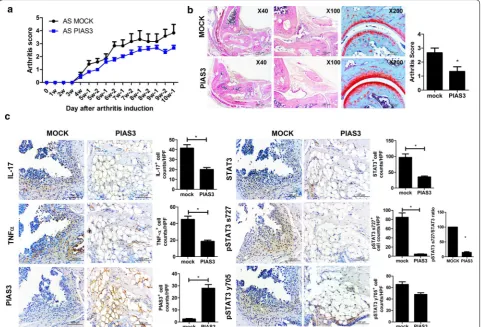
Beneficial effects of PIAS on peripheral arthritis in mice with SpA
The clinical and histologic scores of peripheral arthri-tis were assessed to determine the preventive effects of PIAS3 on SpA mice. The clinical scores of PIAS3-treated mice showed tendency to be suppressed than Mock-treated mice (n = 5 for each group, Fig. 1a). The arthritis score (histologic score of peripheral joints) of PIAS3-treated mice was significantly lower than that of Mock-treated mice, and a gross histology image revealed that the joint structure of PIAS3-treated mice was more preserved than that of Mock-treated mice (Fig. 1b). The pro-inflammatory cytokines IL-17, and TNF-α were substantially suppressed in PIAS3-treated mice. Total STAT3, pSTAT3 s727, and ratio of pSTAT3 s727 to total STAT3 were effectively suppressed in PIAS3-treated mice (Fig. 1c). These results indicate that PIAS3 attenu-ates the induction of peripheral arthritis in mice with SpA by reducing STAT3 signaling and the secretion of pro-inflammatory cytokines.
Beneficial effects of PIAS3 on gut inflammation in mice with SpA
Gut inflammation was assessed by measuring the total gut length, colitis score, ileitis score, and cytokine expres-sion in gut tissues. The difference in total gut length was minimal between the two groups; however, both ile-itis and colile-itis scores were significantly lower in
PIAS3-treated mice (Fig. 3a, b). Upon immunohistochemical
staining of the gut, the expression of IL-17, TNF-α, and total STAT3 was significantly reduced in PIAS3-treated mice relative to Mock-treated mice (Fig. 3c). On confocal staining analysis, Th17 population was significantly sup-pressed in PIAS3-treated mice (Fig. 3d). These findings are important because the SpA model from SKG mice exhibits Crohn’s disease-like features, indicating that PIAS3 attenuates the progression of gut inflammation by regulating the STAT3 pathway.
Effects of PIAS3 on Th17 and Treg cell differentiation in spleen
After sacrifice, spleen tissues were obtained to assess Th cell differentiation. The number of Th cells was counted, and CD4+IL-17+, CD4+p-STAT3(S727)+,
CD4+p-STAT3(Y705)+ cells, and ratio of
CD4+p-STAT3(S727)+ cells to CD4+STAT3+ cells were significantly diminished in PIAS3-treated mice relative to
Mock-treated mice, whereas CD4+CD25+FOXP3+ and
CD4+p-STAT5+ cells were increased in PIAS3-treated
mice relative to Mock-treated mice (Fig. 4a). Flow cyto-metric analysis showed downregulation of Th17 and upregulation of Treg in PIAS3-treated group than Mock-treated group (Fig. 4b, n = 11 for each group). Th1 dif-ferentiation did not show difference between two groups (Fig. 4b). PIAS3 was well encoded in CD4+ cell of spleen (Fig. 4c).
[image:4.595.58.540.89.416.2]Discussion
This study proved that PIAS3 has inhibitory effects on the progression of SpA in the aspect of peripheral arthritis and gut inflammation. The histologic scores of peripheral joints, the small intestine, and colon were all decreased in PIAS3-treated mice relative to Mock-treated mice. In addition, the expression of pro-inflammatory cytokines in peripheral joints and the gut was effectively reduced by PIAS3 treatment in mice with SpA. Treatment with PIAS3 decreased the expression of pro-inflammatory cytokines and osteoproliferative molecules in the axial joints. These findings could be explained by several mechanisms. First, PIAS3 decreased IL-17 and TNF-α secretion in the joints and gut. Second, PIAS3 regulated Th17 and Treg cells in the spleen.
IL-23 and IL-17 play crucial roles in the pathogenesis of SpA [20]. The major sources of IL-23 are monocytes and dendritic cells, and IL-23 controls immune cells of the
adaptive and innate immune systems to differentiate into IL-17-secreting cells [20]. IL-17-secreting cells vary and express other pathologic cytokines such as TNF-α, IL-22, and IFN-γ. Among these pathologic cytokines, IL-17 and TNF-α are assumed to be direct inductors of arthritis, enthesitis, and gut inflammation [2]. Blocking agents of IL-17 and TNF-α are already used as a therapeutic choice in axial SpA [21]. PIAS3 substantially suppressed IL-17 and TNF-α in the axial joints, peripheral joints, and gut. Therefore, direct inhibition of STAT3 may be a promising therapeutic choice for SpA treatment in the future.
STAT3 is a major transcriptional factor for Th17 differ-entiation, whereas STAT5 is a major transcriptional fac-tor for Treg differentiation [14]. Activated STAT proteins bind to intracellular genes and influence the epigenetic patterns and specify naïve CD4+ T cells to differentiate
into certain Th cells [13]. Activated STAT3 stimulates
[image:5.595.57.540.86.431.2]nuclear receptor γt, which is essential for Th17 induction, and suppresses the transcriptional factor Foxp3, which
induces Treg differentiation [22]. STAT3 and STAT5
share multiple binding sites on Il17 loci and compete for binding [22]. The relative ratio of activated STAT3 and STAT5 regulates Th17 cell lineage differentiation [22]. This study revealed that PIAS3 controls the expression of STAT3 and STAT5 molecules and regulates Th17 and Treg cell differentiation.
IL-17-producing cells vary, and many ongoing studies are working towards discovering major cell types that secrete IL-17 and the relative importance of the adaptive/ innate immune response in SpA pathogenesis. Myeloper-oxidase-positive neutrophils are dominant IL-17-positive cells in the facet joint, whereas c-Kit-positive mast cells comprise the major IL-17-positive cell population in the synovium [23, 24]. A recent study showed that mucosa-associated invariant T (MAIT) cells were elevated in the synovial fluid of patients with AS and presented the IL-17 phenotype [25]. Circulating Th17 cells were elevated in
patients with AS and even in patients with early axial SpA [7, 26, 27]. Previous studies have shown that a responder to TNF-α inhibition caused a decrease in circulating Th17 cells and an increase in circulating Treg cells and vice versa in a TNF-α inhibitor non-responder [28]. The transmission of SpA features to SCID mice occurs by transferring CD4+ cells from curdlan-induced SpA mice
[18]. The major pathologic immune cell in SpA remains
uncertain, but Th17 cells are one of the main pathologic cells. STAT3 signaling affects not only Th17 cell differen-tiation, but also MAIT and NKT cell differentiation [29]. We revealed that PIAS3 has an inhibitory function on Th17 cell polarization and the expression of pathologic cytokines. Although we focused on the Th cell subset in this study, further research studies should determine whether PIAS3 suppresses other IL-17-secreting cells, such as MAIT cells.
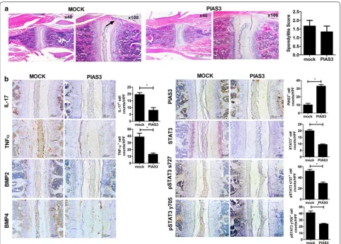
[image:6.595.58.541.89.420.2]eventually form bamboo spine. Syndesmophyte forma-tion is followed by several stages: inflammaforma-tion, erosion, and ankyloses [30]. It is assumed that the main patho-logic cytokines differ at each stage, and that Wnt and BMP signaling are the main pathways that act on the final syndesmophyte formation stage [30]. Observational studies have revealed that a TNF-α inhibitor slowed the radiologic damage of the spine in AS, albeit with some limitations [31–33]. First, these results were not from a randomized control trial. Second, enrolled patients were heterogenic in terms of disease duration. Sequential observation of the spinal magnetic resonance imaging study revealed that mature lesions (fatty degeneration) were at a higher risk for developing syndesmophyte than acute inflammatory lesions, and the results supported that SpA patients with mature lesions in the spine were
more susceptible to developing syndesmophyte [34].
Third, spinal progression was assessed by the modified Stoke ankylosing spondylitis Spine Score, which is not precise when detecting radiologic progression in AS. The effectiveness of TNF-α inhibition on preventing new bone formation is still under debate, and agents that defi-nitely block spinal progression have not been reported until now [35]. Mesenchymal stem cells from AS patients were superior to those from healthy controls in terms of osteogenic differentiation, and osteogenic differentiation capacity was regulated by BMP-2 and Noggin secretion [36]. Chen et al. reported that AS patients with spinal fusion had higher serum BMP-2, BMP-4, and BMP-7 lev-els than those without spinal fusion [37]. Recent research studies revealed that BMP may play a pivotal role in the “inflammation-driven syndesmophyte” theory by show-ing increased expression of BMP genes by TNF-α or IL-1β stimulation [38]. In this study, the expression of Fig. 4 PIAS3 reciprocally regulates the Th17/Treg cell imbalance. Spleen tissues were isolated from mice in each group at 10 weeks after curdlan injection. a Tissues were stained with specific Abs against CD4 (red), CD25 (blue), IL‑17 (green), FOXP3 (green), p‑STAT3 y705 (green), p‑STAT3 s727 (green), p‑STAT5 (green), and PIAS3 (green). Positive cells are shown in the bar graphs. b, c Splenocytes were stained with Abs against CD4–PerCP, IL‑17–FITC, IFNγ‑PE, CD25‑APC, FOXP3–FITC, and PIAS3‑APC to determine the presence of Th17, Tregs and PIAS3 expression on CD4+ T cells. Data
[image:7.595.58.539.86.436.2]BMP and TNF-α was reduced by PIAS3 in spine tissue. Further studies are needed to clarify the potential effects of PIAS3 on exaggerated new bone formation in SpA.
Conclusions
In conclusion, PIAS3 had beneficial effects in our curd-lan-induced SpA mouse model. PIAS3 diminished the histologic scores of peripheral arthritis and gut inflam-mation, and reduced the expression of pro-inflammatory cytokines in the axial/peripheral joints and the gut. In addition, BMP expression was attenuated by PIAS3 in axial joints, and PIAS3 reciprocally regulated Th17/Treg cell differentiation. These findings suggest that PIAS3 could serve as a therapeutic agent for SpA treatment.
Abbreviations
Abs: antibodies; APC: allophycocyanin; AS: ankylosing spondylitis; BMP: bone morphogenic protein; CD: cluster of differentiation; FITC: fluorescein isothio‑ cyanate; FOXP3: forkhead box P3; H&E: hematoxylin and eosin; IFN: interferon; IL: interleukin; i.p: intraperitoneal; MAIT: mucosa‑associated invariant T; NKT: natural killer T cells; perCP: peridin chlorophyll protein; PIAS3: protein inhibitor of activated STAT3; PE: phycoerythrin; RA: rheumatoid arthritis; SCID: severe combined immunodeficiency; SpA: spondyloarthritis; STAT : signal transducer and activator of transcription; Th: helper T cell; TNF‑α: tumor necrosis factor‑α; Treg: regulatory T cell.
Authors’ contributions
HKM designed the study, and drafted the manuscript. JWC collected data, and performed the analysis. SYL collected the data, helped the data analysis, and helped to draft the manuscript. HBS collected data, and performed analysis. KAJ collected data, and performed analysis. HSN helped the data analysis, and performed the statistical analysis. JGR collected data, and performed analysis. SKK supervised the study, and contributed to draft the manuscript. MLC participated in the study design, supervised the study. SHP supervised the study, and contributed to draft the manuscript. All authors read and approved the final manuscript.
Author details
1 Division of Rheumatology, Department of Internal Medicine, College of Med‑ icine, Seoul St. Mary’s Hospital, The Catholic University of Korea, Seoul 137‑070, South Korea. 2 Rheumatism Research Center, Catholic Research Institute of Medical Science, The Catholic University of Korea, Seoul 137‑040, South Korea. 3 Impact Biotech, Korea 505 Banpo‑Dong, Seocho‑Ku, Seoul 137‑040, South Korea. 4 Laboratory of Immune Network, Conversant Research Consor‑ tium in Immunologic Disease, College of Medicine, The Catholic University of Korea, Seoul, South Korea. 5 Rheumatism Research Center, Catholic Insti‑ tutes of Medical Science, The Catholic University of Korea, 222 Banpo‑Daero, Seocho‑gu, Seoul 137‑701, South Korea. 6 Division of Rheumatology, Depart‑ ment of Internal Medicine, College of Medicine, Seoul St. Mary’s Hospital, The Catholic University of Korea, 222 Banpo‑Daero, Seocho‑gu, Seoul 137‑701, South Korea.
Acknowledgements
None.
Competing interests
The authors declare that they have no competing interests.
Availability of data and materials
All data generated or analysed during this study are included in this published article.
Consent for publication
Not applicable.
Ethics approval and consent to participate
Not applicable.
Funding
This research was supported by a grant from the Korea Health Technology R&D Project through the Korea Health Industry Development Institute (KHIDI), funded by the Ministry of Health & Welfare, Republic of Korea (HI15C1062), Basic Science Research Program through the National Research Foundation of Korea (NRF) funded by the Ministry of Science, ICT & Future Planning (No. NRF‑ 2015R1C1A2A01051677) and the National Research Foundation of Korea (NRF) grant funded by the Korea government (MSIP) (NRF‑2017R1A2B3007688).
Publisher’s Note
Springer Nature remains neutral with regard to jurisdictional claims in pub‑ lished maps and institutional affiliations.
Received: 2 October 2018 Accepted: 4 January 2019
References
1. Ranganathan V, Gracey E, Brown MA, Inman RD, Haroon N. Pathogenesis of ankylosing spondylitis—recent advances and future directions. Nat Rev Rheumatol. 2017;13:359–67.
2. Smith JA, Colbert RA. Review: the interleukin‑23/interleukin‑17 axis in spondyloarthritis pathogenesis: Th17 and beyond. Arthritis Rheumatol. 2014;66:231–41.
3. Lubberts E. The IL‑23‑IL‑17 axis in inflammatory arthritis. Nat Rev Rheuma‑ tol. 2015;11:415–29.
4. Baeten D, Sieper J, Braun J, Baraliakos X, Dougados M, Emery P, et al. Secukinumab, an Interleukin‑17A inhibitor, in ankylosing spondylitis. N Engl J Med. 2015;373:2534–48.
5. Marzo‑Ortega H, Sieper J, Kivitz A, Blanco R, Cohen M, Martin R, et al. Secukinumab and sustained improvement in signs and symptoms of patients with active ankylosing spondylitis through two years: results from a phase III study. Arthritis Care Res (Hoboken). 2017;69:1020–9. 6. Zhang L, Li YG, Li YH, Qi L, Liu XG, Yuan CZ, et al. Increased frequencies
of Th22 cells as well as Th17 cells in the peripheral blood of patients with ankylosing spondylitis and rheumatoid arthritis. PLoS ONE. 2012;7:e31000.
7. Shen H, Goodall JC, Hill Gaston JS. Frequency and phenotype of periph‑ eral blood Th17 cells in ankylosing spondylitis and rheumatoid arthritis. Arthritis Rheum. 2009;60:1647–56.
8. Grant CR, Liberal R, Mieli‑Vergani G, Vergani D, Longhi MS. Regulatory T‑cells in autoimmune diseases: challenges, controversies and‑yet‑unan‑ swered questions. Autoimmun Rev. 2015;14:105–16.
9. Ji W, Li H, Gao F, Chen Y, Zhong L, Wang D. Effects of Tripterygium glycosides on interleukin‑17 and CD4(+)CD25(+)CD127(low) regulatory T‑cell expression in the peripheral blood of patients with ankylosing spondylitis. Biomed Rep. 2014;2:517–20.
10. Duan Z, Gui Y, Li C, Lin J, Gober HJ, Qin J, et al. The immune dysfunction in ankylosing spondylitis patients. Biosci Trends. 2017;11:69–76.
11. Bidad K, Salehi E, Jamshidi A, Saboor‑Yaraghi AA, Oraei M, Meysamie A, et al. Effect of all‑transretinoic acid on Th17 and T regulatory cell subsets in patients with ankylosing spondylitis. J Rheumatol. 2013;40:476–83. 12. Min HK, Kim JK, Lee SY, Kim EK, Lee SH, Lee J, et al. Rebamipide prevents
peripheral arthritis and intestinal inflammation by reciprocally regulating Th17/Treg cell imbalance in mice with curdlan‑induced spondyloarthritis. J Transl Med. 2016;14:190.
13. O’Shea JJ, Lahesmaa R, Vahedi G, Laurence A, Kanno Y. Genomic views of STAT function in CD4 + T helper cell differentiation. Nat Rev Immunol. 2011;11:239–50.
14. Dong C. TH17 cells in development: an updated view of their molecular identity and genetic programming. Nat Rev Immunol. 2008;8:337–48. 15. Miossec P, Korn T, Kuchroo VK. Interleukin‑17 and type 17 helper T cells. N
•fast, convenient online submission •
thorough peer review by experienced researchers in your field • rapid publication on acceptance
• support for research data, including large and complex data types •
gold Open Access which fosters wider collaboration and increased citations maximum visibility for your research: over 100M website views per year •
At BMC, research is always in progress.
Learn more biomedcentral.com/submissions
Ready to submit your research? Choose BMC and benefit from:
16. Lee SH, Moon SJ, Park MJ, Kim EK, Moon YM, Cho ML. PIAS3 sup‑ presses acute graft‑versus‑host disease by modulating effector T and B cell subsets through inhibition of STAT3 activation. Immunol Lett. 2014;160:79–88.
17. Tang X, Yin K, Zhu H, Tian J, Shen D, Yi L, et al. Correlation between the expression of microRNA‑301a‑3p and the proportion of Th17 cells in patients with rheumatoid arthritis. Inflammation. 2016;39:759–67. 18. Ruutu M, Thomas G, Steck R, Degli‑Esposti MA, Zinkernagel MS, Alexander
K, et al. beta‑glucan triggers spondylarthritis and Crohn’s disease‑like ileitis in SKG mice. Arthritis Rheum. 2012;64:2211–22.
19. Han ES, Oh JY, Park HJ. Cordyceps militaris extract suppresses dextran sodium sulfate‑induced acute colitis in mice and production of inflam‑ matory mediators from macrophages and mast cells. J Ethnopharmacol. 2011;134:703–10.
20. Burns RC, Rivera‑Nieves J, Moskaluk CA, Matsumoto S, Cominelli F, Ley K. Antibody blockade of ICAM‑1 and VCAM‑1 ameliorates inflammation in the SAMP‑1/Yit adoptive transfer model of Crohn’s disease in mice. Gastroenterology. 2001;121:1428–36.
21. van der Heijde D, Ramiro S, Landewe R, Baraliakos X, Van den Bosch F, Sepriano A, et al. 2016 update of the ASAS‑EULAR management recom‑ mendations for axial spondyloarthritis. Ann Rheum Dis. 2017;76:978–91. 22. Yang XP, Ghoreschi K, Steward‑Tharp SM, Rodriguez‑Canales J, Zhu
J, Grainger JR, et al. Opposing regulation of the locus encoding IL‑17 through direct, reciprocal actions of STAT3 and STAT5. Nat Immunol. 2011;12:247–54.
23. Appel H, Maier R, Wu P, Scheer R, Hempfing A, Kayser R, et al. Analysis of IL‑17(+) cells in facet joints of patients with spondyloarthritis sug‑ gests that the innate immune pathway might be of greater relevance than the Th17‑mediated adaptive immune response. Arthritis Res Ther. 2011;13:R95.
24. Noordenbos T, Yeremenko N, Gofita I, van de Sande M, Tak PP, Canete JD, et al. Interleukin‑17‑positive mast cells contribute to synovial inflamma‑ tion in spondylarthritis. Arthritis Rheum. 2012;64:99–109.
25. Gracey E, Qaiyum Z, Almaghlouth I, Lawson D, Karki S, Avvaru N, et al. IL‑7 primes IL‑17 in mucosal‑associated invariant T (MAIT) cells, which contribute to the Th17‑axis in ankylosing spondylitis. Ann Rheum Dis. 2016;75:2124–32.
26. Jandus C, Bioley G, Rivals JP, Dudler J, Speiser D, Romero P. Increased numbers of circulating polyfunctional Th17 memory cells in patients with seronegative spondylarthritides. Arthritis Rheum. 2008;58:2307–17. 27. Jansen DT, Hameetman M, van Bergen J, Huizinga TW, van der Heijde D,
Toes RE, et al. IL‑17‑producing CD4 + T cells are increased in early, active
axial spondyloarthritis including patients without imaging abnormalities. Rheumatology (Oxford). 2015;54:728–35.
28. Xueyi L, Lina C, Zhenbiao W, Qing H, Qiang L, Zhu P. Levels of circulat‑ ing Th17 cells and regulatory T cells in ankylosing spondylitis patients with an inadequate response to anti‑TNF‑alpha therapy. J Clin Immunol. 2013;33:151–61.
29. Wilson RP, Ives ML, Rao G, Lau A, Payne K, Kobayashi M, et al. STAT3 is a critical cell‑intrinsic regulator of human unconventional T cell numbers and function. J Exp Med. 2015;212:855–64.
30. Tam LS, Gu J, Yu D. Pathogenesis of ankylosing spondylitis. Nat Rev Rheu‑ matol. 2010;6:399–405.
31. Haroon N, Inman RD, Learch TJ, Weisman MH, Lee M, Rahbar MH, et al. The impact of tumor necrosis factor alpha inhibitors on radiographic progression in ankylosing spondylitis. Arthritis Rheum. 2013;65:2645–54. 32. Baraliakos X, Haibel H, Listing J, Sieper J, Braun J. Continuous long‑term
anti‑TNF therapy does not lead to an increase in the rate of new bone for‑ mation over 8 years in patients with ankylosing spondylitis. Ann Rheum Dis. 2014;73:710–5.
33. Molnar C, Scherer A, Baraliakos X, de Hooge M, Micheroli R, Exer P, et al. TNF blockers inhibit spinal radiographic progression in ankylosing spon‑ dylitis by reducing disease activity: results from the Swiss Clinical Quality Management cohort. Ann Rheum Dis. 2018;77:63–9.
34. Baraliakos X, Heldmann F, Callhoff J, Listing J, Appelboom T, Brandt J, et al. Which spinal lesions are associated with new bone formation in patients with ankylosing spondylitis treated with anti‑TNF agents? A long‑term observational study using MRI and conventional radiography. Ann Rheum Dis. 2014;73:1819–25.
35. Magrey MN, Khan MA. The paradox of bone formation and bone loss in ankylosing spondylitis: evolving new concepts of bone formation and future trends in management. Curr Rheumatol Rep. 2017;19:17. 36. Xie Z, Wang P, Li Y, Deng W, Zhang X, Su H, et al. Imbalance between
bone morphogenetic protein 2 and noggin induces abnormal osteo‑ genic differentiation of mesenchymal stem cells in ankylosing spondylitis. Arthritis Rheumatol. 2016;68:430–40.
37. Chen HA, Chen CH, Lin YJ, Chen PC, Chen WS, Lu CL, et al. Association of bone morphogenetic proteins with spinal fusion in ankylosing spondyli‑ tis. J Rheumatol. 2010;37:2126–32.