R E S E A R C H
Open Access
Increase of plasma IL-9 and decrease of plasma IL-5,
IL-7, and IFN-g
in patients with chronic heart failure
Claudia Cappuzzello
1†, Luca Di Vito
2†, Roberta Melchionna
3, Guido Melillo
3, Lorena Silvestri
4, Eleonora Cesareo
5,
Filippo Crea
2, Giovanna Liuzzo
2, Antonio Facchiano
3, Maurizio C Capogrossi
3, Monica Napolitano
3*Abstract
Background:Several cytokines are associated with the development and/or progression of chronic heart failure (CHF). Our aim was to look more closely at the cytokine networks involved in CHF, and to assess whether disease etiology affects cytokine expression. The study population was comprised of a) 69 patients with stable CHF, New York Heart Association (NYHA) II/IV classes, secondary to ischaemic (ICM) and non ischaemic dilated (NIDCM) cardiomyopathy and b) 16 control subjects. We analyzed and compared the plasma levels of 27 pro- and anti-inflammatory mediators, in the study population and assessed for any possible correlation with echocardiographic parameters and disease duration.
Methods:27 cytokines and growth factors were analyzed in the plasma of ICM- (n = 42) and NIDCM (n = 27) NYHA class II-IV patients vs age- and gender-matched controls (n = 16) by a beadbased multiplex immunoassay. Statistical analysis was performed by ANOVA followed by Tukey post-hoc test for multiple comparison.
Results:Macrophage inflammatory protein (MIP)-1b, Vascular endothelial growth factor (VEGF), interleukin (IL)-9, Monocyte chemotactic protein (MCP)-1, and IL-8 plasma levels were increased in both ICM and NIDCM groups vs controls. In contrast, IL-7, IL-5, and Interferon (IFN)-gwere decreased in both ICM and NIDCM groups as compared to controls. Plasma IL-6 and IL-1bwere increased in ICM and decreased in NIDCM, vs controls, respectively. IL-9 levels inversely correlated, in ICM patients, with left ventricular ejection fraction (LVEF) while IL-5 plasma levels inversely correlated with disease duration, in NYHA III/IV ICM patients.
This is the first time that both an increase of plasma IL-9, and a decrease of plasma IL-5, IL-7 and IFN-ghave been reported in ICM as well as in NIDCM groups, vs controls. Interestingly, such cytokines are part of a network of genes whose expression levels change during chronic heart failure. The altered expression levels of MIP-1b, VEGF, MCP-1, IL-1b, IL-6, and IL-8, found in this study, are in keeping with previous reports.
Conclusions:The increase of plasma IL-9, and the decrease of plasma IL-5, IL-7 and IFN-gin ICM as well as in NIDCM groups vs controls may contribute to get further insights into the inflammatory pathways involved in CHF.
Background
The role of inflammation in the pathogenesis and pro-gression of chronic heart failure (CHF) is well estab-lished [1,2]. Typical hallmarks for the involvement of immune mechanisms in CHF pathogenesis are the acti-vation/migration of leukocytes from the circulation to the areas of myocardial inflammation and an increased expression of proinflammatory cytokines, such as tumor
necrosis factora, interleukin-1, and interleukin-6 from a damaged myocardium [3-6]. In heart failure such cyto-kine levels increase in association with disease severity and may contribute to impair cardiac function through cardiomyocyte apoptosis, inflammatory response, cardiac hypertrophy and matrix metalloproteinase activation [1,7-9].
Proinflammatory cytokines may cause myocyte apop-tosis and necrosis; interleukin-6 induces a hypertrophic response in myocytes [10], whereas TNF-acauses left ventricular dilatation, apparently through the activation of matrix metalloproteinases [11]. Elevated levels of CXC and C-C chemokines such as GROa, IL-8, MCP-1,
* Correspondence: [email protected] †Contributed equally
3
Laboratorio di Patologia Vascolare, Istituto Dermopatico dell’ Immacolata-IRCCS, Rome, Italy
Full list of author information is available at the end of the article
RANTES and MIP-1a were also found in the most severe cases of heart failure, which would suggest a pos-sible role for such molecules in CHF progression [12,13]. Peripheral blood mononuclear cells are impor-tant players in the CHF inflammatory process [14,15]. However, the mechanisms that, in heart disease, ulti-mately cause the transition from cardiac disease to heart failure still need further investigation.
The identification of the cytokines associated with chronic heart failure is not only important to get deeper insights into the inflammatory pathways involved, but it could also lead to the identification of disease’s biomar-kers. The biomarkers found in heart failure include BNP, NT pro-BNP, C-reactive protein, tumor necrosis factora, interleukins 1 and 6, TNF-a, matrix metallo-proteinases, neuroepinephrine, renin, galectin-3 [16-18], some of which are also important tools in the diagnosis and pathogenesis of heart failure, in the identification of subjects at risk of heart failure, risk stratification, thera-peutic agents response monitoring [17].
The aim of this study was to investigate the inflamma-tory pathways associated with CHF, and to assess whether disease etiology affects cytokine cascades. To this end, we analyzed plasma cytokine levels in patients with stable ischaemic or non ischaemic dilated cardio-myopathy CHF vs healthy donors, by a multiplex cyto-kine/growth factor immunoassay, and then assessed their relationship to echocardiographic parameters and disease duration.
Materials and methods
Subjects
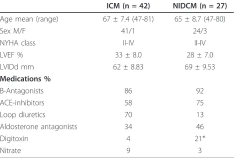
Sixty-nine patients with chronic stable heart failure for six consecutive months were enrolled at the Cardiology Unit of the Catholic University of Sacred Heart of Rome out of 210 patients admitted to the unit during the same period. Baseline patient demographic variables, heart failure classification and echocardiographic para-meters of patients are shown in Table 1. CHF patients were classified in New York Heart Association (NYHA) functional class II/IV. The causes of heart failure were classified as 1) ICM in patients (n = 42) with a history of myocardial infarction and coronary atherosclerosis with a stenosis >70% in at least one major coronary artery branch or 2) NIDCM in patients (n = 27) with no history of myocardial infarction and angiografically nor-mal coronary arteries. NIDCM is defined as patients with dilated cardiomyopathy, with LVEF less than 40%, in the absence of ischaemic or congenital heart disease, or cardiac valve disease. LVEF and left ventricular inter-nal diastolic diameter (LVIDd) were calculated from 2-D and M-mode transthoracic echocardiographic images. Duration of disease was calculated as the time between the first ICM or NIDCM diagnosis and enrolment in
our study. Exclusion criteria were infections, malignan-cies, autoimmune disorders, diabetes, pulmonary disease, myocardial infarction, unstable angina or myocarditis during the 6 months prior to enrollment in the study and blood sample collection. Sixteen age- and sex-matched controls were recruited at the Clinical Labora-tory at IDI-IRCCS from randomly chosen individuals with no overt cardiac disease and dyslipidemia. Inflam-matory indexes, i.e. C-reactive protein (CRP) and erytro-cyte sedimentation rate (ESR), were within physiologic ranges and no leukocytosis was observed.
The study complies with The Helsinki Declaration and was approved by the Ethics Committee of the Catholic University and all subjects gave written informed consent.
Sample collection and management
Venous blood samples (20 ml) from ICM and NIDCM patients and controls were collected in pyrogen-free, heparinized tubes between 8.00 am and 10.00 am and immediately centrifuged at 3000 rpm for 15 minutes. Plasma samples were aliquoted and stored at -80°C. Samples were frozen and thawed only once.
Multiplex immunoassay
We used a human cytokine 27-Bio-Plex assay kit (BioRad Laboratories, Milan, Italy), a beadbased multi-plex immunoassay. This technology has the capacity to measure several cytokines/cytokine receptors and growth factors simultaneously in small volumes of plasma or serum with a greater detection dynamic range (~ 1-10,000 pg/mL) than single ELISAs [15-18] with high accuracy and sensitivity [19-22].
[image:2.595.305.540.101.259.2]The factors analysed were: IL1-b, IL-1 receptor antagonist (ra), IL-2, IL-4, IL-5, IL-6, IL-7, IL-8, IL-9, IL-10, IL-12, IL-15, IL-16, 1L-17, basic fibroblast growth factor (FGF), eotaxin, granulocyte colony stimulating Table 1 Patient characteristics
ICM (n = 42) NIDCM (n = 27)
Age mean (range) 67 ± 7.4 (47-81) 65 ± 8.7 (47-80)
Sex M/F 41/1 24/3
NYHA class II-IV II-IV
LVEF % 33 ± 8.0 28 ± 7.0
LVIDd mm 62 ± 8.83 69 ± 9.53
Medications %
Β-Antagonists 86 92
ACE-inhibitors 58 75
Loop diuretics 70 13
Aldosterone antagonists 34 46
Digitoxin 4 21*
Nitrate 9 3
factor (G-CSF), granulocyte-macrophage colony stimu-lating factor (GM-CSF), IFN-g, Interferong-inducible protein (IP)-10, MCP-1, MIP-1a, MIP-1 b, Platelet-derived growth factor (PDGF)-BB, Regulated on Activa-tion Normal T-cell-Expressed and Secreted (RANTES), Tumor necrosis factor (TNF) a and VEGF. The lower detection limit was 0.2-19.3 pg/ml (see http://www.bio-rad.com/webroot/web/pdf/lsr/literature/Bulletin_3157. pdf). We tested the plasma concentrations of the above molecules in the whole study population. The plasma samples were diluted 1:4 and tested in duplicates. The samples were read on a Bio-Plex 200 instrument equipped with the software Bioplex Manager, version 4.1, using a five-parameters not-linear regression for-mula to compute sample concentrations from the stan-dard curves.
The pubgene program http://www.pubgene.org was used to evaluate the expression and/or functional corre-lation between cytokines and growth factors.
Statistical analysis
All the major study variables evaluated in the present study were not normally distributed; thus logarithmic transformation was applied to the data to allow para-metric techniques to be used. The Bivariate Pearson correlation coefficient (r) was calculated between cyto-kine plasma levels and echocardiografic parameters or disease duration. Statistical analysis between ICM, NIDCM and CTRL was performed by ANOVA followed by Tukey’s post-hoc test for multiple comparison. No significant differences were detected in the three groups in terms of age and gender (chi-square p value >0.05). The cytokines and growth factor values have been expressed as median and interquartile range and a p value <0.05 was considered statistically significant. Pro-portions (Table 1), were compared by Fisher’s exact test.
Results
Plasma cytokine profiles in ICM and NIDCM CHF patients vs controls
Multiplex immunoassay screening for cytokine and growth factor protein expression, by means of the multi-plex XMAP technology, was performed on ICM and NIDCM CHF patients (Table 1) and on control plasma.
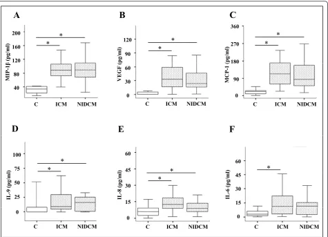
The first group of factors was composed of MIP-1b, VEGF, MCP-1, IL-9, and IL-8, (Figure 1A-E); the levels of these cytokines were increased in patients with ICM as well as in NIDCM with respect to controls (p < 0.05). Further, the IL-6 cytokine (panel F) was significantly increased in ICM patients vs healthy controls (p < 0.05). The cytokines and growth factor values have been expressed as median and interquartile range.
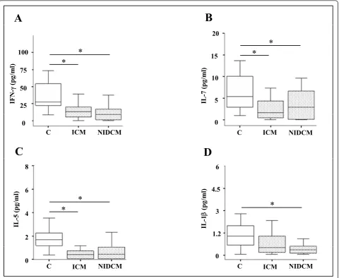
The second group included IFN-g, IL-7, and IL-5, whose levels were significantly lower in the plasma of
ICM and NIDCM patients vs healthy individuals, see Figure 2 (panel A-C). IL-1 b, on the other hand, was only decreased in the NIDCM patients vs controls (Fig-ure 2 panel D) (*p < 0.05).
In our study, several factors, i.e. MIP-1 b, VEGF, MCP-1, IL-8 and IL-6 were found to be modulated, in CHF patients, in accordance to previous reports [9,19]. Only a few of the 27 cytokines and growth factors, i.e. IL-2, IL-15, IL-17, bFGF, and MIP-1ashowed undetect-able levels in all three groups, except for sporadic cases. All other detectable molecules, except the ones descri-bedFigure 1 and 2 showed no statistically significant var-iation between either ICM or NIDCM groups, and controls (data not shown).
However, what was noteworthy, and described here for the first time, was the increase of IL-9, and decrease of IL-5, IL-7 and IFN-g plasma levels in patients with chronic heart failure.
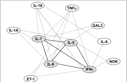
Using the bioinformatic analysis from the http://www. pubgene.org program, we showed, in Figure 3 that the molecules newly associated with chronic heart failure (gray ovals), are linked to each other in terms of expres-sion and/or function (black thick lines), and to a num-ber of molecules, namely Norepinephrine, Endothelin-1, IL-1A, Galectin-3, TNFa, IL-6 and IL-18 [13], which play a key role in CHF (white ovals).
Correlation of cytokine plasma leves with
echocardiographic parameters and disease duration
In order to assess if our findings were clinically signifi-cant, we correlated IL-5, IL-7, IL-9 and IFN-gplasma levels with the patients’echocardiographic parameters, such as LVEF and LVIDd, and with disease duration.
Interestingly, in ICM patients, IL-9 levels inversely correlated with left ventricular ejection fraction (LVEF) (Table 2). Furthermore, IL-5 plasma levels inversely cor-related with disease duration in NYHA III/IV ICM patients (Table 2). Neither IL-7 nor IFN ginstead corre-lated with echocardiographic parameters or disease duration, and no correlation was found between NIDCM patients and the above mentioned parameters. Further, no significant association was found between modulated cytokines and LVIDd (data not shown).
Discussion
In our study, we described two different modulation patterns, i.e. increased or decreased cytokine plasma levels, in chronic heart failure (ICM and NIDCM) patients as compared to controls. The first group was composed of MIP-1b, MCP-1, IL-8, VEGF and IL-9, that were increased in both the ICM and NIDCM patients vs controls.
The second group was composed of IL-5, IL-7, IFN-g, which showed lower plasma levels in both the ICM and NIDCM patients as compared to controls. Furthermore, IL-6 increased only in ICM while IL-1bdecreased only in NIDCM, both changes with respect to controls. Pre-vious study results on IL-1b systemic level changes in heart failure have been inconsistent and reported as either unchanged [25], increased [26], or decreased (pre-sent study) in such patients.
Several previous reports have described the role of cytokines and growth factors in the development and/or progression of chronic heart failure [1,5,9,11]; further, a number of such cytokines are regarded as disease
biomarkers [17,18]. It is worth noting that our study, for the first time, has associated IL-5, IL-7, IL-9, and IFN-g plasma levels with chronic heart failure.
It has been reported that IL-5 cardiac protein levels increase four days after Myocardial Infarction (MI) in a mouse model [27]; on the contrary, as yet no data is available on IL-5 serum or plasma levels in chronic heart failure patients. IL-7 is produced by stromal cells in lymphoid tissues and is required for T cell develop-ment and persistence in the periphery. Interestingly, local production of IL-7 has been associated with the maintenance and predominance of CD8+ T cells, which cause tissue damage in human chronic Chagas’disease cardiomyopathy, an inflammatory-dilated cardiomyopa-thy [28]. IL-9 is a mostly T-cell produced cytokine that has a functional role in allergic disease and resistance to intestinal nematodes [29], but no data is yet available on its role in heart disease. IFN-g is an important pro-inflammatory cytokine produced by Th1 cells, that increases the expression of MHC class I and class II
360
A
B
C
160 200
120 90
120
270 360
180
ȕ
(pg/ml) (pg/ml) (pg/ml)
*
*
*
*
*
*
40 80
0 30 60
0 90 180
MIP-1
ȕ
VEGF
(
MCP-1
(
C ICM NIDCM C ICM NIDCM C ICM NIDCM
F
E
D
75 100
45 60
60
)
*
) l)*
*
*
25 50
45
15 30
45
15 30
IL-9 (pg/ml
)
IL-8 (pg/ml
)
IL-6 (pg/m
l
*
C ICM NIDCM C ICM NIDCM
C ICM NIDCM 0
[image:4.595.63.539.88.431.2]0 0
molecules. An increase in the percentage of IFN-g -posi-tive CD4 (+) T cells, has previously been described in patients with CHF [30]. Furthermore, an increase in IFN
gserum levels has also been described [31], in a mouse model, one week after MI, i.e. not in chronic heart fail-ure experimental conditions.
Our study recorded decreased IL-5 plasma levels in both ICM and NIDCM groups vs controls. Interestingly, IL-5 plasma levels inversely correlated with disease duration in NYHA III/IV ICM patients. In fact, patients with longer disease duration had smaller concentrations of plasmatic IL-5 in comparison with patients more recently diagnosed. Furthermore, a statistically signifi-cant correlation with IL-5 plasma levels was found only among NYHA III/IV ICM patients, but not among patients with less severe chronic heart failure. This
would suggest that decreased levels of this cytokine are associated with disease progression.
We here showed an increase in IL-9 levels in both ICM and NIDCM groups vs controls and that IL-9 levels inversely correlated with ejection fraction in ICM patients, but not in NIDCM patients. This would sug-gest that increased levels of this cytokine are associated with left ventricular dysfunction and, thus, with disease progression toward organ failure in ICM patients. Further, we reported a decrease in plasma IFN-glevels and of IL-7 in both ICM and NIDCM CHF patients vs controls, but no correlation with echocardiographic parameters or duration of disease was found (not shown).
Nevertheless, our conclusions cannot broadly apply to the entire chronic heart failure population as only a
A
B
100
75
20
15
m
l)
*
)*
*
*
75
50
25
10
5
IFN-J
(pg/
m
IL-7 (pg/ml
)
0 0
C ICM NIDCM C ICM NIDCM
D
C
8
6
6
4.5
/ml)
*
g/ml)*
4
2
3
1.2
IL-5 (pg
/
IL-1
E
(p
g
*
*
0 0
[image:5.595.59.540.89.483.2]C ICM NIDCM C ICM NIDCM
Figure 2Cytokine plasma levels decreased in ICM and NIDCM CHF patients vs controls. Plasma levels of IFN-g(A), IL-7 (B), IL-5 (C) and IL-1
small number of women were enrolled and analysed in both the ICM and NIDCM groups.
Interestingly, the cytokines that were identified in the present study are part of a gene network of expression/ function regulation that link them to Norepinephrine, Endothelin-1, IL-1A, Galectin-3, TNFa, IL-6 and IL-18 [13], which are molecules associated with chronic heart failure [1,4,7,10,11,17,18]; thus we hypothesize that IL-5,
IL-7, IL-9 and IFN-g are part of signaling cascades that contribute to the development and/or progression of chronic heart failure.
Notwithstanding the ICM and NIDCM difference in pathogenetic causes there was a similar cytokine expres-sion pattern when compared to controls. In fact, among all cytokines previously reported as being associated with CHF, our study showed only IL-6 and IL-1b modi-fication to be associated uniquely with ICM or NIDCM, respectively. All the other cytokines instead similarly increased or decreased, vs controls, regardless of the etiology. Further, we found that IL-1Ra was able to dis-criminate between the two patient groups, since it was statistically higher in the ICM group, although only a small increase was observed (data not shown).
In conclusion, the novelty in our study results is the increase of IL-9 and decrease of IL-5, IL-7, and IFN-g plasma levels, in both ICM and NIDCM CHF patients vs controls and the inverse correlation of IL-5 and IL-9 plasma levels with duration of disease and LVEF, respec-tively, in ICM patients. Further studies are needed to assess whether the modification of IL-5, IL-7, IL-9 and
IL-18
TNF
D
IL-1A
GAL3
IL-5
IL-6
IL-7
IFN
J
IL-9
NOR
IFN
J
[image:6.595.55.540.89.403.2]ET-1
Figure 3Gene networks in CHF. The lines link genes whose expression or function is affected by neighbouring genes. The gene network has been generated using the PUBGENE program http://www.pubgene.org. White ovals: genes previously known to be modulated in CHF. Grey ovals: genes found to be regulated in CHF in the present study. Endothelin-1 (ET-1), Galectin 3 (GAL3), Norepinephrine (NOR), Tumor necrosis factor (TNF)a, Interferon (IFN)-g, Interleukin (IL) 1A, 5, 6, 7, 9, 18.
Table 2 Correlation between cytokine plasma levels and duration of disease or ejection fraction
Cytokine IL-5 IL-9
Duration of disease r = -0.475 (p = 0.05) n.s. Left Ventricular Ejection
Fraction
n.s. r = -0.364 (p = 0.02)
A negative Pearson’s correlation coefficient (r) indicates that cytokine expression inversely correlates with the variable in exam (e.g. : patients with longer disease duration had smaller concentrations of plasmatic IL-5 in comparison with patients more recently diagnosed).
[image:6.595.56.291.611.658.2]IFN-gplasma levels has a null, a protective, or a patho-genetic role in chronic heart failure and may constitute a potential target for therapeutic intervention.
Acknowledgements
This work was supported by Ministry of Health [grant # RF 05 conv 66 to M. C.C.]. We thank Ann Anthony for excellent editorial assistance.
Author details
1Laboratorio di Biologia Vascolare e Medicina Rigenerativa, Centro
Cardiologico Monzino-IRCCS, Milan, Italy.2Institute of Cardiology, Catholic University, Rome, Italy.3Laboratorio di Patologia Vascolare, Istituto
Dermopatico dell’Immacolata-IRCCS, Rome, Italy.4Laboratorio di Analisi,
Istituto Dermopatico dell’Immacolata-IRCCS, Rome, Italy.5Laboratorio di
Ingegneria tissutale e Fisiopatologia cutanea, Istituto Dermopatico dell’Immacolata-IRCCS, Rome, Italy.
Authors’contributions
MN conceived the study design, coordinated the study and drafted the manuscript. CC isolated plasmas, performed statistical analysis and contributed to drafting the manuscript. LDV collected the clinical data of patients and performed statistical analysis. FC, GL, MCC and RM critically revised the manuscript’s draft. GM performed statistical analysis, LS enrolled patients. EC and AF performed and interpreted the multiplex assay. All authors read and approved the final manuscript.
Competing interests
The authors declare that they have no competing interests.
This article conforms to ethics in the authorship and publishing of scientific articles [32].
Received: 6 December 2010 Accepted: 21 March 2011 Published: 21 March 2011
References
1. Levine B, Kalman J, Mayer L, Fillit HM, Packer M:Elevated circulating levels
of tumor necrosis factor in severe chronic heart failure.N Engl J Med
1990,323:236-236.
2. Mann DL, Young JB:Basic mechanisms in congestive heart failure.
Recognizing the role of proinflammatory cytokines.Chest1994,
105:897-904.
3. Torre-Amione G:Immune activation in chronic heart failure.Am J Cardiol 2005,95:3C-8C, discussion 38C-40C.
4. Naito Y, Tsujino T, Fujioka Y, Ohyanagi M, Okamura H, Iwasaki T:Increased
circulating interleukin-18 in patients with congestive heart failure.Heart
2002,88:296-7.
5. Boerrigter G, Costello-Boerrigter LC, Burnett JC Jr:Natriuretic peptides in
the diagnosis and management of chronic heart failure.Heart Fail Clin
2009,5:501-14.
6. Marriott JB, Goldman JH, Keeling PJ, Baig MK, Dalgleish AG, McKenna WJ: Abnormal cytokine profiles in patients with idiopathic dilated
cardiomyopathy and their asymptomatic relatives.Heart1996,75:287-90.
7. Aukrust P, Ueland T, Lien E, Bendtzen K, Muller F, Andreassen AK,et al: Cytokine network in congestive heart failure secondary to ischemic or
idiopathic dilated cardiomyopathy.Am J Cardiol1999,83:376-82.
8. Rauchhaus M, Doehner W, Francis DP, Davos C, Kemp M, Liebenthal C, et al:Plasma cytokine parameters and mortality in patients with chronic
heart failure.Circulation2000,102:3060-7.
9. Seta Y, Shan K, Bozkurt B, Oral H, Mann DL:Basic mechanisms in heart
failure: the cytokine hypothesis.J Card Fail1996,2:243-9.
10. Janssen SP, Gayan-Ramirez G, Van den Bergh A, Herijgers P, Maes K, Verbeken E,et al:Interleukin-6 causes myocardial failure and skeletal
muscle atrophy in rats.Circulation2005,111:996-1005.
11. Bradham WS, Bozkurt B, Gunasinghe H, Mann D, Spinale FG:Tumor necrosis factor-alpha and myocardial remodeling in progression of heart
failure: a current perspective.Cardiovasc Res2002,53:822-30.
12. Aukrust P, Ueland T, Muller F, Andreassen AK, Nordoy I, Aas H,et al: Elevated circulating levels of C-C chemokines in patients with
congestive heart failure.Circulation1998,97:1136-43.
13. Hayashidani S, Tsutsui H, Shiomi T, Ikeuchi M, Matsusaka H, Suematsu N, et al:Anti-monocyte chemoattractant protein-1 gene therapy attenuates left ventricular remodeling and failure after experimental myocardial
infarction.Circulation2003,108:2134-40.
14. Yndestad A, Damas J, Geir Eiken H, Holm T, Haug T, Simonsen S,et al: Increased gene expression of tumor necrosis factor superfamily ligands in peripheral blood mononuclear cells during chronic heart failure. Cardiovasc Res2002,54:175-82.
15. Cappuzzello C, Napolitano M, Arcelli D, Melillo G, Melchionna R, Di Vito L, et al:Gene expression profiles in peripheral blood mononuclear cells of
chronic heart failure patients.Physiol Genomics2009,38:233-40.
16. Anand IS, Latini R, Florea VG, Kuskowski MA, Rector T, Masson S,et al: C-reactive protein in heart failure: prognostic value and the effect of
valsartan.Circulation2005,112:1428-34.
17. Braunwald E:Biomarkers in heart failure.N Engl J Med2008,358:2148-59. 18. Emdin M, Vittorini S, Passino C, Clerico A:Old and new biomarkers of
heart failure.Eur J Heart Fail2009,11:331-5.
19. Elshal MF, McCoy JP:Multiplex bead array assays: performance evaluation
and comparison of sensitivity to ELISA.Methods2006,38:317-23.
20. Ray CA, Bowsher RR, Smith WC, Devanarayan V, willey MB, Brandt JT, Dean RA:Development, validation, and implementation of a multiplex immunoassay for the simultaneous determination of five cytokines in
human serum.J Pharm Biomed Anal2005,36:1037-44.
21. Khan SS, Smith MS, Reda D, Suffredini AF, McCoy JP Jr:Multiplex bead array assays for detection of soluble cytokines: comparisons of sensitivity and quantitative values among kits from multiple
manufacturers.Cytometry B Clin Cytom2004,61:35-9.
22. Chowdhury F, Williams A, Johnson P:Validation and comparison of two multiplex technologies, Luminex and Mesoscale Discovery, for human
cytokine profiling.J Immunol Methods2009,340:55-64.
23. Inoue T, Komoda H, Nonaka M, Kameda M, Uchida T, Node K:Interleukin-8 as an independent predictor of long-term clinical outcome in patients
with coronary artery disease.Int J Cardiol2008,124:319-25.
24. Hoffman SA, Wang L, Shah CV, Ahya VN, Pochettino A, Olthoff K,et al: Plasma cytokines and chemokines in primary graft dysfunction
post-lung transplantation.Am J Transplant2009,9:389-96.
25. Munger MA, Johnson B, Amber IJ, Callahan KS, Gilbert EM:Circulating concentrations of proinflammatory cytokines in mild or moderate heart
failure secondary to ischemic or idiopathic dilated cardiomyopathy.Am J
Cardiol1996,77:723-7.
26. Testa M, Yeh M, Lee P, Fanelli R, Loperfido F, Berman JW,et al:Circulating levels of cytokines and their endogenous modulators in patients with mild to severe congestive heart failure due to coronary artery disease or
hypertension.J Am Coll Cardiol1996,28:964-71.
27. Timmers L, Sluijter JP, van Keulen JK, Hoefer IE, Nederhoff MG, Goumans MJ,et al:Toll-like receptor 4 mediates maladaptive left ventricular remodeling and impairs cardiac function after myocardial
infarction.Circ Res2008,102:257-64.
28. Fonseca SG, Reis MM, Coelho V, Nogueira LG, Monteiro SM, Mairena EC, et al:Locally produced survival cytokines IL-15 and IL-7 may be associated to the predominance of CD8+ T cells at heart lesions of
human chronic Chagas disease cardiomyopathy.Scand J Immunol2007,
66:362-71.
29. Soroosh P, Doherty TA:Th9 and allergic disease.Immunology2009, 127:450-8.
30. Fukunaga T, Soejima H, Irie A, Sugamura K, Oe Y, Tanaka T,et al:Expression of interferon-gamma and interleukin-4 production in CD4+ T cells in
patients with chronic heart failure.Heart Vessels2007,22:178-83.
31. Vistnes M, waehre A, Nygard S, Sjaastad I, Andersson KB, Husberg C, Christensen G:Circulating cytokine levels in mice with heart failure are
etiology dependent.J Appl Physiol2010,108:1357-64.
32. Shewan LG, Coats AJ:Ethics in the authorship and publishing of scientific
articles.Int J Cardiol2010,144:1-2.
doi:10.1186/1479-5876-9-28
Cite this article as:Cappuzzelloet al.:Increase of plasma IL-9 and