RESEARCH
A prospective study on the changes
and clinical significance of pre-operative
and post-operative circulating tumor cells
in resectable gastric cancer
Qiyue Zhang
1†, Fei Shan
2†, Ziyu Li
2, Jing Gao
1, Yilin Li
1, Lin Shen
1, Jiafu Ji
2*and Ming Lu
1*Abstract
Background: Circulating tumor cells (CTCs) have been suggested as potential prognostic indicators for multiple tumors, including gastric cancer; however, pre- and post-operative CTC changes in resectable gastric cancer and pos-sible correlations to post-operative recurrence have not been evaluated.
Methods: Subjects (n = 93) with resectable gastric cancer were prospectively reviewed from July 2013 to December 2014 at Peking University Cancer Hospital. The proportion of CTCs were evaluated before (n = 93) and after (n = 63) radical operation using a standardized CellSearch system.
Results: CTCs ≥ 1 were measured in the pre-operative blood of 31 (33.3%) patients and in the post-operative blood of 21 patients (33.3%). Patients with relatively poor clinicopathological features had more pre- and post-operative CTCs. The 3-year disease-free survival (DFS) rate for patients with CTCs ≥ 5/7.5 ml was significantly lower than for patients with CTCs < 5/7.5 ml (40.0% vs 66.4%, p < 0.001 for pre-surgery; 25.0% vs 62.2%, p < 0.001 for post-surgery). Patients with CTCs ≥ 5/7.5 ml in post-operative blood had significantly shorter mean DFS (1.28 vs 31.6 months; p = 0.002) and overall survival (OS; 10.0 vs 34.9 months; p = 0.001) than other patients. Among the 10 patients with hematogenous recurrence, 3 had post-operative CTCs ≥ 2/7.5 ml and had early recurrence (DFS 1.1, 1.1, 1.4 months). Moreover, DFS for the seven patients was 20.2, 11.9, 20.0, 6.0, 15.5, 25.9, 30.0 months, respectively. DFS for the three patients with increased CTCs after surgery was shorter than for patients with mildly increased, stable, or decreased CTCs.
Conclusions: Pre- and post-operative CTCs are promising prognostic markers for resectable gastric cancer. Our study further suggests that increased post-operative CTCs may be correlated with hematogenous recurrence.
Trial registration (ClinicalTrials.gov Identifier: NCT01848015). Registered 7 May 2013. https ://clini caltr ials.gov/ct2/show/ NCT01 84801 5
Keywords: CellSearch, Circulating tumor cells, Hematogenous metastasis, Recurrence, Resectable gastric cancer
© The Author(s) 2018. This article is distributed under the terms of the Creative Commons Attribution 4.0 International License (http://creat iveco mmons .org/licen ses/by/4.0/), which permits unrestricted use, distribution, and reproduction in any medium, provided you give appropriate credit to the original author(s) and the source, provide a link to the Creative Commons license, and indicate if changes were made. The Creative Commons Public Domain Dedication waiver (http://creat iveco mmons .org/ publi cdoma in/zero/1.0/) applies to the data made available in this article, unless otherwise stated.
Open Access
*Correspondence: jiafuj@hotmail.com; qiminglu_mail@126.com †Qiyue Zhang and Fei Shan are co-first authors
1 Key Laboratory of Carcinogenesis and Translational Research (Ministry of Education/Beijing), Department of GI Oncology, Peking University Cancer Hospital & Institute, Fucheng Road 52, Haidian District, Beijing 100142, China
Background
Gastric cancer is a highly malignant disease, with high morbidity and mortality rates worldwide, especially in China [1]. Surgery is the most common curative approach for resectable gastric cancer; however, many patients, especially those with local advanced gastric cancer suffer from recurrence after radical gastrectomy, contributing to poor prognosis [2, 3]. Presently, poor clinicopatho-logical characteristics (such as TNM classification, depth of tumor penetration, and lymph node metastasis) and high carcinoembryonic antigen (CEA) are predictive of high-risk post-operative tumor recurrence; but their use is limited due to low specificity and accuracy [4–7]. In addition to predicting patient prognosis, monitoring recurrence patterns of gastric cancer patients is vital. Hematogenous distant metastasis is a chief pattern of recurrence for gastric cancer patients. Yoo and colleagues confirmed that the mean time for hematogenous recur-rence was the shortest [8]; however, TNM classification could not predict recurrence patterns of gastric cancer accurately. Therefore, the identification of effective mark-ers is necessary to predict disease recurrence.
Circulating tumor cells (CTCs) are important markers of malignant tumor metastasis [9, 10] that can predict chemotherapeutic response and prognosis for multiple metastatic tumors [11–17]. In addition, pre- or post-operative CTCs have been reported to be correlated to post-operative recurrence of bladder, prostate, breast, and colorectal cancers, but data reported differ among studies [18–22]. Gazzaniga’s group reported that CTCs may predict decreased time to first tumor recurrence for stage I bladder cancer [21], but Pal and colleagues reported no correlation existed between CTCs and tumor recurrence for prostate cancer [22].
Correlations in pre- or operative CTCs to post-operation recurrence and prognosis for gastric can-cer are also limited. Uenosono and colleagues analyzed CTCs in pre-operative peripheral blood samples of 148 patients who underwent gastrectomy, and intact CTCs in preoperative blood were significantly correlated with poor clinicopathological characteristics (depth of tumor invasion, lymph node metastasis, etc.) [23]. In addi-tion, relapse-free and overall survival for patients with CTCs was significantly lower than for patients with-out CTCs (p < 0.001) [23]. Of note, pre-operative CTCs was an independent factor for overall survival (OS) for patients with gastric cancer according to multivariate analysis (p = 0.024). However, whether post-operative CTCs and changes in CTCs during surgery could be assessed for resectable gastric cancer (stage I–III) and any correlations with prognosis is unclear. Thus, we con-ducted this prospective study (ClinicalTrials.gov Identi-fier: NCT01848015), and measured CTCs in pre- and
post-operative blood from patients with resectable gas-tric cancer and analyzed correlations of pre- and post-operative CTCs and further evaluated changes in CTCs during surgery with clinicopathological characteristics, prognosis, and recurrence patterns.
Methods
Patients and sample collection
From July 2013 to December 2014, 93 subjects with his-tologically confirmed gastric cancer who underwent a radical gastrectomy at the Department of Surgery in Peking University Cancer Hospital were enrolled. All of the patients were diagnosed with stage I–III gastric can-cer. We excluded patients with distant metastases or those had undergone therapy prior to surgery, and 63 subjects had paired preoperative and postoperative blood samples. Peripheral blood was sampled within 1 week prior to sur-gery and post-operative samples were acquired a week after surgery. CTCs were counted for all of the subjects.
Isolation and enumeration of circulating tumor cells
Circulating tumor cells were counted with a CellSearch system (Veridex, New Jersey, U.S.) according to previously published methods [17], which included a CellSearch Epi-thelial Cell Kit, CellPrep System, and CellSpotter Analyzer. In brief, 7.5 ml blood was drawn into 10-ml customized Cell Save Vacutainer tubes (Becton–Dickinson, New Jer-sey, U.S.), including EDTA and a cell fixative, followed by CTC enumeration within 72 h. Reagents used included anti-epithelial cell adhesion molecule (anti-EpCAM) antibody-coated magnetic beads, fluorescent dye-labeled anti-CD45 antibody and anti-cytokeratins (CK-8, -18, and -19) antibody, and 4′, 6-diamidino-2-phenylindole (DAPI). CTCs identified with the CellSearch system were CK-positive, DAPI-positive, and CD45-negative.
Statistical analysis and illustrations
Results
Patient characteristics
Patients (n = 93) with resectable gastric cancer were prospectively assessed. Patient characteristics are presented in Table 1. In total, 68 male and 25 female patients were included in this study, with the median
age of 60 years (26–82 years). Twenty-nine (31.2%) patients had primary tumors located in gastroesopha-geal junction, and 71 patients (76.3%) had tumors with poor differentiation (including low differentia-tion, mucinous adenocarcinoma, and signet-ring cell carcinoma). The number of patients with stage I, II,
Table 1 Characteristics of patients
a EGJ, gastroesophageal junction
b Good, including high or moderate differentiation; Poor, including low differentiation, mucinous adenocarcinoma, and signet-ring cell carcinoma c carcinoembryonic antigen
d non-available
Characteristics Pre-operation (n = 93) Post-operation (n = 63)
No. of Patients (%) No. of Patients (%)
Gender
Male 68 (73.1%) 46 (73.0%)
Female 25 (26.9%) 17 (27.0%)
Age (years)
≤ 45 16 (17.2%) 13 (20.6%)
> 45 77 (82.8%) 50 (79.4%)
Primary sitea
EGJ 29 (31.2%) 19 (30.2%)
Non-EGJ 64 (68.8%) 44 (69.8%)
Differentiationb
Good 22 (23.7%) 15 (23.8%)
Poor 71 (76.3%) 48 (76.2%)
Lauren
Intestinal 36 (38.7%) 25 (39.7%)
Diffuse 23 (24.7%) 16 (25.4%)
Mixed 34 (36.6%) 22 (34.9%)
TNM classification
I 24 (25.8%) 15 (23.8%)
II 24 (25.8%) 16 (25.4%)
III 45 (48.4%) 32 (50.8%)
Depth of penetration
T1/T2 30 (32.3%) 19 (30.2%)
T3 34 (36.6%) 25 (39.7%)
T4 29 (31.2%) 19 (30.2%)
Lymph node
N0 31 (33.3%) 16 (25.4%)
N1/N2 31 (33.3%) 26 (41.3%)
N3 31 (33.3%) 21 (33.3%)
Lymph-vascular invasion
Yes 45 (48.4%) 34 (54.0%)
No 48 (51.6%) 29 (46.0%)
CEAc level before operation (ng/ml)
< 5 75 (80.6%) 51 (81.0%)
≥ 5 17 (18.3%) 12 (19.0%)
NAd 1 (1.1%) 0 (0.0%)
Adjuvant chemotherapy
Yes 56 (60.2%) 42 (66.7%)
[image:3.595.58.541.194.680.2]and III were 24 (25.8%), 24 (25.8%), and 45 (48.4%), respectively.
Correlations of CTCs to clinicopathological characteristics
For preoperation blood collection, ≥ 1, ≥ 2, ≥ 3, ≥ 4, and ≥ 5 CTCs/7.5 ml were found in 31 (33.3%), 13 (14.0%), 9 (9.7%), 6 (6.5%), and 5 (5.4%) patients, respectively. In addition, for postoperation blood collection, ≥ 1, ≥ 2, ≥ 3, ≥ 4, and ≥ 5 CTCs/7.5 ml were found in 21 (33.3%), 11 (17.5%), 10 (15.9%), 8 (12.7%), and 4 (6.3%) patients, respectively. Preoperative CTCs data are presented in Table 2 and Fig. 1. CTCs were significantly (p < 0.05) correlated with Lauren, TNM classification, depth of penetration, and lymph-vascular invasion (Fig. 1a–d). However, there was no significant (p > 0.05) correla-tion between CTCs and age, differentiacorrela-tion, lymph node metastasis, and CEA levels prior to surgery (Fig. 1e–h). Moreover, patients no older than 45 years-of-age, with low differentiation and lymph node metastasis were more likely to have more CTCs. Additional file 1: Table S1 depicts patients CTCs data pre- and post-surgery. Post-operative CTCs were correlated with patient characteris-tics in a manner similar to pre-operative CTCs (Table 3). Patients with relatively poor clinicopathological features had more post-operative CTCs, and this was significantly correlated with depth of penetration (p < 0.05) and non-significantly correlated with TNM classification (p > 0.05; Fig. 2). Ten patients had ≥ 3 CTCs/7.5 ml in post-oper-ative samples and most had poor differentiation (n = 8), stage IIB/III (n = 10), or T3/T4 (n = 10) penetration.
Correlations of CTCs enumeration with DFS and OS in resectable gastric cancer
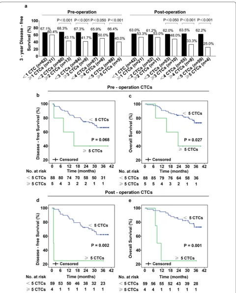
Until November 2016, median follow-up was 36.4 [inter-quartile 33.9–38.7] months. In total, 31 (33.3%) patients relapsed, 24 (25.8%) patients died. Patients CTC and DFS data are presented in Additional file 2: Table S2. In our study, patients with gastroesophageal junc-tion, T3/T4 penetrajunc-tion, lymph node metastases, CEA level ≥ 5 ng/ml and higher pre- or postoperative CTCs had lower 3-year DFS (Table 2 and Fig. 3a). The 3-year DFS was 100.0% in patients with stage I tumors, 68.3% in patients with stage II tumors, 44.0% in patients with stage III tumors (Table 2). As shown in Fig. 3a, patients who had more CTCs had lower 3-year DFS. Obviously, as the threshold increased, the 3-year DFS of patients with post-operative CTCs above each cut-off value decreased markedly. Moreover, 3-year DFS in patients with CTCs ≥ 5/7.5 ml was significantly lower than in patients with CTCs < 5/7.5 ml (40.0% vs 66.4%, p < 0.001 for preop-eration CTCs; 25.0% vs 62.2%, p < 0.001 for postoppreop-eration CTCs; Fig. 3a). In addition, patients with ≥ 5 CTCs/7.5 ml blood pre-operatively had shorter DFS (p = 0.068) and OS
(p = 0.027) as assessed by univariate analyses (Fig. 3b, c). Likewise, patients with CTCs ≥ 5/7.5 ml in postoperative blood had significantly shorter DFS (1.28 vs 31.6 months; p = 0.002) and OS (10.0 vs 34.9 months; p = 0.001) than patients with CTCs < 5/7.5 ml (Fig. 3d, e). These results indicated that postoperative CTCs may be an effective prognostic marker in resectable gastric cancer; there-fore, more attention and proper adjuvant chemotherapy should be given to patients who have higher post-opera-tive CTCs.
Changes of CTCs during surgery to recurrence pattern
Among the 31 patients who relapsed, 10 patients had hematogenous (osseous, hepatic and lung) metastases, and 14 patients had non-hematogenous (peritoneal and locoregional) metastases. The remaining seven patients had no identified hematogenous recurrence or non-hematogenous recurrence until death. We found that pre-operative CTCs were not correlated with the recur-rence pattern (Additional file 3: Figure S1). Patients with hematogenous metastases who had post-operative CTCs ≥ 2/7.5 ml had markedly shorter DFS (Fig. 4a). The DFS of three patients with hematogenous metasta-ses who had markedly increased CTCs after surgery was short (1.1, 1.1, and 1.4 months, respectively, Fig. 4b); however, the DFS of other patients was relatively longer (25.9, 20.2, 11.9, 6.0, 30.0, 20.0 and 15.5 months, respec-tively). For patients with non-hematogenous metastases, the post-operative CTCs and changes of CTCs during surgery were not correlated with DFS. Post-operative CTC and markedly increased CTC during surgery may have potential association with hematogenous metastasis and suggest early recurrence.
Discussion
Surgery is the first-line therapy for gastric cancer, espe-cially for patients with resectable gastric cancer; however, tumor recurrence within 5 years suggests a poor progno-sis [2]. The 5-year DFS for patients with stage II, IIIA, and IIIB gastric cancer are 64.4, 50.0, and 34.4%, respectively [2]. Poor clinicopathological features and high CEA and CA-19-9 tumor markers are risk factors for post-opera-tive tumor recurrence, even though these markers have limited clinical significance [4–7]. Thus, there is a need to distinguish patients with high post-operative recurrence risk with effective biomarkers.
Table 2 Correlation of preoperative CTC number to clinicopathological characteristics
Italic values indicate that there are significant differences in pre-operative CTCs between different clinicopathological types
a EGJ, gastroesophageal junction
b Good, including high or moderate differentiation; Poor, including low differentiation, mucinous adenocarcinoma, and signet-ring cell carcinoma c carcinoembryonic antigen
d non-available
Characteristics 3-year recurrence
rate% No. of patients (%)
CTC ≥ 1/7.5 ml CTC ≥ 2/7.5 ml CTC ≥ 3/7.5 ml CTC ≥ 4/7.5 ml CTC ≥ 5/7.5 ml
Gender
Male (n = 68) 65.6 23 (33.8%) 9 (13.2%) 6 (8.8%) 5 (7.4%) 5 (7.4%)
Female (n = 25) 63.1 8 (32.0%) 4 (16.0%) 3 (12.0%) 1 (4.0%) 0 (0.0%)
p value 0.658 0.869 0.997 0.949 1.000 0.319
Age
≤ 45 (n = 16) 58.3 6 (37.5%) 4 (25.0%) 4 (25.0%) 2 (12.5%) 1 (6.3%)
> 45 (n = 77) 66.2 25 (32.5%) 9 (11.7%) 5 (6.5%) 4 (5.2%) 4 (5.2%)
p value 0.244 0.698 0.317 0.070 0.274 1.000
Primary sitea
EGJ (n = 29) 50.4 9 (31.0%) 3 (10.3%) 2 (6.9%) 2 (6.9%) 2 (6.9%)
Non-EGJ (n = 64) 71.2 22 (34.3%) 10 (15.6%) 7 (10.9%) 4 (6.3%) 3 (4.7%)
p value 0.002 0.873 0.787 0.873 1.000 0.635
Differentiationb
Good (n = 22) 71.8 4 (18.2%) 1 (4.5%) 1 (4.5%) 0 (0.0%) 0 (0.0%)
Poor (n = 71) 62.6 27 (38.0%) 12 (16.9%) 8 (11.3%) 6 (8.5%) 5 (7.0%)
p value 0.174 0.084 0.268 0.604 0.330 0.335
Lauren
Intestinal (n = 36) 68.0 8 (22.2%) 2 (5.6%) 1 (2.8%) 0 (0.0%) 0 (0.0%)
Diffuse (n = 23) 67.4 6 (26.1%) 5 (21.7%) 5 (21.7%) 3 (13.0%) 2 (8.7%)
Mixed (n = 34) 60.0 17 (50.0%) 6 (17.6%) 3 (8.8%) 3 (8.8%) 3 (8.8%)
p value 0.434 0.033 0.118 0.060 0.068 0.148
TNM classification
I (n = 24) 100.0 6 (25.0%) 0 (0.0%) 0 (0.0%) 0 (0.0%) 0 (0.0%)
II (n = 24) 68.3 8 (33.3%) 4 (16.7%) 2 (8.3%) 1 (4.2%) 1 (4.2%)
III (n = 45) 44.0 17 (37.8%) 9 (20.0%) 7 (15.6%) 5 (11.1%) 4 (8.9%)
p value 0.000 0.563 0.047 0.103 0.306 0.443
Depth of penetration
T1/T2 (n = 30) 90.0 8 (26.7%) 1 (3.3%) 1 (3.3%) 1 (3.3%) 1 (3.3%)
T3 (n = 34) 56.9 12 (35.3%) 5 (14.7%) 1 (2.9%) 0 (0.0%) 0 (0.0%)
T4 (n = 29) 47.7 11 (37.9%) 7 (24.1%) 7 (24.1%) 5 (17.2%) 4 (13.8%)
p value 0.000 0.627 0.064 0.007 0.009 0.026
Lymph node
N0 (n = 31) 96.8 7 (22.6%) 2 (6.5%) 1 (3.2%) 0 (0.0%) 0 (0.0%)
N1/N2 (n = 31) 58.4 9 (29.0%) 3 (9.7%) 2 (6.5%) 2 (6.5%) 2 (6.5%)
N3 (n = 31) 38.9 15 (48.4%) 8 (25.8%) 6 (19.4%) 4 (12.9%) 3 (9.7%)
p value 0.000 0.081 0.109 0.133 0.160 0.362
Lymph-vascular invasion
Yes (n = 45) 53.0 20 (44.4%) 9 (20.0%) 6 (13.3%) 3 (6.7%) 3 (6.7%)
No (n = 48) 75.8 11 (22.9%) 4 (8.3%) 3 (6.3%) 3 (6.3%) 2 (4.2%)
p value 0.001 0.028 0.105 0.307 1.000 0.671
CEAc level before operation (ng/ml)
< 5 (n = 75) 68.8 26 (34.7%) 12 (16.0%) 9 (12.0%) 6 (8.0%) 5 (6.7%)
≥ 5 (n = 17) 46.3 5 (29.4%) 1 (5.9%) 0 (0.0%) 0 (0.0%) 0 (0.0%)
NAd (n = 1) 0 (0.0%) 0 (0.0%) 0 (0.0%) 0 (0.0%) 0 (0.0%)
[image:5.595.57.536.112.666.2]with non-detectable CTCs [23]. Therefore, we measured pre- and post-operative CTCs and changes in CTCs dur-ing surgery in patients with resectable gastric cancer and correlated these changes with clinicopathological fea-tures, post-operative tumor recurrence, and recurrence patterns.
Circulating tumor cells measured in 31 patients prior to surgery were greater than those reported in another study using a prospective cohort [23]. Differences in clin-icopathological features between our and other cohorts may explain this discrepancy, such as differences in TNM classification and depth of tumor penetration. In our study, 63 patients had paired pre- and opera-tive samples and CTCs were found in 21 patients post-operatively. These data demonstrated that CTCs may still appear in circulation after radical surgery. Thus, long-term monitoring CTCs after surgery may predict post-operative tumor recurrence.
Patients with relatively poor clinicopathological fea-tures had greater pre- and post-operative CTCs. Poor clinicopathological features were correlated with high post-operative tumor recurrence risk and poor prog-nosis, suggesting that patients with higher pre- or post-operative CTCs had earlier recurrence. Until now, studies of correlations in CTCs with recurrence of resectable gastric cancer have been limited, so there is no standard threshold. To address this issue, we set CTC counts of 1, 2, 3, 4, and 5 as thresholds and found that patients with pre-surgical CTCs ≥ 4 had obviously but not significantly shorter DFS (p = 0.459) and OS (p = 0.209) than patients without CTC or patients with 1–3 CTCs (Additional file 4: Figure S2). The median DFS of patients without CTC, 1–3 CTCs, or more than 4 CTCs post-operatively were 33.87, 34.33, and 20.98 months respectively. As the threshold increased, we observed that pre-surgical
CTCs ≥ 5 were significantly associated with worse out-comes (Fig. 3a–e). For this reason, we set 5 as the thresh-old value.
Since the prognostic value of pre-operative CTCs is likely to be affected by surgery, we suggest that post-operative CTCs might be a more direct and efficient recurrence marker than pre-operative CTCs. However, few reports have questioned whether monitoring post-operative CTCs is useful for predicting prognosis. Our results demonstrated that patients with post-operative CTCs ≥ 4–5 had shorter DFS, OS, and less 3-years DFS (Fig. 3a–e, Additional file 4: Figure S2). In addition, act-ing as the “seed” of hematogenous metastasis, CTCs can travel in the circulation and form metastases [9, 10]. High post-operative CTCs and markedly increased post-oper-ative CTCs were correlated with hematogenous metas-tasis, while pre-operative CTCs were not correlated with recurrence patterns. Our results also confirmed that post-operative CTCs might be a more direct post-opera-tive recurrence marker especially for patients who experi-enced hematogenous recurrence. As a result, monitoring the recurrence patterns of gastric cancer is important for better post-operative adjuvant treatment decisions.
There are several limitations in our study. First, our study aimed to analyze the clinical significance of CTCs in radical gastric cancer, and patients with stage I and II accounted for 51.6% of all of the patients. Furthermore, CTCs ≥ 3–5 were identified in a few patients. For future work, more subjects are needed to determine an optimal threshold, and a sufficient sample size to adequately perform Kaplan–Meier analyses. Second, mesenchymal-like cancer cells are likely to escape from CellSearch system detection, which is based on epithelial markers, such as EpCAM and CKs [17]. Nonetheless, in future studies, we can
[image:7.595.58.540.88.253.2]Table 3 Correlation of postoperative CTC number to clinicopathological characteristics
Italic values indicate that there are significant differences in post-operative CTCs between different clinicopathological types
a EGJ, gastroesophageal junction
b Good, including high or moderate differentiation; Poor, including low differentiation, mucinous adenocarcinoma, and signet-ring cell carcinoma c carcinoembryonic antigen
Characteristics 3-year recurrence rate%
No. of patients (%)
CTC ≥ 1/7.5 ml CTC ≥ 2/7.5 ml CTC ≥ 3/7.5 ml CTC ≥ 4/7.5 ml CTC ≥ 5/7.5 ml
Gender
Male (n = 46) 60.9 18 (39.1%) 10 (21.7%) 9 (19.6%) 7 (15.2%) 3 (6.5%) Female (n = 17) 57.5 3 (17.6%) 1 (5.9%) 1 (5.9%) 1 (5.9%) 1 (5.9%)
p value 0.606 0.108 0.272 0.352 0.574 1.000 Age
≤ 45 (n = 13) 57.1 5 (38.5%) 2 (15.4%) 2 (15.4%) 2 (15.4%) 1 (7.7%) > 45 (n = 50) 60.6 16 (32.0%) 9 (18.0%) 8 (16.0%) 6 (12.0%) 3 (6.0%)
p value 0.565 0.912 1.000 1.000 1.000 1.000 Primary sitea
EGJ (n = 19) 43.5 7 (36.8%) 3 (15.8%) 3 (15.8%) 3 (15.8%) 1 (5.3%) Non-EGJ (n = 44) 67.2 14 (31.8%) 8 (18.2%) 7 (15.9%) 5 (11.4%) 3 (6.8%)
p value 0.001 0.698 1.000 1.000 0.943 1.000 Differentiationb
Good (n = 15) 66.0 5 (33.3%) 3 (20.0%) 2 (13.3%) 1 (6.7%) 0 (0.0%) Poor (n = 48) 57.8 16 (33.3%) 8 (16.7%) 8 (16.7%) 7 (14.6%) 4 (8.3%)
p value 0.244 1.000 1.000 1.000 0.719 0.564 Lauren
Intestinal (n = 25) 62.0 8 (32.0%) 5 (20.0%) 4 (16.0%) 2 (8.0%) 0 (0.0%) Diffuse (n = 16) 65.7 4 (25.0%) 3 (18.8%) 3 (18.8%) 3 (18.8%) 2 (12.5%) Mixed (n = 22) 52.4 9 (40.9%) 3 (13.6%) 3 (13.6%) 3 (13.6%) 2 (9.1%)
p value 0.115 0.580 0.844 0.913 0.505 0.167 TNM classification
I (n = 15) 100.0 2 (13.3%) 0 (0.0%) 0 (0.0%) 0 (0.0%) 0 (0.0%) II (n = 16) 54.7 5 (31.3%) 4 (25.0%) 3 (18.8%) 2 (12.5%) 1 (6.3%) III (n = 32) 43.1 14 (43.8%) 7 (21.9%) 7 (21.9%) 6 (18.8%) 3 (9.4%)
p value 0.062 0.117 0.106 0.161 0.249 0.798 Depth of penetration
T1/T2 (n = 19) 89.5 2 (10.5%) 0 (0.0%) 0 (0.0%) 0 (0.0%) 0 (0.0%) T3 (n = 25) 51.2 10 (40.0%) 4 (16.0%) 3 (12.0%) 3 (12.0%) 1 (4.0%) T4 (n = 19) 41.4 9 (47.4%) 7 (36.8%) 7 (36.8%) 5 (26.3%) 3 (15.8%)
p value 0.000 0.036 0.008 0.005 0.043 0.178 Lymph node
N0 (n = 16) 100.0 4 (25.0%) 2 (12.5%) 1 (6.3%) 0 (0.0%) 0 (0.0%) N1/N2 (n = 26) 54.5 8 (30.8%) 6 (23.1%) 6 (23.1%) 5 (19.2%) 2 (7.7%) N3 (n = 21) 33.3 9 (42.9%) 3 (14.3%) 3 (14.3%) 3 (14.3%) 2 (9.5%)
p value 0.001 0.488 0.761 0.375 0.185 0.663 Lymph-vascular invasion
Yes (n = 34) 49.8 10 (29.4%) 5 (14.7%) 5 (14.7%) 3 (8.8%) 2 (5.9%) No (n = 29) 71.4 11 (37.9%) 6 (20.7%) 5 (17.2%) 5 (17.2%) 2 (6.9%)
p value 0.002 0.475 0.533 1.000 0.453 1.000 CEAc level before operation (ng/ml)
< 5 (n = 51) 62.6 18 (35.3%) 9 (17.6%) 9 (17.6%) 8 (15.7%) 4 (7.8%)
≥ 5 (n = 12) 48.6 3 (25.0%) 2 (16.7%) 1 (8.3%) 0 (0.0%) 0 (0.0%)
[image:8.595.59.537.99.677.2]combine other methods of molecular analysis to bet-ter identify the prognostic value of CTCs in resect-able gastric cancer. Third, numerous studies agree that intra-operative CTCs shedding occurs with tumor manipulation [24]. However, our data suggested that post-operative CTC enumeration within 1 week after surgery was not increased by surgery. Although intra-operative CTCs were not detected in our study, stud-ies of other tumors reported that CTC detection rate and not CTC enumeration increased in intra-operative
samples [24]. Moreover, studies also demonstrated that increased intra-operative CTCs normalize post-operatively within days to weeks [24]. Fourth, we col-lected post-operative samples within 1 week after surgery (2–7 days after surgery). We are also interested in how CTCs can change when the length of obser-vation is increased to two or 3 weeks, which we will address in follow-up studies. Interestingly, Krag and colleagues reported that CTCs rapidly declined during 48 h post-operatively in most patients with operable
[image:10.595.61.540.86.549.2]breast cancer [25]. Animal studies also found the simi-lar phenomenon [26]. Krag et al. also demonstrated that 30% patients had detectable CTCs persistently up to 14 days after surgery. These results are consist-ent with the notion that only a minority of CTCs can survive and have capacity to be clonogenic. In addi-tion, those authors further put forward a view, which is similar to us, namely, the persistent presence of cancer cells after radical surgery is a strong indicator of recurrence. Therefore, we hypothesize that CTCs are stable from 2 days to 2 weeks, and CTCs should be monitored before and after surgery to predict can-cer recurrence and for disease staging and treatment management.
Conclusion
In summary, we analyzed the utility of post-operative CTCs in resectable gastric cancer. In addition, to the best of our knowledge, this is the first prospective study that has analyzed the correlations between post-operative CTCs and changes in CTCs during surgery with clinicopathological characteristics, prognosis, and recurrence patterns. We found that post-opera-tive CTCs might be a more direct and efficient recur-rence marker than pre-operative CTCs and increased post-operative CTCs might be correlated with hema-togenous recurrence. Monitoring the post-operative CTCs and changes in CTCs during surgery is of great importance for better post-operative adjuvant treat-ment decisions.
Abbreviations
CTC : circulating tumor cell; CEA: carcinoembryonic antigen; OS: overall survival; EpCAM: epithelial cell adhesion molecule; CK: cytokeratin; DAPI: 4′, 6-diamidino-2-phenylindole; DFS: disease-free survival; 3-year DFS: 3-year disease-free survival rate.
Authors’ contributions
ML designed the study. QZ and FS summarized the data and drafted the manuscript. All of the authors contributed to the revision of manuscript. All authors read and approved the final manuscript.
Additional files
Additional file 1: Table S1. Patient characteristics and CTC numbers in 63 patients.
Additional file 2: Table S2. Patient characteristics and CTC numbers in 31 relapsed patients.
Additional file 3: Figure S1. Correlations of pre-operative CTCs with patient recurrence patterns.
Additional file 4: Figure S2. Patients who have ≥ 4 CTCs/7.5 ml blood
pre-operatively had shorter DFS (a, c) and OS (b, d).
Acknowledgements
We thank LetPub for its linguistic assistance during the preparation of this manuscript.
Competing interests
The authors declare that they have no competing interests.
Availability of data
The data sets analyzed within the current study are available from the cor-responding author upon reasonable request.
Consent for publication Not applicable.
Ethics approval and consent to participate
All of the procedures followed were in accordance with the ethical standards of the responsible committee on human experimentation (institutional and national) and with the Helsinki Declaration of 1964 and later versions. Informed consent or an appropriate substitute was obtained from all of the patients prior to inclusion in the study.
Funding
This work was funded by Capital Health Research and Development (No. 2014-4-1023), the Beijing Natural Science Foundation (7161002), and the Beijing Municipal Administration of Hospital Clinical Medicine Development of Special Funding Support (ZYLX201701).
Publisher’s Note
Springer Nature remains neutral with regard to jurisdictional claims in pub-lished maps and institutional affiliations.
Received: 26 January 2018 Accepted: 7 June 2018
References
1. Siegel R, Ma J, Zou Z, Jemal A. Cancer statistics, 2014. CA Cancer J Clin. 2014;64:9–29.
2. Sasako M, Sakuramoto S, Katai H, et al. Five-year outcomes of a randomized phase III trial comparing adjuvant chemotherapy with S-1 versus surgery alone in stage II or III gastric cancer. J Clin Oncol. 2011;29:4387–93.
3. Park SH, Sohn TS, Lee J, et al. Phase III trial to compare adjuvant chemo-therapy with capecitabine and cisplatin versus concurrent chemoradio-therapy in gastric cancer: final report of the adjuvant chemoradiochemoradio-therapy in stomach tumors trial, including survival and subset analyses. J Clin Oncol. 2015;33:3130–6.
4. Sun Z, Wang ZN, Zhu Z, et al. Evaluation of the seventh edition of American Joint Committee on Cancer TNM staging system for gastric cancer: results from a Chinese monoinstitutional study. Ann Surg Oncol. 2012;19:1918–27.
5. Li Z, Zou X, Xie L, et al. Personalizing risk stratification by addition of PAK1 expression to TNM staging: improving the accuracy of clinical decision for gastroesophageal junction adenocarcinoma. Int J Cancer. 2015;136:1636–45.
6. Duffy MJ, Lamerz R, Haglund C, et al. Tumor markers in colorectal cancer, gastric cancer and gastrointestinal stromal cancers: european group on tumor markers 2014 guidelines update. Int J Cancer. 2014;134:2513–22. 7. Cristescu R, Lee J, Nebozhyn M, et al. Molecular analysis of gastric cancer
identifies subtypes associated with distinct clinical outcomes. Nat Med. 2015;21:449–56.
8. Yoo CH, Noh SH, Shin DW, Choi SH, Min JS. Recurrence following curative resection for gastric carcinoma. Br J Surg. 2000;87:236–42.
9. Reymond N, d’Água BB, Ridley AJ. Crossing the endothelial barrier during metastasis. Nat Rev Cancer. 2013;13:858–70.
•fast, convenient online submission •
thorough peer review by experienced researchers in your field • rapid publication on acceptance
• support for research data, including large and complex data types •
gold Open Access which fosters wider collaboration and increased citations maximum visibility for your research: over 100M website views per year •
At BMC, research is always in progress.
Learn more biomedcentral.com/submissions
Ready to submit your research? Choose BMC and benefit from:
11. Cristofanilli M, Budd GT, Ellis MJ, et al. Circulating tumor cells, disease progression, and survival in metastatic breast cancer. N Engl J Med. 2004;351:781–91.
12. Cohen SJ, Punt CJ, Iannotti N, et al. Relationship of circulating tumor cells to tumor response, progression-free survival, and overall survival in patients with metastatic colorectal cancer. J Clin Oncol. 2008;26:3213–21. 13. de Bono JS, Scher HI, Montgomery RB, et al. Circulating tumor cells
predict survival benefit from treatment in metastatic castration-resistant prostate cancer. Clin Cancer Res. 2008;14:6302–9.
14. Krebs MG, Sloane R, Priest L, et al. Evaluation and prognostic significance of circulating tumor cells in patients with non-small-cell lung cancer. J Clin Oncol. 2011;29:1556–63.
15. Yu M, Bardia A, Aceto N, et al. Cancer therapy. Ex vivo culture of circulat-ing breast tumor cells for individualized testcirculat-ing of drug susceptibility. Science. 2014;345:216–20.
16. Liu MC, Shields PG, Warren RD, et al. Circulating tumor cells: a useful predictor of treatment efficacy in metastatic breast cancer. J Clin Oncol. 2009;27:5153–9.
17. Li Y, Zhang X, Ge S, et al. Clinical significance of phenotyping and karyo-typing of circulating tumor cells in patients with advanced gastric cancer. Oncotarget. 2014;5:6594–602.
18. Sotelo MJ, Sastre J, Maestro ML, et al. Role of circulating tumor cells as prognostic marker in resected stage III colorectal cancer. Ann Oncol. 2015;26:535–41.
19. Iinuma H, Watanabe T, Mimori K, et al. Clinical significance of circulat-ing tumor cells, includcirculat-ing cancer stem-like cells, in peripheral blood for recurrence and prognosis in patients with Dukes’ stage B and C colorectal cancer. J Clin Oncol. 2011;29:1547–55.
20. Lucci A, Hall CS, Lodhi AK, et al. Circulating tumour cells in non-metastatic breast cancer: a prospective study. Lancet Oncol. 2012;13:688–95. 21. Gazzaniga P, de Berardinis E, Raimondi C, et al. Circulating tumor cells
detection has independent prognostic impact in high-risk non-muscle invasive bladder cancer. Int J Cancer. 2014;135:1978–82.
22. Pal SK, He M, Wilson T, et al. Detection and phenotyping of circulating tumor cells in high-risk localized prostate cancer. Clin Genitourin Cancer. 2015;13:130–6.
23. Uenosono Y, Arigami T, Kozono T, et al. Clinical significance of circulating tumor cells in peripheral blood from patients with gastric cancer. Cancer. 2013;119:3984–91.
24. Kauffman EC, Lee MJ, Alarcon SV, et al. Lack of impact of robotic-assisted laparoscopic radical prostatectomy on intraoperative levels of prostate cancer circulating tumor cells. J Urol. 2015;195:1136–42.
25. Krag DN, Ashikaga T, Moss TJ, et al. Breast cancer cells in the blood: a pilot study. Breast J. 1999;5:354–8.