Usefulness of two-level PECS block in providing
opioid-free analgesia for intra and post-operative cancer-related
breast surgery-A randomized controlled trial
Dissertation submitted in partial fulfilment of the requirement of the
Tamil Nadu Dr. M.G.R Medical University for the M.D. Branch X
Usefulness of two-level PECS block in providing
opioid-free analgesia for intra and post-operative cancer-related
breast surgery-A randomized controlled trial
Dr. Roshan K. Kurien | M.D. Anaesthesiology | Registration number: 201620361
Department of Anaesthesia, Christian Medical College,
CERTIFICATE
This is to certify that the dissertation titled “Usefulness of two-level PECS
block in providing opioid-free analgesia for intra and post-operative
cancer-related breast surgery- A randomized controlled trial” is a
bonafide original work of Dr. Roshan K. Kurien in partial fulfilment of the
requirements for the MD Anaesthesiology examination (Branch X) of The
Tamil Nadu Dr. M.G.R University Examination to be held in May 2019.
Guide
Dr. Serina Ruth Salins,
Associate Professor, Department of Anaesthesia, Christian Medical College, Vellore, Tamil Nadu.
Head of the Department Dr. Sajan Philip George,
Professor and Head, Department of Anaesthesia, Christian Medical College, Vellore, Tamil Nadu.
Principal
Dr. Anna B. Pulimood,
Professor, Department of Pathology,
Candidate Certification
This is to certify that the dissertation entitled “Usefulness of two-level PECS
block in providing opioid-free analgesia for intra and post-operative
cancer-related breast surgery- A randomized controlled trial” is a bonafide
original work by me, towards the M.D. Branch X (Anaesthesiology) Degree
examination of the Tamil Nadu Dr. M.G.R. Medical University, Chennai, to be
conducted in May 2019 comprises only of my original work and due
acknowledgement has been made in the text to all material used.
Dr. Roshan K. Kurien, Post Graduate Resident, Christian Medical College, Vellore-632004,
URKUND CERTIFICATE (ANTI-PLAGIARISM)
This is to certify that this dissertation work titled entitled “Usefulness of two-level PECS Block In Providing Opioid-Free Analgesia For Intra and Post-Operative Cancer-Related Breast Surgery: A randomized controlled trial”, of the candidate Dr. Roshan K. Kurien(201620361) in the branch of Anaesthesiology has been submitted for verification. I personally verified the urkund.com website for the purpose of plagiarism check. I found that the uploaded thesis file contains from introduction to conclusion pages and result shows percentage of plagiarism in the dissertation.
Guide
Dr. Serina Ruth Salins,
Associate Professor,
Department of Anaesthesia.
Christian Medical College,
Table of Contents
INTRODUCTION ---09
AIMS AND OBJECTIVES---12
LITERATURE REVIEW---13
METHODS---56
RESULTS---65
DISCUSSION---80
LIMITATIONS---87
SUMMARY---90
CONCLUSIONS---92
BIBLIOGRAPHY---94
ANNEXURES---99
1. IRB approval letter---100
2. Funding--- ---104
3. Data Spread sheet---105
4. Performa---116
5. Patient information Sheet---117
Acknowledgement
• I would like to thank God Almighty, who has brought me safely this
far, and will surely lead me onward.
• I would like to express my sincere gratitude to my guide Dr. Serina
Ruth Salins, Associate Professor, Department of Anaesthesiology, for
her meticulous guidance, immense patience, and valuable suggestions while guiding me through my dissertation.
• I would like to thank Dr. Sajan Philip George, Professor and Head,
Department of Anaesthesiology, for his encouragement and support.
• I am grateful to the entire Department of Anaesthesiology, including
faculty, colleagues, and technicians, for all the support rendered in collecting the data for my dissertation.
• I extend my thanks to Mrs. Mahasampathi Gowri from the
Department of Biostatistics, for her help with the statistics and study
design and Dr. Kurien Thomas, Retired Professor Department of
Medicine, for his help in the statistical analysis.
• I wish to thank all my patients for their co-operation in this study.
• My special thanks to my Parents, wife and son for their love,
Breast cancer is the most common cancer among women and its incidence is on
the rise. According to GLOBOCAN statistics, there are 1.7 million women
diagnosed with breast cancer and 522,000 related deaths. American cancer
society reports that one in eight women will develop breast cancer in her
lifetime(1). India along with U.S.A and China account for one-third of the
global breast cancer burden. Among the Indian population, it has surpassed that
of cervical cancer and has become the most common cancer with 25.8 per
100,000 and mortality of 12.7 per 100,000 . Despite the improved efficiency in
surgical treatment of breast cancer, peri-mastectomy pain syndrome remains a
persistent challenging concern.
The standard of anaesthetic care for patients undergoing mastectomy
surgery is general anaesthesia with a multimodal approach for analgesia, such
as opioids, paracetamol and, NSAIDs (2). Excessive morphine consumption is
an additional concern in the management, as there is growing evidence of an
increase in cancer recurrence with the use of morphine (3). Advances in
anaesthesia in the previous two decades have given rise to regional anesthesia
techniques, such as thoracic epidural and paravertebral block which are now
considered as ‘gold standard’ regional anaesthetic techniques for these patients.
Although evidence shows that they are effective, associated significant
complications have been reported with these procedures (4). Blanco R in 2011,
PECS, this study was initiated to provide further evidence to the efficacy and
safety of this recently introduced technique towards pain management in
mastectomy surgeries. Our aim was to study the potential advantages of
pectoralis nerve block for providing analgesia in intra and postoperative
mastectomy surgery period by assessing reduction in pain, improved analgesia
in the axilla and upper limb and for reduced morphine consumption, besides
being less invasive, easier and safer to perform as it is done under ultrasound
Aim:
To study the benefit of two-level Pectoral Nerve block (PECS-I and II ) in
providing analgesia for intra and post-operative cancer-related breast surgery
Primary Objective: To study the intra and postoperative, safety and efficacy
of the block, and the total morphine consumption.
Secondary Objective: To determine the occurrence of chronic pain and cancer
Persistent pain associated with breast cancer surgery is said to be about 40% to
50%. Initially reported by Wood in 1978 and described by the International
Association for The Study of Pain (IASP) , as chronic pain in the anterior
aspect of the thorax, axilla and/or the upper half of the arm beginning after
mastectomy and persisting beyond three months of surgery. Four subtypes of
neuropathic pain have been described during the surgical procedure(6).
1. This is a painful sensation on the breast that has been removed
2. Intercostobrachial neuralgia- pain in the distribution of the
intercostobrachial nerve with or without axillary dissection. Sensorial
symptoms vary according to where the nerve was severed. Severing the
nerve at its origin results more frequently in paresthesia than pain.
Damage to this nerve has been the most frequent cause of chronic pain.
3. Pain secondary to the presence of a Neuroma
4. Other nerve injury pain resulting from damage to the medial, lateral
pectoral, long thoracic, or thoracodorsal nerves.
.
Surgical treatment for breast cancer surgery has been in constant evolution
since the Halsted radical mastectomy (19th century) which involved large
incisions and extensive tissue ablation. By 1972, the Madden modified radical
sentinel node biopsy with or without axillary lymph node dissection for
positive sentinel lymph nodes or radical mastectomy (removal of the complete
breast with axillary dissection of levels I and II) with or without breast
reconstruction and sentinel node biopsy with or without axillary lymph node
dissection for positive sentinel lymph nodes. Despite the improved efficiency
in surgical treatment, complications still exist, particularly post mastectomy
pain syndrome.
Search for an ideal anaesthetic technique is still an ongoing process with
limited literature. Nonetheless, anaesthetic techniques are hypothesized to
play a role in breast cancer recurrence through immunological mechanisms.
Larger doses of opioids are shown to inhibit or suppress the natural killer
cells (NK) activity, one of the body's main defense mechanism against
metastasis. Animal studies have shown that regional anaesthesia technique
along with propofol has been proven beneficial against the metastasis of
cancer by the preservation of the innate immune function along with direct
anti-inflammatory effect (8). General anaesthesia along with opioids for
analgesia is the standard of care for patients coming for mastectomy. The
thoracic epidural block was considered the "gold standard" regional
anaesthesia technique for patients undergoing breast surgery, until the single
shot or multilevel paravertebral block was introduced. This technique
provided decreased stress response, post-operative pain and vomiting with
significantly shorter hospital stay. Paravertebral blocks have also shown to
surgery under general anaesthesia alone versus 21% with the block. It is also
a very effective block with a failure rate of less than 13% (9). Although
these blocks are simple to learn and when applied properly, have fewer
side-affects then general anaesthesia, they may result in serious complications
such as pneumothorax, severe hypotension, Horner’s syndrome and epidural
spread (10). Recent studies show that paravertebral blocks do not cover the
required dermatomal supply. Many patients still complain of pain in the
axilla and the upper limb. This is because the paravertebral block does not
block the medial and lateral pectoral nerve(MPN, LPN) as effectively as the
thoracodorsal(TDN) and the long thoracic nerve(LTN) as they arise from the
brachial plexus, leading to inadequate analgesia (11). A recent meta-analysis
done by Terkawi et al reported that their effect on postoperative pain
intensity, opioid consumption and prevention of chronic postsurgical pain
varied substantially across studies. Variability may result from the use of
different drugs and techniques. Besides, improved analgesia was noted only
with addition of fentanyl along with performing multilevel blocks (11). The
duration to perform block along with its associated complications, brought
anaesthetists to a crossroads to rethink technique for regional anaesthesia that
is simpler, faster to perform and associated with less complication. Blanco.R
described the PECS block in 2011(5), illustrating its effectiveness in
on either side. He used a 50mm nerve block needle and a dose of 0.4ml/kg of
0.25% levobupivacaine. Hydro-dissection from the block also provides
improved surgical dissection avoiding damage to surrounding nerves.
Blanco.R emphasized on the requirement of more randomized official
ANATOMY
Innervation of the breast and the superficial tissue
There are several distinct nerves that innervate the breast and surrounding
tissues. The majority of the cutaneous sensation to the breast is derived from
the intercostal nerves. Upon exiting the intervertebral foramina, the thoracic
spinal nerves divide into dorsal and ventral rami. The dorsal rami innervate the
skin and muscles over the medial back (Fig. 1). The ventral rami pass through
the paravertebral space and become the intercostal nerves, which travel in the
intercostal space just below the inferior border of the superior rib and are
accompanied by an intercostal vein and artery. Much like the abdominal
musculature, the intercostal region is composed of 3 muscle planes. From
superficial to deep, the muscular planes are formed by the external intercostal
muscle, the internal or intermediate intercostal muscle, and an innermost layer
composed of the subcostal (posterior), innermost intercostal, and transversus
thoracic (anterior) muscles. The intercostal nerves travel in the plane between
the innermost layer and the internal intercostal muscle (Fig. 1) Near the
midpoint between the spine and sternum, at approximately the angle of the rib
and mid-axillary line, a lateral cutaneous branch(LCB) arises from the
lateral chest (Figs. 1–3). The continuation of the intercostal nerve terminates as
an anterior cutaneous branch (ACB) by piercing the fascial extension of the
external intercostal muscle close to the lateral edge of the sternum, providing
cutaneous innervation to the medial chest and sternum (Figs. 1–3).
The breast is essentially a subcutaneous organ that receives innervation from
anterior and lateral cutaneous branches of intercostal nerves, as well as
supraclavicular nerves. Published descriptions of the specific nerves involved
and their courses vary significantly, likely because of both anatomic variability
and differences in research methodology. The most commonly described
pattern of innervation of the medial breast is by the ACBs of the T2 through T5
intercostal nerves with variable involvement of T1 and T6 and innervation of
the lateral breast by the lateral cutaneous branches of the T2 through T5
intercostal nerves with variable involvement of T1, T6, and T7 (Figs. 2, 3).
Innervation of the chest wall
Although the cutaneous innervation of the breast is derived from the intercostal
nerves with a small contribution from the supraclavicular nerves, the brachial
plexus supplies the innervation to the muscles of the chest wall (other than the
intercostal muscles, which derive their innervation from the intercostal nerves).
muscles. The upper portion of the PM muscle is supplied by the lateral pectoral
nerve (LPN), whereas the medial pectoral nerve (MPN) innervates the
pectoralis minor (Pm) muscle and the lower portion of PM. These nerves arise
from the brachial plexus at variable locations and take a variable course
through the internal and external intercostal muscles, the lateral cutaneous
branches of the intercostal nerves penetrate through the slips of origin of the
SAM. The anterior divisions of these nerves course over the lateral edge of the
pectoralis major (PM) muscle to reach the cutaneous tissue of the chest (Figs.
2, 3). The T4 and T5 lateral cutaneous branches may also give rise to a deep
branch that pierces the PM muscle before reaching the breast.
Special consideration should be given to the course of the lateral cutaneous
branch arising from the T2 intercostal nerve, termed the intercostobrachial
nerve(ICBN). As with the other lateral cutaneous branches, this nerve branches
off the intercostal nerve around the angle of the rib. The lateral aspect of the T2
rib lies in the axilla. After piercing the intercostal and SAMs, the majority of
the lateral cutaneous branch of T2 travels laterally along the floor of the base of
the axilla to reach the upper medial arm. The intercostobrachial nerve(ICBN)
provides cutaneous innervation to the axillary tail of the breast, the axilla, and
the medial up- per arm. The extrathoracic anatomy of this nerve is highly
variable. It may receive contributions from other intercostal branches (T1, T3,
This nerve is often implicated in post mastectomy pain, particularly after
axillary dissection or lymph node sampling. Its variable anatomy may account
for the conflicting reports of post–nerve injury symptoms.
Disruption, stretching, or spasm of the pectoral muscles or associated fascia
can be a significant source of myofascial pain after breast surgery. Although
labelled simply as motor nerves, LPN and MPN have been described to carry
both nociceptive and proprioceptive fibres. In addition to proprioception, all of
the motor nerves to the chest wall carry postganglionic fibres from the cervical
and thoracic ganglion, which may be another mechanism for communication of
pain (12).
The crucial nerves that arise from the brachial plexus and innervate the
pectoralis muscles are the lateral and medial pectoral nerves. The former's
origins can be traced from the lateral cord of brachial plexus as well as cervical
roots C5-C7. It then penetrates the coracoclavicular fascia and innervates the
pectoralis major muscle, acromioclavicular joint, subacromial bursa as well as
costoclavicular ligaments. The latter begins from roots C8, T1 and the medial
cord of brachial plexus. This nerve supplies the pectoralis minor muscle, chest
wall, axilla (along with intercostobrachial nerve); (Figure 3)
Another strategic group of nerves that need to be anesthetized is the thoracic
intercostals (T2- T6). These take root between the posterior intercostal
membrane and pleural surfaces, traverse the intercostal muscles and reach
lateral divisions terminate as anterior and posterior limbs, after perforating
the serratus anterior and the intercostal externii muscles. An exception is the
nerve from the T2 nerve; this does not divide and is named as the
intercostobrachialis nerve. The medial portion of the breast is innervated by
the anterior limb, that pierces the intercostal internal muscles. (Figure 2).
Ansa pectoralis is a connection between both the pectoral nerves, lying
immediately next to the thoracoacromial artery. Occasionally, there may be a
link between the medial pectoral nerve and the intercosto-brachial nerve as
well.
The long thoracic nerve(LTN) along with the thoracodorsal nerve(TDN),
also need to be targeted for completion of the anesthetic blockade. C5- C7
roots give rise to the long thoracic nerve (nerve to serratus anterior), which
passes posterior to the brachial plexus and penetrates the axilla(figure 4).
Disruption of this nerve causes a winging of the scapula, that is brought
about due to lack of innervation of the serratus anterior muscle.
The posterior cord of the brachial plexus gives origin to the thoracodorsal
nerve. The main area supplied is the posterior part of the axilla and the
latissimus dorsi(LD) muscle. This is of consequence mainly in reconstructive
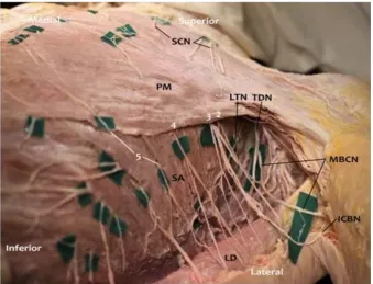
Figure 1: ANATOMY OF PECTORAL NERVES(12)
[image:23.595.135.455.435.692.2][image:24.595.155.465.417.689.2]
At the pectoral level, the fascia creates four potential compartments for the
injection of local anesthetic:
• 1. Between the superficial and deep pectoral fascial layers
• 2. Between the pectoral fascia and the clavipectoral fascia
• 3. Between the clavipectoral fascia and the superficial border of the
serratus anterior muscle
• 4. Between the serratus anterior muscle and the exothoracic fascia
The first two compartments are in the pectoral region, but the third and fourth
communicate with the axillary region. The nerves and vessels in this region
create communications by crossing the compartments. The nerves of the
pectoral region are mainly the lateral and medial pectoral nerves, but there is
also an important innervation from the supraclavicular nerve(SCN)(Figure 3)
and from the lateral and anterior branches of the intercostal nerves. The LPN
crosses the axillary artery anteriorly and penetrates the clavipectoral fascia and
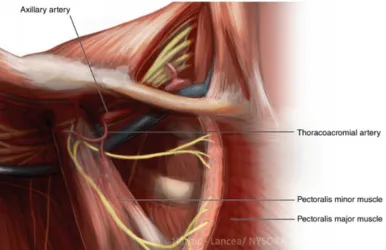
continues to remain close to the thoracoacromial artery(Figure 5). The LPN lies
medial to the pectoralis minor before piercing the pectoralis major muscle; it
communicates across the axillary artery with the medial pectoral nerve and,
through this communication (via ansa pectoralis), supplies the pectoralis minor.
The medial pectoral nerve arises from the medial cord fibres from C8–T1,
behind the axillary artery at the level below the clavicle, and passes through the
innervates pectoralis major. Both pectoral nerves enter the deep surface of the
pectoralis major, and neither has a cutaneous branch(14).
Relationship between the lateral pectoral nerve and the acromiothoracic artery
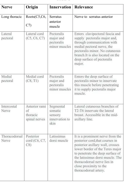
[image:26.595.91.477.145.395.2]Table-1 The relationship of various nerves, its innervation and
[image:27.595.91.496.179.770.2]relevance
Table-1
Nerve Origin Innervation Relevance Long thoracic Roots(C5,C6,
C7)
Serratus anterior muscle
Nerve to serratus anterior
Lateral pectoral
Lateral cord (C5, C6, C7)
Pectoralis major and pectoralis minor muscles
Enters clavipectoral fascia and supply pectoralis major and, through communication with medial pectoral nerve, the pectoralis minor. No cutaneous branch.It is also located on the deep surface of pectoralis major.
Medial
pectoral Medial cord (C8, T1) Pectoralis major and pectoralis minor muscles
Enters the deep surface of pectoralis minor to innervate this muscle before penetrating it to supply pectoralis major muscle. Intercostal Nerve Anterior rami of thoracic spinal nerves Segmental somatic sensory innervation to skin
Lateral cutaneous branches of T2-T6 innervate the lateral breast. Accessible in the mid-axillary line.
Thoracodorsal Nerve
Posterior cord (C6, C7, C8)
Latissimus dorsi muscle
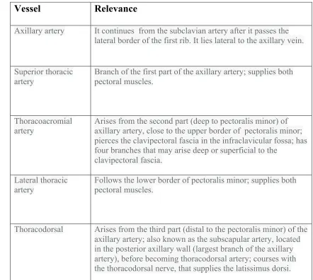
Table-2 Vessels relevant to PECS I and II Block
Vessel Relevance
Axillary artery It continues from the subclavian artery after it passes the lateral border of the first rib. It lies lateral to the axillary vein.
Superior thoracic artery
Branch of the first part of the axillary artery; supplies both pectoral muscles.
Thoracoacromial artery
Arises from the second part (deep to pectoralis minor) of axillary artery, close to the upper border of pectoralis minor; pierces the clavipectoral fascia in the infraclavicular fossa; has four branches that may arise deep or superficial to the
clavipectoral fascia.
Lateral thoracic
artery Follows the lower border of pectoralis minor; supplies both pectoral muscles.
ULTRASOUND BASICS
Ultrasound guidance is rapidly becoming the “gold Standard” for regional
anaesthesia. The image of the target nerve can now be directly viewed, the
needle can be guided under real-time observation, and be navigated away from
sensitive anatomy. The quality and quantity of spread of local anesthetic
spread can thus be observed. Use of ultrasound in daily clinical practice
requires high-level ultra-sonographic equipment and training. The accuracy of
performance of nerve block under direct ultra-sonographic guidance vary with
the operator's skill depending on various regional anesthetic techniques. There
are many potential advantages of using ultrasound guidance over conventional
methods such as direct visualization of anatomical structures, direct and
indirect spread of local aesthetic, reduction of the dose of local anaesthetic,
faster onset time and longer duration of the block (15). Ultrasound imaging is
based on sound waves that are transmitted from and received by, a transducer
utilizing frequencies of 2– 15 MHz (human hearing operates at 1 – 20 kHz).
Most transducers use artificial polycrystalline ferroelectric materials (ceramics)
[e.g. lead zirconate titanate (PZT)], which have piezoelectric properties. When
a current is applied across the crystal, it expands and contracts as the polarity of
the voltage changes. This results in a series of pressure waves (sound waves).
This also works in reverse; when the sound wave returns, it squeezes and
stretches the crystal generating a voltage change across its surface which is
amplified and forms the receiving signal. The speed of sound in tissue varies
reflected at the interface between different structures, reflectivity being
dependent on the difference in acoustic impedance between those structures
and the angle of the US beam. These interface reflections form the basis of the
grey US image which is presented as a matrix of picture elements or pixels.
When there are small differences in acoustic impedance, more of the US beam
will be transmitted through to deeper structures. However, if there is a large
difference in acoustic impedance between tissues, a greater proportion of the
beam is reflected, obscuring deeper tissues (16).
Structure of Transducer
The structure of the transducer is designed to produce and transmit
ultrasound waves in the most effective manner. It essentially consists of three
layers- the backing material, piezoelectric crystals and matching layer;
[image:30.595.106.458.477.630.2](Figures 3 a & b)
Figure 3 b
The piezoelectric crystals are placed in between the matching and backing
layer. Multiple piezoelectric crystals are arranged methodically and
interconnected electronically. The thickness of individual crystals
determines the frequency of the transducer. The backing material contracts
the response of the piezoelectric crystals after which the brief excitation
pulse absorbs the backward energy and laterally transmits to ultrasound
energy. The matching layer which is placed just beneath the footprint (the
area of the transducer which meets the skin) of the transducer minimizes the
reflection and refraction of the ultrasound beam as it exits the transducer.
Types of transducers
There are various types of transducers to suit various applications.
Most of the ultrasound machines use phased array transducers, which consist
of many small carefully, arranged piezoelectric crystals. The frequency of
the transducer is determined by the thickness of these crystals and the shape
of the transducer determines the shape of the ultrasound beam. For example,
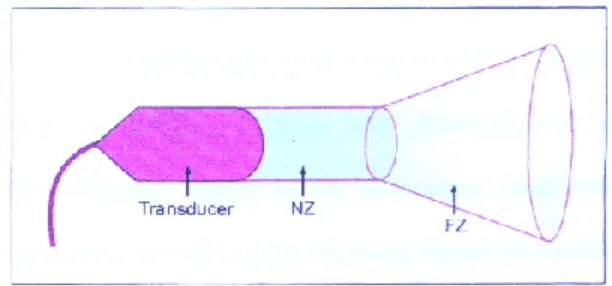
the skin) will produce a cylindrical beam; (Figure 4a) (17) . The transducer
with rectangular (linear) footprint will produce a big rectangular beam;
(Figure 4b)
Transducer with curved (round) footprint produces a cylindrical beam. NZ = near zone, FZ = Far zone
Figure 4 a
[image:32.595.137.443.165.308.2]
Transducer with rectangular (linear) footprint produces a rectangular beam. NZ = near zone, FZ = Far zone
Ultrasound wave frequency, image resolution, and penetration
High-frequency probes (8–12 MHz) and midrange frequency probes (5–10
MHz) provide better resolution but have less penetration. It is due to this
that high-frequency probes are preferentially chosen for US imaging of
superficial structures (2–4 cm), while midrange frequency probes are
preferred for slightly deeper structures (5–6 cm). However, when US
imaging of deep structures (for example, a proximal sciatic nerve that can be
as much as 10 cm deep) is required, a low-frequency probe (2–5 MHz) is
preferred, although the quality of the image would be substantially poor.
When determining the correct choice between probes with different US
frequencies, choose the one that would provide the best resolution for the
required depth. Most practitioners have several different probes for more
flexibility.
Modern US machines have useful “nerve”, “vascular” or “musculoskeletal ”
modes. The “focus” function available on these machines may help to
improve visualization of the targeted structure, although it is rarely needed
for superficial structures if the depth is set correctly(16).
Depth settings
It advised to begin with a somewhat higher depth setting in order to first get
a more magnified picture, and then gradually decrease the depth when the
targeted structure is found. For US-guided injection, the depth should be set
importance, such as a vessel or pleura, is situated below the target, the depth
should be adjusted accordingly to produce a good view where those are
involved in the field to improve safety.
By knowing the target depth and its position on the screen, the initial angle
of needle advancement can be estimated even before visualizing the needle
on the screen. This angle should be adjusted as soon as the needle is viewed
on the screen and its trajectory is clear.
Probe orientation
Probe orientation is important because the US probe can be easily rotated
around (180°) while the position of the monitor remains unchanged, which
may create confusion in the direction of probe manipulation and needle
advancement and placement. Therefore, it is always useful to confirm which
side of the probe corresponds to a particular side of the screen in order to
identify the correct orientation of the image. All transducers have an
orientation marker that corresponds to the marker on the screen (17).
Probe (transducer) movements
Sliding, angulation, and rotation are the most commonly used probe
Needle and probe relationship
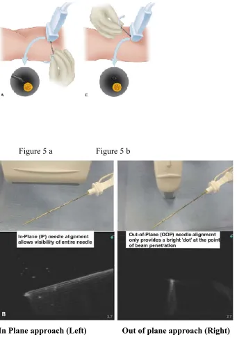
'In-Plane' approach. The needle is inserted in the plane of the ultrasound
beam. The entire needle shaft along with the tip is visualized approaching
the target with this technique.
Out- of- plane' approach
The needle insertion path crosses the plane of the ultrasound beam. The
entire needle shaft along with the tip is not visualized by this technique. The
needle tip in transverse section is visualized moving towards the required
target.
Different blocks need different approaches. With experience, the
same block can be given with both approaches. With the 'in plane"
technique more tissue distance is traversed which is an advantage in
continuous blocks as more tissue anchorage can be achieved (catheter
fixations); (Figure 5a, 6a). The tissue distance traversed by the needle is less
Figure 5 a Figure 5 b
Equipment
Any conventional Ultrasound machine (SONOSITE), in which the user is
familiar with can be used (figure 7a and b). The high-frequency linear probe
has to be selected. Knobology
Figure 7 a Figure 7 b
Sterility of the Probe
The probe can be aseptically prepared by covering the surface with
non-sterile ultrasound gel slipping it into the non-sterile camera cover and then
covered with sterile gel. It is important to avoid the entrapment of air bubbles
in between the cover and the transducer surface. For single shot blocks,
application of surface disinfectants may be used instead of the gel.
Ergonomics of ultrasound-guided procedure
An ultrasound machine is to be positioned on the contralateral side of the
blocked or examined. The transducer is usually held in the operator’s
non-dominant hand, with the needle in the non-dominant hand. The transducer is
gently held quite low on the probe, close to the scanning surface, rather than
harshly gripped on the top of the handle. When planning to use the in-plane
approach, it is preferable to place the probe directly perpendicular to the
skin.
Coupling Medium
Ultrasound does not travel through the air; gel provides a medium for sound
wave into the body by removing the air gap. Sterile saline, sterile water, or
sterile gel can be used as coupling agents.
Choice of Needle
Needle shaft and tip identification are major perquisites to avoid nerve or
blood vessel damage. Any needle can be visualized well under ultrasound if
positioned properly, although the larger the bore the easier it is to be
visualized. If a catheter has to be inserted then 18 gauge Tuhoys needle is to
be inserted. The length of the needle depends on the depth of the target
nerve. Both 5cm and 10cm block needles are available. Most of the needles
do not provide the desired reflective qualities to allow safe penetration
especially at a steep angle. For our study we used the Sonoplex echogenic
needle. Under ultrasound the image is greatly enhanced due to the
Figure 8 a Figure 8 b
Nerve Identification
Nerve identification is the basis of ultrasound-guided regional anaesthesia
and therefore merits a detailed discussion.
It is possible to scan the nerves either in transverse sections or longitudinal.
It is best to scan nerves in the transverse section for block procedures
because transverse scans are easier to work in, than longitudinal scans. In
transverse section, the nerves cast a typical circular to oval "honeycomb" like
appearance. The "honeycomb" appearance of the nerve is due to the
fascicles cut in transverse section, which appear like hypo-echoic images
with the intervening hyper-echoic tissue. The perineurium surrounding the
fascicles imparts a hyper-echoic interface. It is worthwhile to trace the nerve
along its course to identify the path and differentiate it from the adjacent
tendons, which disappear in the muscle mass when traced proximally (18).
TECHNIQUE OF BLOCK (18)
The patient is positioned supine, and the ultrasound machine is placed such
that maximum ergonomic comfort is obtained for the person performing the
block. It is advised to do a scout scan of the chest wall and breast to identify
the structures. The probe was positioned under the outer third of the
clavicle(Figure 9). The axillary artery and the axillary vein are identified, the
identified as a hyper-echoic thin line that slides with respiration( Figure 11
and b). Using the in-plane technique, keeping the tip of the needle in view,
the needle is advanced to the tissue plane between the Pectoralis minor and
the Serratus anterior (PECS 11). Two to three ml of the saline is first
deposited, noting the spread in the correct interfacial plane which is
confirmed with easy separation of the two muscle layers and the pleura
moving downwards. Then 0.4ml/kg of the drug is deposited above the
Figure 9 : Positioning probe at time of block
.
Figure 11 a Ultrasound image of in-plane technique targeting PECS 11
Keeping the needle and the probe in the same position the needle is then slowly brought out, with continuous visualization of the tip and shaft into the tissue plane between the Pectoralis major and and minor. Two to three ml of saline is infiltrated to confirm the position and separation of the two muscle layers. Then 0.2ml/kg of the drug is deposited between the two muscle layers. (Figures 11 a and b)
[image:43.595.133.400.71.237.2]:
Figure 11 b Ultrasound image of in-plane technique targeting PECS
PHARMACOLOGY
Bupivacaine, an amino amide local anesthetic was first synthesized in
Sweden by A.F Ekenstam and his colleagues in 1957. The first report of its
use was in 1963 by L. J Teluvio. It is one of the long-acting local anesthetic
agents available, which is extensively used for intrathecal, extradural and
peripheral nerve blocks. It is a white crystalline powder soluble in water
(19).
Chemical nomenclature of Bupivacaine
1-butyl-n-(2, 6-dimethylphenyl)
piperidine-2-carboxamide.
Chemical structure of Bupivacaine
Physiochemical properties
Molecular formula C18H28N2O
Molecular weight 288.43 g/mol
Solubility in water 25mg/ml
The darker side of bupivacaine was exposed by its potential for cardiac toxicity
and local anaesthetic toxicity which manifests as a progression from mild
symptoms to convulsions, eventual cardiac depression, and cardiac arrest.
Besides, there are reports of sudden cardiac arrest without prodromal central
nervous system (CNS) symptoms, prolonged and difficult resuscitations and a
high proportion of maternal deaths accounting for 76% of fatalities (20).
Subsequent changes in anaesthetic practice, such as slow incremental dosing
and the use of test doses, were able to reduce the incidence of severe toxicity
but such measures could not guarantee safety in the event of accidental
intravascular injection. Bupivacaine has the unusual property of
stereoisomerism due to the presence of an asymmetric carbon. This behaves as
a chiral center. Levorotatory isomers under experimental conditions showed
safer pharmacological profile with less cardiac and neurotoxic effects. The
reduced toxicity of levobupivacaine is also due to its faster protein binding rate.
The pure S (−) enantiomers of bupivacaine, i.e., Ropivacaine and
Levobupivacaine (Figure 11) were thus utilized into the clinical anaesthesia
practice. Evidence based data has established the safety of levobupivacaine
over bupivacaine when used in regional anesthesia. This is clear from the
decreased incidence of adverse outcomes when using the former as compared
to the latter(21). It was also noted that the incidence of adverse cardiac and
neurological events was significantly higher with bupivacaine as compared to
has been recently introduced into the Indian market and is being widely used in
[image:46.595.128.273.494.645.2]various health care fields.
Figure 11. The molecular structures of levobupivacaine and dextro-bupivacaine.
Figure 12:
Levobupivacaine ([2S]-1-butyl-N-[2, 6-dimethylphenyl] piperidine-2-carboxamide)
Mechanism of action of levobupivacaine is similar to that of any other local
anesthetic;(Figure 12.) The primary action of local anesthetics is on the cell
membrane axon, myelinated nerves being blocked through exposure at the
Nodes of Ranvier more readily then unmyelinated nerves and small nerves
being blocked more readily than larger ones. In general, the progression of
anesthesia is related to the diameter, myelination and conduction velocity of
the affected nerve fiber. The drug binds to the intracellular portion of the
sodium channels and blocks the sodium influx into the nerve cells. This, in
turn, prevents electrical depolarization and the eventual generation of the
conduction of the nerve impulse. This action of levobupivacaine is due to its
direct interaction with the voltage-gated sodium channels. As the anesthetic
action progressively develops in a nerve, the threshold for electrical
excitability gradually decreases, the rate of rising of the action potential
declines, impulse conduction slows, and the safety factor for conduction
decreases, these factors decrease the probability of propagation of the action
potential and nerve conduction fails (22,23). The mechanism by which local
anesthetics block sodium conductance is as follows:
1. Local anesthetics in the cationic form act on the receptors
within the sodium channels on the cell membrane and block it. The
local anesthetics can reach the sodium channel either via the
lipophilic pathway directly across the lipid membrane or via the
axoplasmic opening. This mechanism accounts for 90% of the nerve
2. The second mechanism of action is by membrane expansion. This
is a non-specific drug-receptor interaction.
Pharmacokinetics
The dose, and the route of administration of levobupivacaine, contributes to the
plasma concentration. Absorption dependends upon the vascularity of the
tissue. With epidural administration, Levobupivacaine, absorption is biphasic.
There is rapid absorption of minute quantity of drug into the circulation
initially. Following which there is a much slower absorption of the majority of
the remaining drug. Due to this the peak levels of levobupivacaine in the blood
reaches approximately 30 min after epidural administration and doses up to 150
mg had resulted in mean Cmax levels up to 1.2 µg/mL. Epidural absorption
depends on age as the fraction absorbed decreases and the fast absorption phase
is shorter in older (aged > 70 years) compared with the younger (aged 18-44
years) patients. Because of this older patients have higher spread of drug ~ 3
dermatomes. Therefore, in the elderly patients a lower dose of levobupivacaine,
according to their physical status should be recommended. In the study done by
Burlacu et al he showed that the volume of distribution is estimated at 66.91 ±
18.23 L (after intravenous administration of 40 mg in healthy volunteers). The
pKa of levobupivacaine is 8.1, similar to the pKa of the racemic bupivacaine.
The half-life is 3.3 h. The rate of clearance is 39.06 ± 13.29 L/h (after
than 3% of free drug in the plasma. The free proportion of the drug can cause
toxicity on the other tissues, leading to unwanted side-effects and worsening
toxic manifestations. In a newborn and in protein-deficient states like
undernutrition and nephrotic syndrome, there is less protein binding resulting
in higher levels of the free drug. This decreases the therapeutic range of safety
resulting in toxic effects at lower doses (23) (24). Levobupivacaine is
extensively metabolized with no active metobolite in urine or faeces. In vitro
studies using levobupivacaine showed that cytochrome CYP3A4 isoform and
CYP1A2 isoform mediate the metabolism of levobupivacaine to inactive
metabolites, desbutyl levobupivacaine, and 3-hydroxy levobupivacaine,
respectively. In vivo, the 3-hydroxy levobupivacaine appears to undergo
further transformation to glucuronide and sulfate conjugates, which are
excreted in urine. Metabolic inversion of levobupivacaine to R (+)-bupivacaine
was not evident both in vitro and in vivo Following intravenous
administration, recovery of the radiolabeled dose of levobupivacaine was
essentially quantitative with a mean total of about 95% being recovered in
urine and faeces in 48 hours. Of this 95%, about 71% was in urine while 24%
was in faeces (23,24).
Peripheral Nerve Blocks
Various studies have compared levobupivacaine, ropivacaine and bupivacaine
in brachial plexus block for upper limb surgery[(25)(26)]. Levobupivacaine is a
provides a significantly longer duration of analgesia.[(19)] The return of motor
activity is earlier with ropivacaine.[(21)] The long duration of the sensory
block associated with good analgesia and less toxicity of levobupivacaine
makes it a better choice for upper extremity blocks.[(25)] Levobupivacaine
0.5% provides a longer duration of sensory block after sciatic nerve block
using the Labat approach than the same dose of ropivacaine in foot and ankle
surgery.[(26)] Levo-bupivacaine 0.5% is as effective as bupivacaine 0.5% and
is recommended for the 3-in-1 block (27).. The use of a single dose of 0.5%
levobupivacaine to block the tibial and peroneal nerves for hallux valgus
surgery using popliteal approach is preferable over 0.5% ropivacaine for good
anesthesia and better control of post-operative pain.[(27)] . The quality and
duration of peripheral nerve block are improved with the use of higher
concentrations of levobupivacaine, (0.5-0.75%).[(28)] Levobupivacaine
administered via a peripheral nerve block continuous catheter provides
excellent postoperative analgesia and decreases the postoperative systemic
opioids requirements.[(29)] The addition of adjuvants to the local anesthetics in
peripheral nerve blocks such as epinephrine, clonidine or opioids improve the
quality of analgesia and provide a dose-sparing effect, thereby decreasing the
potential for systemic toxicity. Epinephrine does not add to the inherent long
block provide excellent analgesia and local anaesthetic sparing effect and
decrease post-operative systemic morphine requirement.[(30)] Similarly, the
addition of tramadol to levobupivacaine in middle interscalene block
significantly increases the duration of the sensory block.[(31)]
Toxicity
Systemic toxic reactions can occur, usually owing to accidental intrathecal or
intravascular injections or to an administration of an excessive dose of
bupivacaine. It involves mainly the CNS. Allergic reactions to amide type of
local anesthetics are rare. The toxic plasma concentration of bupivacaine is
4-5 mcg/ml.The earliest signs of CNS toxicity are circumoral or tongue
numbness, tinnitus, restlessness, lightheadedness and dizziness, confusion,
small muscle twitches involving the face and the distal part of the
extremities, which may progress to generalized tonic-clonic seizures. In later
stages, it progresses to respiratory arrest (32). The cardiotoxicity of
bupivacaine is different from other local anaesthetics. The ratio of the dose
required for irreversible cardiovascular collapse and the dose needed to
produce CNS toxicity (CC/CNS dose ratio) is lower for bupivacaine which is
z3.7±0.5. [CC/CNS dose ratio of 7.1 ± 1.1 was found for lidocaine]. The
higher the CC/CNS ratio the better the safety margin. The dose of
bupivacaine required for irreversible cardiovascular collapse compared to
that required for CNS toxicity is lower than that of lignocaine (CC: CNS
ratio 2.0:7.1 for the two LA). The cardiovascular manifestations of
hypotension, bradycardia, and syncope. cardiac resuscitation is more difficult
following bupivacaine-induced cardiovascular collapse. Acidosis and
hypoxia potentiate the cardiotoxicity of bupivacaine.
Levobupivacaine produces the same adverse effects as seen with racemic
bupivacaine and other local anesthetics. The most common adverse drug
reaction reported is hypotension (31%) followed by nausea (21%), vomiting
(14%), headache (9%), procedural pain (8%) and dizziness (6%) (32). The
cardiac toxicity, neurological injury after peripheral nerve block
and unwanted CNS effects, may be lower than bupivacaine. Allergic-type
reactions are rare and range in severity from urticaria to anaphylactoid-like
reaction. Levobupivacaine has a safety margin of 1.3, which means toxic
effects are not seen until the concentration rises by 30%. The concentration
necessary to produce cardiac and neurotoxicity is higher for levobupivacaine
than for racemic bupivacaine. There are three case reports of successful
resuscitation after inadvertent intravenous injection. The presentations were
severe hypotension and bradycardia (following drug error); loss of
consciousness, convulsions, hypotension, and changes in the QRS pattern of
ECG (presumed intravenous injection during lumbar plexus block) and loss of
consciousness with convulsions (following spinal, sciatic nerve and continuous
lumbar plexus blocks). In all cases, resuscitation was successful with
Recently, studies have been carried out comparing the beneficial effects of
vasopressor drugs and lipid therapy in local anesthetic systemic toxicity
(LAST). Epinephrine is to be used in small doses (10-100 µg) in adults. The
use of vasopressin is not recommended. Lipid emulsion therapy is to be
considered at the first signs of LAST, after airway management.[(33)]
Successful resuscitation has been reported with intralipid emulsions in a
peri-arrest condition following use of levobupivacaine in lumbar plexus block.(32)
Table-3:
DOSING
The recommended dose of levobupivacaine is a maximum single dose of 150
mg or 400mg over 24hrs. The increased safety margin of levobupivacaine
might suggest that more drug could be administered in divided doses to
patients. A study of 20 patients undergoing axillary brachial plexus blocks
using levobupivacaine 0.5% [(33)] reported the highest single administration
dose evaluated in the levobupivacaine clinical programme (300 mg, ). Despite
cardiovascular toxicity was observed, indicating that levobupivacaine was
tolerated at the high dose evaluated in this study. Fifty-nine patients have safely
received levobupivacaine in a dose of F 400 mg for postoperative analgesia
over 24 h, of whom eight received F 500 mg and six F 600 mg, with no signs
of toxicity. (36)
In conclusion, this review shows that levobupivacaine is a major advantage in
regional anaesthesia as it has a similar efficacy but an enhanced safety profile
when compared to bupivacaine, It is to be hoped that the introduction of this
new single-isomer local anaesthetic will be a stepping stone towards safer
regional anaesthetic practice.
Efficacy of Block
The spread of local anesthetic into the axilla has been demonstrated by
dissection of cadavers and contrast distribution(35)..It is doubtful whether a
single blocking method can provide adequate analgesia throughout the entire
breast area. The targets of PECS II block include the medial and lateral
pectoral nerves, including the lateral cutaneous branches of the thoracic
intercostal nerves (TICNs) . Local anesthetics cannot reach the anterior
cutaneous branches of the TICNs by piercing the external and internal
intercostal muscles. Therefore, they cannot block anterior cutaneous branches
Although various studieshave shown better pain relief and a significant
reduction in opioid consumption when the Thoracic paravertebral block s
(TPVB) was combined with general anaesthesia (38), patients having radical
mastectomy under TPVB frequently complain of pain in the axilla and upper
limb(39), because the TPVB does not block the medial and lateral pectoral
nerves as effectively as the long thoracic and thoracodorsal nerves, leading to
inadequate analgesia. And patients undergoing TPVB had higher complications
related to block such as vascular puncture 3.8%, hypotension 4.6%, pleural
puncture 1.1% and pneumothorax 0.5%.
In conclusion, this review shows that levobupivacaine is a major
advantage in regional anaesthesia as it has similar efficacy but an enhanced
safety profile when compared to bupivacaine. It is hoped that the introduction
of this single-isomer local anaesthetic along with PECS block be effective for
This study included all patients of ASA 1 & 2 undergoing mastectomy surgery
with axillary clearance admitted under the Department of Endocrine Surgery
and General Surgery at the Christian Medical College, Vellore, Tamil Nadu,
during the period of 2017 to 2018. It was cleared by the IRB-N0.10390 and
registered in the National Registry
(CTRI-No 010630). A randomized control study design was used for
evaluation of the objectives.
A.
Inclusion Criteria : -All ASA 1 & 2 consenting patients -breastcancer surgery with axillary clearance
B.
Exclusion Criteria:a. Non-consenting patients
b. ASA 3 & 4 physical status
c. Bilateral mastectomy
d. Allery to local and general anaesthetic drugs
e. Chest wall anatomical abnormality
f. Presence of infection
g. Patients on anti-coagulants or anti-platelet drugs /coagulopathy
Method of randomization: Block randomization was performed using
computed generated random numbers used to ensure equality of numbers in
Recruitment:
The study included patients who fulfilled the inclusion criteria. Before
proceeding with the study, a written and informed consent was taken from
these patients. The same was explained to their bystanders. The
confidentiality of the patient was maintained.
Patients underwent routine pre-anesthetic evaluation and all patients
recruited were either ASA 1 or 2. Fasting protocols were followed. All
patients were kept nil per oral (NPO) for 6 hours for solids and milk; 2 hours
for clear fluids like coconut water, or water.
On the morning of surgery, patients were reassessed and taken in. The
anaesthesia machine, emergency oxygen source, pipeline oxygen supply,
working laryngoscope were checked. Working suction apparatus with
suction catheter, airway equipment, intravenous fluids and all emergency
drugs were kept ready for emergency resuscitation.
Blinding: Randomization was concealed using opaque envelopes till time of
recruitment. However, intervention was not concealed. Evaluation of the
outcome was performed by an independent pain nurse in the ward who was
Intervention:
Group-1: Received general anaesthesia followed by ultrasound-guided
PECS-I and II block.
Group-2 : Received only general anaesthesia.
Both Groups were given standard anaesthetic care for mastectomy surgery
including, oxygen, opioids(fentanyl, morphine),muscle relaxant(vecuronium
bomide),inhalational agent(Isofluranre-Mac=1), additionally, paracetamol,
anti- inflammatory drugs (NSAIDs), calculated as per kilogram body weight ,
noting the base line response from time of incision, following which, drug
administered based on adequacy of depth of anaesthesia and analgesia.
Flow chart of intervention:
A Mayo trolley containing sterile drapes, two 10 ml syringes, a 10cm
Sonoplex needle and ultrasound sterile sleeve along with the 0.25%
levobupivicaine was kept ready. Ultrasound machine (Sonosite), with high
frequency linear (8-12 MHZ) probe and ultrasound gel were organized.
ASA 1 and 2 patients
Random-ization
GA + Block* 30 GA 30 Paracetamol, NSAIDS, opioids, as required
Intra and post operative pain assessment along with arm abduction pain within 24hrs
* Ultrasound-guided PECS-I (0.2ml/kg) and II (0.4ml/kg)block post
All patients received general anaesthesia. After establishing intravenous
access in the operation theatre, routine monitors were established (ECG,
pulse oximetry, noninvasive BP). Oxygen was applied via facemask at 3 L/
min. Anaesthesia was induced with fentanyl 2mcg/kg, propofol 2-3 mg/kg
and vecuronium bromide 0.1mg/kg was used to facilitate endotracheal
intubation. Anaesthesia was maintained with isoflurane and air with oxygen
in the ratio 2:2. Neuromuscular blockade was maintained with vecuronium.
Ventilatory parameters were set to maintain normocarbia. Depth anaesthesia
was maintained at a MAC of 1. Group-1 patients were then prepared
accordingly receiving the block prior to commencement of surgery. The
block was performed with the patient in supine position and the arm
abducted 90 degrees. The landmarks under US was identified that is the
Pectoralis major, Pectoralis minor and the pectoral branch of the
thoracoacromial artery. The transducer was rotated slightly to allow in-plane
needle trajectory from medical to lateral side. The proper fascial plane was
confirmed by hydro-dissection with saline to open the space between the
pectoral muscles. First the plane between serratus anterior and pectoralis
minor, was identified and 0.4ml/kg of levobupivicaine was instilled,
followed by the plane and between pectoralis major and minor 0.2ml/kg of
drug was instilled as the needle was withdrawn. At all times, the shaft of the
Outcome Measurements:
Intra-operative Period:
Any increases in heart rate and blood pressure to more than 20% of the
baseline was treated as a pain response after the patient was under adequate
plane of anesthesia. All patients remained hemodynamically stable.
Paracetamol(15mg/kg) was started towards the end of surgery. Intraoperative
vitals were monitored on a intervals of 5, 10 ,15 30, 45, 60, 90, 120, 160 and
180. Systolic and Diastolic blood pressure along with heart rate ET Co2 and
saturation was recorded.
After completion of surgery, neuromuscular blockade was reversed with
neostigmine 0.05mg/kg and glycopyrrolate 0.01mg/kg and patients were
extubated once the criteria for extubation were met. Total morphine and
fentanyl consumption was also recorded.
Post-operative Period:
After surgery, patients were transferred to post-anaesthesia care unit
(PACU), Pain score was documented against Numeric Rating Scale scale
every 2 hours for the initial 24 hours and every 6 hours for the next 24 hours
Pain Assessment Tool: Numerical Rating Scale :
The Numerical Rating Scale has shown a high correlations as compared to
other pain-assessment tools(40).The patients were asked to circle the number
between 0 and 10, zero representing “no pain at all” whereas the upper limit
represents “the worst pain possible”
No pain Worst Pain
0 1 2 3 4 5 6 7 8 9 10
[image:62.595.84.527.495.708.2]Functional Activity Score (Arm Abduction Score)- was used to assess ability for arm abduction postoperative.
Table-4
No:
Comment
Score
1 No Limitation of Relevant Activity
A
2 Mild Limitation of Relevant Activity
B
3 Severe Limitation of Relevant Activity
C
Source: Victoria Quality Council. Acute Pain management measurement (Fourth Edition). A Practical guide(41)
Statistical Methods Sample size:
Following formula was used- n(per arm)=2x [Z1-α/2+Z1-β/2]2 x SD2
(mean difference)2
Since in the study, pain was treated with morphine, the amount of
morphine consumed was noted as one of the primary outcome.
The following tables gives various sample sizes for potential differences
in two groups with possible Standard Deviations of the dose of morphine.
Table-5: Two Means - Hypothesis testing for two means
Standard deviation in group-I 5 4 4
Standard deviation in group-II 5 4 4
Mean difference 3 3 2
Effect size 0.6 0.75 0.5
Alpha error (%) 5 5 5
Power (1- beta) % 80 80 80
1 or 2 sided 2 2 2
Required sample size per group 44 28 63
The sample size varied from 28 to 63 for various potential differences in
Standard Deviation to detect a difference of 2 mgs and 3 mgs between two
groups intra operatively. For the present study it was decided to choose 30
subjects per group.
Analytical Methods: SPSS software was used for analysis of data.
Base line data was described using frequency, Mean and 95% Confidence
Interval in the two study groups. Continues variables in the data was
at different times using ‘Mixed Model Analysis of Co-Variance’ with
‘Group’ as between group variable and ‘Time’ as repeated measures.
Baseline data was used as the ‘Co-variate’. Since most of the surgery took
less than 180 minutes, ANOVA measurement included only periods(Time)
where outcomes where more than 50% of patients were available.
Total dose of morphine and Fentanyl were compared using student t test.
There were 60 patients recruited as per inclusion and exclusion criteria during
the study period from Nov 2016 to August 2017. These patients were admitted
for breast cancer surgery in the hospital. There was 1 male and 59 females. The
mean age in the study was 49+10 years.
The base line distribution of of Age, Sex, Weight and ASA grade in the test
and control groups are given in Table-6
Table-6 Baseline distribution (Age, Sex, Weight, ASA grade)
No Parameter Test Group Control Group P Value
Value Variability Value Variability
1 Sex Ratio M:F 0:30 - 1:29 - 0.5
2 Age (Years} 51 47-55 47 44-51 0.14
3 Weight (Kg) 59 56-62 63 59-67 0.1
4 ASA grade Ratio 1: 2 11:19 - 17:13 - 0.12
There was no statistically significant difference noted in the Sex, Age, weight and ASA grade in the test and control groups.
Table -7 Type of Surgery
GROUP Total
Block Non block
SURGERY
MRM (Left) 12 16 28
MRM (Right) 17 11 28
Simple Mastectomy (Left) 1 1 2 Simple Mastectomy (Right) 0 2 2
Total 30 30 60
Intra-operative Data:
Table-8 shows duration of surgery in 60 patients. The surgical time lasted for
more than 60, 90, 120, 150, 180 minutes for all 60, 58, 38, 12, 3 patients,
respectively. Mean Duration of Surgery was 115 minutes with Standard
Deviation of 27 minutes.
Table-8 Duration of Surgery in 60 patients
5 mins 10 mins 15 mins 30 mins 45 mins 60 mins 90 mins 120 mins 150 mins 180 mins
Valid 60 60 60 60 60 60 58 38 12 3
Missing 0 0 0 0 0 0 2 22 48 57
Time to perform block in 30 subjects
The time for performing the block varied from 3 to 26 minutes with mean
value of 7.9 minutes and Standard Deviation of 4.4 minutes. Table-9 gives the
[image:67.595.130.463.497.721.2]details in 30 subjects in whom the block was performed.
Table-9 Time for Block
Time to perform block GROUP Block Total
Anaesthesia (time to perform block)
3 Min 1 1
4 Min 3 3
5 Min 5 5
6 Min 3 3
7 Min 7 7
8 Min 2 2
10 Min 4 4
11 Min 1 1
12 Min 1 1
13 Min 1 1
14 Min 1 1
26 Min 1 1
Heart Rate(HR):
Table-10 Heart Rate Mean and Std. Deviation during Surgery for all patients:
N Mean Std.
Deviation Error Std. Interval for Mean 95% Confidence Minimum Maximum Lower
Bound Bound Upper
HR baseline
Block 30 91.20 11.830 2.160 86.78 95.62 69 114 Non
block
30 94.47 15.220 2.779 88.78 100.15 60 122
Total 60 92.83 13.615 1.758 89.32 96.35 60 122
HR 5 mins
Block 30 90.13 10.683 1.950 86.14 94.12 70 110 Non
block
30 90.33 14.933 2.726 84.76 95.91 50 113
Total 60 90.23 12.873 1.662 86.91 93.56 50 113
HR 10 mins
Block 30 87.77 10.588 1.933 83.81 91.72 62 110 Non
block 30 90.80 13.952 2.547 85.59 96.01 54 112 Total 60 89.28 12.374 1.598 86.09 92.48 54 112
HR 15 mins
Block 30 85.23 11.104 2.027 81.09 89.38 62 111 Non
block
30 88.60 14.481 2.644 83.19 94.01 58 110
Total 60 86.92 12.906 1.666 83.58 90.25 58 111
HR 30 mins
Block 30 79.77 14.097 2.574 74.50 85.03 49 114 Non
block 30 85.93 15.056 2.749 80.31 91.56 58 119 Total 60 82.85 14.791 1.910 79.03 86.67 49 119
HR 45 mins
Block 30 79.13 12.356 2.256 74.52 83.75 54 106 Non
block 30 78.07 13.859 2.530 72.89 83.24 56 103 Total 60 78.60 13.028 1.682 75.23 81.97 54 106
HR 60 mins
Block 30 79.30 14.690 2.682 73.81 84.79 48 107 Non
block 30 80.27 13.856 2.530 75.09 85.44 50 112 Total 60 79.78 14.166 1.829 76.12 83.44 48 112
HR 90 mins
Block 29 76.62 11.992 2.227 72.06 81.18 55 97 Non
block
29 81.52 10.829 2.011 77.40 85.64 60 104
Total 58 79.07 11.591 1.522 76.02 82.12 55 104
HR 120 mins
Block 21 74.24 12.377 2.701 68.60 79.87 60 103 Non
block
17 83.71 13.873 3.365 76.57 90.84 58 109
Total 38 78.47 13.740 2.229 73.96 82.99 58 109
HR 150 mins
Block 7 76.14 17.180 6.493 60.25 92.03 54 99 Non
block
5 85.40 10.065 4.501 72.90 97.90 72 98
Total 12 80.00 14.851 4.287 70.56 89.44 54 99
HR 180 mins
Block 2 71.00 1.414 1.000 58.29 83.71 70 72 Non
block
1 102.00 . . . . 102 102
Graph-1 Heart rate by Time duration of Surgery:
Table -11 Overall Mean HR
GROUP Mean Std. Error 95% Confidence Interval Lower Bound Upper Bound
Block 82.752a 1.698 79.302 86.202
Non block 86.084a 1.959 82.101 90.066
a. Covariates appearing in the model are evaluated at the following values: HR baseline = 94.16.
b. P value Time = 0.01, P value between Group = 0.36: P Interaction TxG =0.86
The graph-1 and Table 11 shows that both test and control group HR decreased
by time during surgery. This was significant at P=0.01. There was a difference
of mean HR of nearly 3 beats between group, however, this did not reach
statistical significance with P value of 0.36. There was no significant change
between group during the surgery time as shown by Treatment to Time