Activation
Chun Mathers,aXenia Schafer,aLuis Martínez-Sobrido,bJoshua Mungera
Department of Biochemistry and Biophysicsaand Department of Microbiology and Immunology,bUniversity of Rochester Medical Center, Rochester, New York, USA
ABSTRACT
Viral infection frequently triggers activation of host innate immune pathways that attempt to limit viral spread. The NF-
B
pathway is a critical component that governs this response. We have found that the human cytomegalovirus (HCMV) U
L26
pro-tein antagonizes NF-
B activation. Upon infection, an HCMV strain lacking the U
L26 gene (
⌬
U
L26) induced the nuclear
translo-cation of the NF-
B RelB subunit and activated expression and secretion of interleukin-6 (IL-6), an NF-
B target gene. The
⌬
U
L26 mutant was also more sensitive to challenge with tumor necrosis factor alpha (TNF-
␣
), a canonical NF-
B inducer.
Fur-ther, expression of U
L26 in the absence of other viral proteins blocked NF-
B activation induced by either TNF-
␣
treatment or
infection with Sendai virus (SeV). Our results indicate that U
L26 expression is sufficient to block TNF-
␣
-induced NF-
B nuclear
translocation and I
B degradation. Last, U
L26 blocks TNF-
␣
-induced I
B-kinase (IKK) phosphorylation, a key step in NF-
B
activation. Combined, our results indicate that U
L26 is part of a viral program to antagonize innate immunity through
modula-tion of NF-
B signaling.
IMPORTANCE
The NF-
B signaling pathway regulates innate immunity, an integral host process that limits viral pathogenesis. Viruses have
evolved mechanisms to modulate NF-
B signaling to ensure their replication. HCMV is a major cause of birth defects and
dis-ease in immunosuppressed populations. HCMV is known to actively target the NF-
B pathway, which is important for HCMV
infection. Our results indicate that the HCMV U
L26 gene is a key modulator of NF-
B pathway activity. We find the U
L26 gene is
both necessary and sufficient to block NF-
B activation upon challenge with antiviral cytokines. Further, U
L26 attenuates the
phosphorylation and activation of a key NF-
B activating kinase complex, IKK. Our study provides new insight into how HCMV
targets the NF-
B pathway. Given its importance to viral infection, the mechanisms through which viruses target the NF-
B
pathway highlight areas of vulnerability that could be therapeutically targeted to attenuate viral replication.
H
uman cytomegalovirus (HCMV) is a widely disseminated
op-portunistic pathogen that causes lifelong infection but rarely
results in significant disease in mature, healthy individuals (
1
).
However, congenital HCMV infection causes substantial disease,
with approximately one in a thousand children born suffering
from permanent HCMV-induced disabilities, including hearing
and vision loss, cerebral palsy, and cognitive disability (
2–4
).
Fur-ther, HCMV causes serious disease in immunosuppressed
popu-lations, including transplant recipients, AIDS patients, and cancer
patients receiving immunosuppressive therapies (
1
,
5
).
HCMV, a betaherpesvirus, contains a large double-stranded
DNA genome of approximately 230 kb, which encodes over 200
open reading frames (
7
,
8
). HCMV virions are enveloped and
contain an icosahedral capsid which houses the viral genome. A
layer of diverse proteins, termed the tegument layer, is located
between the capsid and the envelope. Upon initial fusion of the
viral envelope with the host-cell plasma membrane, many of these
tegument proteins are delivered to the cytoplasm, where they
per-form a variety of functions that instill an environment conducive
to viral replication. These functions include attenuating innate
immunity, inducing mitogenic signal transduction pathways, and
activating viral gene transcription (
9–13
).
The protein encoded by the U
L26 gene is a tegument protein
critical for high-titer HCMV replication. Translation of U
L26 can
initiate from one of two in-frame methionines, producing long
and short isoforms with molecular masses of 27 and 21 kDa,
re-spectively (
14
). The U
L26 protein is present throughout the viral
life cycle; immediately upon envelope fusion, tegument-derived
U
L26 protein is delivered to the cytoplasm (
14
), and soon after,
de
novo
U
L26 is transcribed as an early gene (
13
). At the onset of
infection, the majority of U
L26 protein localizes to the nucleus
(
15
). As the infectious cycle progresses, U
L26 becomes
increas-ingly cytoplasmic and eventually localizes to viral assembly sites
(
15
). Viral mutants containing deletions of the U
L26 gene
repli-cate with slower kinetics, produce plaques of reduced size, and
grow to lower final titers (
15–17
). However, the mechanisms
through which U
L26 contributes to HCMV replication have
largely remained elusive.
The NF-
B pathway is a central regulator of a host cell’s early
response to viral infection. A variety of inflammatory events,
in-cluding viral infection and exposure to inflammatory molecules,
can induce NF-
B’s transcriptional activity, which subsequently
drives expression of a number of different cytokines, chemokines,
and proinflammatory enzymes. The canonical NF-
B pathway is
controlled through a signaling cascade that can initiate upon
tu-Received4 September 2014Accepted24 September 2014
Published ahead of print1 October 2014
Editor:L. Hutt-Fletcher
Address correspondence Joshua Munger, josh.munger@rochester.edu.
Copyright © 2014, American Society for Microbiology. All Rights Reserved. doi:10.1128/JVI.02552-14
on November 7, 2019 by guest
http://jvi.asm.org/
mor necrosis factor alpha (TNF-
␣
) receptor binding, resulting in
increased linear ubiquitination of NEMO and subsequent
assem-bly and activation of the inhibitor of
B kinase (IKK) (
18
,
19
).
Activated IKK phosphorylates the inhibitor of
B (IKB), resulting
in its ubiquitin-mediated degradation (
20
,
21
). In the absence of
IKB, the RelA and p50 NF-
B subunits are free to translocate to
the nucleus, and they subsequently activate transcription of genes
containing
B binding sites (
20
). Noncanonical NF-
B signaling
occurs upon IKK
␣
activation, which induces nuclear
transloca-tion of p52-RelB dimers (
22–24
). The nature of the transcriptional
response induced by these different NF-
B pathways varies
de-pending on the subunit composition and posttranslational
mod-ifications of the specific NF-
B dimers activated (
25
).
Here, we explored a role for U
L26 in inhibiting innate immune
signal transduction. We find that the HCMV U
L26 protein is an
inhibitor of NF-
B activation. Infection with a mutant harboring
a deletion of U
L26 (
⌬
U
L26) induces noncanonical NF-
B
activa-tion and fails to block canonical TNF-
␣
-induced NF-
B
activa-tion. Further, we find that expression of U
L26 in the absence of
other viral proteins is sufficient to block NF-
B activation upon
TNF-
␣
treatment. This inhibition of NF-
B activity appears to
result from a U
L26-mediated attenuation of IKK phosphorylation
and subsequent stabilization of IKB. In summary, our results
indicate that the HCMV U
L26 protein is a viral factor that
attenuates the innate immune response through inhibition of
NF-
B signaling.
MATERIALS AND METHODS
Cell culture, viruses, and chemicals.MRC5 fibroblasts (passages 23 to 29) were cultured in Dulbecco’s modified Eagle medium (DMEM; Invit-rogen) supplemented with 10% fetal bovine serum. The wild-type (WT) HCMV strain used in this study was BADwt, a bacterial artificial chromo-some (BAC) clone of Ad169 (26,27). The⌬UL26 mutant in these studies
is a BADwtderivative that was previously described (15,28). Viral stocks were prepared through combining clarified infected cell medium (3,000⫻g) with the remaining clarified infected-cell lysate. For the lysate, the scraped cells were resuspended in 5 ml of infected media, sonicated, and clarified through centrifugation at 3,000⫻g. Titers of all stocks were determined by plaque assay. For all infections, cells were grown to a con-fluence of⬃3.2⫻104cells per cm2prior to infection. Once confluent,
medium was removed and serum-free medium was added for 24 h. In all infections, viral inocula were added to cells for a 2-h adsorption period and then the viral inocula were aspirated. For all experiments at a multi-plicity of infection (MOI) of 3.0, the extent of infection was monitored through analysis of either green fluorescent protein (GFP) fluorescence (for the⌬UL26 mutant that expresses GFP in place of UL26 [15]) or
through the appearance of HCMV’s distinctive cytopathic effect. In either case, based on these measures, infection at an MOI of 3.0 resulted in ⬃100% infection rates. In experiments utilizing UV-irradiated virus, the viral inocula were exposed to 254-nm light at 0 or 1,920 mJ/cm2with a
model 2400 Stratalinker UV cross-linker prior to infection. TNF-␣was purchased from Sigma, and alpha interferon (IFN-␣) was purchased from PBL Assay Science.
Plaque formation assay.Serum-starved confluent MRC5 fibroblasts were treated with TNF-␣or IFN-␣at various concentrations in 12-well dishes. The cells were then incubated at 37°C for 4 h prior to infection. After removal of the cytokine-containing medium, a fixed number of PFU from freshly thawed virus stocks was seeded. The infection was followed by a conventional plaque assay gel overlay. At 10 days postinfection (dpi), the number of plaques in each well was counted. The number of plaques at each cytokine concentration was plotted as a percentage of the non-cyto-kine-treated control. Linear or exponential regression analysis was
subse-quently performed using Origin 8. The resulting best-fit curves were uti-lized to extrapolate 50% inhibitory concentrations (IC50).
Confocal microscopy.For analysis of endogenous RelA, RelB, and IRF3 localization, serum-starved MRC5 fibroblasts were grown on glass coverslips. For RelA analysis, TNF-␣was added at 48 h postinfection (hpi) for 1 h. At various time points postinfection, cells were washed once with phosphate-buffered saline (PBS), fixed with 2% paraformaldehyde in PBS for 20 min, washed three times with PBS, permeabilized with 0.1% Triton X-100 and 0.1% SDS for 15 min, and washed twice with PBS containing 0.05% Tween 20. For IRF3, at 6 hpi, cells were fixed and permeabilized. Cells were subsequently blocked by overnight incubation in PBS contain-ing 2% bovine serum albumin (BSA), 5% goat serum, 5% human serum, and 0.3% Triton X-100. Cells were incubated with primary antibody to RelA (C-20; Santa Cruz) or RelB (C-19; Santa Cruz), diluted in PBS plus 0.05% Tween 20 for 1 h, washed with PBS containing 0.01% Tween 20 three times, incubated with fluorochrome-conjugated anti-rabbit sec-ondary (Invitrogen) antibody for 1 h, and washed three times in the same buffer lacking antibody. Coverslips were mounted in SlowFade Gold an-tifade reagent (Molecular Probes) and DAPI (4=,6=-diamidino-2-phe-nylindole). Confocal images were captured with an FV1000 Olympus la-ser scanning confocal microscope. All images were captured under identical confocal settings.
For confocal analysis of endogenous RelA with UL26 transfection,
293T cells were transfected with either 2g pAC empty vector or 2g pAC-UL26 expression plasmid using Oligofectamine (Invitrogen). At 24 h posttransfection, cells were treated with 10 ng/ml TNF-␣for 24 h. Cells were washed once with PBS, fixed, permeabilized, and blocked as indi-cated above prior to immunostaining and confocal analysis. For confocal analysis of RelA and IRF3, 30 to 40% confluent 293T cells were cotrans-fected with 2g of either pCAGGS GFP-RelA or pEGFP-C1-hIRF3 and 2 g of pAC empty vector or pAC-UL26 expression plasmid. At 24 hpi, cells were treated with 10 ng/ml TNF-␣for 24 h. Cells were then washed once with PBS, fixed, permeabilized, and blocked as indicated above prior to immunostaining and confocal analysis. The IRF3 (17C2) antibody was purchased from KeraFAST.
For confocal analysis of RelA and IRF3 at various times post-TNF-␣ treatment, 30 to 40% confluent 293T cells were cotransfected with 2g of either pCAGGS GFP-p65 or pEGFP-C1-hIRF3 and 2g of pAC empty vector or pAC UL26 overexpression plasmid. At 24 hpi, cells were treated with 10 ng/ml TNF-␣for various time periods. GFP-positive cells were counted using a Nikon Eclipse TE200 microscope. The percentage of cells containing nuclear GFP was calculated in comparison to the total number of cells that contained detectable GFP.
NF-B and IRF3 luciferase assays.TNF-␣or Sendai virus (SeV) in-duction of the NF-B-dependent reporter plasmid pNF-B–FF (RelA-specific vector) was done as described previously (29). SeV induction of the IRF3-dependent reporter plasmid (p55C1B-FF) was described previ-ously (30). Briefly, 30 to 40% confluent 293T cells in 6-well plates were cotransfected using calcium phosphate (Stratagene) with 1 g of pNFB-FF or 1g p55C1B-FF, 1g of pAC empty vector, or 1g pAC UL26 overexpression vector (0.25, 0.5, or 1.0g for the dose-dependent
assay) and 1g of an expression plasmid encodingRenillaluciferase (RL) under the control of a simian virus 40 promoter (pSV40-RL) to normalize transfection efficiencies. For TNF-␣-mediated NF-B activation experi-ments, cells were treated 24 h posttransfection with 10 ng/ml TNF-␣for 24 h, before harvesting for analysis of luciferase activity. For SeV infec-tions, cells were mock or SeV infected (MOI⫽3.0) for 1 h at room temperature in 1⫻PBS, and cell lysates were prepared 16 to 18 h post-treatment. Luciferase activities were determined using a Promega dual-luciferase reporter assay and a Lumicount luminometer (Hewlett Pack-ard). Reporter gene activation was calculated relative to RL activity to control for transfection efficiency.
Real-time qPCR.At various times postinfection, medium was aspirated from cells, total RNA was extracted with TRIzol, and cDNA was synthesized using SuperScript II reverse transcriptase (Invitrogen). Quantitative PCR Mathers et al.
on November 7, 2019 by guest
http://jvi.asm.org/
(qPCR) was performed using Fast SYBR green master mix, a model 7500 Fast real-time PCR system, and Fast 7500 software (Applied Biosystems) accord-ing to the manufacturer’s instructions. Gene expression levels relative to glyc-eraldehyde-3-phosphate dehydrogenase (GAPDH) were determined accord-ing to the 2⫺⌬⌬CT method. Specific primer pairs used are as follows:
interleukin-6 (IL-6), 5=-AAA-TTC-GGT-ACA-TCC-TCG-ACG-GCA-3= (forward) and 5=-AGT-GCC-TCT-TTG-CTG-CTT-TCA-CAC-3=(reverse); IL-8, 5=-AGA-AAC-CAC-CGG-AAG-GAA-CCA-TCT-3=(forward) and AGA-GCT-GCA-GAA-ATC-AGG-AAG-GCT-3=(reverse); GAPDH,
5=-CAT-GTT-CGT-CAT-GGG-TGT-GAA-CCA-3= (forward) and
5=-ATG-GCA-TGG-ACT-GTG-GTC-ATG-AGT-3=(reverse).
Protein gel electrophoresis and Western blot analysis.Protein from cell lysates was solubilized in disruption buffer (50 mM Tris [pH 7.0], 2% SDS, 5% 2-mercapoethanol, and 2.75% sucrose), separated by either 10% or 15% SDS-PAGE, and transferred to nitrocellulose in Tris-glycine trans-fer buftrans-fer. Blots were then stained with Ponceau S to visualize protein bands and ensure equal protein loading. The membranes were blocked in 5% milk in Tris-buffered saline-Tween 20 (TBST), followed by incuba-tion in primary antibody. After subsequent washes, blots were treated with secondary antibody and protein bands were visualized using the enhanced chemiluminescence (ECL) system (Pierce). The primary anti-bodies were specific for IB␣(Cell Signaling), cellular protein tubulin (Epitomics), phosphor-IKK␣(Ser176)/IKK(Ser177) (Cell Signaling), total IKK␣/(Santa Cruz), NF-B p50 (H-119; Santa Cruz), p52/P100 (18D10; Cell Signaling), GAPDH (Cell Signaling), and the viral protein UL26 (7H19) (31). The secondary antibodies were rabbit polyclonal
(Santa Cruz Biotechnology, Inc.) and mouse monoclonal (Abcam). Quantification of the relative abundance of Western blot bands was per-formed using a ChemiDoc XRS⫹, a charge-coupled-device (CCD)-based fluorescent quantification system, and ImageLab software, both from Bio-Rad. All Western blot bands were within the linear range of the CCD camera.
ELISA.The quantities of IL-6 and TNF production in cells were mea-sured by commercial enzyme-linked immunosorbent assay (ELISA) kits (PeproTech) by following the manufacturer’s instructions. Values are means plus standard errors (SE;n⫽3 replicates).
Statistical analysis.For all of the Western analyses, a representative blot from at least 3 independent experiments is shown, with the exception of those shown inFig. 3CandD, which are representative of 2 indepen-dent experiments. For all of the Western quantifications, the data are expressed as averages plus standard errors of the means (SEM) from at least two independent biological replicates, which were subsequently an-alyzed technically in duplicate, i.e., a separate SDS-PAGE gel, blot, and ChemiDoc scan. For all luciferase experiments, the data are representative of at least three biological replicates. Where indicated, the significance of the results was assessed by Student’sttest. APvalue of⬍0.05 was consid-ered statistically significant.
RESULTS
A U
L26-deletion mutant activates the noncanonical NF-
B
pathway.
HCMV appears to have a complex relationship with the
NF-
B pathway. Several reports indicate that HCMV can induce
NF-
B activity (
32–37
), while others indicate that HCMV
antag-onizes NF-
B activation (
12
,
38
,
39
). To explore whether U
L26
plays a role in HCMV’s modulation of NF-
B activity, we
exam-ined the localization of the RelA NF-
B subunit after infection
with WT HCMV or the
⌬
U
L26 mutant. Consistent with previous
reports (
35
), WT HCMV induced higher levels of RelA than those
seen with mock-infected cells (
Fig. 1A
and
B
). A similar induction
of RelA was observed with
⌬
U
L26-infected cells (
Fig. 1A
and
B
). In
canonical NF-
B signaling, RelA dimerizes with NF-
B p50,
which is a proteolytic product of a p105 protein precursor (
22
).
The total amounts of p105 and its processed p50 product were
similar between WT and
⌬
U
L26 infection, although both were
enhanced in comparison to results with mock-infected cells
(
Fig. 1B
). Further, there was little difference between WT- and
⌬
U
L26-infected cells with respect to RelA or p50 localization.
RelA localized primarily to the cytoplasm (
Fig. 1A
), where it is
transcriptionally inactive, while p50 localized primarily in the
nu-cleus in both mock- and HCMV-infected cells, regardless of
whether U
L26 was present (
Fig. 1C
). Thus, our results indicate
that deletion of U
L26 had little impact on the levels and
localiza-tion of the canonical NF-
B subunits RelA and p50.
In contrast to RelA, immunofluorescence of RelB differed
sub-stantially between WT and
⌬
U
L26 infection (
Fig. 1D
). At 4 h
post-⌬
U
L26 infection, RelB was primarily localized in the nucleus, in
contrast to results with WT infection. A similar trend was
ob-served at 48 hpi, where RelB immunofluorescence was almost
en-tirely nuclear during
⌬
U
L26 infection in contrast to results with
WT infection (
Fig. 1D
). Infection with both WT and
⌬
U
L26
vi-ruses induced higher RelB levels than those for mock-infected
cells, with
⌬
U
L26-infected cells exhibiting an increase in RelB in
comparison to results for WT-infected cells (
Fig. 1E
). This
in-crease was most notable at 48 hpi, a time at which RelB levels in
⌬
U
L26-infected cells were
⬎
2-fold higher than those in
WT-in-fected cells (
Fig. 1E
). The NF-
B p100 protein is processed to
generate the p52 subunit which forms the noncanonical NF-
B
dimer with RelB (
22
). Both WT and
⌬
U
L26 infections induced
higher levels of p100 than those for mock-infected cells (
Fig. 1E
),
but the deletion of U
L26 had little impact on p52 levels or
local-ization relative to results with the WT (
Fig. 1E
and
F
, respectively).
The finding that
⌬
U
L26 infection induces RelB levels and nuclear
translocation suggests that deletion of U
L26 induces noncanonical
NF-
B activation.
To examine the potential impact of RelB nuclear translocation
during
⌬
U
L26 infection, we analyzed the mRNA levels of IL-6 and
IL-8, genes known to be regulated by NF-
B (
40–43
). Infection
with the
⌬
U
L26 mutant substantially induced accumulation of
IL-6 mRNA in comparison to either mock or WT infection (
Fig.
2A
), which is consistent with increased NF-
B activation during
⌬
U
L26 infection. In contrast, the levels of IL-8 mRNA at 24 hpi
were actually reduced during
⌬
U
L26 infection, but they were not
significantly different between WT and
⌬
U
L26 infection at 48 or
72 hpi (
Fig. 2B
). To determine whether the induction of IL-6
mRNA observed during
⌬
U
L26 infection correlates to an
induc-tion of IL-6 producinduc-tion, we measured the amount of IL-6
pro-duced during viral infection. Consistent with the mRNA data,
infection with the
⌬
U
L26 mutant significantly increased cellular
IL-6 secretion over that seen with WT-infected cells (
Fig. 2C
).
Combined, these results indicate that the deletion of U
L26 induces
RelB nuclear translocation, and the induction of IL-6 expression
and secretion.
A U
L26-deletion mutant is more sensitive than the WT to
treatment with antiviral cytokines.
Since the
⌬
U
L26 mutant
in-duces RelB nuclear translocation indicating noncanonical NF-
B
activation, we hypothesized that the
⌬
U
L26 mutant might be
sen-sitive to innate immune challenge. To explore whether U
L26 is
important for modulation of innate immunity, we compared the
abilities of WT HCMV and a U
L26-deletion mutant (
⌬
U
L26) to
initiate infection after challenge with NF-
B activating cytokines.
After cells were treated with various concentrations of TNF-
␣
or
IFN-
␣
, they were infected with an equivalent number of WT or
⌬
U
L26 PFU. Increasing concentrations of antiviral cytokines
re-duced the ability of either virus to initiate a productive infection as
assessed by plaque formation, with TNF-
␣
treatment inhibiting
on November 7, 2019 by guest
http://jvi.asm.org/
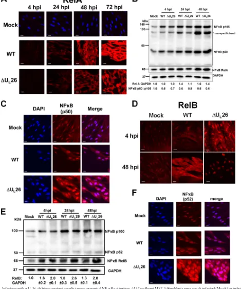
FIG 1Infection with a UL26-deletion mutant results in noncanonical NF-B activation. (A) Confluent MRC5 fibroblasts were mock infected (Mock) or infected
with wild-type (WT) or⌬UL26 HCMV (MOI⫽3.0), fixed at the indicated times postinfection, and processed for RelA-specific immunofluorescence (A; red).
(B) Cells infected as in panel A were harvested at various times postinfection and processed for Western analysis using the indicated antibodies. The relative ratios of RelA to GAPDH and p50 to p105 are indicated. (C and D) Cells infected as in panel A were harvested at 48 hpi and processed for p50-specific immunofluo-rescence (C) or harvested at the indicated times for analysis of RelB-specific immunofluoimmunofluo-rescence (D). (E) Cells infected as in panel A were harvested at the indicated times postinfection and processed for Western analysis using antibodies specific for RelB, p52/p100, or GAPDH. The relative ratios of RelB to GAPDH and p52 to p100 are indicated (averages⫾SEM). (F) Cells were infected as in panel A. At 48 hpi, cells were fixed, permeabilized, and immunostained with antibodies specific for NF-B p52 (red). For all immunofluorescence images, DAPI fluorescence is shown as blue staining; this signal was omitted in panels A and D so as to not obscure nuclear NF-B subunit staining. Scale bars⫽20m. For Western and immunofluorescence experiments, representative images and blots are shown.
on November 7, 2019 by guest
http://jvi.asm.org/
[image:4.585.58.527.44.608.2]the establishment of infection at substantially lower
concentra-tions than those for IFN-
␣
(
Fig. 2D
and
E
). For both cytokines
tested, the
⌬
U
L26 virus was more sensitive to treatment than WT
HCMV (
Fig. 2D
and
E
). However, the sensitivity of
⌬
U
L26 was
more pronounced in response to TNF-
␣
than IFN-
␣
. After
TNF-
␣
treatment, the
⌬
U
L26 mutant exhibited a
⬎
2.5-fold
de-crease in the IC
50of plaque formation (
P
⬍
0.01) (
Fig. 2D
and
E
).
To explore the possibility that deletion of U
L26 might impact
TNF-
␣
production, we assayed for TNF-
␣
levels after mock, WT,
or
⌬
U
L26 infection. WT HCMV infection induced more TNF-a
production than mock-infected cells (
Fig. 2F
), which agrees with
previous reports (
44
,
45
). Cells infected with
⌬
U
L26 induced a
similar amount of TNF-
␣
(
Fig. 2F
), suggesting that increased
TNF-
␣
production does not explain the increased sensitivity of
⌬
U
L26 virus. Rather, these results suggest that in cells exposed to
TNF-
␣
, the U
L26 protein is important for initiation of infection.
The HCMV U
L26 protein antagonizes TNF-
␣
-induced
NF-
B activation.
One of the major consequences of TNF-
␣
treatment is NF-
B pathway activation (reviewed in reference
46
).
Given that the
⌬
U
L26 mutant induces noncanonical NF-
B
activ-ity as well as sensitivactiv-ity to TNF-
␣
treatment, we wanted to explore
potential mechanistic links between these phenotypes. We tested
the impact of U
L26 deletion on NF-
B activity after challenge with
TNF-
␣
. After TNF-
␣
challenge, RelA localized to the nucleus in
mock-infected cells (
Fig. 3A
). Infection with WT HCMV blocked
TNF-
␣
-induced RelA nuclear translocation (
Fig. 3A
). In contrast,
⌬
U
L26-infected cells failed to block TNF-
␣
-induced RelA nuclear
accumulation (
Fig. 3A
). While WT HCMV infection blocked
RelA nuclear translocation upon TNF-
␣
treatment (
Fig. 3A
), it
failed to block RelB translocation upon TNF-
␣
treatment (
Fig.
3B
). Interestingly, TNF-
␣
treatment induced more RelB nuclear
translocation in WT-infected cells than in mock-infected cells
(
Fig. 3B
). As expected,
⌬
U
L26 infection induced RelB nuclear
translocation regardless of whether cells had been treated with
TNF-
␣
.
As RelA nuclear translocation is induced by I
B degradation,
we examined I
B levels during HCMV infection. In the absence of
TNF-
␣
treatment, infection with WT HCMV or
⌬
U
L26 induced
an increase in total I
B
␣
levels relative to results seen with mock
infection (
Fig. 3C
). However, there was little difference in total
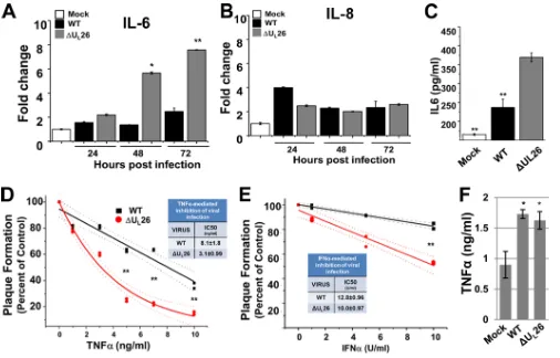
FIG 2A UL26-deletion mutant is more sensitive to treatment with antiviral cytokines. (A and B) MRC5 cells were mock infected or infected with WT or⌬UL26
HCMV (MOI⫽3.0) and harvested at the indicated time points. Real-time PCR was performed using primers specific for IL-6, IL-8, or GAPDH (nⱖ3, averages⫾SEM; signals normalized to GAPDH and compared to those of mock-infected cells at 24 hpi). (C) The amount of IL-6 produced was determined at 24 h after mock, WT, or⌬UL26 infection by ELISA analysis (nⱖ3, averages⫾SEM). (D and E) MRC5 cells were pretreated with different concentrations of TNF-␣(D) or IFN-␣
(E) for 4 h. After removal of the cytokines, a fixed number of PFU from freshly thawed wild-type HCMV (black) or⌬UL26 (red) viral stocks was plated. The
percentage of plaque formation at the different doses of cytokine pretreatment is plotted relative to that of the control (untreated). Best-fit curves were plotted and utilized to estimate IC50(dotted lines represent 95% confidence intervals of the best-fit curves). (F) The amount of TNF-␣produced at 48 h after mock, WT,
or⌬UL26 infection by ELISA analysis (averages⫾SEM). For panels A, B, D, and E, statistical comparisons were made between WT and⌬UL26 infection at the
same time point. For panel C, comparisons were made between mock or WT infection versus⌬UL26 infection. For panel F, the comparison was made between
WT and mock infection (Studentttest; *,P⬍0.05; **,P⬍0.01).
on November 7, 2019 by guest
http://jvi.asm.org/
[image:5.585.39.545.64.388.2]I
B
␣
levels in WT- or
⌬
U
L26-infected cells, with the exception of
72 hpi, when there was more I
B
␣
present in WT- than in
⌬
U
L26-infected cells (
Fig. 3C
). With TNF-
␣
treatment, there was little
difference in the amount of I
B
␣
levels between WT- and
⌬
U
L26-infected cells at 24 hpi (
Fig. 3D
). In contrast, at 48 hpi, TNF-
␣
treatment induced significantly more I
B
␣
degradation in
⌬
U
L26-infected and mock-infected cells than in cells infected with
WT HCMV (
Fig. 3D
). These results suggest that the U
L26 protein
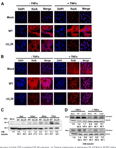
FIG 3UL26 is necessary to block TNF-␣-mediated NF-B activation. (A) Nuclear translocation of endogenous NF-B RelA in HCMV-infected cells upon
TNF-␣treatment. Confluent MRC5 fibroblasts were mock infected (Mock) or infected with WT or⌬UL26 HCMV (MOI⫽3.0). At 48 hpi, cells were treated with
TNF-␣(10 ng/ml) for 1 h, followed by fixation and processing for RelA immunofluorescence (red) and nuclear DAPI staining (blue). Representative images are shown. Bars⫽20m. (B) Cells were infected and treated with TNF-␣as in panel A, followed by analysis of RelB immunofluorescence (red) and nuclear DAPI staining (blue). Representative images are shown. Bars⫽20m. (C) Protein levels of IB␣during mock, WT, or⌬UL26 infection (MOI⫽3.0). Cells were
harvested for SDS-PAGE and Western analysis at the indicated times (averages⫾SEM). (D) At 24 hpi or 48 hpi, cells were treated with TNF-␣(10 ng/ml) for 1 h, harvested, and processed for Western analysis using IB␣- or tubulin-specific antibodies. The relative ratios of IB to tubulin are indicated below each blot (B and C) (averages⫾SEM). For Western and immunofluorescence experiments, representative images/blots are shown.
Mathers et al.
on November 7, 2019 by guest
http://jvi.asm.org/
[image:6.585.89.496.60.577.2]is important for HCMV-mediated inhibition of TNF-
␣
-induced
NF-
B activity inasmuch as its deletion results in increased RelA
nuclear translocation and I
B degradation.
While we found the U
L26 protein to be necessary for
HCMV-mediated inhibition of TNF-
␣
-induced RelA nuclear
transloca-tion, we wanted to test whether it was sufficient to block TNF-
␣
-induced NF-
B activity in the absence of other HCMV viral
proteins. Expression of U
L26 blocked TNF-
␣
-induced NF-
B-de-pendent luciferase activity (
Fig. 4A
). The inhibition of TNF-
␣
-induced NF-
B activity was largely U
L26 dose dependent (
Fig.
4B
). To determine whether U
L26 expression was sufficient to
block NF-
B nuclear translocation upon TNF-
␣
treatment, we
examined the localization of GFP-tagged RelA. Shortly after
TNF-
␣
treatment, GFP-RelA migrated to the nucleus in vector
control-transfected cells (
Fig. 4C
). TNF-
␣
-induced GFP-RelA
translocation was largely blocked by coexpression of the U
L26
protein (
Fig. 4C
). Similar results were observed with the analysis
of endogenous RelA; U
L26 attenuated the translocation of
endog-enous NF-
B to the nucleus upon TNF-
␣
treatment (
Fig. 4D
).
Analysis of I
B levels indicated that U
L26 expression stabilized
I
B after TNF-
␣
treatment (
Fig. 4E
). Taken together, these results
indicate that U
L26 expression is sufficient to stabilize I
B and
block NF-
B activation after TNF-
␣
treatment.
The HCMV U
L26 protein blocks NF-
B activity induced by
Sendai virus infection.
To delineate whether U
L26-mediated
in-hibition of NF-
B activation was specific for TNF-
␣
, we sought to
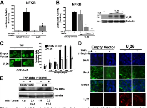
FIG 4UL26 is sufficient to bock TNF-␣-induced NF-B activation. (A) 293T cells were cotransfected with an NF-B-dependent luciferase construct, aRenilla
expression construct, and equal amounts of either empty vector or UL26 overexpression vector (1g). Twenty-four hours posttransfection, cells were mock or
TNF-␣treated (10 ng/ml), incubated for 24 h, harvested, and assayed for firefly luciferase (Fluc) andRenillaluciferase (Rluc) activity. Averages are plotted after normalization toRenillaluciferase activity with SEM (nⱖ3; *,P⬍0.05; **,P⬍0.01 [UL26 versus vector transfected]). (B) 293T cells were transfected and treated
as in panel A but with various amounts of UL26 as indicated (0.25, 0.5, and 1.0g). Averages are plotted after normalization toRenillaluciferase activity with SEM
(nⱖ3; *,P⬍0.05; **,P⬍0.01). The UL26 protein levels in transfected 293T cells were determined by Western analysis. (C) 293T cells were cotransfected with
plasmids expressing a GFP-RelA protein with either UL26 expression plasmid or an empty vector. At 24 h posttransfection, cells were treated with TNF-␣(10
ng/ml) for 24 h. The subcellular localization of GFP-RelA was assessed under a fluorescence microscope. GFP-RelA nuclear translocation was quantified at various times post-TNF-␣treatment (**,P⬍0.01 [UL26 versus vector transfected at the same time point]). (D) 293T cells were transfected as in panel A. At 24
h posttransfection, cells were treated with TNF-␣(10 ng/ml) for 24 h, and the subcellular localization of endogenous RelA or UL26 was assessed by confocal
microscopy. Cellular nuclei were stained with DAPI (blue). Representative images are shown. (E) 293T cells were transfected as in panel A. Twenty-four hours posttransfection, cells were treated with TNF-␣(10 ng/ml) for 24 h prior to processing for Western analysis with antibodies specific for IB␣or tubulin. The relative ratios of IB to tubulin are indicated below the blot (averages⫾SEM). For all Western and immunofluorescence experiments, representative images/ blots are shown.
on November 7, 2019 by guest
http://jvi.asm.org/
[image:7.585.40.540.65.433.2]test if U
L26 inhibited NF-
B activation in response to other
NF-
B inducers. Toward this end, we infected cells with Sendai
virus (SeV), which induces both NF-
B and IRF3 activation
through a RIGI-dependent pathway (
47
) (
Fig. 5A
). SeV-infected
cells exhibited pronounced NF-
B-dependent luciferase activity
(
Fig. 5B
). This induction was substantially reduced by transfection
with U
L26 (
Fig. 5B
). These results demonstrate that in addition to
blocking TNF-
␣
-induced NF-
B activation, expression of U
L26
blocks NF-
B activation mediated by an alternative pathway,
in-dicating that U
L26 is acting at a conserved downstream point
within these pathways.
SeV infection also resulted in a strong elevation of
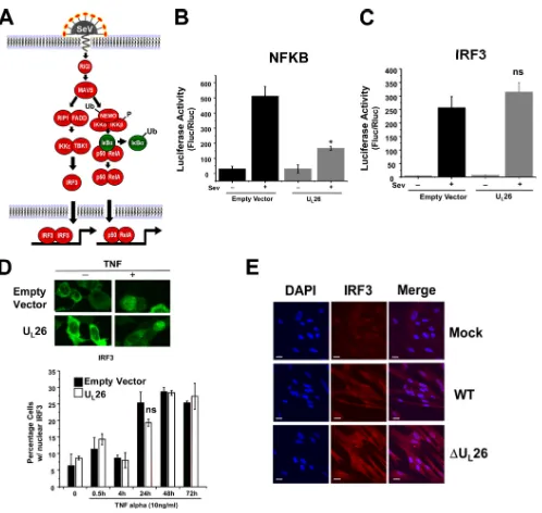
IRF3-depen-FIG 5UL26 blocks Sendai virus-induced NF-B but not IRF3 activation. (A) Schematic of Sendai virus (SeV)-induced IRF3 and NF-B activation. Ub,
ubiquitination; P, phosphorylation. (B) 293T cells were cotransfected with an NF-B-dependent luciferase construct, aRenillaexpression construct, and equal amounts of either empty vector or UL26 overexpression vector (1g). At 24 h posttransfection, cells were either mock infected or infected with SeV (MOI⫽3.0).
Lysates were harvested at 16 hpi and assessed for luciferase activity (averages are plotted after normalization toRenillaluciferase activity (with SEM;nⱖ3; *, P⬍0.05 [UL26 versus vector transfected]). (C) 293T cells were treated as in panel B, with the exception that an IRF3-dependent luciferase construct was utilized
(ns, not significant). (D) 293T cells were cotransfected with a GFP-tagged IRF3 expression plasmid and with either an empty vector or UL26 expression plasmid.
At 24 h posttransfection, cells were treated with TNF-␣(10 ng/ml) for 24 h, followed by assessment of GFP-IRF3 subcellular localization by fluorescence microscopy. GFP-IRF3 nuclear translocation was quantified at various times post-TNF-␣treatment as indicated (ns, not significant). (E) Confluent MRC5 fibroblasts were mock infected or infected with WT or⌬UL26 HCMV (MOI⫽3.0). At 6 hpi, cells were fixed, permeabilized, and immunostained with antibodies
to endogenous IRF3 (red). Cellular nuclei were stained with DAPI (blue). Representative images are shown. Mathers et al.
on November 7, 2019 by guest
http://jvi.asm.org/
[image:8.585.42.540.59.531.2]dent luciferase activity (
Fig. 5C
). This induction of IRF3 activity
was unaffected by expression of U
L26 (
Fig. 5C
). U
L26 expression
also had no effect on TNF-
␣
-induced IRF3 nuclear translocation
(
Fig. 5D
). These results suggest that U
L26-mediated inhibition of
NF-
B signaling occurs downstream of the divergence between
TNF-
␣
’s and SeV’s stimulation of IRF3 and NF-
B signaling.
Fur-ther supporting a lack of impact on IRF3 signaling, infection with
a U
L26-deletion mutant did not impact IRF3 localization during
HCMV infection (
Fig. 5E
).
The HCMV U
L26 protein blocks phosphorylation of the IKK
complex.
Our findings that U
L26 blocks NF-
B activation
in-duced by either TNF-
␣
or SeV infection suggest that U
L26 blocks
NF-
B activation downstream of where these pathways converge,
which occurs at the activation of the IKK complex. In
combina-tion with the findings that U
L26 maintains I
B levels in the
pres-ence of TNF-
␣
signaling (
Fig. 4E
), our results suggest that the
U
L26 site of action is either I
B or the IKK complex. The IKK
complex is active when IKK
␣
/

is serine phosphorylated in its
activation loop, which is mediated either through an upstream
kinase or through autophosphorylation (
48
). We examined IKK
phosphorylation in WT- or
⌬
U
L26-infected cells. At 48 hpi, in the
absence of TNF-
␣
, WT- and
⌬
U
L26-infected cells exhibited
roughly equivalent amounts of IKK phosphorylation (
Fig. 6A
).
The levels of IKK phosphorylation during infection with either
virus were increased in comparison to levels seen with
mock-in-fected cells, suggesting a higher level of basal phosphorylation
during infection (
Fig. 6A
). At this same time point, upon TNF-
␣
treatment, WT-infected cells exhibited similar amounts of IKK
phosphorylation as untreated cells, consistent with an
HCMV-induced block to TNF-
␣
-induced IKK phosphorylation and
acti-vation. In contrast, mock- or
⌬
U
L26-infected cells showed an
in-crease in IKK phosphorylation upon TNF-
␣
treatment, consistent
with IKK activation (
Fig. 6A
). These results suggest
HCMV-me-diated attenuation of TNF-
␣
-induced IKK phosphorylation
re-quires functional U
L26. It has been previously reported that
HCMV blocks TNF-
␣
-induced NF-
B signaling at 48 hpi, but not
prior to 24 hpi (
39
). If such is the case, it would suggest that
tegument protein delivery, which would include the U
L26 protein,
is not sufficient to block TNF-
␣
-induced NF-
B activation. To
further explore this issue, we tested whether WT or UV-irradiated
HCMV was sufficient to block TNF-
␣
-induced IKK
phosphory-lation at various times postinfection. As shown in
Fig. 6B
, neither
WT HCMV nor UV-HCMV was capable of blocking TNF-
␣
-in-duced IKK phosphorylation at 6 hpi. At 24 hpi, TNF-
␣
-induced
IKK phosphorylation was slightly reduced in HCMV-infected
cells relative to TNF-
␣
-treated mock-infected cells (
Fig. 6B
). In
contrast, at 48 hpi, WT HCMV infection, but not that with
UV-irradiated HCMV, resulted in a complete block in IKK
phosphor-ylation (
Fig. 6B
). These results suggest that HCMV-mediated
teg-ument protein delivery is not sufficient to block TNF-
␣
-induced
IKK phosphorylation. To determine if U
L26 expression is
suffi-cient to block TNF-
␣
-induced IKK phosphorylation in the
ab-sence of other viral proteins, we transfected cells with U
L26,
treated them with TNF-
␣
, and assessed IKK phosphorylation.
Ex-pression of the U
L26 protein in the absence of other HCMV
proteins blocked TNF-
␣
-induced IKK phosphorylation (
Fig.
6C
). These results suggest that the U
L26 protein blocks
TNF-␣
-induced IKK phosphorylation, thereby attenuating NF-
B
signaling.
DISCUSSION
The NF-
B pathway is central to innate immunity and defense
against viruses. NF-
B activation can induce an antiviral state that
limits viral replication; thus, many different viruses attenuate
NF-
B signaling as part of their infectious program (
49
). We have
found that the U
L26 gene blocks many aspects of NF-
B signaling,
including TNF-
␣
-induced IKK phosphorylation, I
B
degrada-tion, and RelA nuclear translocation. Further, in contrast to WT
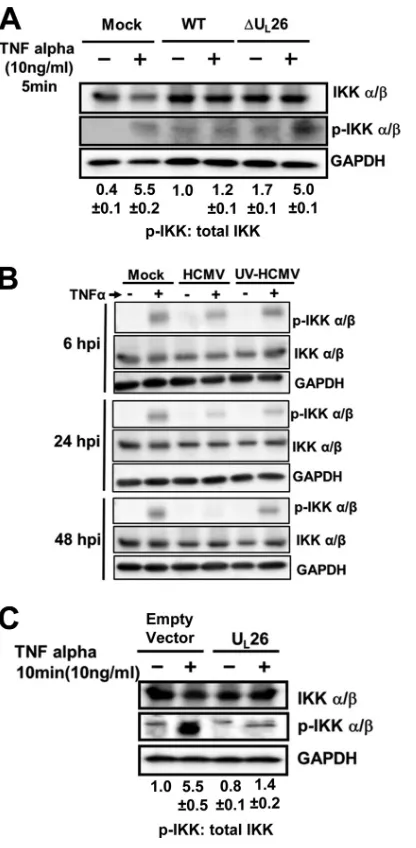
FIG 6UL26 blocks IKK phosphorylation. (A) MRC5 cells were mock infected
or infected with WT or⌬UL26 HCMV (MOI⫽3.0). At 48 hpi, cells were
treated with TNF-␣(10 ng/ml) for 5 min and then processed for Western analysis using the indicated antibodies. Ratios of pIKK to total IKK are shown under each blot (averages⫾SEM). (B) MRC5 cells were mock infected or infected with mock-irradiated or UV-irradiated WT HCMV (MOI⫽3.0), treated with TNF-␣(10 ng/ml) for 5 min at the indicated hpi, and processed for Western analysis using the indicated antibodies. (C) 293T cells were trans-fected with either an empty vector or a UL26 expression plasmid. Twenty-four
hours posttransfection, cells were treated with TNF-␣(10 ng/ml) for 10 min and then processed for Western analysis using the indicated antibodies. The relative ratios of pIKK to total IKK are indicated below each blot (B and C) (averages⫾SEM).
on November 7, 2019 by guest
http://jvi.asm.org/
[image:9.585.315.523.64.488.2]HCMV, a U
L26 deletion mutant fails to block TNF-
␣
-induced
NF-
B activation, and it is more sensitive to challenge with
TNF-
␣
. U
L26 was also sufficient to block NF-
B activation
in-duced by SeV infection, suggesting that the impact of inhibition of
NF-
B mediated by U
L26 is not TNF-
␣
specific. In the absence of
an exogenous NF-
B inducer, infection with a U
L26-deletion
mu-tant resulted in the activation of noncanonical NF-
B signaling,
typified by RelB nuclear translocation. Together, our results
indi-cate that the HCMV U
L26 gene is an important viral modulator of
NF-
B signaling.
We found that the U
L26 protein can impact both the canonical
and noncanonical NF-
B pathways. In the context of HCMV
in-fection, the lack of U
L26 resulted in RelB nuclear translocation,
with concomitant induction of IL-6. Interestingly, in the absence
of infection, HCMV IE1 expression has been reported to induce
RelB nuclear translocation (
50
). Given this finding, U
L26 may be
necessary to block RelB nuclear translocation that is induced by
IE1 activity during infection. Despite U
L26 being necessary and
sufficient to block RelA nuclear translocation upon TNF-
␣
treat-ment, infection with
⌬
U
L26 in the absence of TNF-
␣
did not
in-duce RelA nuclear translocation. The lack of RelA nuclear
trans-location suggests that infection with Ad169 does not activate the
canonical NF-
B pathway. Clinical HCMV strains would likely
behave differently, as contrary to laboratory adapted strains, they
express U
L144, which has been demonstrated to induce NF-
B
activation (
51
,
52
), and U
L138, which sensitizes cells to TNF-
␣
treatment (
53
,
54
). Additionally, both laboratory and clinical
strains encode other factors that antagonize canonical NF-
B
sig-naling. The IE2 protein (IE86) and the major tegument protein
pp65 have been reported to inhibit the DNA binding of NF-
B
subunits in the nucleus (
12
,
55
). The deletion of the U
L26 protein
has been shown to impact virion tegumentation (
15
), which
sug-gests the possibility that defective tegumentation could impact
NF-
B-related phenotypes early during infection. However, our
experiments indicate that the HCMV-mediated inhibition of
TNF-
␣
-induced IKK phosphorylation did not occur until 48 hpi
and does not occur upon UV inactivation, suggesting that
de novo
protein synthesis, and not merely delivery of tegument proteins, is
required for preventing IKK phosphorylation upon TNF-
␣
treat-ment. Further, the finding that U
L26 is sufficient in the absence of
other genes to block TNF-
␣
-induced NF-
B activation strongly
suggests that these TNF-
␣
-associated phenotypes are attributable
to U
L26. However, it is possible that different mechanisms are
responsible for the two main NF-
B phenotypes that we have
found to be associated with
⌬
U
L26 infection, i.e., increased RelB
nuclear translocation and increased TNF-
␣
-mediated NF-
B
sig-naling. The RelB nuclear translocation occurs at an earlier time
during
⌬
U
L26 infection than does the protection from TNF-
␣
signaling. For the RelB translocation phenotype, the interplay of
U
L26 with other viral factors in the virion could therefore be
im-portant. Alternatively, differentially secreted factors present in the
initial inoculum, e.g., IL-6, could play a role in the RelB
pheno-type, as our viral stocks contain conditioned media from the
in-fection responsible for viral stock creation. Future work will need
to examine both how different viral factors cooperate to modulate
NF-
B activity and how differential cytokine excretion resulting
from U
L26 deletion could impact infection.
Both TNF-
␣
treatment and SeV infection induce NF-
B
acti-vation, but their upstream signaling components differ. TNF-
␣
induces TRAF assembly with the TNF-
␣
receptors and subsequent
recruitment and activation of TAK1, which in turn activates the
IKK complex (
46
). SeV induces NF-
B activation through MAVS/
RIGI-dependent activation of IKK (
47
). Our finding that U
L26
blocks NF-
B activation induced by either TNF-
␣
or SeV
infec-tion suggests that U
L26 likely acts downstream of where these
pathways converge. The convergence of these pathways at IKK,
combined with our results indicating that U
L26 blocks induction
of IKK phosphorylation, together implicate the IKK complex as
the site of U
L26 modulation. U
L26-mediated modulation of IKK
activity is also consistent with U
L26 impacting both the canonical
RelA and noncanonical RelB pathway, as these two pathways
share a dependence on IKK activity.
It has been reported that HCMV induces IKK activity, which is
important for viral replication in certain settings (
32–34
).
Consis-tent with these reports, we find that the basal level of IKK
phos-phorylation is induced by WT HCMV infection. U
L26 does not
appear to be important for this observed increase in IKK
phos-phorylation, but rather is only necessary to prevent
hyperphos-phorylation upon exposure to external NF-
B activators.
To-gether, these studies, in combination with ours, suggest that
HCMV may benefit from a controlled activation of IKK, yet has
evolved mechanisms to antagonize the robust IKK activity
associ-ated with antiviral cytokines. This notion fits into a model of what
appears to be a complex relationship between HCMV and NF-
B.
Several studies have found that extrinsic NF-
B activators inhibit
HCMV infection (
56–58
), while others have found that inhibition
of NF-
B attenuates HCMV infection (
32–34
). Further, reports
indicate that HCMV infection induces NF-
B transcriptional
ac-tivity (
35
,
52
), while others indicate that HCMV infection blocks
NF-
B activation (
12
,
38
,
39
,
55
,
59
). On the surface, these reports
appear to contradict each other, yet they likely reflect the
com-plexity of NF-
B signaling as well as the multifaceted nature of
HCMV-mediated NF-
B modulation. For example, NF-
B
acti-vation can induce HCMV promoters (
35
,
37
,
60
) as well as induce
expression of host NF-
B targets that are important for HCMV
replication (
61
). This suggests that limited activation of NF-
B
could benefit viral replication. On the other hand, extrinsic
acti-vators of NF-
B lead to an antiviral state that is known to inhibit
HCMV replication (
58
), and thus HCMV has evolved gene
prod-ucts to attenuate these pathways. The central question becomes
how, mechanistically, HCMV shapes NF-
B transcriptional
out-put to induce proviral NF-
B targets while attenuating the
expres-sion of antiviral NF-
B targets. Our results suggest that U
L26 is a
key player in this process.
In summary, we find that the U
L26 gene is an important
com-ponent in HCMV’s effort to modulate NF-
B activity. Diverse
viral families attenuate innate immune pathways to enable
high-titer replication. This virus-host, innate immune interaction
represents a critical evolutionary battleground that shapes the
outcome of viral infection. Viral factors, such as U
L26, that
manipulate innate immune responses are therefore potentially
at-tractive targets to limit viral spread. Further, given the central
importance of these pathways to infection in general, information
garnered from their analysis can potentially be widely applicable
to diverse viral families.
ACKNOWLEDGMENTS
This work was supported by a grant to J.M. from the National Institute of Allergy and Infectious Diseases (R01AI081773). J.M. is a Damon Runyon-Rachleff Innovator supported (in part) by the Damon Runyon Cancer Mathers et al.
on November 7, 2019 by guest
http://jvi.asm.org/
Research Foundation (DRR-09-10). Research in L.M.-S.’s laboratory is funded by NIH grants RO1 AI077719 and R03AI099681-01A1, the NIAID Centers of Excellence for Influenza Research and Surveillance (HHSN266200700008C), and The University of Rochester Center for Biodefense Immune Modeling (HHSN272201000055C).
REFERENCES
1.Mocarski ES, Shenk T, Pass RF.2007. Cytomegaloviruses, p 2701–2757. InKnipe DM, Howley PM, Griffin DE, Lamb RA, Martin MA, Roizman BR, Straus SE (ed), Fields virology, 5th ed. Lippincott Williams & Wilkins, New York, NY.
2.Cannon MJ.2009. Congenital cytomegalovirus (CMV) epidemiology and awareness. J. Clin. Virol.46(Suppl 4):S6 –S10.http://dx.doi.org/10.1016/j .jcv.2009.09.002.
3.Andrei G, De Clercq E, Snoeck R.2008. Novel inhibitors of human CMV. Curr. Opin. Investig. Drugs9:132–145.
4.Grosse SD, Ross DS, Dollard SC. 2008. Congenital cytomegalovirus (CMV) infection as a cause of permanent bilateral hearing loss: a quanti-tative assessment. J. Clin. Virol.41:57– 62.http://dx.doi.org/10.1016/j.jcv .2007.09.004.
5.Gerna G, Baldanti F, Revello MG.2004. Pathogenesis of human cyto-megalovirus infection and cellular targets. Hum. Immunol.65:381–386.
http://dx.doi.org/10.1016/j.humimm.2004.02.009. 6. Reference deleted.
7.Murphy E, Rigoutsos I, Shibuya T, Shenk TE.2003. Reevaluation of human cytomegalovirus coding potential. Proc. Natl. Acad. Sci. U. S. A.
100:13585–13590.http://dx.doi.org/10.1073/pnas.1735466100. 8.Stern-Ginossar N, Weisburd B, Michalski A, Le VT, Hein MY, Huang
SX, Ma M, Shen B, Qian SB, Hengel H, Mann M, Ingolia NT, Weiss-man JS. 2012. Decoding human cytomegalovirus. Science 338:1088 – 1093.http://dx.doi.org/10.1126/science.1227919.
9.Kalejta RF, Bechtel JT, Shenk T.2003. Human cytomegalovirus pp71 stim-ulates cell cycle progression by inducing the proteasome-dependent degrada-tion of the retinoblastoma family of tumor suppressors. Mol. Cell. Biol.23:
1885–1895.http://dx.doi.org/10.1128/MCB.23.6.1885-1895.2003. 10. Baldick CJ, Jr, Marchini A, Patterson CE, Shenk T. 1997. Human
cytomegalovirus tegument protein pp71 (ppUL82) enhances the infectiv-ity of viral DNA and accelerates the infectious cycle. J. Virol.71:4400 – 4408.
11. Bresnahan WA, Shenk TE.2000. UL82 virion protein activates expres-sion of immediate early viral genes in human cytomegalovirus-infected cells. Proc. Natl. Acad. Sci. U. S. A.97:14506 –14511.http://dx.doi.org/10 .1073/pnas.97.26.14506.
12. Browne EP, Shenk T.2003. Human cytomegalovirus UL83-coded pp65 virion protein inhibits antiviral gene expression in infected cells. Proc. Natl. Acad. Sci. U. S. A.100:11439 –11444.http://dx.doi.org/10.1073/pnas .1534570100.
13. Abate DA, Watanabe S, Mocarski ES.2004. Major human cytomegalo-virus structural protein pp65 (ppUL83) prevents interferon response fac-tor 3 activation in the interferon response. J. Virol.78:10995–11006.http: //dx.doi.org/10.1128/JVI.78.20.10995-11006.2004.
14. Stamminger T, Gstaiger M, Weinzierl K, Lorz K, Winkler M, Schaffner W.2002. Open reading frame UL26 of human cytomegalovirus encodes a novel tegument protein that contains a strong transcriptional activation domain. J. Virol. 76:4836 – 4847. http://dx.doi.org/10.1128/JVI.76.10 .4836-4847.2002.
15. Munger J, Yu D, Shenk T.2006. UL26-deficient human cytomegalovirus produces virions with hypophosphorylated pp28 tegument protein that is unstable within newly infected cells. J. Virol.80:3541–3548.http://dx.doi .org/10.1128/JVI.80.7.3541-3548.2006.
16. Yu D, Silva MC, Shenk T.2003. Functional map of human cytomegalo-virus AD169 defined by global mutational analysis. Proc. Natl. Acad. Sci. U. S. A.100:12396 –12401.http://dx.doi.org/10.1073/pnas.1635160100. 17. Lorz K, Hofmann H, Berndt A, Tavalai N, Mueller R,
Schlotzer-Schrehardt U, Stamminger T. 2006. Deletion of open reading frame UL26 from the human cytomegalovirus genome results in reduced viral growth, which involves impaired stability of viral particles. J. Virol.80:
5423–5434.http://dx.doi.org/10.1128/JVI.02585-05.
18. Haas TL, Emmerich CH, Gerlach B, Schmukle AC, Cordier SM, Rieser E, Feltham R, Vince J, Warnken U, Wenger T, Koschny R, Komander D, Silke J, Walczak H.2009. Recruitment of the linear ubiquitin chain assembly complex stabilizes the TNF-R1 signaling complex and is
re-quired for TNF-mediated gene induction. Mol. Cell36:831– 844.http://dx .doi.org/10.1016/j.molcel.2009.10.013.
19. Tokunaga F, Sakata S, Saeki Y, Satomi Y, Kirisako T, Kamei K, Naka-gawa T, Kato M, Murata S, Yamaoka S, Yamamoto M, Akira S, Takao T, Tanaka K, Iwai K.2009. Involvement of linear polyubiquitylation of NEMO in NF-kappaB activation. Nature Cell Biol.11:123–132.http://dx .doi.org/10.1038/ncb1821.
20. Chen Z, Hagler J, Palombella VJ, Melandri F, Scherer D, Ballard D, Maniatis T.1995. Signal-induced site-specific phosphorylation targets I kappa B alpha to the ubiquitin-proteasome pathway. Genes Dev.9:1586 – 1597.http://dx.doi.org/10.1101/gad.9.13.1586.
21. DiDonato J, Mercurio F, Rosette C, Wu-Li J, Suyang H, Ghosh S, Karin M.1996. Mapping of the inducible IkappaB phosphorylation sites that signal its ubiquitination and degradation. Mol. Cell. Biol.16:1295–1304. 22. Oeckinghaus A, Ghosh S.2009. The NF-kappaB family of transcription
factors and its regulation. Cold Spring Harb. Perspect. Biol.1:a000034.
http://dx.doi.org/10.1101/cshperspect.a000034.
23. Sun SC.2012. The noncanonical NF-kappaB pathway. Immunol. Rev.
246:125–140.http://dx.doi.org/10.1111/j.1600-065X.2011.01088.x. 24. Perkins ND.2007. Integrating cell-signalling pathways with NF-kappaB
and IKK function. Nature Rev. 8:49 – 62. http://dx.doi.org/10.1038 /nrm2083.
25. Smale ST.2011. Hierarchies of NF-kappaB target-gene regulation. Nat. Immunol.12:689 – 694.http://dx.doi.org/10.1038/ni.2070.
26. Rowe WP, Hartley JW, Waterman S, Turner HC, Huebner RJ.1956. Cytopathogenic agent resembling human salivary gland virus recovered from tissue cultures of human adenoids. Proc. Soc. Exp. Biol. Med.92:
418 – 424.http://dx.doi.org/10.3181/00379727-92-22497.
27. Yu D, Smith GA, Enquist LW, Shenk T.2002. Construction of a self-excisable bacterial artificial chromosome containing the human cytomeg-alovirus genome and mutagenesis of the diploid TRL/IRL13 gene. J. Virol.
76:2316 –2328.http://dx.doi.org/10.1128/jvi.76.5.2316-2328.2002. 28. Mathers C, Spencer CM, Munger J.2014. Distinct domains within the
human cytomegalovirus U(L)26 protein are important for wildtype viral replication and virion stability. PLoS One9:e88101.http://dx.doi.org/10 .1371/journal.pone.0088101.
29. Rodrigo WW, Ortiz-Riaño E, Pythoud C, Kunz S, de la Torre JC, Martínez-Sobrido L.2012. Arenavirus nucleoproteins prevent activation of nuclear factor kappa B. J. Virol.86:8185– 8197.http://dx.doi.org/10 .1128/JVI.07240-11.
30. Martínez-Sobrido L, Zuniga EI, Rosario D, Garcia-Sastre A, de la Torre JC.2006. Inhibition of the type I interferon response by the nucleoprotein of the prototypic arenavirus lymphocytic choriomeningitis virus. J. Virol.
80:9192–9199.http://dx.doi.org/10.1128/JVI.00555-06.
31. Munger J, Bajad SU, Coller HA, Shenk T, Rabinowitz JD.2006. Dynamics of the cellular metabolome during human cytomegalovirus infection. PLoS Pathog.2:e132.http://dx.doi.org/10.1371/journal.ppat.0020132.
32. Caposio P, Dreano M, Garotta G, Gribaudo G, Landolfo S. 2004. Human cytomegalovirus stimulates cellular IKK2 activity and requires the enzyme for productive replication. J. Virol.78:3190 –3195.http://dx.doi .org/10.1128/JVI.78.6.3190-3195.2004.
33. Caposio P, Luganini A, Hahn G, Landolfo S, Gribaudo G.2007. Acti-vation of the virus-induced IKK/NF-kappaB signalling axis is critical for the replication of human cytomegalovirus in quiescent cells. Cell. Micro-biol.9:2040 –2054.http://dx.doi.org/10.1111/j.1462-5822.2007.00936.x. 34. Caposio P, Musso T, Luganini A, Inoue H, Gariglio M, Landolfo S,
Gribaudo G.2007. Targeting the NF-kappaB pathway through pharma-cological inhibition of IKK2 prevents human cytomegalovirus replication and virus-induced inflammatory response in infected endothelial cells. Antiviral Res. 73:175–184. http://dx.doi.org/10.1016/j.antiviral.2006.10 .001.
35. Yurochko AD, Kowalik TF, Huong SM, Huang ES. 1995. Human cytomegalovirus upregulates NF-kappa B activity by transactivating the NF-kappa B p105/p50 and p65 promoters. J. Virol.69:5391–5400. 36. DeMeritt IB, Milford LE, Yurochko AD.2004. Activation of the
NF-kappaB pathway in human cytomegalovirus-infected cells is necessary for efficient transactivation of the major immediate-early promoter. J. Virol.
78:4498 – 4507.http://dx.doi.org/10.1128/JVI.78.9.4498-4507.2004. 37. DeMeritt IB, Podduturi JP, Tilley AM, Nogalski MT, Yurochko AD.
2006. Prolonged activation of NF-kappaB by human cytomegalovirus promotes efficient viral replication and late gene expression. Virology346:
15–31.http://dx.doi.org/10.1016/j.virol.2005.09.065.
38. Jarvis MA, Borton JA, Keech AM, Wong J, Britt WJ, Magun BE, Nelson
on November 7, 2019 by guest
http://jvi.asm.org/
JA.2006. Human cytomegalovirus attenuates interleukin-1beta and tu-mor necrosis factor alpha proinflammatory signaling by inhibition of NF-kappaB activation. J. Virol.80:5588 –5598.http://dx.doi.org/10.1128/JVI .00060-06.
39. Montag C, Wagner J, Gruska I, Hagemeier C.2006. Human cytomeg-alovirus blocks tumor necrosis factor alpha- and interleukin-1beta-mediated NF-kappaB signaling. J. Virol.80:11686 –11698.http://dx.doi .org/10.1128/JVI.01168-06.
40. Kunsch C, Lang RK, Rosen CA, Shannon MF.1994. Synergistic tran-scriptional activation of the IL-8 gene by NF-kappa B p65 (RelA) and NF-IL-6. J. Immunol.153:153–164.
41. Kannabiran C, Zeng X, Vales LD.1997. The mammalian transcriptional repressor RBP (CBF1) regulates interleukin-6 gene expression. Mol. Cell. Biol.17:1–9.
42. Kang HB, Kim YE, Kwon HJ, Sok DE, Lee Y.2007. Enhancement of NF-kappaB expression and activity upon differentiation of human embry-onic stem cell line SNUhES3. Stem Cells Dev.16:615– 623.http://dx.doi .org/10.1089/scd.2007.0014.
43. Kunsch C, Rosen CA.1993. NF-kappa B subunit-specific regulation of the interleukin-8 promoter. Mol. Cell. Biol.13:6137– 6146.
44. Geist LJ, Monick MM, Stinski MF, Hunninghake GW.1994. The im-mediate early genes of human cytomegalovirus upregulate tumor necrosis factor-alpha gene expression. J. Clin. Invest.93:474 – 478.http://dx.doi .org/10.1172/JCI116995.
45. Smith PD, Saini SS, Raffeld M, Manischewitz JF, Wahl SM. 1992. Cytomegalovirus induction of tumor necrosis factor-alpha by human monocytes and mucosal macrophages. J. Clin. Invest.90:1642–1648.http: //dx.doi.org/10.1172/JCI116035.
46. Varfolomeev EE, Ashkenazi A.2004. Tumor necrosis factor: an apoptosis JuNKie? Cell116:491–497.http://dx.doi.org/10.1016/S0092-8674(04)00166-7. 47. Elco CP, Guenther JM, Williams BR, Sen GC.2005. Analysis of genes
induced by Sendai virus infection of mutant cell lines reveals essential roles of interferon regulatory factor 3, NF-kappaB, and interferon but not toll-like receptor 3. J. Virol.79:3920 –3929.http://dx.doi.org/10.1128/JVI.79.7 .3920-3929.2005.
48. Israël A.2010. The IKK complex, a central regulator of NF-kappaB acti-vation. Cold Spring Harb. Perspect. Biol.2:a000158.http://dx.doi.org/10 .1101/cshperspect.a000158.
49. Hiscott J, Nguyen TL, Arguello M, Nakhaei P, Paz S.2006. Manipula-tion of the nuclear factor-kappaB pathway and the innate immune re-sponse by viruses. Oncogene25:6844 – 6867.http://dx.doi.org/10.1038/sj .onc.1209941.
50. Jiang HY, Petrovas C, Sonenshein GE.2002. RelB-p50 NF-B complexes are selectively induced by cytomegalovirus immediate-early protein 1: dif-ferential regulation of Bcl-xLpromoter activity by NF-B family members.
J. Virol. 76:5737–5747. http://dx.doi.org/10.1128/JVI.76.11.5737-5747 .2002.
51. Poole E, Groves I, MacDonald A, Pang Y, Alcami A, Sinclair J.2009. Identification of TRIM23 as a cofactor involved in the regulation of NF-kappaB by human cytomegalovirus. J. Virol.83:3581–3590.http://dx.doi .org/10.1128/JVI.02072-08.
52. Poole E, King CA, Sinclair JH, Alcami A.2006. The UL144 gene product of human cytomegalovirus activates NFkappaB via a TRAF6-dependent mechanism. EMBO J.25:4390 – 4399.http://dx.doi.org/10.1038/sj.emboj .7601287.
53. Le VT, Trilling M, Hengel H.2011. The cytomegaloviral protein pUL138 acts as potentiator of tumor necrosis factor (TNF) receptor 1 surface den-sity to enhance ULb=-encoded modulation of TNF-␣signaling. J. Virol.
85:13260 –13270.http://dx.doi.org/10.1128/JVI.06005-11.
54. Montag C, Wagner JA, Gruska I, Vetter B, Wiebusch L, Hagemeier C.
2011. The latency-associated UL138 gene product of human cytomegalo-virus sensitizes cells to tumor necrosis factor alpha (TNF-alpha) signaling by upregulating TNF-alpha receptor 1 cell surface expression. J. Virol.
85:11409 –11421.http://dx.doi.org/10.1128/JVI.05028-11.
55. Taylor RT, Bresnahan WA.2006. Human cytomegalovirus IE86 attenu-ates virus- and tumor necrosis factor alpha-induced NFkappaB-dependent gene expression. J. Virol.80:10763–10771.http://dx.doi.org /10.1128/JVI.01195-06.
56. Allan-Yorke J, Record M, de Preval C, Davrinche C, Davignon JL.1998. Distinct pathways for tumor necrosis factor alpha and ceramides in hu-man cytomegalovirus infection. J. Virol.72:2316 –2322.
57. Davignon JL, Castanie P, Yorke JA, Gautier N, Clement D, Davrinche C.1996. Anti-human cytomegalovirus activity of cytokines produced by CD4⫹T-cell clones specifically activated by IE1 peptides in vitro. J. Virol.
70:2162–2169.
58. Pavic´ I, Polic´ B, Crnkovic´ I, Lucin P, Jonjic´ S, Koszinowski UH.1993. Participation of endogenous tumour necrosis factor alpha in host resis-tance to cytomegalovirus infection. J. Gen. Virol.74(Part 10):2215–2223.
http://dx.doi.org/10.1099/0022-1317-74-10-2215.
59. Baillie J, Sahlender DA, Sinclair JH. 2003. Human cytomegalovirus infection inhibits tumor necrosis factor alpha (TNF-alpha) signaling by targeting the 55-kilodalton TNF-alpha receptor. J. Virol.77:7007–7016.
http://dx.doi.org/10.1128/JVI.77.12.7007-7016.2003.
60. Chan G, Bivins-Smith ER, Smith MS, Yurochko AD.2008. Transcrip-tome analysis of NF-kappaB- and phosphatidylinositol 3-kinase-regulated genes in human cytomegalovirus-infected monocytes. J. Virol.
82:1040 –1046.http://dx.doi.org/10.1128/JVI.00864-07.
61. Zhu H, Cong JP, Yu D, Bresnahan WA, Shenk TE.2002. Inhibition of cyclooxygenase 2 blocks human cytomegalovirus replication. Proc. Natl. Acad. Sci. U. S. A.99:3932–3937.http://dx.doi.org/10.1073/pnas .052713799.
Mathers et al.
on November 7, 2019 by guest
http://jvi.asm.org/