Evaluation of expression systems of recombinant human interferon gamma
145
0
0
Full text
(2) Evaluation of Expression Systems of Recombinant Human Interferon Gamma. Thesis submitted by Ali Razaghi MSc, (SLU, Uppsala, Sweden) Submitted in 2017 for the degree of Doctor of Philosophy (PhD) College of Science & Engineering, James Cook University (JCU) Townsville, Australia.
(3) Acknowledgements My interest in science and nature rooted from the early childhood and forged when I watched a movie about young Thomas Edison who established a lab in his basement, so I did the same at ten years old, then at 16 years old, I was one of the finalists in national practical chemistry Olympiad in Iran. Thereafter, I walked a long way, and my motivation was evolved from chemistry to microbial biology and up to this point in medical biotechnology and cancer therapeutics. I acknowledge project support by the Advanced Manufacturing Cooperative Research Centre (AMCRC), funded through the Australian Government’s Cooperative Research Centre Scheme and the JCU Postgraduate Research Scholarship (JCUPRS) which was granted by Graduate Research School. I particularly appreciate my primary advisor, A/Prof. Kirsten Heimann for providing me the opportunity to follow my genuine desire in research in medical biotechnology and cancer biology/therapeutic and secondary advisor A/Prof. Leigh Owens for his bright idea to work with human interferon gamma which shaped my thesis during this research. I am thankful to Dr Roger Heurlimann for mentoring me as a friend with many laboratory techniques from qPCR to gel electrophoresis; he was an invaluable help during my work. To Dr Jose Domingos for sharing his knowledge with me for RNA extraction and analysis, to Mrs Narges Mashkour for training me with mammalian culturing techniques, to A/Prof. Patrick Schaeffer for permitting me to work at his laboratory and troubleshooting my protein analysis and to Dr Jennifer Elliman for training me with some instruments in the lab. I acknowledge people at the NQAIF, MEEL and Virology Laboratories, especially, Dr Florian Berner, Dr Nick Von Alvensleben, Dr David Jones, Danilo Malara and Dr Samuel Cires for being nice friends to me. Finally, I would like to especially thank my special mother and father, “Parvin” and “Rasoul” for their never-ending supports and kindness which was always present through my whole life. I would also like to remember my late grandmother “Aaliyah” herself from a scholar household, who believed that I would be a “doctor” one day!. I.
(4) Statement of the Contribution of Others Funding of PhD •. The Advanced Manufacturing Cooperative Research Centre (AMCRC).. •. Tuition fee waiver, James Cook University. •. Doctoral completion Award, Graduate Research School, JCU. •. JCU Postgraduate Research Scholarship (JCUPRS). Funding of the project •. AMCRC-MBD Energy Ltd linkage grant. •. JCU-HDR Rnhancement Scheme Grant for Research to Ali Razaghi. •. Private funding by Ali Razaghi for cancer research. •. NQAIF Culturing Facility, College of Science and Engineering, JCU. •. Private funding by Stan Hudson and Kirsten Heimann for expression studies at the Protein Expression Facility at the University of Queensland. •. Provision of consumables at the Virology Laboratory for mammalian culturing facility, College of Public Health, Medical & Vet Sciences, JCU. Intellectual contribution and data collection Chapter 1. Ali Razaghi wrote a literature review. Kirsten Heimann and Leigh Owens provided the supervision and editorial assistance. Chapter 2. Ali Razaghi, Kirsten Heimann, Obulisamy Parthiba Karthikeyan designed the experiments, collected the data, performed the data analysis and wrote the drafts. Kirsten Heimann and Leigh Owens provided the supervision and editorial assistance. Chapter 3. Ali Razaghi designed the experiments in collaboration with Linda Lua and Kirsten Heimann, collected the data, performed the data analysis and wrote the draft. Obulisamy parthiba Karthikeyan and Emilyn Tan provided technical assistance. Kirsten Heimann and Leigh Owens provided the supervision and editorial assistance. Chapter 4. Ali Razaghi designed the experiments, collected the data, performed the data analysis and wrote the draft. Roger Huerlimann provided technical assistance. Kirsten Heimann and Leigh Owens provided the supervision and editorial assistance. Chapter 5. Ali Razaghi designed the experiments with cellular biology and signalling pathway input by Kirsten Heimann, collected the data, performed the data analysis and. II.
(5) wrote the draft. Kirsten Heimann and Leigh Owens provided the supervision and editorial assistance. Chapter 6. Ali Razaghi wrote the draft. Kirsten Heimann provided the supervision and editorial assistance.. III.
(6) Abstract Human interferon gamma (hIFNγ) is a cytokine belonging to a diverse group of interferons which have a crucial immunological function against mycobacteria and a wide variety of viral infections. Specifically, recombinant hIFNγ has been shown to be an effective biopharmaceutical, against a wide range of viral, immuno-suppressive diseases with promising prospects in cancer immunotherapy resulting in a strong increase in demand and price. To date, it has been approved for treatment of chronic granulomatous disease and malignant osteopetrosis. hIFNγ is commonly expressed in Escherichia coli, marketed as ACTIMMUNE®. However, the resulting product of the prokaryotic expression system is unglycosylated with a short half-life in the bloodstream; the purification process is tedious and makes the product costlier. To solve these limitations; recombinant hIFNγ, as a lucrative biopharmaceutical, has been engineered in different expression systems including prokaryotic, protozoan, fungal (yeasts), plant, insect, and mammalian cells. Other expression systems also did not show satisfactory results regarding yields, the biological activity of the protein or economic viability. This thesis aimed to 1) lower the cost of production by using cheap C1 carbon sources (e.g. methane) from agricultural activities (e.g. intensive dairy, piggeries, etc.) for the cultivation of transformed yeast and 2) to assess the therapeutic efficacy of recombinant hIFNγ in its glycosylated and non-glycosylated form from different expression systems against ovarian cancer cells. Chapter 1 of the thesis gives a comprehensive review of expression and production of recombinant hIFNγ leading to the aims of the research. The second chapter investigated the potential of Rhodotorula glutinis; a yeast once reported as a methane-oxidizing yeast, for growth on cheap C1 carbon sources (methane and methanol) to evaluate the species potential for lowering production costs of recombinant immuno-therapeutics. In contrast to previous reports, R. glutinis did not utilise any C1 carbon sources even under near-identical experimental conditions to those reported. It also failed to grow on intermediates of the methane oxidation pathway (methanol, formaldehyde and formate) and only grew on C2 or more complex carbon sources. It is therefore concluded that R. glutinis is neither a methanotrophic nor methylotrophic yeast and not suitable as a cheap carbon-sustained expression system. This result led the research to look for an alternative yeast species with a proven ability to utilise a C1 carbon source (i.e. methanol). Among these alternative expression systems, Pichia pastoris was chosen as a proven methylotrophic (i.e. methanol-utilising) heterologous expression system. Six months after choosing this expression system, efficient expression of hIFNγ was reported by Wang et al (2014). Therefore, the third chapter replicated hIFNγ expression in P. pastoris similar to the. IV.
(7) previous study and expanded on it by using four different strains (X33: wild type; GS115: HIS-Mut+; KM71H: Arg+, Mut- and CBS7435: MutS) and three different vectors (pPICZαA, pPIC9, and pPpT4αS). In addition, the native sequence (NS) and two codon-optimised sequences (COS1 and COS2) for P. pastoris were used. Methanol induction yielded no expression/ secretion of hIFNγ in X33; highest levels were recorded for CBS7435: MutS (~16 µg L-1). mRNA copy number calculations acquired from RT-qPCR for GS115-pPIC9-COS1 proved low abundance of mRNA. A 10-fold increase in expression of hIFNγ was achieved by lowering the minimal free energy of the mRNA and a 100-fold by using the MutS phenotypes, but yields were substantially lower than reported by Wang et al (2014). The results show that commercial production of low cost, eukaryotic recombinant hIFNγ is not an economically viable in P. pastoris. In the fouth chapter, the aim was to study how selective pressure on the Histidinol dehydrogenase gene (HIS4), using amino acid starvation, affects the level of expression and secretion of the adjacent hIFNγ in the transformed P. pastoris GS115 strain, a histidine-deficient mutant. hIFNγ was cloned into the pPIC9 vector adjacent to the HIS4 gene, a gene essential for histidine biosynthesis, which was then transformed into P. pastoris. Under amino acid starvation, only successfully transformed cells (hIFNγ –HIS4+) can synthesise histidine and therefore thrive. As shown by ELISA, amino acid starvation-induced selective pressure on HIS4 improved expression and secretion of the adjacent hIFNγ by 55% compared to unchallenged cells. RT-qPCR showed that there was also a positive correlation between duration of amino acid starvation and increased levels of the hIFNγ RNA transcripts. According to these results, it is suggested that these adjacent genes (hIFNγ and HIS4) in the transformed P. pastoris are transcriptionally co-regulated and their expression is synchronised. To the best of the knowledge of the authors; this is the first study demonstrating that amino acid starvation-induced selective pressure on HIS4 can alter the regulation pattern of adjacent genes in HIS4+ P. pastoris strains. The aim of the fifth chapter was to determine the effect of glycosylation and expression platform of hIFNγ on ovarian carcinoma cell lines; PEO1 & SKOV3. Additionally, signalling transduction pathway for cytostasis and cell death were explored. The results showed that hIFNγ affected both PEO1 and SKOV3, but the E. coli-derived product was not effective against SKOV3, while the mammalian expressed was effective against both cancer cell lines. The primary effect was through cytostasis by cell cycle arrest and to a lesser extent through cytotoxicity, whilst the cell death mechanism was not apoptotic. Mammalian expressed hIFNγ, particularly when expressed in HEK293 (human embryonic kidney 293), showed better cytostatic effectiveness for both cell lines and higher cytotoxicity towards SKOV3. Furthermore, deglycosylation only slightly. V.
(8) reduced the cytostatic and cytotoxic effects of the CHO-expressed hIFNγ. In general; mammalian expressed hIFNγ may be advantageous for inhibiting the growth of ovarian carcinomas more effectively, particularly for drug-resistant cell lines. We also suggested for the first time that upregulation of FADD in SKOV3 can be the reason of anti-apoptotic behaviour and drug resistance in this cell line, which may present a novel therapeutic target. In conclusion, expression of hIFNγ in C1 carbon utilising yeast yielded insufficient product to be commercially viable. I, therefore, recommend exploring different mammalian expression systems e.g. CHO, HEK293, PER.C6, and CAP/CAP-T for the production of this biopharmaceutical because these expression systems are highly productive, cost-efficient, possess human-like post-translation glycosylation outcomes which increase biological activity and half-life of the protein in the bloodstream. Achieving the milestone of improved quality and lowered costs can also facilitate uptake of mammalian-expressed recombinant hIFNγ for clinical trials particularly due to a strong potential in cancer immunotherapy. .. VI.
(9) Table of Contents Acknowledgements ................................................................................................ I Statement of the Contribution of Others ................................................................ II Abstract ................................................................................................................ IV List of Tables ........................................................................................................ XI List of Figures..................................................................................................... XIII Abbreviations ..................................................................................................... XVI Chapter 1. General introduction ................................................................................... 1 1.1 Abstract .............................................................................................................. 2 1.2 Preamble ............................................................................................................ 3 1.3 Introduction ......................................................................................................... 4 1.4 Overview on interferons ...................................................................................... 5 1.5 Characteristics of human IFNγ ............................................................................ 5 1.6 Genomics & proteomics ...................................................................................... 6 1.7 Interactomics ...................................................................................................... 7 1.8 Production of recombinant hIFNγ ........................................................................ 8 1.8.1 Production in E. coli ...................................................................................... 8 1.8.2 Purification of E. coli recombinant hIFNγ ...................................................... 9 1.8.3 Limitations of the hIFNγ E. coli expression system ......................................11 1.8.4 Comparison of recombinant hIFNγ expressed in E. coli with native hIFNγ...12 1.9 Expression of recombinant hIFNγ in other protein production systems ..............13 1.10 Glycosylation ...................................................................................................15 1.11 Medical applications.........................................................................................17 1.11.1 Market prospect.........................................................................................17 1.11.2 Therapeutics & side-effects .......................................................................17 1.11.3 Gene therapy.............................................................................................18 1.11.4 Prospect for cancer immunotherapy ..........................................................20 1.11.5 Diagnostics ................................................................................................21 1.12 Thesis objective and structure .........................................................................23. VII.
(10) Chapter 2. Methane oxidation by the oleaginous yeast Rhodotorula glutinis – fact or fiction?.........................................................................................................................25 2.1 Abstract .............................................................................................................26 2.2 Introduction ........................................................................................................27 2.3 Material and methods ........................................................................................29 2.3.1 Cultivation ...................................................................................................29 2.3.2 Growth on different carbon substrates .........................................................29 2.3.3 Methane fixation assessment ......................................................................29 2.3.4 Analytical procedures and reagents.............................................................30 2.4 Results and Discussion ......................................................................................31 2.5 Conclusion .........................................................................................................32 Chapter 3. Is Pichia pastoris a realistic platform for industrial production of recombinant human interferon gamma? ......................................................................35 3.1 Abstract .............................................................................................................36 3.2 Introduction ........................................................................................................37 3.3 Material and Methods ........................................................................................39 3.3.1 Strains, sequences, vectors and cloning......................................................39 3.3.2 Transformation into Pichia pastoris..............................................................42 3.3.3 Expression of hIFNγ ....................................................................................44 3.3.4 Cell lysis for protein extraction .....................................................................45 3.3.5 SDS-PAGE and western blotting .................................................................46 3.3.6 ELISA ..........................................................................................................46 3.3.7 Detection and determination of mRNA copy number by RT-qPCR ..............46 3.3.8 Prediction of mRNA secondary structure .....................................................47 3.4 Results...............................................................................................................48 3.4.1 Confirmation of integration into P. pastoris ..................................................48 3.4.2 SDS-PAGE & Western blotting ....................................................................48 3.4.3 ELISA ..........................................................................................................48 3.4.4 RNA analysis ...............................................................................................49 3.5 Discussion .........................................................................................................50. VIII.
(11) 3.6 Conclusions .......................................................................................................53 Chapter 4. Increased expression and secretion of recombinant hIFNγ through amino acid starvation-induced selective pressure on the adjacent HIS4 gene in Pichia pastoris ....................................................................................................................................55 4.1 Abstract .............................................................................................................56 4.2 Introduction ........................................................................................................57 4.3 Material and methods ........................................................................................59 4.3.1 Cloning and transformation..........................................................................59 4.3.2 Confirmation of integration to genomic DNA by PCR ...................................59 4.3.3 Protein expression under amino acid starvation-induced selective pressure on HIS4 ................................................................................................................61 4.3.4 ELISA ..........................................................................................................62 4.3.5 Immuno-blotting...........................................................................................62 4.3.6 qPCR, RNA extraction & RT-qPCR .............................................................63 4.3.7 Statistical analysis .......................................................................................64 4.4 Results...............................................................................................................64 4.4.1 Transformation and confirmation of integration ............................................64 4.4.2 Protein expression under amino acid starvation-induced selective pressure on HIS4 ................................................................................................................65 4.4.3 Gene quantification and gene copy number analysis ...................................66 4.4.4 Transcriptional analysis of hIFNγ RNA ........................................................66 4.5 Discussion .........................................................................................................67 4.6 Conclusion .........................................................................................................69 Chapter 5. Therapeutic efficacy of recombinant human interferon-γ is improved by mammalian expression system in the drug-resistant ovarian cancer cell line SKOV3..70 5.1 Abstract .............................................................................................................71 5.2 Introduction ........................................................................................................72 5.3 Material & methods ............................................................................................77 5.3.1 Ovarian carcinoma cell lines & cultivation ....................................................77 5.3.2 Recombinant hIFNγ .....................................................................................77. IX.
(12) 5.3.3 Deglycosylation ...........................................................................................77 5.3.4 In-vitro treatment .........................................................................................78 5.3.5 Cytotoxic & cytostatic measurements ..........................................................78 5.3.6 Protein extraction & determination ...............................................................79 5.3.7 Denaturing polyacrylamide gel electrophoresis (SDS-PAGE) & Western blot analysis ................................................................................................................79 5.3.8 Antibodies ...................................................................................................80 5.3.9 Dose-response assay ..................................................................................80 5.3.10 Microscopy ................................................................................................80 5.3.11 Statistics ....................................................................................................81 5.4 Results...............................................................................................................81 5.4.1 Cytotoxic effect of recombinant hIFNγ on PEO1 and SKOV3 ......................81 5.4.2 Cytostatic effect of recombinant hIFNγ on PEO1 and SKOV3 .....................84 5.4.3 Dose-effect of hIFNγ-1b and hIFNγ-HEK on growth of PEO1 and SKOV3 ..85 5.5 Discussion .........................................................................................................87 5.5.1 hIFNγ efficacy in SKOV3 .............................................................................87 5.5.2 hIFNγ efficacy in PEO1 ...............................................................................88 5.5.3 Differences in responses of SKOV3 and PEO1 to treatment with hIFNγ ......90 Chapter 6. General discussion & conclusion...............................................................92 6.1 Synopsis of major conclusions and outcomes ....................................................93 6.2 Synthesis of research outcomes ........................................................................95 6.2.1 Expression comparison of hIFNγ to other interferons achieved in P. pastoris .............................................................................................................................96 6.2.2 Productivity and cost-effectiveness comparison of P. pastoris to other expression systems ..............................................................................................97 6.3 Future research directions .................................................................................99 Bibliography .....................................................................................................102. X.
(13) List of Tables Table 1.1 Impact of cultivation mode for E. coli on yields of recombinant hIFNγ……….9 Table 1.2 Methods for purification of recombinant hIFNγ from inclusion bodies in E. coli………………………………………………………………………………...10 Table 1.3 Comparison between native hIFNγ and hIFNγ 1b…………………………....12 Table 1.4 Effect of expression systems on yield and activity of recombinant hIFNγ….14 Table 1.5 Effects of recombinant hIFNγ on different cancers……………………….…..22 Table 3.1 Combinational order of expression systems, strains, vectors and sequences which have been used for cloning and transformation……………………...42 Table 3.2 Primer sequences for each vector and their hybridising points on the target DNA………………………………………………………………………………44 Table 3.3 Maximal yield of secreted hIFNγ expressed in P. pastoris after 72 hpi …...49 Table 3.4 hIFNγ cDNA (= mRNA) copy number of GS115-pPIC9-COS1 P. pastoris transformants (Mean ± SD, n = 3)………………………………………........49 Table 4.1. Primer design for qPCR /RT-qPCR…………………………….…….…….….64 Table 4.2. Summary of one-way ANOVA results for 5 serial passages of transformed P. pastoris producing hIFNγ……………………………………………….......66 Table 4.3. Approximate hIFNγ gene copy number and hIFNγ DNA amplicon concentration [ng] of serial passages 1, 3 and 5 of hIFNγ –HIS4+ Mut+ P. pastoris transformants under amino acid starvation……………………..….66 Table 4.4. C(t) values of RT-qPCR for quantification of hIFNγ RNA and calculated initial concentration of the cDNA amplicons (Mean ± SD, n = 2)…………..66 Table 5.1. Summary of preclinical treatments of ovarian cancer cell lines with hIFNγ1b…………………………………………………………………………………76. XI.
(14) Table 6.1 Summary of yield, economic viability (modelling bioprocess costs) and glycosylation similarity of different expression systems………………………98. XII.
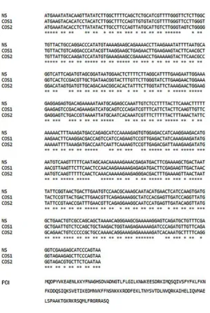
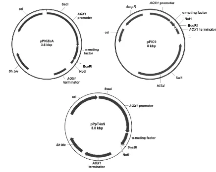
(15) List of Figures Figure 1.1 Schematic diagram depicting the amino acid sequence, N-glycosylation sites, and signal peptide of the hIFNγ precursor…………………….………..6 Figure 1.2 N-glycan structures associated with N25 and N97 glycosylation sites of recombinant hIFNγ expressed in different host systems (James et al, 1995; Sareneva et al, 1996). A: Core fucosylated plus a varied degree of sialyation, B: Non-fucosylated complex high mannose oligosaccharide chain, C: Core fucosylated plus tri-mannosyl, D: Oligomannose (Man 5), E: Core fucosylated. F: Oligomannose (varied), G: Core fucosylated, H: Core fucosylated…………………………………………………………………….…19 Figure 2.1 Methane fixation assessment of Rhodotorula glutinis. Headspace CH4 concentrations were analysed by gas chromatography–mass spectrometry (GC-MS) as an indication of methane consumption. BM without an inoculum was used as a CH4 dissolution control (Mean ± SD. n = 5)….…33 Figure 2.2 Differential interference micrograph of budding Rhodotorula glutinis cells (1,000x magnification, on an Olympus CX21LED, Philippines) …………...33 Figure 2.3 Nineteen-day growth trial of Rhodotorula glutinis on five carbon substrates (acetate, ethanol, glycerol, methanol and methane). Growth is presented as a number of cells per millilitre of medium (Mean ± SD. n = 3)………..……34 Figure 3.1 DNA sequences of hIFNγ; NS: Native sequence, COS1: Codon-optimised sequence 1, COS2: Codon-optimised sequence 2. POI: Protein of interest i.e. an amino acid sequence of hIFNγ. (*): Presence of this symbol shows the similarity in the bases. The first 23 amino acid sequence (eq. 69 bp nucleotides) is the native secretion signal at the N-terminal of the amino acid sequence………………………………………………………….….…….40 Figure 3.2 Generic plasmid vector maps of pPIC9, pPICZαA, pPpT4αS. Ori: the origin of replication, for more information consult the text. 6His-tag: polyhistidine tag. Sh ble: The Zeocin™ resistance gene AOX: alcohol oxidase gene….41 Figure 3.3 Bi-dimensional modelling of mRNA secondary structure predicted based on the MFE. NS: Native sequence, COS1: Codon-optimised sequence 1, COS2: Codon-optimised sequence 2…………………………………………50 Figure 4.1. Placement of the two adjacent genes, hIFNγ and HIS4, as part of the pPIC9-hIFNγ vector (a) and result of the integration of the vector between. XIII.
(16) the 3´AOX into the intact AOX1 locus (Mut+) and the gain of promoter 5’ AOX1, hIFNγ gene, and HIS4 (expression cassette) (b). 5’AOX1: 5’ Alcohol oxidase promotor gene which requires methanol for induction, S: α-factor secretion signal, hIFNγ: optimised human interferon gamma gene for P. pastoris, 3’AOX (TT): Alcohol oxidase transcription terminator, HIS4: Histidinol dehydrogenase gene which is essential for histidine biosynthesis, pBR322: origins from E. coli, Amp: Ampicillin resistance gene…….….…..60 Figure 4.2. Diagram showing continuous amino acid starvation over 10 days in buffered Minimal Glycerol (BMG) medium (a) and protein expression in buffered methanol-complex (BMMY) medium (b). S: Serial passage… …61 Figure 4.3. Dot blot is showing hIFNγ positive cultivation media of two cultures exposed to amino acid starvation (a) and supernatant of cell culture of untransformed P. pastoris GS115 (negative control) (b)………………...…65 Figure 4.4. Amino acid starvation-induced levels of secreted hIFNγ over 5 serial passages of P. pastoris GS115 transformed with hIFNγ and HIS4 (Mean ± SD, n = 2) ……………….…………………………………………………...….65 Figure 5.1. hIFnγ-induced signal transduction in ovarian carcinoma cells. Involvement of the FADD pathway is hypothetical. The figure has been composed based on information obtained from (Alappat et al, 2005; Barton et al, 2005; Bell et al, 2008; Boselli et al, 2007; Burke et al, 1999; Jean et al, 1999; Kim et al, 2002; Lee et al, 2012; Li et al, 2011; Park et al, 2004; Pasparakis & Vandenabeele, 2015; Patel et al, 2014; Pyo et al, 2005; Schinske et al, 2011; Thapa et al, 2011; Wall et al, 2003; Xu et al, 1998). Bax, Bcl-2associated X protein, Bid, BH3 interacting domain death agonist, CASP1, 3, 7, 8, 9, Caspase1, 3, 7, 8, 9; CYT-C, Cytochrome-C; c-FLIP, Cellular FLICE (FADD-like IL-1β-converting enzyme)-inhibitory protein; FADD, FasAssociated Death Domain Protein; Fas, Cell surface death receptor; FasL, Fas Ligand; GAS, Gamma interferon-activated sequence; IRF-1, Interferonregulated factor-1; Jak, Janus kinase; NF-κB, Nuclear factor kappa-lightchain-enhancer of activated B cells; PARP, Poly (ADP-ribose) polymerase; STAT1, Signal transducer & activator of transcription-1; TRAIL, TNF-related apoptosis-inducing ligand; DR, Death receptor…………………….…….….75 Figure 5.2. Cytotoxic and cytostatic effect of hIFNγ on PEO1 and SKOV3 (A) Percentage of total dead cells, and (B) percent TUNEL-positive cells of dead cells, (C) and cytostasis after 72 h exposure to recombinant hIFNγ. XIV.
(17) from three different expression systems and their deglycosylated forms (Mean ± SD, n= 3)……………………………………………..................……82 Figure 5.3. Western analysis of recombinant hIFNγ-induced signalling molecules in SKOV3 and PEO1. (A, D) procaspase-3 (inactive form of caspase-3), (B) FADD, (C, E) Cdk2 (as an indication of G1/S phase), Histone H3 (as a biomarker of M phase), Untreated cells (control) were used to determine un-induced signalling molecule levels and β-actin, a housekeeping protein, was used as a loading control to obtain relative intensity histograms with Image J………………………………………………………...............….……83 Figure 5.4. Cell cycle analysis of PEO1 (A) and SKOV3 (B) following a 72 h exposure to recombinant hIFNγ from three different expression platforms and their deglycosylated forms. (Mean ± SD, n= 3) ……………….…………….…... 85 Figure 5.5. Recombinant hIFNγ induces cell elongation in SKOV3 cells. Phase contrast micrographs of SKOV3 cells after 72 h treatment with the elongated thin shape. A) Control B) hIFNγ-1b C) hIFNγ-CHO D) deglycohIFNγ-CHO E) hIFNγ-HEK F). deglyco-hIFNγ-HEK. White arrows point to elongated thin-shaped cells……………………………………….…….…….86 Figure 5.6. 48h-dose-response to treatment with hIFNγ-1b and hIFNγ-HEK on the growth of PEO1 (A) and SOKV3 (B). Growth is expressed as a fraction of control values. (Mean ± SD, n=3)…………………………………...……..…86. XV.
(18) Abbreviations 3D, three dimensional AGRF, Australian Genome Research Facility AOX, alcohol oxidase APCs, antigen-presenting cells Atg5, autophagy protein 5 Bax, Bcl-2-associated X protein Bid, BH3 interacting domain death agonist BIIC, Baculovirus-infected insect cells BMGY, buffered glycerol complex medium BMMY, buffered methanol-complex medium CASP1, 3, 7, 8, 9, Caspase1, 3, 7, 8, 9 CC, codon context c-FLIP, cellular FLICE (FADD-like IL-1β-converting enzyme)-inhibitory protein CHO, Chinese hamster ovary CoG, cost of goods COS1, codon-optimised sequence 1 COS2, codon-optimised sequence 2 CYT-C, Cytochrome-C GHG, greenhouse gas DCW, dry cell weight DR, death receptor FADD, Fas-Associated Death Domain Protein Fas, cell surface death receptor. XVI.
(19) FasL, Fas Ligand FBS, fetal bovine serum FDA, Food and Drug Administration GAS, Gamma interferon-activated sequence GOI, gene of interest HCDC, high cell density cultivation HCV, hepatitis C virus HEK293, human embryonic kidney 293 hIFNγ, human interferon gamma HIS4, histidinol dehydrogenase gene hpi, hours post induction HPLC, high-pressure liquid chromatography ICU, individual codon usage IFN, interferon IFNAR, interferon-α/β receptor IFNG, interferon gamma gene precursor IFNGR, interferon gamma receptor IFNGR-α, interferon gamma receptor alpha IFNGR-β, interferon gamma receptor beta IGRA, interferon gamma release assays IRF-1, interferon regulatory factor 1 ISGs, IFN-stimulated genes IU, international unit JAK, Janus kinase. XVII.
(20) LC3, microtubule-associated protein 1 light chain 3 LTB, latent tuberculosis MD, minimal Dextrose MFE, minimum free energy MGY, minimal glycerol medium MM, minimal methanol medium MMO, methane monooxygenases MS, multiple sclerosis Mut, methanol utilisation Mut+, methanol utilisation plus Muts, slow methanol utilisation NF-κB, nuclear factor kappa-light-chain-enhancer of activated B cells NK cells, natural killer cells NKT cells, natural killer T cells NS, native sequence PARP, poly (ADP-ribose) polymerase PTM, post-translational modification RIPK3, receptor-interacting kinase 3 STAT1, signal transducer & activator of transcription-1 STATs, signal transducers and activators of transcription SVR sustained virological response TAG, triacylglycerol TB, tuberculosis Th1, T helper cell type 1. XVIII.
(21) Tm, melting temperature TM, transgenic mice TNFα, tumour necrosis factor alpha TRAIL, TNF-related apoptosis-inducing ligand TT, transcription terminator TUNEL, terminal deoxynucleotidyl transferase (TdT) dUTP Nick-End labelling assay YMB, yeast malt broth YNB, yeast nitrogen base. XIX.
(22) Chapter 1. General introduction. The following chapter is a collaborative effort of which each author’s contribution has been detailed at the start of the thesis. The publication has been modified to fit the thesis format, and specific sections have been moved to the general discussion, as the information presented there was not available when the research and its approach was conceived. The scope was also broadened to introduce the motivation for the research.. Published: Razaghi Ali, Leigh Owens, and Kirsten Heimann. "Review of the recombinant human interferon gamma as an immunotherapeutic: Impacts of production platforms and glycosylation." Journal of Biotechnology 240 (2016): 48-60..
(23) Chapter I. 1.1 Abstract Human interferon gamma is a cytokine belonging to a diverse group of interferons which have crucial immunological functions against mycobacteria and a wide variety of viral infections. To date, it has been approved for treatment of chronic granulomatous disease and malignant osteopetrosis, and its application as an immunotherapeutic agent against cancer is an increasing prospect. Recombinant human interferon gamma, as a lucrative biopharmaceutical, has been engineered in different expression systems including prokaryotic, protozoan, fungal (yeasts), plant, insect, and mammalian cells. Human interferon gamma is commonly expressed in Escherichia coli, marketed as ACTIMMUNE ®. However, the resulting product of the prokaryotic expression system is unglycosylated with a short half-life in the bloodstream; the purification process is tedious and makes the product costlier. Other expression systems also did not show satisfactory results in terms of yields, the biological activity of the protein or economic viability. Thus, the review aims to synthesise available information from previous studies on the production of human interferon gamma and its glycosylation patterns in different expression systems, to provide direction for future research in this field.. 2.
(24) Chapter I. 1.2 Preamble Cancer and viral disease are a growing burden for health care systems. Among the various cancers, viral-induced liver cancers are of particular concern, globally. In 2012, more than half a million people worldwide were diagnosed with liver cancer. The incidence is rising globally at an alarming rate, more than 80% of liver cancer cases occur in developing countries, largely owing to the widespread infection of hepatitis C virus (HCV) which is becoming a growing serious health challenge worldwide. Chronic infection with HCV is the main causative for liver disease including cirrhosis and liver cancer (Averhoff et al, 2012). It is currently estimated that more than 170 million people worldwide are infected with HCV (Harnois, 2012). One of the potential pharmaceuticals proposed to limit the impact of hepatitis is recombinant human interferon gamma (hIFNγ). Some clinical trials showed that recombinant hIFNγ therapy is beneficial, safe and well-tolerated to chronic hepatitis C-infected patients (Kokordelis et al, 2014; Muir et al, 2006), but in vitro studies showed that responses to treatment in liver cancers were minimal (Li et al, 2012). One of the most severe limitations for conducting more clinical trials with hIFNγ is the cost of production, partially linked to feedstock and purification of the product, and quality of the biopharmaceutical (Razaghi et al, 2016b) (see sections 1.8.3 & 1.11.1). This research aimed to tackle the cost of production bottleneck by using cheap C1-carbon (methane (CH4) and methanol) utilising expression yeast systems. In addition, the research aimed to unravel whether expression platform and glycosylation status affected the pharmaceutical potency of recombinant hIFNγ. Ovarian cancer cell lines were chosen for this investigation because they have been well studied for hIFNγ therapeutic potential, which was an important aspect for achieving this objective. The following part of the introduction provides the background knowledge necessary to understand the experimental approach chosen and ends by providing a succinct outline for the specific aims of each chapter.. 3.
(25) Chapter I. 1.3 Introduction According to the European Union regulations definition, biopharmaceuticals are proteins or nucleic acid constituents which are formulated using biotechnological approaches for therapeutic in vivo use (Borden et al, 2007). Many substances including vaccines, enzymes, antibodies, and antibiotics have been commercialised under the biopharmaceutical term, among them, interferons (IFNs) are noticeable due to their therapeutic importance against a wide variety of diseases (Borden et al, 2007; Meager, 2006; Samuel, 2001). Interferons are macromolecules which were discovered separately by two research groups in the 1950s and named after the aptitude of these molecules to interfere with viral replication of the flu virus in infected cells (Fensterl & Sen, 2009). In the following decades, IFNs have been studied in fine detail including the mechanisms of transcriptional induction, their antiviral properties, mode of action, viral countermeasures and therapeutic applications against a range of diseases (Fensterl & Sen, 2009; Marciano et al, 2004). Subsequently, efforts for cloning and expression of IFN genes were carried out in many different protein production systems viz., Escherichia coli, mammalian cells, yeasts, protozoan and transgenic plants, but only E. coli expression systems were at the centre of attention due to high productivities (Chen et al, 2011). The main IFN genes (α, β, and γ) have been predominantly expressed in E. coli at industrial scale and approved by the FDA (Food and Drug Administration, USA) and marketed under trade-names of ROFERON-A®, ALFERON-N®, INFERON-A®, and AVENOX® (exceptionally produced in hamster ovary cells) for human IFNα , BETASERON® for human IFNβ and both ACTIMMUNE® and γ-IMMUNEX® for hIFNγ (Jonasch & Haluska, 2001; Panahi et al, 2012). Notwithstanding the importance of hIFNγ and the presence of many articles about this biopharmaceutical, no review has specifically dealt with the expression of hIFNγ in different host cells. Thus, the objective of this review is to synthesize outcomes of previous efforts on the whole process of expression, optimisation and purification of hIFNγ in different host cells, and the effect of expression host on glycosylation patterns, in order to discern which protein production system might be more desirable for future studies and applications e.g. cancer immunotherapy.. 4.
(26) Chapter I. 1.4 Overview on interferons Interferons are cytokines which are expressed by a diverse group of genes and have been cloned from different vertebrates including mammals, birds, fish and even amphibians (Qi et al, 2010). Translated proteins of these genes generally vary in size between 165 and 208 amino acids and the protein moieties are further modified by post-translational glycosylation. IFNs are produced in reaction to viral infections harnessing host cells to non-specifically inhibit viral replication (Samuel, 2001; Takaoka & Yanai, 2006). Mammalian IFNs are broadly classified into three groups, according to amino acid sequence homology and their receptors: Type I IFNs, also known as viral IFNs, as they are induced by viral infection, contain many subtypes of IFNα (13 in humans originating from leukocytes), one IFNβ (originating from fibroblasts), IFNω (originating from leukocytes), IFNτ (originating from ovine trophoblasts), IFNε, IFNκ, and IFNζ. All type I IFN genes are located in a cluster on human chromosome 9 and all interact with the heterodimeric IFNα/β receptor (IFNAR) (Jonasch & Haluska, 2001; Samuel, 2001). Type II IFNs, also known as immune IFNs, are represented solely by IFNγ, which is distinctly dissimilar to other IFNs and uses a distinct heterodimeric IFNγ receptor (IFNGR) (Samuel, 2001; Takaoka & Yanai, 2006). This type of IFN is induced by either IFNα and β (in the case of viral infection) or IFNγ (in the case of mitogenic or antigenic stimuli) (Samuel, 2001). IFNγ proteins show similar biological activities inherent also to other IFNs; but has the advantage of being 100-10,000 more active as an immunomodulator than the other IFNs (Farrar & Schreiber, 1993). Type III IFNs, have been identified lately, containing IFNλ 1, 2, and 3, previously known as Interleukin 29, 28A, and 28B, respectively (Vilcek, 2003). Their genes are located in a cluster on human chromosome 19 and use the heterodimeric IFNλ receptor IL10R2/IFNLR1 (Fensterl & Sen, 2009). This type of IFN is induced directly by viruses or stimulated with IFNα or λ. Thus, they are identified as IFN-stimulated genes (Ank et al, 2006).. 1.5 Characteristics of human IFNγ Native hIFNγ is naturally synthesised by CD4+ T helper cell type 1 (Th1) lymphocytes, CD8+ cytotoxic lymphocytes and natural killer (NK) cells (Bach et al, 1997). It is also secreted by other cells, such as B cells, NKT cells, and professional antigen-presenting cells (APCs) (Frucht et al, 2001). Secretion of hIFNγ by NK cells and APCs is important. 5.
(27) Chapter I in early host reactions against infection, while production of hIFNγ by T lymphocytes is important in the adaptive immune response (Frucht et al, 2001).. 1.6 Genomics & proteomics hIFNγ is encoded by the IFNG gene precursor "NCBI: NM_000610.2" which consists of 1240 bp nucleotides on chromosome 12q24.1 with four exons (Chevillard et al, 2002). The resultant protein "UniProtKB: P01579" is a symmetrical homodimeric glycoprotein with 143 amino acid residues (precursor of native hIFNγ composed of 166 amino acids including 23 residues as the N-terminal secretory signal peptide), two glycosylation sites, a total molecular size of approximately 38.8 kDa in a dimeric structure which is an essential structure for its functional biological active mode and no sulphide bridge (Fig. 1.1) (Borden et al, 2007; Crisafulli et al, 2008; Younes & Amsden, 2002).. Figure 1.1 Schematic diagram depicting the amino acid sequence, N-glycosylation sites, and signal peptide of the hIFNγ precursor.. The folding pattern of hIFNγ is also unique. Each monomer of recombinant hIFNγ consists of six α helices ranging in length from 9-21 residues. Four helices (A, B, C, and D) from one subunit and two from the other (E' and F') interact to form one of two distinct, symmetrical domains of the protein. The two domains lie at a 55° angle, separated by a V-shaped cleft and a large random coil-structured surface loop (residues 16-27) connects the N-terminal helices A and B (Ealick et al, 1991). The functional importance of the N-terminal helix A and the AB-loop has been proven for the unfolding pathway and thermodynamic stability of recombinant hIFNγ (Waschutza et al, 1996).. 6.
(28) Chapter I Helix A is also essential for interaction with receptor-ligand and hence biological activity of hIFNγ (Lundell & Narula, 1994). Three regions have been found to be important for receptor binding: a long loop connecting the A and B helices, (histidine) H111 (Fig. 1.1) in the F helix and a conserved section of the flexible C-terminal. These three regions may form one continuous binding domain (Lundell & Narula, 1994). The C-terminal of native hIFNγ is highly variable and extends from (proline) P122 to (glutamine) Q143. It has been shown that truncation of the C-terminus decreases yields due to increased solubility of the recombinant protein produced in E. coli. Furthermore, truncation of the entire C-terminal domain or deletion of more than 9 amino acids decreased the biological activity of the recombinant protein, yet removal of the last 3, 6, and 9 C-terminal amino acids increased the biological activity of the recombinant protein up to 10-fold (Nacheva et al, 2003). The protein is also heat-sensitive and is irreversibly denatured in solution at a temperature range of 40–65°C (Younes & Amsden, 2002).. 1.7 Interactomics In general terms, hIFNγ has a wide range of antiviral and antitumor activity and is involved in complex interactions of cellular metabolism and differentiation (Jonasch & Haluska, 2001). There are two receptor subunits for hIFNγ, known as IFNGR-α (also known as IFNGR1) which provides binding affinity and IFNGR-β (also known as IFNGR2) which is involved in signal transduction. It has been proposed that the receptor has a tetrameric structure composed of two IFNGR-α and IFNGR-β molecules each (Crisafulli et al, 2008). Both subunits bind to Janus Kinase 1 and 2 binding domains (JAK1 and JAK2, respectively). Oligomerisation occurs after ligand-receptor binding, concomitant with trans-phosphorylation of JAKs which is followed by phosphorylation of the cytoplasmic tails of the receptor molecules. This prepares a docking site for the signal transducers and activators of transcription (STATs) which subsequently are phosphorylated by the JAKs; The receptor molecules release the phosphorylated STAT dimers which are translocated to the nucleus to then activate transcription of IFN-stimulated genes (ISGs) (Borden et al, 2007; Jonasch & Haluska, 2001; Schroder et al, 2004) or IFN-regulated factor 1 (IRF1) (Li et al, 2012). The ISG products restrict viral infection and boost host immunity. Once the virus is cleared from the cells; the IFN response will be dampened by an inhibitory feedback loop before it. 7.
(29) Chapter I becomes detrimental to the host. More detailed information about this topic has been published elsewhere (Schroder et al, 2004). Recently, the crystal structure of IFNGR-β also revealed the importance of Nglycosylation for the stability of this protein and approved the structural basis for receptor specificity (Mikulecky et al, 2016).. 1.8 Production of recombinant hIFNγ 1.8.1 Production in E. coli In the 1980s, hIFNγ was only produced by exposing human T-lymphocytes to mitogenic stimuli or by translating mRNA in oocytes which resulted in low expression and activity of 102-104 IU mL-1. In addition, there were also some problems with purification due to the formation of cytoplasmic aggresomes and costly denaturation processes (Arbabi et al, 2003). However; with the development of recombinant DNA technology, hIFNγ cDNA was successfully cloned and expressed in E. coli in 1982 (Gray et al, 1982). Escherichia coli is one of the most frequently used expression systems for the production of heterologous proteins owing to its simple nutrient requirement, high growth rate, and its well-understood physiology and molecular genetics (Babaeipour et al, 2010). Attempts for the production of recombinant hIFNγ in E. coli were followed by many studies to improve yields (Khalilzadeh et al, 2004; Perez et al, 1990; Rojas Contreras et al, 2010). The expression of recombinant hIFNγ in E. coli, like other heterologous expression systems, is strongly affected by many factors and their interactions. These factors include the composition of media, temperature, inducer concentration and induction time (Balderas Hernández et al, 2008). Thus, determination of optimal culture conditions is necessary to attain higher expression levels (Perez et al, 1990). The objective of optimising the production of recombinant proteins is to produce the highest amount of functional product per unit volume per unit time (Choi et al, 2006). Thus far, four strategies have been applied to optimise the production of recombinant proteins in E. coli, including, choice of culture media, mode of cultivation, strain development, and expression system control (Babaeipour et al, 2010). To date, the production of recombinant hIFNγ in E. coli grown on glucose as a carbon substrate is the prime method for providing this recombinant protein on a large scale (Gray et al, 1982; Hu & Ivashkiv, 2009). Furthermore, production of recombinant hIFNγ in E. coli optimised by response surface methodology and a Box-Behnken. 8.
(30) Chapter I design - compared to un-optimised conditions improved yields up to 13 times (Balderas Hernández et al, 2008). Large-scale production platforms of recombinant hIFNγ in E. coli included batch, fedbatch and continuous cultivation modes (Babaeipour et al, 2010; Babaeipour et al, 2007; Khalilzadeh et al, 2003; Vaiphei et al, 2009). Among them, fed-batch cultivation is the most productive in terms of biomass and protein production (Table1.1) (Babaeipour et al, 2007). Fed-batch cultivation using high cell density cultivation (HCDC) is most often used to obtain high specific productivity of E. coli (mg protein per g dry cell weight (DCW)) (Table 1.1) (Babaeipour et al, 2007; Khalilzadeh et al, 2003). Feeding strategies are critical for achieving HCDC, because of effects on maximum attainable cell concentration and formation of by-products (Babaeipour et al, 2007).. Table 1.1 Impact of cultivation mode for E. coli on yields of recombinant hIFNγ Overall Yield Biomass g L-1 Cultivation productivity mg g-1 Reference [DCW] mg L-1 h-1 DCW (Babaeipour et al, 420 300 14 2010) Batch (Varedi et al, 2006) 200 330 7 (Babaeipour et al, 2570 370 115 2007) (Khalilzadeh et al, 900 350 100 Fed-batch 2004) (HCDC) (Varedi et al, 2006) 2500 330 127 (Babaeipour et al, 3000 400 127 2013) Continuous (Vaiphei et al, 2009) 75 182 4.8. 1.8.2 Purification of E. coli recombinant hIFNγ Although the hIFNγ gene is expressed very effectively in E. coli, the product is accumulated in the form of dense particles, refractile inclusion bodies (denatured proteins usually aggregate in prokaryotic cytoplasm upon targeted gene overexpression), within the cell’s cytoplasm which requires complicated extraction and costly denaturation and refolding processes (Lee et al, 2005; Petrov et al, 2010). A standard procedure for extraction and purification of biologically active proteins, like recombinant hIFNγ, from inclusion bodies, consists of the following steps: (1) the inclusion bodies are solubilised in high concentrations of guanidinium hydrochloride (GnHCl) or urea; (2) the denatured proteins are purified (3) proteins are re-folded and (4) the re-folded proteins are purified. During this procedure, the re-folding is the most critical step for the final yield, stability and biological activity of the protein. A few. 9.
(31) Chapter I external factors govern the successful protein re-folding process, including chaotropic agent concentration, salts concentration (0.75 M urea, 20 mM Tris-HCl), and pH (8.2 for hINFγ). (Petrov et al, 2010). Recently, a new refolding technique proposed to increase the effectiveness of re-folding process in vitro up to 21 times by using a novel type of hairy particles made up of submicron polystyrene cores and brushes of thermosensitive poly(N-isopropyl acrylamide) grafted onto the cores (Huang et al, 2013). To date, solubilised recombinant hIFNγ has been purified using a range of chromatographic and affinity techniques, including size exclusion gel filtration (Reddy et al, 2007; Vandenbroeck et al, 1993), immuno-affinity chromatography by monoclonal antibodies (Honda et al, 1987), and ion exchange chromatography (Haelewyn & De Ley, 1995; Petrov et al, 2010). Approaches for purification of recombinant hIFNγ from inclusion bodies in E. coli are summarised in Table 1.2. All approaches used either GnHCl or urea for solubilisation and their purified product yields ranged from 0.3 to 14 mg g-1 cell mass with biological activities ranging from107–108 IU mg-1 (Petrov et al, 2010; Reddy et al, 2007). As yet, the highest biological activity (2 ×10⁸ IU mg-1) obtained by addition of a labilizing agent, l-arginine, in the re-folding buffer which improved the refolding and purification of recombinant hIFNγ by 10-fold in comparison to other techniques (Table 1.2)(Arora & Khanna, 1996).. Table 1.2 Methods for purification of recombinant hIFNγ from inclusion bodies in E. coli Purification method Immuno-affinity chromatography. Biological activity [IU* mg-1**]. References. 4 ×10⁷. (Novick et al, 1983). Ion exchange chromatography •. Carboxymethyl sepharose. 20 × 107. (Petrov et al, 2010). •. Expanded bed adsorption. 0.8 × 10⁷. (Jin et al, 2006). •. MonoBeads. 1–2 × 10⁷. (Perez et al, 1990). Gel filtration chromatography •. Superdex 75. 1–4 × 10⁷. (Guan et al, 2005; Reddy et al, 2007). •. Sephadex G-75. 1–5 × 10⁷. (Arakawa et al, 1985). •. Sepharose. 20× 107. (Arora & Khanna, 1996). 8.7 × 10⁷. (Geng et al, 2004). Hydrophobic interaction chromatography *International unit, **mg recombinant protein. 10.
(32) Chapter I. Since unglycosylated recombinant hIFNγ monomers contain hydrophobic domains and possess no disulfide bonds, they have a tendency to aggregate in the solution phase, hence it is important to avoid the formation of aggregation in the isolation and purification process in order to re-fold and dimerise the monomers correctly to attain an active structure of the protein, (Jin et al, 2006; Vandenbroeck et al, 1993; Yphantis & Arakawa, 1987). For this reason, some later attempts have used hydrophobic chromatographic column matrices for the re-folding process with the intention of avoiding inactive aggregate formation (Geng et al, 2004; Reddy et al, 2007).. 1.8.3 Limitations of the hIFNγ E. coli expression system In general, for glycoproteins, but specifically for hIFNγ, bacterial production platforms face technical problems for industrial production such as: 1) Recombinant hIFNγ forms insoluble intracellular inclusion bodies in E. coli where the protein is denatured partially or entirely; the latter complicates its purification because a denaturation/renaturation step is required (Petrov et al, 2010). 2) Bacterial expression systems are not capable of assembling glycan branches, resulting in unglycosylated recombinant hIFNγ which has a shorter half-life in the bloodstream circulation in comparison to native hIFNγ (Bocci et al, 1985; Sareneva et al, 1993). 3) Following the release of protein from E. coli, the subsequent solution contains impurities like endotoxins and nucleic acids contaminating the product, which is another disadvantage of this expression system (Mohammadian-Mosaabadi et al, 2007; Rojas Contreras et al, 2010). 4) Recombinant hIFNγ produced in the bacterial system is glycated (a haphazard nonenzymatic process), which impairs the functionality of proteins (Mironova et al, 2003). The formation of advanced glycation end-products (AGEs) causes covalent dimerisation, polymerisation and non-enzymatic proteolysis (degradation of protein due to heat or acidity) which reduces biological activity, shorten the half-life and increases the immunogenicity of recombinant proteins (Mironova et al, 2003). Therefore, to solve the half-life deficiency of recombinant proteins; improvement of glycosylation is necessary (especially fucose-containing glycans).. 11.
(33) Chapter I 1.8.4 Comparison of recombinant hIFNγ expressed in E. coli with native hIFNγ hIFNγ-1b (Trade-name: ACTIMMUNE®) is the generic name for recombinant hIFNγ industrially produced in E. coli (Gray et al, 1982; Koh & Limmathurotsakul, 2010). hIFNγ-1b has the same primary structure as native hIFNγ with a few differences as summarised in (Table 1.3) (Rinderknecht et al, 1984).. Table 1.3 Comparison between native hIFNγ and hIFNγ-1b Native hIFNγ hIFNγ-1b References (Bach et al, 1997; Gray et al, Source E. coli Blood cells 1982) (Farrar & Schreiber, 1993; Perez Modification Glycosylated Unglycosylated et al, 1990; Zhang et al, 1992) (Farrar & Schreiber, 1993; Amino acid length 143 143 Meager, 2006; Miller et al, 2009) (Crisafulli et al, 2008; Farrar & Schreiber, 1993; Malek Sabet et Molecular size (kDa) & Monomeric~25 Monomeric~17 al, 2008; Perez et al, 1990; quaternary structure Dimeric~38.8 Dimeric~35 Younes & Amsden, 2002; Zhang et al, 1992) Physiological state (Gray et al, 1982) Active Active 3×10⁶ IU1 mg(Bouros et al, 2006; Miyata et al, 4-12×107 1= Biological activity IU mg-1 1986; Nathan, 1983) 2×10⁶ IU mL-1 1, IU: International Units. The main difference between native hIFNγ and hIFNγ-1b is attributed to differences in glycosylation. The carbohydrate moiety of native hIFNγ is located at the receptor interaction domain and covers a fairly large surface area of the molecule (Walter et al, 1995). Carbohydrate side chains, in general, have been known to play an important role in many biological processes like protein folding, targeting, stability, clearance and cell to cell interactions (Varki, 1993); However, the absence of carbohydrate moieties in the hIFNγ-1b does not deactivate the molecule but affects its physicochemical and pharmaco-kinetic properties (Younes & Amsden, 2002). Generally, it is assumed that glycosylation of native hIFNγ protects the protein from proteolytic degradation, suggesting that glycans have a potential role in therapeutic applications (Bocci et al, 1985; Sareneva et al, 1996).. 12.
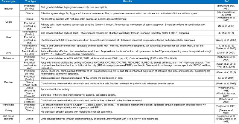
(34) Chapter I. 1.9 Expression of recombinant hIFNγ in other protein production systems Due to the unglycosylated form of recombinant hIFNγ from E. coli which affects the half-life (Bocci et al, 1985), solubility and protease resistance, other expression systems have been used to overcome these problems (Table 1.4) (Leister et al, 2014). Most of these studies were performed at laboratory-scale, and result varied widely between each study. Therefore, a more in-depth investigation is required to justify their pros and cons in comparison to E. coli platform. Methylotrophic yeasts have been demonstrated to deliver large amounts of recombinant protein on an industrial scale (Cereghino & Cregg, 2000). Initially, Wang et al (2014) claimed overexpression of hIFNγ in the methylotrophic yeast P. pastoris which could be promising for industrial production as it lowers the cost of production and the protein would be post-translationally glycosylated. Later, Prabu et al, (2016) and Razaghi et al, (2016) demonstrated the irreproducibility of the results and actual yields achieved were economically not competitive with E. coli (Table 1.4). Productivity in CHO cells is still lower than E. coli, and albeit expression level would be magnified significantly in CHO cells, cultivation still requires bovine serum, which is exorbitantly expensive, and purification of protein from a liquid culture is arduous in the presence of serum (Nakajima et al, 1992), however usage of serum-free culture might be an alternative approach to lower the cost of cultivation, but it is still in the challenging stage of development (Rodrigues et al, 2013). Recently, in order to enhance the expression of hIFNγ codon optimisation approach was conducted to design synthetic hIFNγ coding sequences for heterologous expression in CHO cells based on the fact that recombinant expression of foreign proteins is usually suboptimal due to the usage of non-native codon patterns within the coding sequence (Chung et al, 2013). For codon optimisation, two selected design parameters, codon context (CC), and individual codon usage (ICU) optimisations were used by Chung et al (2013) , they showed that the CC optimised genes exhibited at least a 13-fold increases in expression level compared to the native hIFNγ sequence while approximately a 10-fold increases were observed for the ICU optimised genes. This shows that CC optimisation is comparatively more effective for improving recombinant hIFNγ expression in CHO cells (Chung et al, 2013). Expression of hIFNγ in the baculovirus-infected insect cells (BIIC) and Saccharomyces cerevisiae was not satisfactory due to poor secretion into the culture media, hyperglycosylation, and improper folding. Similarly, in spite of various attempts for. 13.
(35) Chapter I improvement of production, the yield of expression is still unsuitable for industrial production in comparison to expression in E. coli (Davoudi et al, 2011).. Table 1.4 Effect of expression systems on yield and activity of recombinant hIFNγ Yield Molecular Expression system Activity1 Reference [mg L-1] size [kDa] (Bagis et al, 23 × 10-6 1 × 10⁷ 20–25 IU mg-1 2011) 1 × 10⁷- 5 × (Mus spp.) (Lagutin et al, 350-570 107 * Mouse mammary gland 1999) IU mL-1 (Rattus spp.) (Nakajima et al, 4 × 105 * 22-25 Rat cells IU mL-1 1992) 2.0 × 10⁴-1.0 (Haynes & * ×105 22-23 Weissman, 1983) IU mL-1 (Scahill et al, 5.5 × 10⁴ IU (Cricetulus sp.) * 21-25 mL-1 1983) Chinese 8 hamster ovary cells 1-2 × 10 (Mory et al, 1986) * 20-26 IU mg-1 15 * * (McClain, 2010) Spodoptera spp. 2 (Chen et al, 2011) Active* 18-23 (BIIC) (Ebrahimi et al, Solanum lycopersicum * Active* * (Tomato) 2012) Oryzea sativa 17 × 10-3 (Chen et al, 2004) Active* 24-27 (Rice) Bacillus sp. (Rojas Contreras 2-20 Active* 17 (Bacteria) et al, 2010) Leishmania sp. (Davoudi et al, 9.5 Active* 17 (Protozoa) 2011) (Derynck et al, Saccharomyces cerevisiae 2.5 × 10⁴ * Detected E* (Baker’s yeast) IU mL-1 1983) (Razaghi et al, 1-16 × 10 2015; Razaghi et Active* * 3 al, 2017)2 (Prabhu et al, Pichia pastoris 2.5 Active* 17 (Methylotrophic yeast) 2016)2 7 (Wang et al, 1-1.4 × 10 IU 300 15 mg-1 2014)2 -2 6.2 × 10 IU Monkey cells (Gray et al, 1982) * * mL-1 (Leister et al, Homo sapiens 1.93 ×10⁷ IU 6 * (Human tissue culture) mg-1 2014) (Huang et al, E. coli 1700 9 × 10⁷ IU L-1 17 2013) 1 * No data, The antiviral assay for quantifying biological activity of human IFNs is based on the induction of a cellular reaction in the transformed human cell line (WISH); the effectiveness of interferon is assessed by comparing its protective effect against a viral cytopathic effect (usually vesicular stomatitis virus) against a calibrated reference in international unit (IU) (Petrov et al, 2010). 2 These papers were published during the course of my PhD research and are integrated here for completeness.. 14.
(36) Chapter I Despite the fact that expression of recombinant hIFNγ in transgenic mice (TM) was rather comparable to E. coli, expression occurred in live transgenic mice, which is impractical for commercial production (Table 1.4). Comparison of yields in different expression systems reveals that the best results were achieved in prokaryotes followed by mammalian expression system e.g. TM (Table 1.4). One drawback, of studies seeking for an alternative expression system else than E. coli (Table 1.4), is that production parameter (yield, biological activity and molecular size of the recombinant protein) were mostly not considered; for example, neither yields were measured in tomato, Saccharomyces cerevisiae, rat, hamster, and monkey cells nor was the biological activity quantified in Bacillus sp., Leishmania sp, tomato, rice and insect cells. Others studies in tomato, mouse, monkey and human cells did also not determine the molecular size of the recombinant protein. Note: Prior to the start of this thesis in 2012, expression of recombinant hIFNγ in Pichia pastoris was patented by Thill and Davis (1989) with reported yields of 1-10 mg L-1, which surprisingly was not followed up with commercial production. The two following studies were published during the course of my PhD and are being discussed in the relevant data chapters, but the detail is provided here in the introduction for completeness of information. However, later a research article by Wang et al (2014) reported the expression yields of recombinant hIFNγ of 300 mg L-1 in Pichia pastoris, and subsequently, another study by Prabhu et al (2016) resulted in 2.5 mg L-1. None of these studies has resulted in large-scale/industrial scale production yet (see chapter 3).. 1.10 Glycosylation Many of the approved biotherapeutics are glycoproteins (Zhong & Somers, 2012). Glycosylation of glycoproteins can increase therapeutic efficacy through improving protein pharmaco-dynamics and pharmaco-kinetics. Glycosylation is one of the most multifaceted post-translational modifications, found in many eukaryotic proteins which plays an important role in blood transfusion reactions, selectin-mediated leukocyteendothelial adhesion, host-microbe interactions, and numerous ontogenic events, including signalling events by the Notch receptor family (Zhong & Somers, 2012). The nature and content of oligosaccharides affect protein folding, stability, trafficking, immunogenicity, half-life and primary activities of the protein i.e. a lot of sialic acids increases plasma half-life, whilst in contrast, terminal residues of galactose and. 15.
(37) Chapter I mannose shorten the half-life (Zhong & Somers, 2012). Glycoproteins are generally classified into four groups: N-linked, O-linked, glycosaminoglycan, and glycosylphosphatidylinositol-anchored proteins. N-linked glycosylation is the main form of glycosylation and takes place in both the endoplasmic reticulum and Golgi, through the side chain amide nitrogen of a specific asparagine residue which plays a critical role in protein folding and conformation stabilisation and intracellular trafficking (Zhong & Somers, 2012) Native hIFNγ has two N-glycosylated sites at asparagine N25 (fucosylated complex-type oligosaccharides) and N97 (with hybrid and high-mannose structures) (Fig. 1.1) (Farrar & Schreiber, 1993; Kelker et al, 1983; Sareneva et al, 1996; Yip et al, 1982; Younes & Amsden, 2002). It has been shown that native hIFNγ derived from T lymphocytes is heterogeneously glycosylated and doubly, singly, and unglycosylated forms exist resulting in hIFNγ molecules of different molecular masses (16.7-37 kDa) and considerable variation in the carbohydrate structures (>30 different forms) (Sareneva et al, 1996). The glycans at (asparagine) N25 consisted of fucosylated, mainly complextype oligosaccharides, with the highest relative frequency 41% for sugar composition of (N-acetylneuraminic acid, galactose, mannose, N-acetylglucosamine, fucose) which are known to be essential for protease resistance to cathepsin G, granulocyte proteases, plasmin, and purified elastase (Mironova et al, 2003; Sareneva et al, 1996). In contrast, the glycans at N97 were more heterogeneous, with hybrid and highmannose structures with highest relative frequency at 34% for sugar composition of (Nacetylneuraminic acid, galactose, mannose, N-acetylglucosamine) (Fig. 1.2) (Sareneva et al, 1996). The glycosylation pattern of recombinant hIFNγ was also confirmed in three expression systems (CHO, BIIC, and TM) for both N25 and N97 sites. The N97 glycans always showed a non-fucosylated pattern which varied between two types; complex and oligomannose (James et al, 1995). There are, however, minute differences between the N-glycan structure of native and recombinant forms of hIFNγ such as lack of Nacetylneuraminic acid and insertion of N-acetylglucosamine between galactose and mannose in all recombinant forms (Fig 1.2). In conclusion, comparison of glycosylation similarity revealed that glycosylation patterns achieved in the mammalian CHO expression system were most similar to those in human cells (Fig. 1.2). The main obstacle for clinical application of unglycosylated recombinant hIFNγ is primarily due to the short in vivo half-life of the protein. An investigation in the half-life of proteins showed that unglycosylated hIFNγ has a shorter half-life than glycosylated. 16.
Figure
+7


Related documents