Protective Capacity of the Human Anamnestic Antibody Response
during Acute Dengue Virus Infection
Meihui Xu,aRoland Züst,a*Ying Xiu Toh,aJennifer M. Pfaff,bKristen M. Kahle,bEdgar Davidson,bBenjamin J. Doranz,b Sumathy Velumani,aFarhana Tukijan,a,cCheng-I Wang,a Katja Finka,c
Singapore Immunology Network, Agency for Science Technology and Research, Singaporea
; Integral Molecular, Philadelphia, Pennsylvania, USAb
; School of Biological Sciences, Nanyang Technological University, Singaporec
ABSTRACT
Half of the world’s population is exposed to the risk of dengue virus infection. Although a vaccine for dengue virus is now avail-able in a few countries, its reported overall efficacy of about 60% is not ideal. Protective immune correlates following natural dengue virus infection remain undefined, which makes it difficult to predict the efficacy of new vaccines. In this study, we ad-dress the protective capacity of dengue virus-specific antibodies that are produced by plasmablasts a few days after natural sec-ondary infection. Among a panel of 18 dengue virus-reactive human monoclonal antibodies, four groups of antibodies were identified based on their binding properties. While antibodies targeting the fusion loop of the glycoprotein of dengue virus dom-inated the antibody response, two smaller groups of antibodies bound to previously undescribed epitopes in domain II of the E protein. The latter, largely serotype-cross-reactive antibodies, demonstrated increased stability of binding at pH 5. These anti-bodies possessed weak to moderate neutralization capacityin vitrobut were the most efficacious in promoting the survival of infected mice. Our data suggest that the cross-reactive anamnestic antibody response has a protective capacity despite moderate neutralizationin vitroand a moderate decrease of viremiain vivo.
IMPORTANCE
Antibodies can protect from symptomatic dengue virus infection. However, it is not easy to assess which classes of antibodies provide protection becausein vitroassays are not always predictive ofin vivoprotection. During a repeat infection, dengue vi-rus-specific immune memory cells are reactivated and large amounts of antibodies are produced. By studying antibodies cloned from patients with heterologous secondary infection, we tested the protective value of the serotype-cross-reactive “recall” or “anamnestic” response. We found that results fromin vitroneutralization assays did not always correlate with the ability of the antibodies to reduce viremia in a mouse model. In addition, a decrease of viremia in mice did not necessarily improve survival. The most protective antibodies were stable at pH 5, suggesting that antibody binding in the endosomes, after the antibody-virus complex is internalized, might be important to block virus spread in the organism.
M
ultiple studies have characterized the human antibody (Ab)response to natural dengue virus (DENV) infection based on monoclonal antibodies (MAbs) that were isolated from plas-mablasts during the acute phase of infection or from memory B
cells after recovery (1–7). However, antibody-associated
corre-lates of protection and mechanisms of neutralization that prevent or reduce the spread of the virus in the organism are still poorly understood. This was best illustrated by the recent clinical trials of the leading vaccine from Sanofi-Pasteur, for which the overall efficacy across all four DENV serotypes was only 60.3% despite
generally high neutralizing titers in vaccinees (8). Vaccine efficacy
by serotype placed DENV serotype 2 (DENV-2) at the bottom,
with a reported efficacy of only 43% (8). Interestingly, vaccine
efficacy was higher in children above the age of 9 years, and effi-cacy was associated with seropositivity, suggesting that the protec-tive mechanisms of the vaccine are related to the reactivation of specific immune memory cells, or the so-called anamnestic re-sponse.
The aim of this study was to address the protective capacity of antibodies produced during a natural anamnestic response after symptomatic reinfection with a heterologous serotype of DENV. The current literature focuses largely on the description of epitopes of potently neutralizing antibodies. In turn, immuno-dominant epitopes that elicit weakly neutralizing or
nonneutral-izing antibodies and their possible functions and implications for overall disease resolution, or enhancement, have rarely been de-scribed. The envelope (E) glycoprotein is the surface protein of DENV particles and is the primary target of the humoral immune response, eliciting neutralizing antibodies that are necessary to
prevent reinfection (9). Antibodies against the E glycoprotein
have been shown to inhibit virus attachment and infectionin vitro,
Received6 June 2016Accepted26 September 2016 Accepted manuscript posted online5 October 2016
CitationXu M, Züst R, Toh YX, Pfaff JM, Kahle KM, Davidson E, Doranz BJ, Velumani S, Tukijan F, Wang C-I, Fink K. 2016. Protective capacity of the human anamnestic antibody response during acute dengue virus infection. J Virol 90:11122–11131.
doi:10.1128/JVI.01096-16.
Editor:J. U. Jung, University of Southern California
Address correspondence to Katja Fink, Katja_fink@immunol.a-star.edu.sg.
*Present address: Roland Züst, Spiez Laboratory, Federal Office for Civil Protection, Spiez, Switzerland.
Supplemental material for this article may be found athttp://dx.doi.org/10.1128
/JVI.01096-16.
Copyright © 2016 Xu et al. This is an open-access article distributed under the terms of theCreative Commons Attribution 4.0 International license.
on November 7, 2019 by guest
http://jvi.asm.org/
and passive transfer of E glycoprotein-specific antibodies
pro-tected mice from dengue virus challenge (10,11).
The tertiary structure of the E glycoprotein has three domains, EDI, -II, and -III, which fold from a discontinuous primary
pro-tein sequence. EDI forms a central-barrel linking EDII to EDIII
(12). EDII contains a dimerization region that is responsible for
the spontaneous dimer formation of E proteins. EDII also con-tains a fusion loop that is necessary for membrane fusion with host cells during the infection process. EDIII assumes an immunoglob-ulin-like fold and mediates host cell receptor binding, and conse-quently, antibodies against EDIII have been shown to possess
po-tent type-specific neutralization capacities (13). However, dengue
virus has the capacity to escape from these antibodies by mutating
EDIII (10,14,15). A total of 90 E homodimers assume a
“herring-bone” configuration on the mature virus surface. The E glycopro-tein undergoes several drastic conformational changes during the infection cycle to enable host cell infection and production of new
virus progeny (16).
Dengue virus enters susceptible host cells through receptor-mediated endocytosis. Acidification in the endosome promotes dissociation of homodimers into monomers, followed by an irre-versible reassociation of the monomers to form the fusogenic trimer structure that exposes the fusion peptide. The fusogenic trimer structure extends outward from the virion surface toward the host cell membrane to facilitate membrane fusion, releas-ing the viral genome into the host cell’s cytoplasm and triggerreleas-ing translation of nonstructural and structural viral proteins and
propagation of the viral genome (16). Consequently, epitope
ac-cessibility on the E protein is dependent on the pH of the cellular compartment in which the virus is potentially bound by an anti-body. The pH stability of an antibody can therefore be an impor-tant factor determining its protective capacity. Here, we provide
new insight into the correlation of the epitope,in vitro
neutraliza-tion, pH-dependent antibody stability, andin vivoprotective
ca-pacity of human plasmablast-derived antibodies.
MATERIALS AND METHODS
Monoclonal antibodies and virus strains.The panel of human monoclo-nal antibodies used in this study has been described previously (7). The antibody sequences are available in GenBank (see Table S1 in the supple-mental material). The humanized mouse monoclonal antibody 4G2 was a kind gift from Brendon John Hanson, DSO Laboratories, Singapore. An-tibodies 747(4)A11 and 752-2 C8 were produced according to the pub-lished sequences. All dengue virus strains used in this study were propagated in C6/36 mosquito cells. The DENV stocks used were Western Pacific 74 [U88535.1] or DENV-1-D1/SG/05K2916DK1/2005 [EU081234.1], TSV01 [AY037116.1] or DENV-2/SG/D2Y98P-PP1/2009, (JF327392.1), VN32/96 [EU482459], and 2641Y08 [HQ875339.1] for dengue virus serotypes 1, 2, 3, and 4, respectively.
ELISA and competition binding assays.Whole virus particle en-zyme-linked immunosorbent assay (ELISA) was performed by capturing virions from infected C6/36 cell supernatant on 4G2-coated plates. For binding to recombinant E (rE), MaxiSorp plates were coated with 150 ng of purified rE protein in 100l coating buffer at 4°C overnight. rE protein was produced in S2 cells as described previously (17). Antibodies were added at 1g/ml, and binding was detected by adding anti-human IgG-horseradish peroxidase (HRP). For competition ELISAs, biotinylated an-tibodies were added to the plates at 0.01g/ml, while the competing nonlabeled antibodies were added in 100-fold excess. Binding was de-tected by adding streptavidin-HRP and developed by adding 3,3=,5,5= -tetramethylbenzidine (TMB) substrate. The reaction was stopped by
add-ing 1 M HCl. The optical density at 450-nm wavelength (OD450) was measured on an Enspire plate reader.
Neutralization assays (plaque reduction neutralization test [PRNT]/fluorescence-activated cell sorter [FACS]).BHK-21 cells were seeded in each well of a 24-well plate and incubated at 37°C overnight. Antibodies were diluted 4-fold over six dilution steps, starting from 30 g/ml, and the antibody dilutions were then added to a constant amount of virus (multiplicities of infection [MOI] of 0.01 for DENV-1 strain 05K2916 [DENV-1-05K2916] and 0.1 for DENV-2-TSV01, DENV-3-VN32, and DENV-4-2641Y08). The virus-antibody mixtures were incu-bated at 37°C for 1 h prior to infection of cells. A methylcellulose overlay was added, and the plates were incubated for 5 days before plaque visual-ization by crystal violet staining. The flow cytometry-based neutralvisual-ization assay was performed with U937 cells stably expressing DC-SIGN. The number of infected cells was determined by staining all the cells intracel-lularly with 4G2-AlexaFluor 647 and anti-NS1-Alexa 488 as described previously (7). The MOI used were 0.1 for DENV-1-05K2916, 0.5 for DENV-2-TSV01, 0.2 for DENV-3-VN32, and 5 for DENV-4-2641Y08. The 50% plaque reduction neutralization titer (PRNT50) and 50% effec-tive concentration (EC50) values, respectively, were defined as the concen-tration of antibody that results in a 50% reduction of plaques or infected cells, and these values were calculated using a three-parameter nonlinear curve fitted in GraphPad Prism software.
Epitope-mapping studies on recombinant protein.DENV-2 E pro-tein mutants were produced with a QuikChange site-directed mutagene-sis kit (Agilent). A V5 tag at the C terminus of the E protein was used to facilitate immobilization of E protein dimers on ELISA plates (see Fig. S1 in the supplemental material for validation). E proteins were produced in S2 cells and purified as described previously (18). ELISA plates were coated with polyclonal rabbit anti-V5 antibody, and individual E protein mutants were added at a concentration of 5g/ml in 50l coating buffer (half-area ELISA plates; Greiner). After blocking with 3% skim milk and 0.05% Tween 20 in phosphate-buffered saline (PBS), individual antibod-ies were added at a concentration of 1g/ml. Bound antibodies were detected with anti-human IgG-HRP antibody (Sigma), and TMB was used as a substrate for color development.
Antibody epitopes in the postfusion trimeric E protein were illustrated on the DENV-2 structure (Protein Data Bank [PDB] ID3G7T) using Yasara software.
Shotgun mutagenesis epitope mapping. Shotgun mutagenesis epitope mapping (19) was performed using comprehensive mutation li-braries obtained by subjecting DENV-3 (strain CH53489) and DENV-4 (strain 341750) prM/E expression constructs to high-throughput mu-tagenesis. Random mutations were introduced into the DENV-3 prM/E polyprotein, while for DENV-4, each prM/E residue was mutated to ala-nine (and alaala-nines to serine). The mutant plasmids were arrayed in 384-well plates (one mutation per 384-well), transfected into HEK-293T cells, and allowed to express for 22 h. The cells were monodispersed using Cell Stripper (Cellgro), fixed in 4% (vol/vol) paraformaldehyde (PFA), per-meabilized for 20 min with 0.1% (wt/vol) saponin (Sigma-Aldrich) in PBS plus calcium and magnesium (PBS⫹⫹), and then stained for 1 h with purified antibodies diluted in 10% normal goat serum (NGS) (Sigma)-0.1% saponin, pH 9. The primary antibody concentrations were selected using an independent immunofluorescence titration curve against wild-type prM/E to ensure that the signal was within the linear range of detection. Antibodies were detected by incubating with AlexaFluor 488-conjugated secondary antibody (3.75 g/ml; Jackson Immuno-Research Laboratories) in 10% NGS-0.1% saponin for 30 min. The cells were washed 3 times with PBS⫹⫹-0.1 saponin, followed by 2 washes in PBS, and the mean cellular fluorescence was detected using a high-throughput flow cytometer (HTFC) (Intellicyt). Antibody reactivities against each mutant clone were calculated relative to wild-type prM/E protein reactivity by subtracting the signal from mock-transfected con-trols and normalizing it to the signal from wild-type prM/E-transfected controls. Mutations within critical clones were identified as critical to the
on November 7, 2019 by guest
http://jvi.asm.org/
antibody epitope if they did not support reactivity of the test antibody but did support reactivity of other control antibodies. This counterscreen strategy facilitates the exclusion of prM/E mutants that are locally mis-folded or have an expression defect. Control antibodies were selected to represent epitopes over diverse regions on the E protein. The use of con-trol MAbs to confirm the folding of each mutant, combined with mapping each mutant directly in human cells that correctly process prM/E, pro-vides confidence that identified epitope residues are affecting the binding epitope directly and not having indirect effects. Critical amino acids re-quired for antibody binding were visualized on a DENV-2 or DENV-3 Env crystal structure (PDB ID1OANand1UZG).
pH stability assay.Goat anti-human IgG was immobilized on a GLC sensor chip (Bio-Rad) by amine coupling. Antibody analytes were allowed to bind to the immobilized anti-human IgG before the introduction of rE protein. Measurements were recorded at 25°C in running buffer at pH 5 or pH 7 on ProteOn XPR36 equipment (Bio-Rad).
The sandwich ELISA described in “Epitope-mapping studies on re-combinant protein” above was modified as follows. E proteins were buffer exchanged with MES (morpholineethanesulfonic acid) buffer, pH 5, be-fore adding them to the anti-V5-tag-coated plates. After blocking, the antibodies were diluted in MES buffer, pH 5, and incubated for 2 h at room temperature. For pH 7 measurements, E protein in PBS, pH 7.4, was used instead of MES buffer.
Immunofluorescent staining.Antibodies (1g/ml) were incubated with DENV-1 (or DENV-2 [data not shown]) for 1 h at 37°C. BHK-21 cells were grown on chamber slides (ibidi). Antibody-virus mixtures were added to the BHK-21 cells on ice for 20 min for synchronization of anti-body-virus uptake by the cells. The chambers were moved to 37°C for 7 min. The cells were then fixed with 2% PFA, permeabilized, and stained with rabbit anti-EEA Ab. Anti-human IgG AF488 and goat anti-rabbit IgG AF568 (both from Molecular Probes) were used to detect the primary antibodies. Hoechst was used to stain nuclei. Photographs were taken at⫻100 magnification with an Olympus confocal microscope.
Protection assay in mice.AG129 mice were treated with 100g pu-rified antibody injected intravenously (i.v.) 24 h prior to virus challenge, which was administered intraperitoneally. Blood was collected 3 days postinfection, and viremia was quantified by TaqMan reverse transcrip-tion (RT)-PCR (20). The animal experiments were conducted according to the rules and guidelines of the Agri-Food and Veterinary Authority and the National Advisory Committee for Laboratory Animal Research, Sin-gapore. The experiments were reviewed and approved by the Institutional Review Board of the Biological Resource Center, Singapore (Institutional Animal Care and Use Committee; protocol 151099).
RESULTS
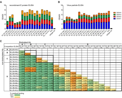
Binding properties identify four main antibody groups in the anamnestic response.Previously, we described the generation of a panel of human monoclonal antibodies from the plasmablasts of two naturally infected dengue patients by single B cell PCR clon-ing. The majority of these plasmablast-derived antibodies were dengue virus specific, and the primary target was found to be the E
protein (7). Here, we further characterized a subset of clonally
distinct antibodies that were selected based on good binding to
both E protein and virus particles (Fig. 1AandB). The relative
ability of each antibody to block binding of all other antibodies individually in a competition ELISA was tested to identify
com-mon antibody epitopes (Fig. 1C). Unlabeled antibodies were used
in 100-fold excess over biotinylated antibodies, and successful competition was defined as a reduction of the normalized strepta-vidin-HRP signal by at least 70%. Four main groups of antibodies, designated A, B, C, and D, were identified based on this competi-tion assay. The largest group, D, comprised more than 50% of the
antibodies tested (Fig. 1C). Interestingly, biotinylated antibodies
from groups A, B, and C appeared to promote binding of
antibod-ies in the same or other groups, as illustrated by a value higher than 1 (Fig. 1C, dark green cells), which is the maximal binding of each biotinylated antibody in the absence of competition.
The acute-phase antibody repertoire is dominated by fusion loop-specific antibodies.In order to identify the footprint of these antibodies on the E protein, another competition ELISA was performed. The same panel of unlabeled antibodies, ordered
ac-cording to their respective groups inFig. 2A, was used to compete
with the well-characterized mouse monoclonal antibody 4G2
(21). The described epitope of 4G2 comprises amino acids G104,
G106, and L107, which lie in the fusion loop of the E protein (22,
23). Competition for binding sites on the immobilized
recombi-nant E protein revealed that 4G2 competed with 12 out of 18
antibodies (Fig. 2B). These antibodies comprised the dominant
group D, and antibodies in this group thus share an epitope with 4G2 in the fusion loop. The dominance of fusion loop-specific
antibodies is consistent with the previous literature (24,25). In
addition, 2 out of 18 antibodies (11%) and 3 out of 18 antibodies (17%) clustered in groups B and C, respectively. Even though the total number of antibodies analyzed in this study is too small to draw general conclusions, the finding indicates that epitopes of groups B and C could also be immunodominant during natural infection.
Epitope-mapping studies reveal that non-solvent-exposed residues of the E protein are critical antibody binding sites.To further dissect the epitopes of antibodies in groups A, B, C, and D,
an alanine scanning mutagenesis approach was used (Fig. 3Aand
B) (26). For these studies, comprehensive mutation libraries of
DENV-3 or DENV-4 prM/E proteins were expressed in HEK-293T cells, and binding of the antibodies was detected by flow cytometry. We selected a representative antibody from each of the four groups for these epitope-mapping studies. Residues critical for each antibody epitope were initially identified as those where prM/E mutations resulted in low reactivity for the antibody of interest (20 to 30% relative to wild-type DENV prM/E) yet greater than 60% of wild-type binding by a control MAb. Antibodies rep-resentative of groups A and B both bound to EDII. For group A antibody 6C-H8L1, we identified residues D215 and P217 as crit-ical for binding; for group B antibody 6C-H8L1, we also identified D215 and P217, as well as residues H209, W212, and A267. The group C antibody 7E-H1L1 was mapped to EDI with critical
res-idues C30, V151, R186, G279, H280, and K282 (Fig. 3A; see Fig.
S2A in the supplemental material). Intriguingly, all the contact residues for group A, B, and C antibodies identified in EDI and EDII are poorly exposed in the available E protein dimer crystal
structures (Fig. 3A).
To help confirm the locations of these epitope residues, we converted group B antibody 2C-H3L2 to a Fab, which was then screened on the Ala scan mutation library. Conversion of anti-body to a Fab weakens binding relative to the full antianti-body, which we have found allows the identification of additional epitope res-idues compared to the full-length MAb. Using the Fab, we identi-fied two additional residues as critical for 2C-H3L2 binding in proximity to the original residues identified (see Fig. S2B in the supplemental material). No residues were identified on the sur-face of the E protein, and similar to the residues identified for the full antibody, the residues identified for the 2C-H3L2 Fab were not exposed on the viral surface in the available E dimer crystal structures. This supports the apparently poorly exposed residues identified here as contributing to the antibody epitopes.
on November 7, 2019 by guest
http://jvi.asm.org/
Additionally, for the mutations at poorly exposed residues, we have compared their binding by several antibodies with epitopes at different sites on the E protein surface (see Table S2 in the supplemental material). The binding of these antibodies was not decreased by residues identified as epitopes for the group A, B, or C antibodies. We note in particular that binding of the quaternary
antibody 5J7 (27) lies on the E protein surface at the DI-DII
inter-face close to the mutations identified on the underside of the E protein for group C antibody 7E-H1L1. However, binding by 5J7 was unaffected by any of these mutations on the underside, further suggesting that the epitope residues identified here for group A, B, C, and D antibodies do not perturb the global structure of the E protein.
Mapping of the group D antibodies identified residues associ-ated with MAbs that bind to the DENV fusion loop, W101, G106,
L107, and F108 (Fig. 3; see Fig. S3 in the supplemental material).
In addition, MAb 1D-H4L1 included residue G78 in the bc loop, adjacent to the fusion loop on DII, a site that includes highly neutralizing epitopes (see Fig. S2 in the supplemental material) (28).
E proteins expressed recombinantly in HEK-293 cells might not form dimers efficiently, or the structure might be different than on virions. This is important, since it has been suggested recently that E dimer-specific antibodies are abundant in the
plas-mablast response (2). To address this potential limitation of the
HEK cell-based screen, we employed an additional ELISA-based epitope-mapping approach using a panel of recombinant E pro-teins with alanine replacement mutations in surface-exposed amino acids that are commonly recognized by human antibodies (2,25) (Fig. 3B). A sandwich approach was used to increase the concentration of E protein dimers, and the assay was validated
with previously published E dimer-specific antibodies (29) (see
Fig. S1 in the supplemental material). This assay confirmed that group D antibodies bound to the fusion loop. The assay also sug-gested that none of the group A, B, and C antibodies bound to virus surface-exposed epitopes that were described previously for
plasmablast-derived antibodies (2). Of note, only correctly folded
E protein mutants that were recognized by the mixture of positive-control antibodies were included in this analysis. Potential anti-body binding sites that could not be mutated due to misfolding of the E protein in drosophila cells (L107A, E161A, I162A, and S274A) were therefore not addressed.
Antibody binding is influenced by pH.To deduce the possible functions of these antibodies, we tested their sensitivity to pH changes by surface plasmon resonance (SPR). The antibodies were immobilized on SPR sensor chips, followed by the flowing of E protein at either pH 7 or pH 5. The dissociation kinetics of the antibodies at either pH 7 or pH 5 were compared. While antibod-ies from groups B and D demonstrated increased stability at pH 5, the reverse was true for antibodies from group C. These antibodies
Biotiylated Ab
Competition ELISA
A B C D
63_6C-H1L1 63_2C-H3L2 63_5D-H1L2 -H1L1 50_7A 50_7H-H1L1 -H1L1 50_7E 50_1B-H1L1 63_8F-H1L1 63_2F-H1L3 -H6L1 63_5A 50_5D-H6L2 50_11E-H1L1 50_3H-H1L1 50_6E-H2L2 50_1D-H8L1 -H2L2 50_9E 50_4B-H2L1 50_2F-H1L3
non-biotinylated Ab
A 63_6C-H1L1 B 63_2C-H3L2 0.877
63_5D-H1L2 0.822 0.177
C 50_7A-H1L1 2.109 1.774 1.092 50_7H-H1L1 2.054 1.77 1.087 0.245 50_7E-H1L1 1.654 1.725 1.072 0.301 0.449
D
50_1B-H1L1 1.643 1.775 1.08 1.467 1.285 1.658 63_8F-H1L1 1.905 1.76 1.095 1.493 1.124 1.571 0.311 63_2F-H1L3 1.265 0.172 0.215 1.271 1.252 1.208 0.399 0.434 63_5A-H6L1 1.532 0.168 0.136 1.319 1.245 1.17 0.155 0.212 0.219 50_5D-H6L2 1.346 1.713 1.1 1.5 1.088 1.042 0.559 0.246 0.622 0.212 50_11E-H1L1 1.761 1.652 1.025 0.968 1.209 1.411 0.799 0.55 0.662 0.358 0.536 50_3H-H1L1 1.75 1.718 1.109 1.507 0.791 2.061 1.194 1.241 0.524 0.206 0.187 0.474 50_6E-H2L2 1.728 1.64 1.058 1.1 1.191 1.115 1.12 1.143 0.501 0.216 0.609 0.45 0.758 50_1D-H8L1 1.668 1.642 1.106 1.177 1.28 0.998 1.193 1.019 0.572 0.243 0.588 0.56 0.875 0.7 50_9E-H2L2 2.144 1.731 1.13 1.401 1.558 1.567 1.013 0.895 0.676 0.287 0.599 0.335 0.794 0.534 0.68 50_4B-H2L1 1.379 1.624 0.981 0.762 1.166 1.402 1.014 0.764 0.651 0.338 0.731 0.341 0.557 0.554 0.735 0.508 50_2F-H1L3 1.717 1.716 1.146 1.273 1.313 1.031 1.351 1.379 0.729 0.341 0.343 0.269 1.096 0.632 0.79 0.428 0.499 % residual binding
100% 50% 10% C A 63_6C -H8L 1 63_2 C-H3L2 63_5D -H1L 2 50_7A -H1L 1 50_7 H-H1L1 50_7E -H1L 1 63_2F -H1L 1 63_5 A-H6L1 63_8 F-H1L1 50_1 B-H1L1 50_1 D-H8L1 50_2F -H1L 3 50_3 H-H1L1 50_4B -H2L 1 50_5D -H6L 1 50_6E -H1L1 50_9 E-H2L2 50_11E -H1L14G2 0 2 4 6 8 10 DENV1 DENV2 DENV3 DENV4 OD 4 5 0
Virus particle ELISA
0 2 4 6 8 10 OD 4 5 0
recombinant E protein ELISA
isotype ctrl. 63_6C -H8L1 63_2C -H3L 2 63_5 D-H1L2 50_7A -H1L 1 50_7 H-H1L1 50_7 E-H1L1 63_2F -H1L 1 63_5 A-H6L1 63_8F -H1L 1 50_1 B-H1L1 50_1D -H8L1 50_2F -H1L 3 50_3 H-H1L1 50_4 B-H2L1 50_5 D-H6L1 50_6 E-H1L1 50_9 E-H2L2 50_11 E-H1L1 4G2 isotype ctrl. B
FIG 1Binding groups of human plasmablast-derived antibodies. (A and B) ELISA of dengue virus-specific antibodies to rE protein (A) and to whole viral particles (B) of all four DENV serotypes. (C) One-way competition ELISA. Nonlabeled antibodies were added in 100-fold excess over biotinylated antibodies to compete for binding sites on the captured rE protein. The values are normalized to antibody binding in the absence of competition. Blocking was defined as reduction in absorbance readings by at least 70%.
on November 7, 2019 by guest
http://jvi.asm.org/
[image:4.585.94.491.66.385.2]showed decreased stability at pH 5. Group A antibodies were not sensitive to pH changes and displayed relatively rapid dissociation
at both pHs (Fig. 4A). To confirm the pH-dependent binding, we
also employed the sandwich ELISA described forFig. 3Band
im-mobilized E proteins that were first equilibrated at pH 5 or 7,
followed by the addition of antibodies at the respective pH (Fig.
4B). Binding at pH 5 was generally lower, possibly due to the less
efficient binding of E protein to the plates at this pH. Nevertheless, a less steep loss of binding with decreasing concentration showed that antibody 2C-H3L2 (group B) was the most stable at pH 5
compared to pH 7 (Fig. 4B), and this was true for all four
sero-types. However, similar to epitope exposure on E dimers (Fig. 3A),
the epitopes of groups A, B, and C were also not surface exposed in the endosome-associated postfusion trimeric E protein structure (Fig. 4C). Interestingly, the stability of antibody 1B-H1L1 (group D) at pH 5 was serotype dependent, despite the completely conserved
epitope of the antibody (Fig. 4B). To test whether antibodies could be
detected in the early endosome after uptake of virus-antibody plexes, we incubated BHK-21 cells with DENV-1–antibody com-plexes for 7 min and detected the comcom-plexes with fluorescent
anti-human IgG antibodies (Fig. 4D). While not all complexes colocalized
in the EEA-1-expressing early endosome, there was evidence for co-localization for at least three of the four antibodies. However, there was no obvious correlation with pH stability.
Fusion loop-specific antibodies are broadly cross-neutraliz-ing but poor in protection, whereas group B antibodies are poor neutralizers but superior in protection.The envelope protein is
the major target of neutralizing antibodies following infection. Neutralization was performed to draw a link between binding of an antibody to a particular epitope and the capacity to block in-fection. Two assays were employed to account for potential differ-ences in antibody-neutralizing capacity depending on the host cell
and the expression of the virus receptor DC-SIGN (30). The
his-torical gold standard for virus neutralization is a PRNT with BHK21 cells. An alternative is a flow cytometry-based assay using
U937 cells expressing DC-SIGN to facilitate infection (7). The
fusion loop antibodies of group D displayed moderate cross-neu-tralization capacities across all four dengue virus serotypes in both
assays (Fig. 5AandB). While antibodies from groups A, B, and C
were not neutralizing by PRNT (Fig. 5A), they showed variable
and weak neutralization in the flow cytometry-based assay (Fig.
5B) (7).
To understand the protective capacity of group A to D
anti-bodies and their potential relevance in resolving an infectionin
vivo, we employed a lethal DENV-2 mouse infection model (31).
Interestingly, antibodies from groups B and D reduced viremia 10- and 100-fold, respectively, but only group B antibodies in-creased survival of the animals significantly. In fact, group D an-tibodies, which led to a higher reduction in viremia than group B antibodies, tended to negatively affect survival. Viremia in ani-mals treated with group C was comparable to that in the isotype control-treated group, and group C antibodies also did not impact survival. Finally, animals treated with a combination of antibodies from all four groups (group E) had a 100-fold reduction in the
viremia load, but the reduction did not promote survival (Fig.
4C). These experiments demonstrated thatin vitroneutralizing
capacity does not necessarily correlate with the capacity to reduce viremia and that lower viremia does not necessarily correlate with longer survival.
DISCUSSION
The ability to elicit neutralizing antibodies to prevent reinfection is a key aspect of immune memory. The human memory response to natural dengue virus infection contains cross-reactive, mostly weakly neutralizing antibodies; serotype-specific potent neutral-izing antibodies; prM-specific antibodies with the potential to both neutralize and enhance infection; and antibodies against the
nonstructural protein NS1 (1,3,4,32). To date, the literature has
largely focused on deciphering the epitopes of potently neutraliz-ing antibodies. However, large quantities of less potently neutral-izing antibodies produced by plasmablasts at a time of infection when viremia is already declining could have an impact on disease progression, and there is a need to characterize these antibodies in greater detail. Dejnirattisai et al. recently described potently neu-tralizing, serotype-cross-reacting antibodies isolated from
plas-mablasts (2). These antibodies were E protein dimer specific (see
Fig. S1 in the supplemental material). Not all patients, however, seem to produce such antibodies at high frequency, and the anti-bodies were isolated from only three out of seven patients in the Dejnirattisai et al. study. This could explain why such antibodies were not among the panel analyzed here. The panel was selected based on good binding in both E protein and virus particle ELISA, which represents the majority of antibodies in our case (88 to
100% of DENV-specific antibodies) (7).
In the context of dengue virus infection, neutralizing-antibody titers do not seem to suffice as a correlate of protection, and we
therefore also tested plasmablast-derived antibodiesin vivo. We
O D450 63_6 C-H8L1 63_2 C-H3L2 63_5 D-H1L2 50_7A -H1L 1 50_7H -H1L 1 50_7 E-H1L1 63_2 F-H1L1 63_5A -H6L 1 63_8F -H1L1 50_1B -H1L 1 50_1 D-H8L1 50_2 F-H1L3 50_3 H-H1L1 50_4 B-H2L1 50_5 D-H6L1 50_6 E-H1L1 50_9 E-H2L2 50_1 1E-H1L1 4G2 4G2-bio only 0.0 0.1 0.2 0.3 0.4 0.5
Epitope A B C D
Clone 63_6C -H8L1 63_2C -H3L2 50_7A -H1L1 63_2F -H1L1 50_1B -H1L1 50_11E -H1L1 50_4B -H2L1 63_5D -H1L2 50_7H -H1L1 63_5A -H6L1 50_3H -H1L1 50_1D -H8L1 50_5D -H6L1 50_7E -H1L1 63_8F -H1L1 50_6E -H1L1 50_2F -H1L3 50_9E -H2L2 A B
4G2 competition ELISA
FIG 2Group D antibodies are DENV fusion loop specific. (A) Summary of antibody clones in each of the four binding groups A, B, C, and D and the color code for each group. (B) 4G2 competition ELISA. Nonlabeled antibodies were added in 100-fold excess over biotinylated 4G2 to compete for binding sites on
the surface of the captured rE.
on November 7, 2019 by guest
http://jvi.asm.org/
[image:5.585.43.287.70.356.2]had tested 9 of the 18 antibodies previously in a mouse model of infection and found that the protective capacity was increased for the serotype of the previous infection of the plasmablast donor compared to the serotype of the ongoing infection, pointing to the
memory B cell origin of the plasmablasts (7). The readout of that
study was viremia, which is often a good indicator of thein vivo
protective capacity of an antibody. In patients, however, viremia is
not always associated with disease severity (33, 34). Secondary
infection is associated with a higher risk of severe disease. How-ever, there is no clear difference in viremia between primary and
secondary infections, at least not in all serotypes (35). We have
found previously in mouse models that viremia in the blood does
not correlate with viremia in all organs (36). A higher viral load in
the lymphatic organs might in fact help to induce an efficient immune response and faster clearance of the virus, as shown for
other viruses (37,38). In the current study, we therefore aimed to
address the protective capacity of antibodies, using not only viremia but also survival as a readout.
Antibodies from groups A and B reduced viremia between 10- and 100-fold, whereas antibodies from group C did not, despite equally poor neutralization capacities of groups A, B,
and Cin vitro. It is important to note that although viremia was
reduced in mice treated with group D antibodies, reduction in viremia did not correlate with longer survival. The epitopes of the non-fusion loop antibodies were mapped to nonexposed residues. We observed that binding by antibodies from groups A, B, and C “promoted” binding of other antibodies. Dengue virus is known to be flexible and to assume variable structures at different temperatures, in contrast to other flaviviruses, such
as Zika virus (39). This flexibility promotes exposure of
epitopes (40) that could then potentially be “locked” by group
A, B, and C antibodies, allowing access to other antibodies. Hence, epitopes that are apparently hidden in structures that were solved under one specific condition may still be accessible to antibodies at increased temperature or different pH. This accessibility might be serotype dependent, as suggested by
dif-FIG 3Groups of antibodies bind to distinct epitopes in EDI or EDII. (A) Amino acid contact residues engaged by antibodies were identified by shotgun mutagenesis mapping. DENV prM/E mutants were expressed in HEK-293T cells, and binding by test antibodies was detected by a fluorescent secondary antibody, normalizing the results to the mean fluorescence intensity. (B) Sandwich ELISA with rE proteins containing alanine replacement mutations in surface-exposed amino acids. The values were normalized to unmutated rE.
on November 7, 2019 by guest
http://jvi.asm.org/
[image:6.585.106.481.65.460.2]FIG 4Antibody groups show different stabilities at low pH. (A) The stability of antibody binding at pH 5 or pH 7 was assessed by surface plasmon resonance. A representative sensorgram from an antibody from each linkage group is shown. (B) Stability of antibody binding at pH 5 or pH 7 for the same antibodies was tested in a sandwich ELISA (see Materials and Methods) for all four serotypes. The ratios of EC50values for pH 7 to the EC50values for pH
5 are shown for each tested antibody. ND, not done; NA, not applicable, since the curve fit for pH 5 was ambiguous. (C) Epitopes of antibodies 6C-H8L1 and 2C-H3L2 illustrated on the trimeric form of the E protein. The three E proteins are shown in blue, light blue, and purple, and the epitope is indicated in only one of the E proteins. (D) BHK-21 cells 7 min after uptake of the indicated antibodies complexed with DENV-1. Anti-human IgG (green) and anti-EEA-1 (red) were used to detect complexes in the early endosomes (yellow). The insets are magnified areas of the main images.
on November 7, 2019 by guest
http://jvi.asm.org/
[image:7.585.63.523.66.589.2]ferential binding of the fusion loop-specific antibody 1B-H1L1
to DENV-1, -2, and -3 at pH 5 (Fig. 4B).
Overall, these observations suggested that neutralization mechanisms other than direct blocking of virus attachment to host cells are crucial for the protective capacity of antibodies. While mouse models cannot replicate all aspects of a human
in-fection,in vivostudies are useful to reveal aspects that are
poten-tially relevant for protection in patients and that cannot be
ob-served in in vitro assays. Interestingly, survival seemed to
correlate with the pH stability of the antibodies (group C Abs
were less stable at pH 5 and not protective compared to group B Abs, which were more stable at pH 5 and more protective). The neutralizing effect could potentially involve virus fusion inhibition in the endosome, as proposed for West Nile
virus-specific antibody E16 (41). However, while blocking of the
fu-sion event may be relevant, fufu-sion loop specificity does not seem to be sufficient to effectively block viremia and prolong
survivalin vivo, as is evident from the data for group D
anti-bodies. More studies are needed to address the relevance of antibody stability and binding at low pH and the possible
0.1 1 10 100
P
RNT
50 (
u
g/
m
l)
0.001 0.01 0.1 1 10 100
U937-DC-SIGN cell- based neutralization assay
Days after infection
Survival A B C D
NT
50 (
ug/
m
l)
BHK21 cell-based plaque reduction neutralization assay
1 2 3 4 1 2 3 4 1 2 3 4 1 2 3 4
DENV
1 2 3 4 1 2 3 4 1 2 3 4 1 2 3 4
DENV
Viremia
A
B
d-1 d0 d3
100ug
Ab mix 105 pfu DENV-2
blood sampling
monitor survival
C
isotype ctrl.
A B C D mix
all
Ab group
***
0 5 10
0 50 100
P
e
rcen
t su
rvi
val
ctrl.
0 1 2 3 4 5 6 7
pf
u e
quiv
a
le
nt
s
(
log)
/m
l s
e
rum
A B C D E
*
**
***
FIG 5Group B antibodies promote survival despite lowin vitroneutralizing capacity. (A) BHK21 cell-based PRNT of all Abs against all four DENV serotypes ordered according to color-coded groups A to D. The highest concentration tested was 100g/ml, indicated by the dashed line. Antibodies that were not neutralizing at 100g/ml were arbitrarily assigned a PRNT50value of 100g/ml. Each dot represents one antibody. (B) U937-DC-SIGN cell-based neutralization
assay of all the Abs against all four DENV serotypes, ordered according to color-coded groups A to D. Each dot represents one antibody (7). (C)In vivoprotection assay in AG129 mice as illustrated in the treatment scheme. Each mouse was treated i.v. with a total of 100g of a mixture of Abs from each group (1 for group A, 2 for group B, 3 for group C, 12 for group D, and 18 for group E [A, B, C, and D]). Viremia was measured 3 days after infection with DENV-2, and mouse survival was monitored until day 12. A Kruskal-Wallis test was used to compare the groups, and aPvalue of⬍0.05 was considered statistically significant. Survival statistics were calculated using a log-rank Mantel-Cox test and applying a Bonferroni-correctedPvalue of 0.01 as the threshold for statistical significance, considering five comparisons of isotype control versus treatment.
on November 7, 2019 by guest
http://jvi.asm.org/
[image:8.585.136.451.62.505.2]blocking of E protein dimer-to-trimer transformation in the context of the systemic virus load and survival.
None of the antibody groups fully protected mice at the
con-centration of 100g that was used per mouse, and these
antibod-ies, when tested in individual groups, cannot be considered very
potentin vivo. Nevertheless, the differential ability of the
antibod-ies to decrease viremia and to prolong survival provides new in-sight into possible mechanisms of antibody-mediated protection, and this could help to define correlates of protection.
ACKNOWLEDGMENTS
This research was funded by the Agency for Science, Technology and Re-search (A*STAR), Singapore and by NIAID contract HHSN272200900055C (B.J.D.).
B.J.D. is a shareholder of Integral Molecular.
FUNDING INFORMATION
This work was funded by HHS | NIH | National Institute of Allergy and Infectious Diseases (NIAID) (HHSN272200900055C). This work was funded by Agency for Science, Technology and Research (A*STAR).
REFERENCES
1. Dejnirattisai W, Jumnainsong A, Onsirisakul N, Fitton P, Va-sanawathana S, Limpitikul W, Puttikhunt C, Edwards C, Duangchinda T, Supasa S, Chawansuntati K, Malasit P, Mongkolsapaya J, Screaton G.
2010. Cross-reacting antibodies enhance dengue virus infection in hu-mans. Science328:745–748.http://dx.doi.org/10.1126/science.1185181. 2.Dejnirattisai W, Wongwiwat W, Supasa S, Zhang X, Dai X, Rouvinski
A, Jumnainsong A, Edwards C, Quyen NT, Duangchinda T, Grimes JM, Tsai WY, Lai CY, Wang WK, Malasit P, Farrar J, Simmons CP, Zhou ZH, Rey FA, Mongkolsapaya J, Screaton GR.2015. A new class of highly potent, broadly neutralizing antibodies isolated from viremic patients in-fected with dengue virus. Nat Immunol16:170 –177.http://dx.doi.org/10 .1038/ni.3058.
3.Beltramello M, Williams KL, Simmons CP, Macagno A, Simonelli L, Quyen NT, Sukupolvi-Petty S, Navarro-Sanchez E, Young PR, de Silva AM, Rey FA, Varani L, Whitehead SS, Diamond MS, Harris E, Lanza-vecchia A, Sallusto F.2010. The human immune response to Dengue virus is dominated by highly cross-reactive antibodies endowed with neu-tralizing and enhancing activity. Cell Host Microbe8:271–283.http://dx .doi.org/10.1016/j.chom.2010.08.007.
4.de Alwis R, Beltramello M, Messer WB, Sukupolvi-Petty S, Wahala WM, Kraus A, Olivarez NP, Pham Q, Brian J, Tsai WY, Wang WK, Halstead S, Kliks S, Diamond MS, Baric R, Lanzavecchia A, Sallusto F, de Silva AM.2011. In-depth analysis of the antibody response of individ-uals exposed to primary dengue virus infection. PLoS Negl Trop Dis
5:e1188.http://dx.doi.org/10.1371/journal.pntd.0001188.
5.de Alwis R, Smith SA, Olivarez NP, Messer WB, Huynh JP, Wahala WM, White LJ, Diamond MS, Baric RS, Crowe JE, Jr, de Silva AM.
2012. Identification of human neutralizing antibodies that bind to com-plex epitopes on dengue virions. Proc Natl Acad Sci U S A109:7439 –7444.
http://dx.doi.org/10.1073/pnas.1200566109.
6.Smith SA, de Alwis AR, Kose N, Jadi RS, de Silva AM, Crowe JE, Jr.
2014. Isolation of dengue virus-specific memory B cells with live virus antigen from human subjects following natural infection reveals the pres-ence of diverse novel functional groups of antibody clones. J Virol88:
12233–12241.http://dx.doi.org/10.1128/JVI.00247-14.
7.Xu M, Hadinoto V, Appanna R, Joensson K, Toh YX, Balakrishnan T, Ong SH, Warter L, Leo YS, Wang CI, Fink K. 2012. Plasmablasts generated during repeated dengue infection are virus glycoprotein-specific and bind to multiple virus serotypes. J Immunol189:5877–5885.
http://dx.doi.org/10.4049/jimmunol.1201688.
8.Hadinegoro SR, Arredondo-Garcia JL, Capeding MR, Deseda C, Chotpitayasunondh T, Dietze R, Hj Muhammad Ismail HI, Reynales H, Limkittikul K, Rivera-Medina DM, Tran HN, Bouckenooghe A, Chansinghakul D, Cortes M, Fanouillere K, Forrat R, Frago C, Gailhardou S, Jackson N, Noriega F, Plennevaux E, Wartel TA, Zambrano B, Saville M, CYD-TDV Dengue Vaccine Working Group. 2015. Efficacy and long-term safety of a dengue vaccine in
regions of endemic disease. N Engl J Med373:1195–1206.http://dx.doi .org/10.1056/NEJMoa1506223.
9.Rothman AL.2011. Immunity to dengue virus: a tale of original antigenic sin and tropical cytokine storms. Nat Rev Immunol11:532–543.http://dx .doi.org/10.1038/nri3014.
10. Teoh EP, Kukkaro P, Teo EW, Lim AP, Tan TT, Yip A, Schul W, Aung M, Kostyuchenko VA, Leo YS, Chan SH, Smith KG, Chan AH, Zou G, Ooi EE, Kemeny DM, Tan GK, Ng JK, Ng ML, Alonso S, Fisher D, Shi PY, Hanson BJ, Lok SM, Macary PA.2012. The structural basis for serotype-specific neutralization of dengue virus by a human antibody. Sci Transl Med4:139ra83.http://dx.doi.org/10.1126/scitranslmed.3003888. 11. Fibriansah G, Tan JL, Smith SA, de Alwis AR, Ng TS, Kostyuchenko
VA, Ibarra KD, Wang J, Harris E, de Silva A, Crowe JE, Jr, Lok SM.
2014. A potent anti-dengue human antibody preferentially recognizes the conformation of E protein monomers assembled on the virus sur-face. EMBO Mol Med 6:358 –371.http://dx.doi.org/10.1002/emmm .201303404.
12. Kuhn RJ, Zhang W, Rossmann MG, Pletnev SV, Corver J, Lenches E, Jones CT, Mukhopadhyay S, Chipman PR, Strauss EG, Baker TS, Strauss JH.2002. Structure of dengue virus: implications for flavivirus organization, maturation, and fusion. Cell108:717–725.http://dx.doi.org /10.1016/S0092-8674(02)00660-8.
13. Sukupolvi-Petty S, Austin SK, Purtha WE, Oliphant T, Nybakken GE, Schlesinger JJ, Roehrig JT, Gromowski GD, Barrett AD, Fremont DH, Diamond MS.2007. Type- and subcomplex-specific neutralizing anti-bodies against domain III of dengue virus type 2 envelope protein recog-nize adjacent epitopes. J Virol81:12816 –12826.http://dx.doi.org/10.1128 /JVI.00432-07.
14. Wahala WM, Huang C, Butrapet S, White L, de Silva AM. 2012. Recombinant dengue type 2 viruses with altered E protein domain III epitopes are efficiently neutralized by human immune sera. J Virol86:
4019 – 4023.http://dx.doi.org/10.1128/JVI.06871-11.
15. Zhou Y, Austin SK, Fremont DH, Yount BL, Huynh JP, de Silva AM, Baric RS, Messer WB.2013. The mechanism of differential neutralization of dengue serotype 3 strains by monoclonal antibody 8A1. Virology439:
57– 64.http://dx.doi.org/10.1016/j.virol.2013.01.022.
16. Modis Y, Ogata S, Clements D, Harrison SC.2004. Structure of the dengue virus envelope protein after membrane fusion. Nature427:313– 319.http://dx.doi.org/10.1038/nature02165.
17. Velumani S, Toh YX, Balasingam S, Archuleta S, Leo YS, Gan VC, Thein TL, Wilder-Smith A, Fink K.2016. Low antibody titers 5 years after vaccination with the CYD-TDV dengue vaccine in both pre-immune and naive vaccinees. Hum Vaccin Immunother12:1265–1273.http://dx .doi.org/10.1080/21645515.2015.1126012.
18. Umashankar M, Sanchez-San Martin C, Liao M, Reilly B, Guo A, Taylor G, Kielian M.2008. Differential cholesterol binding by class II fusion proteins determines membrane fusion properties. J Virol82:9245– 9253.http://dx.doi.org/10.1128/JVI.00975-08.
19. Davidson E, Doranz BJ.2014. A high-throughput shotgun mutagenesis approach to mapping B-cell antibody epitopes. Immunology143:13–20.
http://dx.doi.org/10.1111/imm.12323.
20. Ito M, Katakai Y, Ono F, Akari H, Mukai RZ, Takasaki T, Kotaki A, Kurane I.2011. Serotype-specific and cross-reactive neutralizing anti-body responses in cynomolgus monkeys after infection with multiple den-gue virus serotypes. Arch Virol156:1073–1077.http://dx.doi.org/10.1007 /s00705-011-0959-2.
21. Henchal EA, McCown JM, Burke DS, Seguin MC, Brandt WE.1985. Epitopic analysis of antigenic determinants on the surface of dengue-2 virions using monoclonal antibodies. Am J Trop Med Hyg34:162–169. 22. Crill WD, Chang GJ.2004. Localization and characterization of flavivirus
envelope glycoprotein cross-reactive epitopes. J Virol78:13975–13986.
http://dx.doi.org/10.1128/JVI.78.24.13975-13986.2004.
23. Stiasny K, Kiermayr S, Holzmann H, Heinz FX.2006. Cryptic properties of a cluster of dominant flavivirus cross-reactive antigenic sites. J Virol
80:9557–9568.http://dx.doi.org/10.1128/JVI.00080-06.
24. Lai CY, Tsai WY, Lin SR, Kao CL, Hu HP, King CC, Wu HC, Chang GJ, Wang WK.2008. Antibodies to envelope glycoprotein of dengue virus during the natural course of infection are predominantly cross-reactive and recognize epitopes containing highly conserved residues at the fusion loop of domain II. J Virol82:6631– 6643.http://dx.doi.org/10.1128/JVI .00316-08.
25. Lai CY, Williams KL, Wu YC, Knight S, Balmaseda A, Harris E, Wang WK.2013. Analysis of cross-reactive antibodies recognizing the fusion
on November 7, 2019 by guest
http://jvi.asm.org/
loop of envelope protein and correlation with neutralizing antibody titers in Nicaraguan dengue cases. PLoS Negl Trop Dis7:e2451.http://dx.doi .org/10.1371/journal.pntd.0002451.
26. Christian EA, Kahle KM, Mattia K, Puffer BA, Pfaff JM, Miller A, Paes C, Davidson E, Doranz BJ.2013. Atomic-level functional model of den-gue virus Envelope protein infectivity. Proc Natl Acad Sci U S A110:
18662–18667.http://dx.doi.org/10.1073/pnas.1310962110.
27. Messer WB, Yount BL, Royal SR, de Alwis R, Widman DG, Smith SA, Crowe JE, Jr, Pfaff JM, Kahle KM, Doranz BJ, Ibarra KD, Harris E, de Silva AM, Baric RS.2016. Functional transplant of a dengue virus sero-type 3 (DENV3)-specific human monoclonal antibody epitope into DENV1. J Virol90:5090 –5097.http://dx.doi.org/10.1128/JVI.00155-16. 28. Smith SA, de Alwis AR, Kose N, Harris E, Ibarra KD, Kahle KM, Pfaff
JM, Xiang X, Doranz BJ, de Silva AM, Austin SK, Sukupolvi-Petty S, Diamond MS, Crowe JE, Jr.2013. The potent and broadly neutralizing human dengue virus-specific monoclonal antibody 1C19 reveals a unique cross-reactive epitope on the bc loop of domain II of the envelope protein. mBio4:e00873– 00813.http://dx.doi.org/10.1128/mBio.00873-13. 29. Rouvinski A, Guardado-Calvo P, Barba-Spaeth G, Duquerroy S, Vaney
MC, Kikuti CM, Navarro Sanchez ME, Dejnirattisai W, Wongwiwat W, Haouz A, Girard-Blanc C, Petres S, Shepard WE, Despres P, Arenzana-Seisdedos F, Dussart P, Mongkolsapaya J, Screaton GR, Rey FA.2015. Recognition determinants of broadly neutralizing human antibodies against dengue viruses. Nature520:109 –113.http://dx.doi.org/10.1038 /nature14130.
30. Mukherjee S, Dowd KA, Manhart CJ, Ledgerwood JE, Durbin AP, Whitehead SS, Pierson TC.2014. Mechanism and significance of cell type-dependent neutralization of flaviviruses. J Virol88:7210 –7220.http: //dx.doi.org/10.1128/JVI.03690-13.
31. Tan GK, Ng JK, Trasti SL, Schul W, Yip G, Alonso S.2010. A non mouse-adapted dengue virus strain as a new model of severe dengue in-fection in AG129 mice. PLoS Negl Trop Dis4:e672.http://dx.doi.org/10 .1371/journal.pntd.0000672.
32. Priyamvada L, Cho A, Onlamoon N, Zheng NY, Huang M, Kovalenkov Y, Chokephaibulkit K, Angkasekwinai N, Pattanapanyasat K, Ahmed R, Wilson PC, Wrammert J.2016. B cell responses during secondary dengue infection are dominated by highly cross-reactive, memory-derived plas-mablasts. J Virol90:5574 –5585.http://dx.doi.org/10.1128/JVI.03203-15. 33. Singla M, Kar M, Sethi T, Kabra SK, Lodha R, Chandele A, Medigeshi GR.2016. Immune response to dengue virus infection in pediatric pa-tients in New Delhi, India; association of viremia, inflammatory
media-tors and monocytes with disease severity. PLoS Negl Trop Dis 10:
e0004497.http://dx.doi.org/10.1371/journal.pntd.0004497.
34. Tricou V, Minh NN, Farrar J, Tran HT, Simmons CP.2011. Kinetics of viremia and NS1 antigenemia are shaped by immune status and virus serotype in adults with dengue. PLoS Negl Trop Dis5:e1309.http://dx.doi .org/10.1371/journal.pntd.0001309.
35. Duyen HT, Ngoc TV, Ha DT, Hang VT, Kieu NT, Young PR, Farrar JJ, Simmons CP, Wolbers M, Wills BA.2011. Kinetics of plasma viremia and soluble nonstructural protein 1 concentrations in dengue: differential effects according to serotype and immune status. J Infect Dis203:1292– 1300.http://dx.doi.org/10.1093/infdis/jir014.
36. Zust R, Toh YX, Valdes I, Cerny D, Heinrich J, Hermida L, Marcos E, Guillen G, Kalinke U, Shi PY, Fink K.2014. Type I interferon signals in macrophages and dendritic cells control dengue virus infection: implica-tions for a new mouse model to test dengue vaccines. J Virol88:7276 – 7285.http://dx.doi.org/10.1128/JVI.03827-13.
37. Duhan V, Khairnar V, Friedrich SK, Zhou F, Gassa A, Honke N, Shaabani N, Gailus N, Botezatu L, Khandanpour C, Dittmer U, Hauss-inger D, Recher M, Hardt C, Lang PA, Lang KS.2016. Virus-specific antibodies allow viral replication in the marginal zone, thereby promoting CD8(⫹) T-cell priming and viral control. Sci Rep6:19191.http://dx.doi .org/10.1038/srep19191.
38. Honke N, Shaabani N, Cadeddu G, Sorg UR, Zhang DE, Trilling M, Klingel K, Sauter M, Kandolf R, Gailus N, van Rooijen N, Burkart C, Baldus SE, Grusdat M, Lohning M, Hengel H, Pfeffer K, Tanaka M, Haussinger D, Recher M, Lang PA, Lang KS. 2012. Enforced viral replication activates adaptive immunity and is essential for the control of a cytopathic virus. Nat Immunol13:51–57.http://dx.doi.org/10.1038/ni .2169.
39. Kostyuchenko VA, Lim EX, Zhang S, Fibriansah G, Ng TS, Ooi JS, Shi J, Lok SM.2016. Structure of the thermally stable Zika virus. Nature
533:425– 428.http://dx.doi.org/10.1038/nature17994.
40. Dowd KA, Jost CA, Durbin AP, Whitehead SS, Pierson TC.2011. A dynamic landscape for antibody binding modulates antibody-mediated neutralization of West Nile virus. PLoS Pathog7:e1002111.http://dx.doi .org/10.1371/journal.ppat.1002111.
41. Thompson BS, Moesker B, Smit JM, Wilschut J, Diamond MS, Fre-mont DH.2009. A therapeutic antibody against West Nile virus neutral-izes infection by blocking fusion within endosomes. PLoS Pathog
5:e1000453.http://dx.doi.org/10.1371/journal.ppat.1000453.