Leukemia Virus Type 1: a Role for a tRNA Fragment as a Primer for
Reverse Transcriptase
Katia Ruggero,a* Alessandro Guffanti,bAlberto Corradin,cVarun Kumar Sharma,aGianluca De Bellis,dGiorgio Corti,d* Angela Grassi,e Paola Zanovello,a,eVincenzo Bronte,fVincenzo Ciminale,a,eDonna M. D’Agostinoa
Department of Surgery, Oncology and Gastroenterology, University of Padua, Padua, Italya; Genomnia srl, Lainate (MI), Italyb; Department of Information Engineering, University of Padua, Padua, Italyc; Institute of Biomedical Technologies, National Research Council, Milan, Italyd; Istituto Oncologico Veneto, IRCCS, Padua, Italye; Verona University Hospital and Department of Pathology, Immunology Section, University of Verona, Verona, Italyf
ABSTRACT
The present study employed mass sequencing of small RNA libraries to identify the repertoire of small noncoding RNAs
ex-pressed in normal CD4
ⴙT cells compared to cells transformed with human T-cell leukemia virus type 1 (HTLV-1), the causative
agent of adult T-cell leukemia/lymphoma (ATLL). The results revealed distinct patterns of microRNA expression in
HTLV-1-infected CD4
ⴙT-cell lines with respect to their normal counterparts. In addition, a search for virus-encoded microRNAs yielded
2 sequences that originated from the plus strand of the HTLV-1 genome. Several sequences derived from tRNAs were expressed
at substantial levels in both uninfected and infected cells. One of the most abundant tRNA fragments (tRF-3019) was derived
from the 3
=
end of tRNA-proline. tRF-3019 exhibited perfect sequence complementarity to the primer binding site of HTLV-1.
The results of an
in vitro
reverse transcriptase assay verified that tRF-3019 was capable of priming HTLV-1 reverse transcriptase.
Both tRNA-proline and tRF-3019 were detected in virus particles isolated from HTLV-1-infected cells. These findings suggest
that tRF-3019 may play an important role in priming HTLV-1 reverse transcription and could thus represent a novel target to
control HTLV-1 infection.
IMPORTANCE
Small noncoding RNAs, a growing family of regulatory RNAs that includes microRNAs and tRNA fragments, have recently
emerged as key players in many biological processes, including viral infection and cancer. In the present study, we employed
mass sequencing to identify the repertoire of small noncoding RNAs in normal T cells compared to T cells transformed with
hu-man T-cell leukemia virus type 1 (HTLV-1), a retrovirus that causes adult T-cell leukemia/lymphoma. The results revealed a
dis-tinct pattern of microRNA expression in HTLV-1-infected cells and a tRNA fragment (tRF-3019) that was packaged into virions
and capable of priming HTLV-1 reverse transcription, a key event in the retroviral life cycle. These findings indicate tRF-3019
could represent a novel target for therapies aimed at controlling HTLV-1 infection.
A
dult T-cell leukemia/lymphoma (ATLL) is an aggressive
neo-plasm of mature CD4
⫹cells that is etiologically linked to
infection with human T-cell leukemia virus type 1 (HTLV-1).
About 15 to 25 million people are infected with HTLV-1
world-wide, with infection most prevalent in southwestern Japan and the
Caribbean basin. The virus is transmitted through blood, semen,
and breast milk. While most infected individuals remain
asymp-tomatic, about 3% eventually develop ATLL after decades of
clinical latency. HTLV-1 also causes tropical spastic paraparesis/
HTLV-associated myelopathy (TSP/HAM), a progressive
demy-elinating disease that targets mainly the thoracic spinal cord;
sim-ilar to ATLL, TSP/HAM arises in about 3% of infected individuals,
but after a latency period of years rather than decades (for reviews
of HTLV-1 pathogenesis, see references
1
and
2
).
HTLV-1 was the first human retrovirus to be identified and is
the only one with a direct etiological link to cancer. HTLV-1 is
classified as a “complex” retrovirus, as its genome contains extra
open reading frames (ORFs), in addition to the
gag
,
pol
,
pro
, and
env
genes common to all retroviruses (reviewed in reference
3
).
The extra ORFs in HTLV-1 code for a transcriptional
transactiva-tor named Tax, a posttranscriptional regulatransactiva-tory protein named
Rex, and four accessory proteins named HBZ, p30, p13, and
p12/p8 (
4
).
HTLV-1 is found mainly in CD4
⫹T cells
in vivo
. Infection of
peripheral blood mononuclear cells (PBMC) with HTLV-1 yields
interleukin 2 (IL-2)-dependent immortalized T cells, some of
which progress to a fully transformed phenotype with
IL-2-inde-pendent growth. The immortalizing potential of HTLV-1 is
at-tributable primarily to the viral protein Tax. In addition to
trans-activating the viral promoter, Tax affects the expression and
function of cellular genes controlling signal transduction, cell
growth, apoptosis, and chromosomal stability (
5
) and is able to
Received7 October 2013Accepted30 December 2013
Published ahead of print8 January 2014
Editor:S. R. Ross
Address correspondence to Vincenzo Ciminale, [email protected]. * Present address: Katia Ruggero, Molecular Haematology Unit, Weatherall Institute of Molecular Medicine, University of Oxford, Oxford, United Kingdom; Giorgio Corti, Institute for Cancer Research and Treatment, Turin, Italy.
Supplemental material for this article may be found athttp://dx.doi.org/10.1128 /JVI.02823-13.
Copyright © 2014, American Society for Microbiology. All Rights Reserved.
doi:10.1128/JVI.02823-13
on November 7, 2019 by guest
http://jvi.asm.org/
cause leukemia when expressed as a transgene in mice (
6
). HBZ
also likely contributes to the oncogenic properties of HTLV-1; it is
mitogenic for T cells (
7
) and is able to induce leukemia in
trans-genic mice (
8
). Other accessory proteins may affect viral
transmis-sion and persistence (
9–11
).
In the present study, we investigated the expression repertoire
of small noncoding RNAs, in particular microRNAs (miRNAs)
and tRNA fragments (tRFs), in HTLV-1-infected cells. MicroRNAs
negatively regulate gene expression at the posttranscriptional level
by base pairing to specific target mRNAs in RNA-induced
silenc-ing complexes (RISC) containsilenc-ing Argonaute proteins. Perfect
base pairing leads to degradation of the mRNA in an RNA
inter-ference (RNAi)-like manner, while imperfect base pairing (the
more frequent interaction) results in a block of translation (
12
).
Posttranscriptional regulation of gene expression by microRNAs
is of critical importance in normal cell physiology, and aberrant
expression of microRNAs is emerging as a key component of a
wide range of pathologies, including solid and hematological
tu-mors (
13
).
Expression studies based on quantitative reverse transcription
(RT)-PCR and microarray analysis identified a number of cellular
microRNAs that are downregulated or upregulated in
HTLV-1-infected cell lines and ATLL cells (
14–17
). Viruses may also
produce microRNAs as a means of regulating the expression of
viral or host genes (
18
). Recent studies of bovine leukemia virus
(BLV), a retrovirus in the same subfamily as HTLV-1, revealed the
expression of a cluster of viral microRNAs (
19
,
20
). A
computa-tional analysis of the HTLV-1 genome identified 11 sequences
with potential to form stem-loop structures that could yield viral
microRNAs (
21
).
tRFs are produced from the 3
=
ends of tRNA precursors or
from the 5
=
ends or the 3
=
ends of mature tRNAs and are designated
tRF-1, tRF-5, and tRF-3, respectively (
22
). tRFs have an average
length of 19 nucleotides (nt) (
23
) and, similar to microRNAs, are
produced by specific cleavage events rather than through
degra-dation. tRF-1 sequences are cleaved from tRNA precursors by
RNase Z, while Dicer was shown to be responsible for cleavage to
produce a tRF-5 sequence (
23
) and a tRF-3 sequence (
24
).
Al-though not much is known about the function of tRFs, some are
able to form complexes with Argonaute proteins (
23
,
24
), and one
tRF-3 sequence repressed expression of specific mRNA targets
through a microRNA-like mechanism (
24
).
We employed massive sequencing to identify the repertoire of
microRNAs and tRFs expressed in HTLV-1-infected cells
com-pared to normal CD4
⫹T cells. Comparison of the frequencies of
known microRNAs in the libraries revealed 3 microRNAs that
were differentially expressed in the infected cell lines compared to
CD4 controls, 2 small RNAs that matched HTLV-1 sequences,
and several abundant tRFs. We provide evidence that a tRF
corre-sponding to the 3
=
end of tRNA-proline (Pro) is incorporated into
virus particles and can function as a primer for viral reverse
trans-criptase.
MATERIALS AND METHODS
Cell culture. Cell lines C91PL and MT-2, chronically infected with HTLV-1 (25), were maintained in RPMI (Sigma-Aldrich) supplemented with 10% fetal bovine serum (FBS) (Invitrogen), 2 mM glutamine (Invit-rogen), 100 units/ml penicillin, and 20 units/ml streptomycin (complete RPMI). PBMC were isolated from buffy coat fractions obtained from healthy plasma donors attending the Transfusion Unit of Padua City
Hos-pital by centrifugation through Ficoll-Hypaque (GE Healthcare). Half of the PBMC sample was immediately processed using the MACS CD4⫹T cell Isolation Kit II (Miltenyi Biotec), and the resulting CD4 cells were harvested for total RNA (see below). The other half of the PBMC prepa-ration was placed in complete RPMI (1⫻106cells/ml) supplemented
with 100g/ml phytohemagglutinin (PHA) (Sigma-Aldrich) and cul-tured for 48 h. The culture was then supplemented with 50 U/ml IL-2 (Proleukin-Chiron), incubated for an additional 48 h, and harvested for isolation of CD4 cells with the MACS kit. Flow cytometry analysis revealed that the isolated cell preparations contained more than 99% and 94% CD4 cells in the unstimulated and stimulated preparations, respectively. Total RNA was isolated using TRIzol (Invitrogen). The quality of the RNA was assessed by electrophoresis using the RNA 6000 Nano Assay LabChip Kit and Agilent 2100 Bioanalyzer, and the RNA concentration was measured using a Nanodrop spectrophotometer.
Generation of small RNA libraries.Small RNA libraries were gener-ated according to the method of Lau et al. (26) as follows. A 10-g aliquot of each total RNA preparation was spiked with a32P-labeled 23-nt RNA
tracer and then subjected to polyacrylamide gel electrophoresis (PAGE) through a 15% denaturing gel, along with a32P-labeled aliquot of Decade
RNA ladder (Ambion); the presence of the RNA tracer permitted visual-ization of each modification and purification step after exposure of the gel to a phosphorimaging screen (Storm; GE Healthcare). Species migrating in the⬃18- to 25-nucleotide size range were excised from the gel, eluted, and ethanol precipitated. The RNA was then modified by addition of a 17-nt oligonucleotide linker (miRNA Cloning Linker 1; IDT) at the 3=end with RNA ligase (GE Healthcare), PAGE purified through a 12% denatur-ing gel, modified with a second 17-nt oligonucleotide linker at the 5=end, and PAGE purified again through a 10% denaturing gel. The resulting modified RNA was reverse transcribed and PCR amplified in preparation for sequencing according to a protocol provided by G. Hannon (Cold Spring Harbor Laboratory), as described previously (27). The resulting samples were subjected to 454 massive sequencing using a Roche Life Sciences platform. The total numbers of sequence reads were 7,709 (freshly isolated CD4), 7,818 (stimulated CD4), 7,603 (C91PL), and 6,801 (MT-2). The library prepared from freshly isolated CD4⫹cells was de-scribed previously (27). Lists of sequence reads are available upon request.
Identification of microRNAs and tRFs by bioinformatics analysis.
The sequence reads were trimmed from matches with sequencing primers or linker sequences and subjected to nonredundancy analysis with the NCBI nrdb program of the BLAST suite (28).
The sequences were compared with the mature microRNA subset of the mirBase database, version 18 (http://www.mirbase.org/) using the Smith-Waterman search program from the FastA sequence analysis suite (29), allowing a maximum of one mismatch between the sequence read and the reference mature miRNA. Read counts annotated with the mature microRNA names were tested for differential expression with the Biocon-ductor edgeR package (30). Reads that did not match mature microRNAs were subjected to a further comparison with mirBase precursor sequences. Sequences that did not map to known microRNA precursors were compared with the downloaded FastA sequences from the Genomic tRNA database (http://gtrnadb.ucsc.edu/) using the SHRiMP program (http://compbio.cs .toronto.edu/shrimp/). tRF-3019 and other tRFs were identified by searching sequence lists with Excel tools. The small RNA sequences that did not corre-spond to known microRNAs or tRFs were subjected to novel small RNA prediction with the mirDeep2 pipeline (31), but no suitable novel candidates were found. The analyses were integrated withad hocwritten perl scripts.
To identify potential viral microRNAs, the sequence sets were com-pared to the HTLV-1 genome sequence ATK (GenBank accession no.
J02029and DDBJ accession no.M33896). Sequences withⱖ90% identity with HTLV-1 andⱕ2 gaps andⱕ2 mismatches were further analyzed as described in Results below.
RT assay using tRF-3019. (i) Preparation of RNA template.A DNA fragment corresponding to nt 721 to 822 of HTLV-1 ATK was PCR am-plified using the HTLV-1 molecular clone ACH (32) as a template and
on November 7, 2019 by guest
http://jvi.asm.org/
primers U5-s and Gag-as (see Table S1 in the supplemental material). A 20-nt tail was added to the 5=end of the product with a second round of PCR using primers Tail-U5-s and Gag-as. The 129-nt fragment was cloned into vector pSG5E, which is a modified version of pSG5 (Strat-agene) containing the polylinker of pBluescript (Strat(Strat-agene) 3=to the T7 promoter. The resulting plasmid (pSG-U5-PBS) was linearized 3=to the insert andin vitrotranscribed with T7 RNA polymerase (Invitrogen). After a DNase 1 digestion to eliminate the plasmid, the mixture was ex-tracted with phenol-chloroform and ethanol precipitated to recover the RNA. The resulting pellet was resuspended in distilled H2O and stored at
⫺80°C.
(ii) Preparation of virus particle lysates containing HTLV-1 reverse transcriptase.Confluent cultures of C91PL cells were centrifuged at low speed to remove the cells. The supernatant was passed through a 0.45-m filter (Sartorius) and centrifuged at 24,000 rpm in an SW28 rotor for 2 h. The pelleted material was resuspended in lysis buffer (50 mM Tris-HCl, pH 7.5, 50 mM NaCl, 0.5% Nonidet-P40; 10l per 10 ml centrifuged supernatant) and stored at⫺80°C.
(iii) RT assay.The RT assay was based on a published method (33,34). For each RT assay, a 100-ng aliquot ofin vitro-transcribed RNA was com-bined with 10 pmol of either tRF-3019 RNA, miR-150-5p RNA (negative control), tRF-3019 DNA (positive control), or water instead of primer in a 10.5-l volume; annealed at 70°C for 10 min; and then cooled on ice. The mixtures were brought to a final volume of 20l containing 1 mM each deoxynucleoside triphosphate (dNTP), 10 U RNase inhibitor, RT buffer (25 mM Tris-HCl, pH 8.3, 5 mM MgCl2, 50 mM KCl, 2 mM
di-thiothreitol [DTT]), and 2l virion lysate and incubated at 37°C for 1 h, followed by 95°C for 5 min. A 2.5-l aliquot of the cDNA product was amplified in a final volume of 25l containing 1⫻TaqGold PCR buffer, 2 mM MgCl2, 200M dNTPs, 5 pmol each of primers Tail-s and U5-as,
and 0.5 U AmpliTaq Gold DNA polymerase (Life Technologies). The PCR method consisted of a denaturation step at 94°C for 1 min, followed by 25 cycles of denaturation at 94°C for 30 s, annealing at 58°C for 30 s, and extension at 72°C for 45 s. The products were analyzed on a 6% polyacryl-amide gel and stained with ethidium bromide. Images were obtained us-ing a Bio-Rad Gel Doc XRS system.
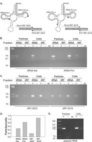
RT-PCR to detect tRFs, tRNAs, and gag/pol RNA.FBS may contain exosomes carrying small RNAs; to avoid this possible source of contami-nation, complete RPMI containing 20% FBS was centrifuged at 24,000 rpm for 4 h using a Beckman-Coulter SW28 rotor to pellet any exosomes. The supernatant medium was then passed through a 0.2-m filter and brought to 10% FBS by adding an equal volume of RPMI containing antibiotics and glutamine. C91PL cells were cultured to confluence in the exosome-depleted medium, and virus particles were recovered as de-scribed above. RNA was isolated from the pelleted particles and the pro-ducer C91PL cells using TRIzol LS (Life Technologies). Aliquots of the RNA were subjected to denaturing PAGE through a 15% polyacrylamide gel to separate species in the size range of full-length tRNAs from the small RNAs, with tRNAs visible in the cellular RNA sample and 5 pmol syn-thetic miR-150-5p serving as size markers for the 2 fractions. The gel was stained with ethidium bromide, and the regions containing tRNAs and small RNAs (about 15 to 30 nt) were excised, crushed, and incubated with gentle mixing in elution buffer (300 mM sodium acetate, pH 5.2, 1 mM EDTA) overnight at 4°C. RNA was ethanol precipitated and resuspended in distilled H2O (10l per 19 ml of original culture supernatant) (seeFig. 6, where the fractions are labeled tRNA and tRF).
The primer sets used to amplify tRF-3019, tRF-3003, and the tRNAs from which they are derived (tRNA-Pro and tRN-Ala, respectively) are listed in Table S1 in the supplemental material (also seeFig. 6A). RT-PCR to detect tRF-3019 and tRF-3003 was based on a protocol for detecting microRNAs (35). Size-fractionated RNA (2l) was annealed with 2 pmol primer RT7-tRF-3019 or RT8-tRF-3003 at 70°C for 10 min in a 7.5-l volume. The mixture was brought to 10l with the addition of 1 mM dNTP, 1⫻RT buffer, and 5 U avian myeloblastosis virus (AMV) reverse transcriptase (Finnzymes) and reverse transcribed at 40°C for 1 h.
Two-microliter aliquots of the resulting cDNAs were PCR amplified in a final volume of 25l containing 1⫻TaqGold PCR buffer, 1.5 mM MgCl2, 200
M dNTP, 0.1 pmol of primer Short-tRF-3019 or Short-tRF-3003, 2.5 pmol each of primers PCR-tRF-s and PCR-tRF-as (specific for tails added by the RT primer and Short-tRF primer), and 0.5 U AmpliTaq Gold DNA polymerase. The PCR method consisted of a denaturation step at 95°C for 10 min and 5 cycles of denaturation at 95°C for 30 s, annealing at 40°C for 45 s, and extension at 72°C for 30 s, followed by 22 cycles of denaturation at 95°C for 30 s, annealing at 60°C for 45 s, and extension at 72°C for 30 s. To detect tRNA-Pro and tRNA-Ala, 1l of size-fractionated RNA was reverse transcribed in a 10-l reaction mixture at 53°C for 50 min using the antisense primer and Superscript III (Life Technologies). The result-ing cDNA (2.5l) was PCR amplified using sense and antisense primers and AmpliTaq Gold, with a denaturation step at 95°C for 8 min, followed by cycles of denaturation at 95°C for 40 s, annealing at 60°C for 40 s, and extension at 72°C for 40 s (30 cycles for Ala and 26 cycles for tRNA-Pro). To detect gag/pol RNA, 0.5l of RNA from virus particles or 200 ng total RNA from producer C91PL cells was reverse transcribed using primer Gag-as and Superscript III and then PCR amplified using primers U5-s and Gag-as (see Table S1 in the supplemental material) and Ampli-Taq Gold as described above for 30 cycles with an annealing temperature of 59°C and a final extension step for 5 min at 72°C. The PCR products were separated on 6% polyacrylamide gels.
RESULTS
Generation, sequencing, annotation, and quantification of
small RNA libraries.
Total RNA was isolated from the
HTLV-1-infected cell lines C91PL and MT-2 (
25
), which have a CD4
⫹phenotype, and from control normal unstimulated and
in vitro
-stimulated CD4
⫹T cells. RNA species of
⬃
18 to 25 nt were
sepa-rated by polyacrylamide gel electrophoresis and modified with
tags at the 5
=
and 3
=
ends according to the method of Lau et al. (
26
)
and sequenced using a Roche 454 Life Sciences platform.
Se-quence reads were mapped against mirBase release 18 to identify
the populations of mature known microRNAs in the investigated
cell lines, and the edgeR Bioconductor statistical package (see
Ma-terials and Methods) was employed to identify statistically
signif-icant differences in the frequencies of known microRNAs.
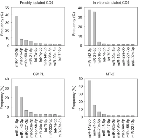
The graphs in
Fig. 1
illustrate the 10 most abundant known
microRNAs identified in the libraries. miR-142-3p, a marker of
hematopoietic cells, stood out as the most abundant microRNA in
freshly isolated CD4
⫹T cells and in the infected cell line MT-2,
with a frequency more than triple that of the other microRNAs.
miR-21, a microRNA known to be linked to T-cell activation and
transformation (
36
), became nearly as frequent as miR-142-3p
upon
in vitro
stimulation of CD4
⫹cells and was also abundant in
the two infected cell lines.
To identify the microRNAs connected with HTLV-1 infection,
we calculated differences in the frequencies of microRNAs in
in-fected cell lines versus resting and stimulated CD4
⫹T cells. Three
microRNAs were differentially expressed in both infected cell lines
compared to control CD4 cells (indicated in boldface in
Table 1
:
miR-34a-5p was upregulated, and miR-150-5p and miR-146b-5p
were both downregulated). An analysis performed using the
miRDeep2 software did not yield any putative new microRNA
candidates among the sequence reads detected in this study.
Small RNAs expressed by HTLV-1.
With the aim of
identify-ing viral microRNAs, the reads obtained from MT-2 and C91PL
cells were aligned to the HTLV-1 genome. This analysis yielded 25
sequences with
ⱖ
90% identity with HTLV-1 and
ⱕ
2 gaps and
ⱕ
2
mismatches (data not shown). Two sequences shown in
Fig. 2A
perfectly matched the primary plus-strand HTLV-1 transcript.
on November 7, 2019 by guest
http://jvi.asm.org/
Both sequences were present only in the MT-2 library. Sequence
MT-2/A was positioned in exon 3, between the stop codons for
p30/p13 and Rex. Sequence MT-2/B was located in the R region, in
a position within stem-loop D of the Rex response element
(RXRE) (
37
). This segment of the HTLV-1 genome was predicted
to form a microRNA precursor hairpin (pre-miRNA)-like
struc-ture and thus to have the potential to give rise to a viral microRNA
(
21
).
Figure 2B
shows the predicted secondary structures of
genomic regions containing MT-2/A and MT-2/B with 5
=
and 3
=
flanking sequences to simulate their position in the 5
=
portion of a
pre-miRNA. Results showed that sequence MT-2/A is likely to be
present mostly in an unstructured region, while sequence MT-2/B
has a high probability to be positioned in a stem.
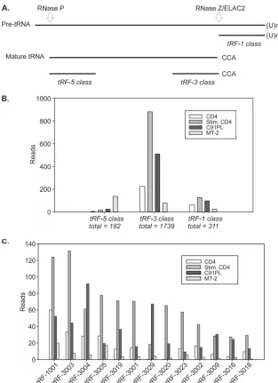
tRFs expressed in HTLV-1-infected cells.
We next tested the
sequences identified in the libraries for perfect matches to the 135
tRFs reported by Lee et al. in a study of prostate cancer cell lines
(
22
). Table S2 in the supplemental material lists the number of
sequence reads for each tRF, as well as isoforms showing
varia-tions at the 5
=
or 3
=
end. Overall, in both normal and
HTLV-1-infected CD4 cells, fragments processed from the 3
=
ends of
ma-ture tRNAs (tRF-3) were considerably more abundant than tRFs
produced from the 3
=
ends of tRNA precursors (tRF-1) or from
the 5
=
ends of mature tRNAs (tRF-5) (
Fig. 3B
).
Figure 3C
shows the most abundant tRFs identified in the
li-braries. Among the 22 previously described 1 sequences,
tRF-1001 was the most abundant. tRF-tRF-1001, as well as the other tRFs,
were upregulated in normal CD4
⫹cells upon mitogenic
stimula-tion. tRF-3004 and tRF-3029 were more abundant in C91PL cells
than in stimulated CD4
⫹controls, and MT-2 cells yielded few tRF
sequences compared to the other 3 cell types.
[image:4.585.119.530.64.350.2]FIG 1Relative abundances of microRNAs identified by sequencing. Shown are the 10 most abundant microRNAs in normal CD4 T cells and HTLV-1-infected cell lines. Frequencies were calculated by dividing the number of se-quence reads for each microRNA by the total number of sese-quence reads for all known microRNAs.
TABLE 1Differentially expressed microRNAsa
Cells MicroRNA Log2FC Pvalue
MT-2 vs. CD4 miR-34a-5p 6.15 0.00015
miR-4448 4.89 0.00366
miR-7-5p 4.35 0.01564
miR-150-5p ⴚ10.08 0.00327
miR-30c-5p ⴚ8.05 0.01329
miR-146b-5p ⴚ7.52 0.03318
miR-29c-3p ⫺6.87 0.03802
C91PL vs. CD4 miR-34a-5p 6.66 0.00001
miR-92b-3p 3.82 0.01313 miR-23a-3p 3.15 0.01166
miR-150-5p ⴚ10.61 0.00127
miR-342-5p ⴚ7.02 0.02719 miR-26a-5p ⴚ6.84 0.00776 miR-20b-5p ⴚ6.74 0.03088
miR-146b-5p ⴚ4.76 0.02268
miR-19b-3p ⴚ3.83 0.03015 miR-16b-5p ⴚ3.65 0.02867
a
MicroRNAs with statistically significant differences in expression are indicated in boldface. FC, fold change.
FIG 2Small RNAs expressed by HTLV-1. (A) Positions and nucleotide se-quences of the two small RNA species identified in MT-2 cells. MT-2/A corre-sponded to nt 7582 to 7602 in exon 3 of the HTLV-1 ATK sequence. MT-2/B corresponded to nt 513 to 530 of the 5=R region and nt 8792 to 8808 of the 3= R region of ATK. (B) Secondary structure predicted by RNAfold (http://rna .tbi.univie.ac.at/cgi-bin/RNAfold.cgi; University of Vienna) for HTLV-1 se-quences containing MT-2/A and MT-2/B with 15 nt added at the 5=end and 50 nt at the 3=end to simulate a pre-miR. The optimal secondary structures and their minimum free energy (MFE) values are indicated. Nucleotides in grey circles have a high probability of occupying the indicated paired or unpaired position and were identified with red or orange circles in the original RNAfold diagram.
on November 7, 2019 by guest
http://jvi.asm.org/
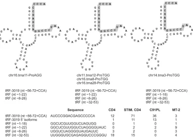
[image:4.585.43.294.66.310.2] [image:4.585.39.286.509.705.2]The tRF-3 class also includes tRF-3019 (
22
). This tRF
corre-sponds to the 3
=
end of tRNA-Pro, the tRNA considered to serve as
the primer for HTLV-1 reverse transcriptase (
38
). tRF-3019 was
the fifth most abundant tRF identified in our libraries and was
most abundant in stimulated CD4
⫹cells. A BLAST search for
tRNA genes able to produce tRF-3019 yielded 21 tRNA-Pro genes
located on chromosomes 1, 5, 6, 11, 14, 16, and 17 (see Table S3 in
the supplemental material). The four libraries contained several
tRF-3019 isoforms with additional nucleotides at the 5
=
end that
perfectly matched the human genome but were not
complemen-tary to the viral genome (
Fig. 4
). The libraries also contained a
small number of reads corresponding to fragments derived from
other portions of tRNA-Pro.
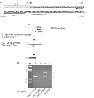
tRF-3019 functions as a primer for HTLV-1 reverse
trans-criptase.
It is noteworthy that only the portion of tRNA-Pro
cor-responding to tRF-3019 is complementary to the HTLV-1 primer
binding site (PBS) (
Fig. 5A
), suggesting that the tRF would be fully
sufficient as a primer for reverse transcription. We thus tested the
primer activity of tRF-3019 in an
in vitro
reverse transcriptase
assay carried out using a synthetic RNA template and the reverse
transcriptase contained in HTLV-1 particles recovered from the
culture supernatant of C91PL cells (
Fig. 5A
). The RT assay
mix-tures contained either no primer, synthetic tRF-3019 RNA, or
tRF-3019 DNA as a positive control or miR-150-5p RNA as a
negative control. The PCR mixture contained a sense primer
spe-cific for a tail sequence present in the synthetic RNA template and
an antisense primer positioned immediately 5
=
to the PBS.
Figure
5B
shows the results of the assays. The RT assay performed using
tRF-3019 RNA yielded the expected 87-bp PCR product, thus
confirming that tRF-3019 can function as a primer for HTLV-1
reverse transcriptase. The assay carried out using a tRF-3019 DNA
primer yielded the 87-bp product, along with a longer product
FIG 3Relative abundances of tRFs. (A) The 3 classes of tRFs aligned to the tRNA precursor. (B) Total numbers of sequence reads with perfect matches to each of the tRF classes, together with 5=and 3=isoforms (see Table S2 in the supplemental material). (C) Sequence reads for tRFs with a total of at least 50 sequence reads summed among the 4 libraries.
on November 7, 2019 by guest
http://jvi.asm.org/
[image:5.585.134.447.63.494.2]indicated by the gray arrow in
Fig. 5B
. This second band
corre-sponded in size to an amplicon produced with the tail primer and
residual tRF-3019 DNA present in the cDNA (i.e., 107 bp).
Inter-estingly, trace amounts of the 87-bp product were also detected in
the assays carried out using C91PL RT and miR-150-5p or no
primer. This amplicon may have originated from cDNA primed
by tRNA-Pro or tRF-3019 present in the viral particle lysate that
was used as a source of reverse transcriptase (see below).
HTLV-1-infected cells release particles containing tRF-3019.
Having established that tRF-3019 is capable of priming HTLV-1
reverse transcription, we next tested for the presence of tRNA-Pro
and tRF-3019 in virus particles recovered from supernatants of
C91PL cultures. As a control, we also assayed for tRF-3003, the
most abundant tRF-3 detected in the 4 libraries, along with its
parent, tRNA-alanine (Ala). As described in Materials and
Meth-ods, RNA isolated from the virus particles and producer cells was
subjected to denaturing PAGE to separate species in the tRF size
range from full-length tRNAs. This was necessary, as the tailed
RT-PCR primers utilized to detect the tRFs also amplified the 3
=
ends of the full-length tRNAs (
Fig. 6A
). RT-PCR products were
separated by PAGE; the intensities of the resulting bands were
measured to estimate relative abundances of the tRNAs and tRFs
in virus particles versus cells.
Figure 6B
shows PAGE analyses of the RT-PCR products
ob-tained for tRNA-Ala and tRNA-Pro. As expected, both tRNA-Ala
and tRNA-Pro were readily detected in the C91PL cells. However,
a plot of the ratios of the band intensities (
Fig. 6D
) revealed that
tRNA-Pro was enriched in virus particles compared to tRNA-Ala.
The RT-PCR products for both tRNAs were much more evident in
the full-length tRNA fraction than in the tRF fraction, indicating
that the denaturing PAGE purification step resulted in acceptable
separation of the 2 size classes.
Figure 6C
shows the results of RT-PCR carried out on the same
samples using primer sets that amplified the tRFs present in the
tRF fraction and the 3
=
ends of the tRNAs in the tRNA fraction.
Calculation of band intensity ratios (
Fig. 6D
) showed that
tRF-3003 was much less abundant in the virus particles than in the
cells, while tRF-3019 was detected at comparable levels in virus
particles and cells. Therefore, we concluded that tRF-3019 was
enriched in virus particles compared to tRF-3003.
These findings indicate that both tRNA-Pro and tRF-3019 are
incorporated into particles released in the supernatant of
HTLV-1-infected cells. It is necessary to point out that these particles may
also contain exosomes, which may also package proteins and
RNA. However, the observation that the particles were enriched
for tRNA-Pro and tRF-3019 compared to tRNA-Ala and tRF-3003
supports a specific packaging process directed by interaction of
the tRNA/tRF with the HTLV-1 PBS. As shown in
Fig. 6E
,
RT-PCR assays on RNA isolated from the particles confirmed that
they contain the HTLV-1 genomic gag/pol mRNA. Therefore,
al-though we cannot exclude the presence of exosomes in the particle
preparations, our findings demonstrate that these particles
con-tain reverse transcriptase activity (
Fig. 5
) and the viral genome and
are enriched for the PBS-specific tRNA-Pro and tRF-3019. Taken
together, these results strongly suggest that tRF-3019 participates
in HTLV-1 reverse transcription.
DISCUSSION
In the present study, we employed mass sequencing to identify the
repertoire of small noncoding RNAs expressed in normal T cells
FIG 4tRFs processed from tRNA-Pro. (Top) Three examples of the 21 tRNA-Pro molecules that are able to produce tRF-3019 (shaded). The diagrams were obtained from the University of California, Santa Cruz, tRNA database and modified by adding the 3=CCA triplet that is present on mature tRNAs and tRF-3 sequences (Fig. 3). (Bottom) Sequences of the tRFs and number of reads identified in each library.
on November 7, 2019 by guest
http://jvi.asm.org/
[image:6.585.102.484.65.335.2]compared to cells transformed with HTLV-1. The results revealed
a distinct pattern of microRNA expression in HTLV-1-infected
cells, two virus-encoded small RNAs, and a number of tRFs.
In-terestingly, the tRNA fragment tRF-3019 was detected in virus
particles and was capable of priming HTLV-1 reverse
transcrip-tion.
Three microRNAs were differentially expressed in both
in-fected cell lines compared to control CD4
⫹cells: miR-34a-5p was
upregulated, and miR-150-5p and miR-146b-5p were both
down-regulated. Their shared pattern of regulation in the two infected
cell lines suggests that these microRNAs play important roles in
HTLV-1 infection/transformation, rather than representing
markers of T-cell activation, which are also present in
HTLV-1-infected cells.
The observation that miR-150-5p expression is reduced in
HTLV-1-infected cell lines is consistent with other studies (
15
,
16
). miR-150-5p undergoes upregulation during T-cell
develop-ment (
27
) but is diminished upon stimulation of normal murine
CD4
⫹T cells (
39
). Expression of miR-150-5p is increased in
sev-eral hematological tumors, including ATLL samples (
15
,
16
), but
is downregulated in the cutaneous CD4
⫹T-cell lymphoma Sézary
syndrome (
40
), in NK/T-cell lymphomas (
41
), and in several
other hematological malignancies (reviewed in reference
42
).
Forced expression of miR-150-5p in B-lymphoma cell lines (
43
),
T-acute lymphoblastic leukemia (T-ALL) cell lines (
27
), and NK
cell lines (
41
) produced antiproliferative and/or proapoptotic
ef-fects. Validated targets of miR-150-5p include the oncogenes
c-Myb (
44
) and NOTCH-3 (
27
), as well as the HIV-1 3
=
untrans-lated region (UTR) (
45
). It is noteworthy that the minus-strand
HTLV-1 transcripts coding for HBZ contain 2 potential binding
sites for miR-150-5p (
46
).
miR-146b-5p is gradually upregulated during T-cell
develop-ment from the double-positive CD4
⫹CD8
⫹to the single-positive
CD4
⫹or CD8
⫹stage (
27
). The sequence of miR-146b-5p is
al-most identical to that of miR-146a, which was identified as
up-regulated through the action of Tax in previous studies of
HTLV-1-infected cell lines (
14
,
47
). miR-146b-5p mRNA targets,
therefore, likely overlap those identified for miR-146a, which
in-clude the Toll-like receptor signaling pathway proteins TRAF6
and IRAK1 (
48
), the apoptosis signaling protein FADD (
49
), and
the chemokine receptor CXCR4 (
50
). miR-146b-5p is
downregu-lated in ATLL (
16
,
17
), Sézary syndrome (
40
), and several other
FIG 5tRF-3019 acts as a primer for HTLV-1 reverse transcriptase. (A) Summary of the RT assay. The template consisted of anin vitro-transcribed RNA spanning HTLV-1 nt 721 to 822 modified by the addition of a 20-nt tail at the 5=end. The template was incubated with HTLV-1 reverse transcriptase present in virus particles recovered from the culture supernatant of C91PL cells and either tRF-3019 RNA, miR-150-5p RNA (negative control), tRF-3019 DNA (positive control), or no primer. The products of the RT reactions were amplified by PCR using PCR primers Tail-s and U5-as and separated by PAGE in a 6% polyacrylamide gel, along with MspI-digested pBluescript as a size marker. (B) Composite of the ethidium bromide-stained gel. The black arrow indicates the position of the 87-bp PCR product expected using primers Tail-s and U5-as. The additional band in lane 3 indicated by the gray arrow was consistent with a product amplified by Tail-s and residual tRF-3019 DNA primer added to the RT assay. The primer sequences are reported in Table S1 in the supplemental material.
on November 7, 2019 by guest
http://jvi.asm.org/
[image:7.585.139.469.64.408.2]hematological malignancies but is upregulated in mycosis
fungoi-des (
51
) and pediatric acute myeloid leukemia (
52
; reviewed in
reference
42
).
miR-34a-5p is known to be upregulated by p53 in response to
genotoxic and oncogenic stresses. miR-34a-5p targets genes
af-fecting cell proliferation and survival, resulting in growth arrest,
senescence, and apoptosis; its downregulation in several solid
tu-mors suggests a tumor suppressor role (
53
). miR-34a-5p was
found to be more abundant in memory versus naive CD4
⫹T cells
(
54
) and is upregulated in Epstein-Barr virus-transformed B
FIG 6RT-PCR to detect tRNAs, tRFs, and gag/pol RNA in virus particles and C91PL cells. (A) As described in Materials and Methods, RNA from virus particles and producer C91PL cells was subjected to denaturing PAGE; regions of the gel containing tRNA and tRFs were excised, and RNA was recovered by passive elution and ethanol precipitation. The resulting fractions were subjected to RT-PCR to detect tRNA-Ala, tRNA-Pro, and their tRF-3 sequences, tRF-3003 and tRF-3019, respectively. (B and C) Images of the RT-PCR products after separation on 6% polyacrylamide gels. The intensities of RT-PCR bands obtained for tRNAs and tRFs (measured in tRNA and tRF fractions, respectively) were measured using a Bio-Rad Gel Doc XRS imager. (D) Plot of ratios of band intensities obtained for virus particles versus cells. The calculated ratios were as follows: tRNA-Ala, particles/cells⫽0.46; tRNA-Pro, particles/cells⫽0.82; tRF-3003, particles/cells⫽0.18; tRF-3019, particles/cells⫽1.07. (E) Results of RT-PCR performed on RNA from the virus particles and producer cells to detect HTLV-1 genomic gag/pol RNA. RT-PCR was carried out using primers U5-s and Gag-as as described in Materials and Methods. The dashed white lines were added to panels B and C to aid in their alignment. The first lane on each gel contained MspI-digested pBluescript as a size marker; band sizes in basepairs are indicated on the left. The plus and minus signs above the lanes indicate RT reactions carried out in the presence (⫹) or absence (⫺) of reverse transcriptase. RNA template was omitted from the RT reaction in lanes labeled nt.
on November 7, 2019 by guest
http://jvi.asm.org/
[image:8.585.135.448.60.541.2]cells (
55
) during latency type III (
56
) and in hepatitis B
virus-associated hepatocellular carcinoma (
57
) and might thus exert
diverse effects depending on the cell context (
58
). The results of
RT-PCR assays indicated strong upregulation of miR-34a-5p
in primary samples from ATLL patients (D. M. D’Agostino,
unpublished data).
Recent studies revealed that BLV, a complex oncogenic
retro-virus related to HTLV-1, encodes a cluster of viral microRNAs
(
19
,
20
). Our deep-sequencing analysis also revealed two
virus-encoded small RNA species (MT-2/A and MT-2/B). However, the
fact that MT-2/A and MT-2/B were detected with only one
sequence read each suggests that, in contrast to BLV, HTLV-1
may not rely on viral microRNAs as a mechanism of
posttran-scriptional regulation. Alternatively, the production of viral
microRNAs might not be favored in cells that are chronically
in-fected, such as MT-2 and C91PL. Therefore, before concluding
that HTLV-1 does not produce microRNAs, it will be important
to measure their levels of expression in the context of primary
samples obtained from infected patients.
The greater representation of tRF-3 sequences than of tRF-1
and tR-5 classes in the libraries is in line with the preponderance of
tRF-3 sequences found in prostate cancer cell lines (see Table S2 in
reference
22
) and in mature B cells (
24
). Previous functional
stud-ies of tRF-1001, which was abundantly expressed in our librarstud-ies,
revealed its elevated expression in cancer cell lines compared to
normal tissue samples and indicated that it is required for cell
proliferation (
22
). Among the tRF-3 sequences abundantly
ex-pressed in the four libraries, functional data are available for
tRF-3018 in the context of B cells (
24
). This tRF, named CU1276 in the
cell study, was differentially expressed in different stages of
B-cell maturation, with the greatest expression found in the
germi-nal center (GC) stage while it was absent in GC-derived
lym-phoma cells. Functional studies of tRF-3018/CU1276 verified its
ability to associate with Argonaute proteins and to repress
expres-sion of RPA1, a protein involved in DNA replication and repair
(
24
).
The present study focused on tRF-3019, as it corresponds to
the 3
=
end of tRNA-Pro, which is generally considered to be the
primer for HTLV-1 reverse transcriptase (
38
). tRF-3019 was
ca-pable of priming HTLV-1 reverse transcription (
Fig. 5
) and was
enriched in virus particles (
Fig. 6
). Taken together, these
observa-tions support a role for tRF-3019 in the life cycle of HTLV-1.
As shown in
Fig. 4
, 12 of the 18 nucleotides of tRNA-Pro that
are complementary to the HTLV-1 PBS are based paired in the
mature tRNA. This positioning of the primer portion of the tRNA
in a closed stem is a characteristic of all retroviral tRNA primers.
These hydrogen bonds must be disrupted in order for the primer
to bind to the PBS, which would not be necessary if a tRF is used as
a primer.
The libraries examined in the present study contained a few
sequence reads for tRF-3015, which represents the 3
=
end of
tRNA-lysine (Lys), the primer for HIV-1 (see Table S2 in the
sup-plemental material). Schopman et al. (
59
) pointed out the
possi-bility that tRFs may serve as primers for reverse transcriptase but
also presented experimental evidence from studies of HIV-1 that
did not support this proposal. Efficient HIV-1 reverse
transcrip-tion requires interactranscrip-tions of tRNA-Lys with the PBS, as well as
other regions of the viral genome. Of particular importance is an
8-nt sequence termed the primer activation signal (PAS) located
in the U5 region that binds to the third stem-loop (T arm) of
tRNA-Lys and promotes initiation of reverse transcription and
elongation of the cDNA (reviewed in reference
60
). Although all
retroviruses are predicted to contain a PAS (
61
), the putative PAS
in HTLV-1, which is positioned approximately 10 nucleotides 5
=
to the PBS, has not yet been functionally characterized.
The secondary structure of the tRNA primer must also be
dis-rupted to allow nucleotides in the T arm to interact with the PAS.
In HIV-1, the NC protein plays an important role in unfolding
tRNA-Lys to allow its binding to the HIV-1 PAS (
62
).
Interest-ingly, a study of NC proteins from several retroviruses indicated
that the HTLV-1 NC protein possesses comparatively weak
nu-cleic acid chaperone activity (
63
). It is possible that another
mech-anism is responsible for unfolding of tRNA-Pro or that the PAS
interaction is not important for HTLV-1.
Alternatively, tRF-3019 may serve as the major primer. In fact,
our
in vitro
assay showed that tRF-3019 permits reverse
transcrip-tion of a segment of HTLV-1 RNA containing the PBS and
pre-dicted PAS. The detailed picture of the interactions between
HIV-1 RNA elements and its tRNA primer raises the possibility
that tRFs representing the 3
=
ends of primer tRNAs might support
the initiation of reverse transcription, but not progressivity, with
failure to proceed to the strand transfer step. In this case, tRF-3019
might inhibit the overall process of reverse transcription, thus
acting as a restriction factor for HTLV-1 replication. Further
stud-ies will be necessary to test these hypotheses by comparing the
abilities of tRF-3019 and tRNA-Pro to prime and support strand
transfer.
ACKNOWLEDGMENTS
We thank Paola Dalla Pria for technical assistance, Katia Basso and Bea-trice Macchi for sharing protocols, Subhamoy Mukherjee for helping with bioinformatics analyses, and Margherita Ghisi, Riccardo Dalla-Favera, Romain Parent, and Luigi Chieco-Bianchi for discussions.
The research was supported by investigator grants from the Associa-zione Italiana per la Ricerca sul Cancro (AIRC; nos. 4175 and 13378), an AIRC-Cariverona Regional grant, and the University of Padua (Ateneo grant no. CPDA124913/12). A. Grassi was supported by a fellowship from the Centro Lincei Interdisciplinare Beniamino Segre, Accademia Nazionale dei Lincei.
REFERENCES
1.Franchini G, Nicot C, Johnson JM.2003. Seizing of T cells by human T-cell leukemia/lymphoma virus type 1. Adv. Cancer Res.89:69 –132.
http://dx.doi.org/10.1016/S0065-230X(03)01003-0.
2.Matsuoka M.2003. Human T-cell leukemia virus type I and adult T-cell leukemia. Oncogene22:5131–5140.http://dx.doi.org/10.1038/sj.onc.12 06551.
3.Lairmore M, Franchini G.2007. Human T-cell leukemia virus types 1 and 2, p 2071–2106.InKnipe DM, Howley PM (ed), Fields virology, 5th ed, vol 2. Lippincott Williams and Wilkins, Philadelphia, PA.
4.Lairmore MD, Anupam R, Bowden N, Haines R, Haynes RAH, Ratner L, Green P.2011. Molecular determinants of human T-lymphotropic virus type 1 transmission and spread. Viruses3:1131–1165.http://dx.doi .org/10.3390/v3071131.
5.Matsuoka M, Jeang KT. 2011. Human T-cell leukemia virus type 1 (HTLV-1) and leukemic transformation: viral infectivity, Tax, HBZ and therapy. Oncogene 30:1379 –1389. http://dx.doi.org/10.1038/onc.2010 .537.
6.Hasegawa H, Sawa H, Lewis MJ, Orba Y, Sheehy N, Yamamoto Y, Ichinohe T, Tsunetsugu-Yokota Y, Katano H, Takahashi H, Matsuda J, Sata T, Kurata T, Nagashima K, Hall WW. 2006. Thymus-derived leukemia-lymphoma in mice transgenic for the Tax gene of human T-lymphotropic virus type I. Nat. Med.12:466 – 472.http://dx.doi.org/10 .1038/nm1389.
7.Satou Y, Yasunaga J, Yoshida M, Matsuoka M.2006. HTLV-I basic
on November 7, 2019 by guest
http://jvi.asm.org/
leucine zipper factor gene mRNA supports proliferation of adult T cell leukemia cells. Proc. Natl. Acad. Sci. U. S. A.103:720 –725.http://dx.doi .org/10.1073/pnas.0507631103.
8.Satou Y, Yasunaga J, Zhao T, Yoshida M, Miyazato P, Takai K, Shimizu K, Ohshima K, Green PL, Ohkura N, Yamaguchi T, Ono M, Sakaguchi S, Matsuoka M.2011. HTLV-1 bZIP factor induces T-cell lymphoma and systemic inflammation in vivo. PLoS Pathog.7:e1001274.http://dx.doi .org/10.1371/journal.ppat.1001274.
9.Van Prooyen N, Andresen V, Gold H, Bialuk I, Pise-Masison C, Franchini G.2010. Hijacking the T-cell communication network by the human T-cell leukemia/lymphoma virus type 1 (HTLV-1) p12 and p8 proteins. Mol. Aspects Med. 31:333–343. http://dx.doi.org/10.1016/j .mam.2010.07.001.
10. Silic-Benussi M, Biasiotto R, Andresen V, Franchini G, D’Agostino DM, Ciminale V.2010. HTLV-1 p13, a small protein with a busy agenda. Mol. Aspects Med.31:350 –358.http://dx.doi.org/10.1016/j.mam.2010.03 .001.
11. Fukumoto R, Andresen V, Bialuk I, Cecchinato V, Walser JC, Valeri VW, Nauroth JM, Gessain A, Nicot C, Franchini G.2009. In vivo genetic mutations define predominant functions of the human T-cell leukemia/ lymphoma virus p12I protein. Blood113:3726 –3734.http://dx.doi.org/10 .1182/blood-2008-04-146928.
12. Huntzinger E, Izaurralde E.2011. Gene silencing by microRNAs: con-tributions of translational repression and mRNA decay. Nat. Rev. Genet.
12:99 –110.http://dx.doi.org/10.1038/nrg2936.
13. Iorio MV, Croce CM.2012. MicroRNA dysregulation in cancer: diagnos-tics, monitoring and therapeutics. A comprehensive review. EMBO Mol. Med.4:143–159.http://dx.doi.org/10.1002/emmm.201100209. 14. Pichler K, Schneider G, Grassmann R.2008. MicroRNA miR-146a and
further oncogenesis-related cellular microRNAs are dysregulated in HTLV-1-transformed T lymphocytes. Retrovirology5:100.http://dx.doi .org/10.1186/1742-4690-5-100.
15. Yeung ML, Yasunaga J, Bennasser Y, Dusetti N, Harris D, Ahmad N, Matsuoka M, Jeang KT.2008. Roles for microRNAs, 93 and miR-130b, and tumor protein 53-induced nuclear protein 1 tumor suppressor in cell growth dysregulation by human T-cell lymphotrophic virus 1. Can-cer Res. 68:8976 – 8985. http://dx.doi.org/10.1158/0008-5472.CAN-08 -0769.
16. Bellon M, Lepelletier Y, Hermine O, Nicot C.2009. Deregulation of microRNA involved in hematopoiesis and the immune response in HTLV-I adult T-cell leukemia. Blood113:4914 – 4917.http://dx.doi.org /10.1182/blood-2008-11-189845.
17. Yamagishi M, Nakano K, Miyake A, Yamochi T, Kagami Y, Tsutsumi A, Matsuda Y, Matsubara A, Muto S, Utsunomiya A, Yamaguchi K, Uchimaru K, Ogawa S, Watanabe T.2012. Polycomb-mediated loss of miR-31 activates NIK-dependent NF-B pathway in adult T-cell leukemia and other cancers. Cancer Cell21:121–135.http://dx.doi.org/10.1016/j .ccr.2011.12.015.
18. Kincaid RP, Sullivan CS.2012. Virus-encoded microRNAs: an overview and a look to the future. PLoS Pathog.8:e1003018.http://dx.doi.org/10 .1371/journal.ppat.1003018.
19. Kincaid RP, Burke JM, Sullivan CS.2012. RNA virus microRNA that mimics a B-cell oncomiR. Proc. Natl. Acad. Sci. U. S. A.109:3077–3082.
http://dx.doi.org/10.1073/pnas.1116107109.
20. Rosewick N, Momont M, Durkin K, Takeda H, Caiment F, Cleuter Y, Vernin C, Mortreux F, Wattel E, Burny A, Georges M, Van den Broeke A.2013. Deep sequencing reveals abundant noncanonical retroviral microRNAs in B-cell leukemia/lymphoma. Proc. Natl. Acad. Sci. U. S. A.
110:2306 –2311.http://dx.doi.org/10.1073/pnas.1213842110.
21. Li SC, Shiau CK, Lin WC. 2008. Vir-Mir db: prediction of viral microRNA candidate hairpins. Nucleic Acids Res.36:D184 –D189.http: //dx.doi.org/10.1093/nar/gkm610.
22. Lee YS, Shibata Y, Malhotra A, Dutta A.2009. A novel class of small RNAs: tRNA-derived RNA fragments (tRFs). Genes Dev.23:2639 –2649.
http://dx.doi.org/10.1101/gad.1837609.
23. Cole C, Sobala A, Lu C, Thatcher SR, Bowman A, Brown JW, Green PJ, Barton GJ, Hutvagner G.2009. Filtering of deep sequencing data reveals the existence of abundant Dicer-dependent small RNAs derived from tRNAs. RNA15:2147–2160.http://dx.doi.org/10.1261/rna.1738409. 24. Maute RL, Schneider C, Sumazin P, Holmes A, Califano A, Basso K,
Dalla-Favera R.2013. tRNA-derived microRNA modulates proliferation and the DNA damage response and is down-regulated in B cell lymphoma.
Proc. Natl. Acad. Sci. U. S. A.110:1404 –1409.http://dx.doi.org/10.1073 /pnas.1206761110.
25. Popovic M, Lange-Wantzin G, Sarin PS, Mann D, Gallo RC. 1983. Transformation of human umbilical cord blood T cells by human T-cell leukemia/lymphoma virus. Proc. Natl. Acad. Sci. U. S. A.80:5402–5406.
http://dx.doi.org/10.1073/pnas.80.17.5402.
26. Lau NC, Lim LP, Weinstein EG, Bartel DP.2001. An abundant class of tiny RNAs with probable regulatory roles in Caenorhabditis elegans. Sci-ence294:858 – 862.http://dx.doi.org/10.1126/science.1065062. 27. Ghisi M, Corradin A, Basso K, Frasson C, Serafin V, Mukherjee S,
Mussolin L, Ruggero K, Bonanno L, Guffanti A, De Bellis G, Gerosa G, Stellin G, D’Agostino DM, Basso G, Bronte V, Indraccolo S, Amadori A, Zanovello P.2011. Modulation of microRNA expression in human T-cell development: targeting of NOTCH3 by miR-150. Blood117:7053– 7062.http://dx.doi.org/10.1182/blood-2010-12-326629.
28. Camacho C, Coulouris G, Avagyan V, Ma N, Papadopoulos J, Bealer K, Madden TL.2009. BLAST⫹: architecture and applications. BMC Bioin-formatics10:421.http://dx.doi.org/10.1186/1471-2105-10-421. 29. Pearson WR, Lipman DJ.1988. Improved tools for biological sequence
comparison. Proc. Natl. Acad. Sci. U. S. A.85:2444 –2448.http://dx.doi .org/10.1073/pnas.85.8.2444.
30. Robinson MD, McCarthy DJ, Smyth GK.2010. edgeR: a Bioconductor package for differential expression analysis of digital gene expression data. Bioinformatics 26:139 –140. http://dx.doi.org/10.1093/bioinformatics /btp616.
31. Friedlander MR, Chen W, Adamidi C, Maaskola J, Einspanier R, Knespel S, Rajewsky N.2008. Discovering microRNAs from deep se-quencing data using miRDeep. Nat. Biotechnol.26:407– 415.http://dx .doi.org/10.1038/nbt1394.
32. Kimata JT, Wong FH, Wang JJ, Ratner L. 1994. Construction and characterization of infectious human T-cell leukemia virus type 1 molec-ular clones. Virology204:656 – 664.http://dx.doi.org/10.1006/viro.1994 .1581.
33. Balestrieri E, Pizzimenti F, Ferlazzo A, Giofre SV, Iannazzo D, Piperno A, Romeo R, Chiacchio MA, Mastino A, Macchi B. 2011. Antiviral activity of seed extract from Citrus bergamia towards human retroviruses. Bioorg. Med. Chem. 19:2084 –2089. http://dx.doi.org/10.1016/j.bmc .2011.01.024.
34. Frezza C, Balestrieri E, Marino-Merlo F, Mastino A, Macchi B.2014. A novel, cell-free PCR-based assay for evaluating the inhibitory activity of antiretroviral compounds against HIV reverse transcriptase. J. Med. Virol.
86:1–7.http://dx.doi.org/10.1002/jmv.23748.
35. Sharbati-Tehrani S, Kutz-Lohroff B, Bergbauer R, Scholven J, Einspanier R.2008. miR-Q: a novel quantitative RT-PCR approach for the expression profiling of small RNA molecules such as miRNAs in a complex sample. BMC Mol. Biol.9:34.http://dx.doi.org/10.1186/1471-2199-9-34.
36. Kumarswamy R, Volkmann I, Thum T.2011. Regulation and function of miRNA-21 in health and disease. RNA Biol.8:706 –713.http://dx.doi.org /10.4161/rna.8.5.16154.
37. Toyoshima H, Itoh M, Inoue J, Seiki M, Takaku F, Yoshida M.1990. Secondary structure of the human T-cell leukemia virus type 1 rex-responsive element is essential for rex regulation of RNA processing and transport of unspliced RNAs. J. Virol.64:2825–2832.
38. Seiki M, Hattori S, Yoshida M. 1982. Human T-cell leukemia virus: molecular cloning of the provirus DNA and the unique terminal structure. Proc. Natl. Acad. Sci. U. S. A.79:6899 – 6902.http://dx.doi.org/10.1073 /pnas.79.22.6899.
39. Cobb BS, Hertweck A, Smith J, O’Connor E, Graf D, Cook T, Smale ST, Sakaguchi S, Livesey FJ, Fisher AG, Merkenschlager M.2006. A role for Dicer in immune regulation. J. Exp. Med.203:2519 –2527.http://dx.doi .org/10.1084/jem.20061692.
40. Ballabio E, Mitchell T, van Kester MS, Taylor S, Dunlop HM, Chi J, Tosi I, Vermeer MH, Tramonti D, Saunders NJ, Boultwood J, Wains-coat JS, Pezzella F, Whittaker SJ, Tensen CP, Hatton CS, Lawrie CH.
2010. MicroRNA expression in Sezary syndrome: identification, function, and diagnostic potential. Blood116:1105–1113.http://dx.doi.org/10.1182 /blood-2009-12-256719.
41. Watanabe A, Tagawa H, Yamashita J, Teshima K, Nara M, Iwamoto K, Kume M, Kameoka Y, Takahashi N, Nakagawa T, Shimizu N, Sawada K.
2011. The role of microRNA-150 as a tumor suppressor in malignant lym-phoma. Leukemia25:1324 –1334.http://dx.doi.org/10.1038/leu.2011.81. 42. D’Agostino DM, Zanovello P, Watanabe T, Ciminale V.2012. The
microRNA regulatory network in normal- and HTLV-1-transformed T
on November 7, 2019 by guest
http://jvi.asm.org/
cells. Adv. Cancer Res.113:45– 83.http://dx.doi.org/10.1016/B978-0-12 -394280-7.00002-6.
43. Chang TC, Yu D, Lee YS, Wentzel EA, Arking DE, West KM, Dang CV, Thomas-Tikhonenko A, Mendell JT.2008. Widespread microRNA re-pression by Myc contributes to tumorigenesis. Nat. Genet.40:43–50.http: //dx.doi.org/10.1038/ng.2007.30.
44. Ramsay RG, Gonda TJ.2008. MYB function in normal and cancer cells. Nat. Rev. Cancer8:523–534.http://dx.doi.org/10.1038/nrc2439. 45. Huang J, Wang F, Argyris E, Chen K, Liang Z, Tian H, Huang W,
Squires K, Verlinghieri G, Zhang H.2007. Cellular microRNAs contrib-ute to HIV-1 latency in resting primary CD4⫹T lymphocytes. Nat. Med.
13:1241–1247.http://dx.doi.org/10.1038/nm1639.
46. Ruggero K, Corradin A, Zanovello P, Amadori A, Bronte V, Ciminale V, D’Agostino DM.2010. Role of microRNAs in HTLV-1 infection and transformation. Mol. Aspects Med.31:367–382.http://dx.doi.org/10.1016 /j.mam.2010.05.001.
47. Tomita M, Tanaka Y, Mori N.2012. MicroRNA miR-146a is induced by HTLV-1 Tax and increases the growth of HTLV-1-infected T-cells. Int. J. Cancer130:2300 –2309.http://dx.doi.org/10.1002/ijc.25115.
48. Taganov KD, Boldin MP, Chang KJ, Baltimore D.2006. NF-kappaB-dependent induction of microRNA miR-146, an inhibitor targeted to sig-naling proteins of innate immune responses. Proc. Natl. Acad. Sci. U. S. A.
103:12481–12486.http://dx.doi.org/10.1073/pnas.0605298103. 49. Curtale G, Citarella F, Carissimi C, Goldoni M, Carucci N, Fulci V,
Franceschini D, Meloni F, Barnaba V, Macino G.2010. An emerging player in the adaptive immune response: microRNA-146a is a modulator of IL-2 expression and activation-induced cell death in T lymphocytes. Blood115:265–273.http://dx.doi.org/10.1182/blood-2009-06-225987. 50. Labbaye C, Spinello I, Quaranta MT, Pelosi E, Pasquini L, Petrucci E,
Biffoni M, Nuzzolo ER, Billi M, Foa R, Brunetti E, Grignani F, Testa U, Peschle C. 2008. A three-step pathway comprising PLZF/miR-146a/ CXCR4 controls megakaryopoiesis. Nat. Cell Biol.10:788 – 801.http://dx .doi.org/10.1038/ncb1741.
51. van Kester MS, Ballabio E, Benner MF, Chen XH, Saunders NJ, van der Fits L, van Doorn R, Vermeer MH, Willemze R, Tensen CP, Lawrie CH.
2011. miRNA expression profiling of mycosis fungoides. Mol. Oncol.
5:273–280.http://dx.doi.org/10.1016/j.molonc.2011.02.003.
52. Zhang H, Luo XQ, Zhang P, Huang LB, Zheng YS, Wu J, Zhou H, Qu LH, Xu L, Chen YQ.2009. MicroRNA patterns associated with clinical prognostic parameters and CNS relapse prediction in pediatric acute leu-kemia. PLoS One 4:e7826. http://dx.doi.org/10.1371/journal.pone.00 07826.
53. Hermeking H.2010. The miR-34 family in cancer and apoptosis. Cell Death Differ.17:193–199.http://dx.doi.org/10.1038/cdd.2009.56.
54. Rossi RL, Rossetti G, Wenandy L, Curti S, Ripamonti A, Bonnal RJ, Birolo RS, Moro M, Crosti MC, Gruarin P, Maglie S, Marabita F, Mascheroni D, Parente V, Comelli M, Trabucchi E, De Francesco R, Geginat J, Abrignani S, Pagani M.2011. Distinct microRNA signatures in human lymphocyte subsets and enforcement of the naive state in CD4⫹T cells by the microRNA miR-125b. Nat. Immunol.12:796 – 803.http://dx .doi.org/10.1038/ni.2057.
55. Mrazek J, Kreutmayer SB, Grasser FA, Polacek N, Huttenhofer A.2007. Subtractive hybridization identifies novel differentially expressed ncRNA species in EBV-infected human B cells. Nucleic Acids Res.35:e73.http: //dx.doi.org/10.1093/nar/gkm244.
56. Cameron JE, Fewell C, Yin Q, McBride J, Wang X, Lin Z, Flemington EK.2008. Epstein-Barr virus growth/latency III program alters cellular microRNA expression. Virology382:257–266.http://dx.doi.org/10.1016/j .virol.2008.09.018.
57. Mizuguchi Y, Mishima T, Yokomuro S, Arima Y, Kawahigashi Y, Shigehara K, Kanda T, Yoshida H, Uchida E, Tajiri T, Takizawa T.
2011. Sequencing and bioinformatics-based analyses of the microRNA transcriptome in hepatitis B-related hepatocellular carcinoma. PLoS One
6:e15304.http://dx.doi.org/10.1371/journal.pone.0015304.
58. Dutta KK, Zhong Y, Liu YT, Yamada T, Akatsuka S, Hu Q, Yoshihara M, Ohara H, Takehashi M, Shinohara T, Masutani H, Onuki J, Toyokuni S.2007. Association of microRNA-34a overexpression with proliferation is cell type-dependent. Cancer Sci.98:1845–1852.http://dx .doi.org/10.1111/j.1349-7006.2007.00619.x.
59. Schopman NC, Heynen S, Haasnoot J, Berkhout B.2010. A miRNA-tRNA mix-up: miRNA-tRNA origin of proposed miRNA. RNA Biol.7:573–576.
http://dx.doi.org/10.4161/rna.7.5.13141.
60. Abbink TE, Berkhout B.2008. HIV-1 reverse transcription initiation: a potential target for novel antivirals? Virus Res.134:4 –18.http://dx.doi.org /10.1016/j.virusres.2007.12.009.
61. Beerens N, Berkhout B.2002. Switching the in vitro tRNA usage of HIV-1 by simultaneous adaptation of the PBS and PAS. RNA8:357–369.http: //dx.doi.org/10.1017/S1355838202028194.
62. Beerens N, Jepsen MD, Nechyporuk-Zloy V, Kruger AC, Darlix JL, Kjems J, Birkedal V.2013. Role of the primer activation signal in tRNA annealing onto the HIV-1 genome studied by single-molecule FRET mi-croscopy. RNA19:517–526.http://dx.doi.org/10.1261/rna.035733.112. 63. Stewart-Maynard KM, Cruceanu M, Wang F, Vo MN, Gorelick RJ,
Wil-liams MC, Rouzina I, Musier-Forsyth K.2008. Retroviral nucleocapsid proteins display nonequivalent levels of nucleic acid chaperone activity. J. Virol.82:10129 –10142.http://dx.doi.org/10.1128/JVI.01169-08.
on November 7, 2019 by guest
http://jvi.asm.org/