by
Kl im Go!1 an
Submitted f o r t h e de gr e e o f Doctor o f Phi l osop hy A u s t r a l i a n Na tional U n i v e r s i t y
The three parts of this thesis are connected by the study of dingo skull morphology. At the outset a study is made of modern populations. The findings of this section are carried over into the morphometric and morphological analysis of fossil dingo populations. In the final section comparisons are made between the dingo morphology of modern and fossil populations and the canids of southeast and southern Asia.
Modern dingoes are taken to represent a benchmark for the study of prehistoric dingo populations. In this study it is found that modern dingo skulls are sexually dimorphic and that this variability in the popu lation is more strongly expressed than variability between regionally defined samples. The statistical technique employed in this investigation is that of Principal Components.
The fossil record shows that the aingo arrived in Australia between 3500-4000 years ago. Examination of a sample of fossil specimens shows that the skull morphology of the dingo has remained essentially unchanged over the last 3000 years. At the same time, there is evidence that part of the prehistoric population has a modified skull morphology and that this may be attributable to a domestication relationship with Aboriginal people.
Abstract
Acknowledgements
Introduction 1
PART 1 MODERN DINGO CRANIAL MORPHOLOGY
Introduction 8
Chapter 1: Modern dingoes; osteological methods
and materials 9
Chapter 2: Sexual dimorphism in the dingo skull;
a method for its determination 26 Chapter 3: Sexual dimorphism and regional varia
bility in modern populations 45
PART 2 FOSSIL DINGO: ITS CRANIAL MORPHOLOGY AND ITS PREHISTORY IN AUSTRALIA
Introduction 51
Chapter 4: Historical introduction to fossil dingo 54 Chapter 5: A first entry date for dingo to Australia 69 Chapter 6: Osteological description of prehistoric
dingo 96
Chapter 7: A statistical treatment of the dingo
morphology 156
Conclusion 174
PART 3 DINGO AFFINITIES WITH NON-AUSTRALIAN CANIDS: QUESTIONS CONCERNING THE ORIGINS OF THE DINGO
Introduction 179
Chapter 8: Theoretical perspectives on the origin
of the dingo 180
Chapter 9: Southeast Asian, New Guinea and Australian
dogs 202
Chapter 10: South Asian dogs and the Australian
connection 229
Conclusion 269
Frontispiece A chained dingo. Photograph by Geoff Carter
1.1 A young dingo (6-9 months) MAC A49 foil. p.15
1.2 Sub-adult dingo (c.12 months) MAC B104 I I
P • 15
1.3 Adult dingo ANU Q06 I I
P • 15
6.1 Fossil dingo WAM 76.9.384 I I
p.103
6.2 Fossil dingo WAM 76.9.385 I I
p.104
6.3 Fossil dingo WAM 60.8.4 I I
p.105
6.4 Fossil dingo WAM 63.7.31 I I
p.107 6.5 Fossil dingo WAM 71.3.12, WAM 72.9.26 I I
p.108
6.6 Fossil dingo WAM 64.2.4A I I
p.109
6.8 Fossil dingo WAM 64.2.4C I I
p.lll
6.9 Fossil dingo NMV FC02 I I
p.112
6.10 Fossil dingo WAM 62.9.5 I I
p.113
6.11 Fossil dingo WAM 64.2.4D I I
p.114
6.12 Fossil dingo WAM 65.12.104 I I
p.115
6.13 Fossil dingo WAM 64.2.4B I I
p.116
6.14 Fossil dingo NMV McEOl I I
p.117
6.15 Fossil dingo WAM 60.8.1 I I
p . 118
6.16 Fossil dingo WAM 65.12.8 I I
p.119
6.17 Fossil dingo WAM 60.8.2 I I
p.120
6.18 Fossil dingo WAM 65.12.61 I I
p . 121
6.19 Fossil dingo WAM 71.10.60 I I
p . 122
6.20 Fossil dingo WAM 63.3.25 I I
p.123
6.21 Fossil dingo WAM F6342 I I
p.124
6.22 Fossil dingo WAM F6342 I I
p.124
6.23 Fossil dingo WAM F6342 I I
p.124
6.24 Fossil dingo NMV FC01 I I
p.125
6.25 Fossil dingo WAM 60.8.3 I I
p.126
6.26 Fossil dingo WAM 67.9.138 I I
p.127 6.27
6.28
Fossil dingo WAM 68.4.1
Fossil dingo mandibles WAM 64.2.4E, WAM
I I
p.128
6.29
64.2.4F, WAM 65.10.117, WAM 68.7.49
Fossil dingo mandibles WAM 65.12.100, WAM 65.12.101, WAM 65.12.102, WAM 66.2.99,
I I
p.129
WAM 71.1.291 I t
p.129
6.30 Fossil dingo SAM Fromm’s Landing I I
p.130
6.31 Fossil dingo SAM Fromm's Landing I I
p.130
6.32 Fossil dingo ANU Milkengay 01 I I
p.131
6.33 Fossil dingo AM BB4/G7 I I
p.133
6.34 Fossil dingo AM BB4/F4 I I
p.134
6.35 Fossil dingo AM BB4/G5B I I
p.135
6.36 Fossil dingo ANU Kioloa 01 I I
p.136
6.37 Fossil dingo ANU MU01 I I
p.138
6.38 Fossil dingo ANU MU02 I I
p.140
6.39 Fossil dingo SHEL MUR01 I I
p.141
6.40 Fossil dingo ANU MUR02 I I
p.143
6.41 Fossil dingo ANU MAL01 I I
p.145
6.42 Fossil dingo ANU MAL02 I I
p.147
8.1 Canis familiaris inostranzwe I I
p.182
8.2 Canis familiaris palustris I I
p.182
8.3 Canis lupus pallipes I t
p.185
8.4 Canis aureus I I
p.185
8.5 Canis familiaris (greyhound) I I
p.198 9.1a, b Iban domestic dog
Canis hallstromi
I I
9.6 C a n i s h a l l s t r o m i AM M8502 I I
p.211
9.7 New Guinea village dog MUN 1966/493 I I
p.213
9.8 New Guinea village dog MUN 1966/492 V I
p.213
9.9 Ne w Guinea village dog ANU W154 I f
p.213
9.10 Thai village dog, Otago THAI01 I I
p.213
9.11 Thai village dog, BER 4094 I I
p.213
9.12 Sarawak village dog KUCH 29.9.59 I t
p.213
9.13 Sumatran Battakspitz BERN 24 I V
p.213
10.1 Indian village dogs V I
p .229
10.2 Indian village dog I t
p.229
10.3 Harappa fossil dog ZSI 10797(d) I I
p.244
10.4 Harappa fossil dog ZSI 1781 I I
p.245
10.5 Harappa fossil dog ZSI SL 1976/1 I t
p.248
10.6 Kalibangan fossil dog ZSI KLB (1)/1 V I
p.250
10.7 Burzahom fossil canid ZSI BZH3135:1 I I
p.253
10.8 Burzahom fossil canid ZSI BZH3136:2 I I
p.254
10.9 Burzahom fossil canid ZSI BZH3136:3 I t
p.255
10.10 Burzahom fossil canid ZSI BZH3135:4 I I
p.256
10.11 Burzahom fossil canid ZSI BZH3288:5 I I
p.257
10.12 . Burzahom fossil canid ZSI BZH4013:6 V I
p.258
10.13 Burzahom fossil canid ZSI BZH3279:7 I I
p.259
10.14 Burzahom fossil canid ZSI BZH3134:8 V I
p.260
10.15 Burzahom fossil canid mandibles ZSI B Z H 3 2 8 8 :5,
B Z H 3 2 7 9 :7 , BZH3136:3 I I
p.261
10.16 Burzahom fossil mandibles BZH3136:7 , Langhnaj
mandible, BZH4013:6 I I
p.261
1.1
FIGURES
Skull dimensions from the lateral view
page
13
1.2 Skull dimensions from the dorsal view 14
1.3 Skull dimensions from the ventral view 15
1.4 Map of sample areas: modern dingoes 24
2.1 Normal densities with principal components 38
2.1b Normal densities, separated on sign values 39
3.1 Modern dingo case scores: regional samples 47
7.1 d ! / x f fossil dingo, sexes separated (cranium) 169
7.2 di/X? fossil dingo, sexes separated (upper teeth) 170
7.3 di/X? fossil dingo, sexes separated (lower teeth) 171
9.1 di/Xi southeast Asian dogs vs dingoes 214
9.2 d i / x f southeast Asian dogs vs dingoes (upper
9.3
teeth)
di/Xf dingoes vs Papuan and pariah dogs (cranium)
215 219
9.4 d dingoes vs Papuan and parish dogs (upper
10.1
teeth)
d l / x l pariahs vs dingoes, sexes separated
220
(cranium) 232
10.2 d i / x f pariahs vs dingoes, sexes separated
(upper teeth) 233
10.3 d
ini
pariahs vs dingoes, sexes separtedTABLES
1.1
2.1
2 . 2
3.1 3.2 5.1 5.2 7.1 7.2 7.3
10.1
Non-metrical traits: dingo, dog, wolf 21 Sample statistics on central Australian dingoes 33 Score rosters for sexing sample 0 42 Variability in dingo and greyhound 48 Variability in dingo, wolf, jackal, dhole 49
Fossil dingoes: group A list 81-87
Fossil dingoes: group B list 88-90
APPENDICES
1. Description of measures on canid crania 289 2. Criteria for the subjective sexing of dingo 300 3. Descriptive statistics on sexed Sample 0 302 4. Information concerning Principal Components
analysis of Sample 0 dingoes 310
5. Score rosters for sexing dingoes using Principal
Components analysis 313
6. Provenance and metrical data on modern dingoes 318
7. Non-metrical data on canid crania 365
8. Principal Components analysis of regionally
defined samples of modern Australian dingoes 372 9. Information concerning the d? analysis 379
10. Metrical data on fossil dingo 385
11. Two comments relating to the dating of the
Lake Mungo canids 396
12. Provenance and metrical data on non-Australian
canids 398
13. Metrical data on Indian fossil canids 453
14. Photographic frame 459
15. Faunal report for fossil dingo Kioloa 01 460
16. A note on the calculation of d? 462
l
17. An 80-variable correlation matrix on data from
This thesis topic was suggested to me by Professor Jack Golson who believed that it was time to reopen investigations into prehistoric dingo. His lieutenants in the prosecution of that goal were my supervisors: Drs Jeannette Hope and Alan Thome. I am still undecided whether to thank them all or to regret the day I got involved with their plans.
I suppose my solid commitment to the problem of the prehistoric dingo was made after Professor N.W.G. Macintosh agreed to meet me in a pub in
Forest Lodge to talk about, as it turned out, my credentials for the job. Later, in a year of meetings in his house overlooking Rose Bay, my child hood, and the Royal Sydney Golf Course, I learnt a lot about dingoes and not a little about the Prehistory Establishment. If I had to settle on one memory of that period it is the distinction I learnt between the wide and the narrow view of the dingo. Macintosh appeared to know something about both.
After Macintosh's death in November 1977, his wife Ann continued to deal with his academic estate, and with me as a latterday adjunct to it. On numerous occasions Ann has helped me with both osteological and biblio graphical material. I consider my debt to Professor and Mrs Macintosh profound.
Amongst the people whose wider view of the research topic I drew on are Duncan Merrilees, Peter Whitehead, Peter Lups and Betty Meehan.
Museum staff, friends and interested parties who have given me particular advice and help are Dr R. Angarmann, Ken Aplin, Sandra Bowdler, Peter Brown, Lucas Chin, Dr A. Clason, Dr Juliet Clutton-Brock, Cliff Hainan, Ian Johnson, Matthew Spriggs, Basil Marlow, Jim Rhoads and John Beaton.
Two scientific institutions have contributed generously in my search for osteological material for this project, the Division of Wildlife, CSIRO, and the Zoological Survey of India (Calcutta). I thank all those who offered support, in particular Dr A. Newsome and Harry Wakefield
acknowledgement in an obscure thesis. Nevertheless herewith is a tradi
tional but-for-whom to Peter Mason, China Gleeson, Philip Frazer, Doug
Yen, Geoff Irwin and Charlie Dortch. Of the multitude of hotel managements
who exchanged my money for a bed I remember, and thank, Mrs Smith of the
Fairlawn Hotel, Calcutta. In the same vein I could hardly forget Jim Allen
and Drusilla Modjeska whose generosity must surely have shaded Sidney
Carton's in that other tale of two cities.
To the final group of people I have a more direct debt, in that I
have been dependent on them for the production of an argument, and later
for the production of the thesis. Jeannette Hope has given me valuable
advice on palaeontological questions and the arguments that are attached
to Holocene faunal sequences in Australia. Sue Wilson, at various times,
has offered constructive criticism of my statistical enterprises; she put
out a number of fires that got a hold in some early chapters. Her theo
retical work underlies the central statistical analysis in this thesis.
Yvonne Pittelkow wrote the main analytic program used in this thesis; she
also sorted out problems in my own programming exercises. In the production
of the thesis I am indebted to Jean Kennedy who unmixed my metaphors and
introduced me to Fowler's English. Dragi Markovic once again has rescued
a PhD program in this Department with his substantial contribution of
photographic services. The more professional of the reproductions in this
thesis are his work. The graphs and illustrations were drawn by Win
Mumford; can there be any doubt that an artist is an artist? Patrick Cook
was also kind enough to interpret some of the mysteries of the dingo
literature. Maureen Johnson typed both drafts of this thesis and hardly
Up in the mountains Woodbarl3 the medicine man3 took
the bones, the kidneys and the head and made two small
dingoes3 one male and one female. He covered them with
skin and blew down the mouths of the dogs until they came to life.
Then Woodbarl said to the male dingo3 'Come on you howl
now ' . The dingo howled. 'All right3 lift up your back
leg now'. The dingo lifted up its back leg.
'All right3 you are a good one. From now on you are a
dingo and you won't eat people. You will be a friend
to man and help him hunt for food'.
Dick Roughsey, The Giant Devil-Dingo
INTRODUCTION
said recently, 'most of it is subjective and legendary, little of it
scientific' (Macintosh 1975). The gaps are at least as interesting as the poor scholarship in the scientific literature. There is, for example, no recognised anatomical description of the dingo; neither soft tissue or bone have been systematically described. There is no comprehensive description or analysis of skeletal morphology of the dingo. While the skull has been partly described by a number of people, as this thesis will elucidate, no distinctive dingo morphology has yet been published. That osteological problem is taken up very early in this thesis, and no apologies are offered to the reader who may feel dumped, unprepared, into morphological details.
The dingo has been treated to a number of specialised zoological studies. Comparative serological analyses (Shaughnessy 1975), karyological description
(Valenti and Levy 1965), preliminary behavioural studies (Corbett and Newsome 1975), and ecological analyses (Newsome et al. 1973; Whitehouse 1977; Coman 1972) have all appeared in the last 20 years. In comparison with studies on other branches of the canids, say, wolves and coyotes, the Australian work is at a very preliminary stage. This observation is high
lighted by the relative abundance of literature on the dingo within the social sciences.
The interest in the dingo as a cultural object has a long history. The early explorers responded to their first sight of the dog in different ways; Vlamingh in 1697 was moved to admire the dog as a surf swimmer;
Dampier in 1703 was impressed with its wolf-like demeanour; Del Prado in 1606 in pragmatic fashion allowed his crew to eat the first Australasian dog they saw.
existing scientific and historical literature. A point has been made in this thesis of tracing the development of the theories that have up till now been thought to provide these answers.
The anthropologists have in the past developed insights into pre historic human/canid relationships. Currently, the place of dingo in Aboriginal culture is the subject of anthropological dispute, and in part that dispute has shaped the structure of this thesis. The two recent articles that set the terms of reference for the argument are Meggitt's (1965) general review and Jones' (1970) historical exploration of dogs in Tasmania.
Meggitt surveyed the first contact literature in Australia in order to assess a theory that had described the relationship between the
European mesolithic hunters and the wild canids as one of 'mutual exploitation' (Downs 1959). From a passably representative range of evidence Meggitt (1) supported Downs' hypothesis of mutual exploitation on the basis of the Aboriginal/dingo relationship; (2) suggested that dingo was never more than a quasi-domesticate whose numbers in camps were maintained by replacement from the wild population, and (3) stated
that the '...tame dingo was by no means an effective hunting dog, and that it contributed relatively little to the Aborigines' larder' (1965: 24). Jones on the other hand advanced the proposition that the Tasmanian Aborigines when first introduced to dogs in the early nineteenth century rapidly learnt to use them in the hunt, building up large numbers in camp packs. While he makes no claims about the hunting qualities of the dingo on the mainland, Jones argues strongly for a natural accommodation between hunters and dogs in a number of different hunting communities. These statements by Meggitt and Jones of near-opposite opinions,
particularly on the question of dog and/or dingo as a hunter for humans, have divided the anthropologists. Not surprisingly the desert and arid country observers (Hamilton 1972; Gould n.d.) have doubted the value of the dog (any dog'.) in the hunting environment where the blind and ambush are primary techniques. Equally the Arnhem Land observers (Meehan n.d. ; Gillespie n.d.) can document highly successful hunting dogs,
. . .Anderabula,
the sweetheart ofL a i a g a j i r r i p a
; she was a great huntress and one of the most anthropologist-oriented dogs going. She should have been made an a^ociate member of the Institute1 where she may have made a more significant contribution than some of the present membership.The common elements of the hunter/dog relationship in the various environ mental regimes of Australia are yet to be revealed, but as Jones (1970) and Hamilton (1972) have averred, the objectification of the dog in the Aboriginal interior landscape may be more important than its public per formance in the hunt.
Meggitt's second point, that true breeding populations of dingoes have never been established in Aboriginal camps, is a widely referenced but rarely discussed claim, and is one that this thesis accordingly takes up in some detail. As a classic question in osteology, the determination of skeletal markers of domestication has a well-developed literature and set of analytic models. At a simple level it is enough to compare the nominally modified dogs against the unmodified or ancestral form to identify the markers of domestication. In Australia there has been a unique opportunity to make just that comparison between the free-range dingo and the fossil dingoes excavated from Aboriginal sites. If sub stantial modification exists in the archaeologically derived fossil group the implication may be drawn that a modified breeding population exists in the camp, and that Meggitt’s hypothesis is false. When I looked at it, the literature was inadequate for such a test; indeed, as I saw it at the beginning of this project, the need existed for detailed study of modern and fossil forms to establish first a basic morphology, and then the sources of variability within it. In this enterprise the thesis has taken its shape, and its connectedness.
The thesis structure is tripartite: it is a three-lump thesis.
Part 1 examines m o d e m dingo morphology. Because there are no recognised dingo diagnostic traits I have drawn up a list using m o d e m cranial series. This appears in Chapter 1, along with an introduction to a metrical data base for crania. In the absence of a recognised sexing technique for dingo crania, I have developed both an unscored subjective test and a multivariate approach with some interesting theoretical novelties. Both are discussed in Chapter 2. On the basis of these techniques the
available m o d e m adult Australian sample, 204 individuals, has been sexed. Chapter 3 looks at an aspect of regional variability in the dingo cranium, namely, the relative strengths of sexual dimorphism and size d i n e s between dingoes from arid and non-arid areas.
The material used in this first part of the thesis was accumulated over the first two and a half years of the project. I started with a sample of 60 central Australian dingoes (CSIRO collection), for which sex, coat colours and collection locality were defined. To these I added progressively from the holdings of the Western Australian Museum
(WAM), the South Australian Museum (SAM), the National Museum of Victoria (NMV), the Australian Museum (AM) and finally the Macintosh collection. I have examined various overseas collections of dingoes, but because provenance, date of collection and other data are usually not known, they have been used very selectively in analysis.
Part 2 of the thesis concerns fossil dingo. The central issue has been to appraise the fossil cranial morphology in terms of its variability and the underlying causes of this variability. Micro-evolutionary change over time in the skull morphology is an obvious starting point in that appraisal, but to calibrate any such change it is necessary to estimate the first entry date for dingo into Australia. In Chapter 4 I have introduced fossil dingo from the historical perspective; then in Chapter 5 I discuss previous opinion and give my own assessment of the first entry date for dingo into Australia. Chapter 6 is essentially a collection of specimen reports for fossil dingo crania. The general character of the sample is discussed and comparative points are made, against the background of the previously defined modern morphology. This largely subjective analysis has generated two hypotheses: (1) that over time the prehistoric dingo has remained morphologically unchanged;
(2) that cranial modification nevertheless exists in some archaeologically derived specimens. The apparent conflict of these hypotheses is examined in Chapter 7, which offers a multivariate statistical model to test both of these hypotheses. The basic technique is a comparison of individual fossils with a sexed modern dingo population using a modification of the generalised distance statistic D 2 . This modification is of theoretical interest.
or by colleagues in the Department of Prehistory, ANU. The museums I visited'-in 1976/77 included WAM, SAM, NMV, AM; subsequent trips have been made to NMV and AM on various occasions. The two excavations I have made were salvage exercises in two different New South Wales locations, Kioloa on the south coast and Lake Mungo in the central west. References to these excavations appear under my name in the bibliography; for this thesis, limited descriptions are supplied in Chapter 6.
From the detailed morphological work of Parts 1 and 2 I have proceeded to investigate the origins of the dingo from an evidential base. The
solidity of evidence highlights the undisguised empirical stance of the thesis up to Chapter 8. This stance contrasts with the very considerable theoretical work overseas on the morphology and taxonomy of the Canidae. From my perspective, nose-deep in the empirical bog, some of these
theoretical developments have seemed heady stuff, particularly in respect of the dingo's central position in many arguments. Chapter 8 reviews a number of these arguments, and I evaluate critically some of the positions taken by past and present authorities on canids. I reassess the notion of dingo as a 'primitive, generalised dog' and find that a reasonable prehistoric origin for ancestral dingoes is to be found in central and/or southern Asia. On the whole, the chapter is speculative, but defensible simply because some explanatory substructure is necessary if the later attempts to establish long-range canid affinities are not to be arbitrary exercises in statistical modelling.
In the final chapters, 9 and 10, I return to the empirical mode with some tests of the likelihood that the population I have defined in
South Asia is ancestral to dingoes. The first problem examined is the entry of a canine breeding population into Australia. Chapter 9 looks at the dogs to the north of Australia for clues of a possible land-based migration through peninsular and island Southeast Asia. Both modern and fossil populations are discussed with particular attention given to the dogs of Papua New Guinea. This varied group has for some time been compared with dingo and some authorities have declared the relationship to be close. My findings do not support such a view.
An entry of dogs into Australia through the Pacific has not been argued seriously since the early part of this century. My work on Hawaiian and New Zealand dog (which is not reported in this thesis)
supports a clear morphological separation of the Polynesian dog and.dingo.
If there was a migration of dingo through the Pacific, there are no traces of it in Polynesia or island Melanesia; nor are the dates consistent with such a migration given that the closest concentration of Pacific dog (in New Zealand) postdates the first dingo by more than 2500 years.
Finally, Chapter 10 looks at the canids of South Asia. Referring back to some of the theoretical issues raised earlier, a number of com parative morphological assessments are made between the dingo and the wTild canids (wolf, jackal and dhole). A conventional finding is reported: of the wild canids, on purely metrical criteria, the wolves are most
closely related to the dingo. In a further comparison between the dingo and the pariah dog the results provide a neat foil to this conventional wisdom. Insofar as dingoes are dogs, they are not wolves. To end with, the important comparison between prehistoric Indian canids and the dingo is made. The fossil material presented includes a number of crania from Indus Valley sites and a series from the Kashmir site of Burzahom. The probability that some of these Indian fossil dogs could be drawn from a population ancestral to dingo is assessed, first from the ’subjective’ point of view and then using the multivariate statistical model previously discussed.
The uncertainties and the unknowns in the solution to the problem of the origin of the dingo are reviewed in a brief conclusion to Part 3 and to the thesis as a whole.
Without doubt, the gathering of data for Part 3 of the thesis has been the most onerous fieldwork I have done. In a period of five months starting in September 1977, I travelled through museums and universities in the following cities: Jakarta, Bogor, Kuching, Calcutta, New Delhi, Bombay, Poona, London, Cambridge, Amsterdam, Leiden, Groningen, Kiel, Hamburg, East and West Berlin, Stuttgart, München, Bern, Basel, Paris, New York, Philadelphia, Tucson, Hawaii, Auckland, Christchurch and
Dunedin. On that trip I examined approximately 1000 canid crania. There are, however, two major gaps in my coverage of the regional holdings of dogs. The first concerns China and Vietnam. I had made representations in 1977 to the appropriate authorities for entry visas to both countries, but the proximity of the war between the two and the cessation of
The three chapters in this first part of the thesis introduce modern dingo and its cranial morphology. The first chapter discusses the data base and its development during the course of the research project. The second chapter analyses m o d e m dingo for morphological variability
attributable to the sex of the individual. The third chapter looks briefly at cranial variability in regionally disparate groups of modern dingo.
When I set out my terms of reference for this thesis I was attracted to making a substantive morphological study of dingo crania. Indeed, I initiated an ambitious factor-analytic study of the crania before asking my first question, which turned out to be ’why?’. From the first furious rush I salvaged a simple project: to produce a metrical sexing criterion for dingo. This has been expanded with the data to include a brief study of that source of variability across the Australian population. As a consequence of this contraction of goals, the first part of the thesis represents a minimal statement of the modern dingo morph. Insofar as the data that is presented is the most comprehensive assembled for the dingo, the value of Part 1 may be measured in terms of that effort rather than in the analytic insights I have developed. Either way, for me, the data and the analysis are prerequisites for the further study of prehistoric dingo in Parts 2 and 3.
< ^
There are three main branches of osteological evidence collected in the course of this research project. The first is measurement data, taken on bone and dental structures using calipers. The second is a record of non-metrical morphological traits on the cranium and teeth, in which a dichotomous scale (present or absent) represents a subjective judgment about the particular form of a cranial or dental structure. The third is a photographic record of the material handled. Standardisation has been the key to this effort.
In a normal run through material I record cranial measures first, then dental measures, non-metrical traits, cranial suture closure, tooth wear and tooth pathology, an estimate of personal age of the individual, and a subjective assessment of the sex of the individual. The measurements, non-metrical traits and photo record I will discuss in some detail below; the other elements are taken up in Chapter 6.
METRIC VARIABLES
At the outset, my view of the dingo cranium was influenced by an ongoing osteometric study (headed by Dr Alan Newsome of the CSIRO Wild life Division). The interests in that program were at that time to provide a substantial metrical data base for dingo crania from which to launch a number of taxonomic studies. From Newsome’s working papers I abstracted about 100 separate cranial and dental measures. To these I added further variables on the skull and major postcranial bones, to make the total up to about 170 separate variables. The literature I consulted to construct this massive and unwieldy list included: Alio (1970), Degerb?$l (1961), Duerst (1926), Giles (1960), Haag (1948), Jolicoeur (1959), Klatt (1950), Lawrence and Bossert (1967), Macintosh
(1964), Tichota (1937).
The redundancy in this data set for most multivariate applications became obvious to me when I experimented with some 7?-mode factor analytic runs directed at the problem of sexual dimorphism. The actual reduction of the set went ahead in the context of those preliminary analyses and could be best described as hit and miss. With 170 variables it was not difficult to hit and in the long run I think I am missing only a few
variables I would, with hindsight, have retained. My present, all-purpose, variable suite includes 80 cranial variables and 42 postcranial variables. None of the postcranial material handled by me is reported in this thesis so I will refrain from describing the variables. The cranial variables are hereafter referred to by the names VI to V 8 0 . The shorthand labels are given below for each of them, and the more important of them are shown diagrammatically in Figures 1.1 to 1.3. A more detailed description of the set is given in Appendix 1.
Metrical variables VI to V80
VI total skull length, prosthion-inion V2 condylobasal length
V3 palate length, prosthion-staphlion
V4 basifacial axis, prosthion-intersphenoid suture V5 tooth-row length, prosthion-posterior M 2 alveolus V6 palatine length
V9 rostral length, prosthion-nasion V10 prosthion-bregma
Vll prosthion-anterior end nasal bone V12 nasal bone length
V13 nasal bone width V14 nasal aperture width V15 least rostral width V16 bizygomatic width
V17 width between posterior alar foramina V18 basicranial width, porion-porion V19 tympanic bulla width
V20 tympanic bulla length
V21 tympanic bulla height, porion-bulla summit V22 between-bulla width
V23 maximum orbit length V24 orbit width
V25 skull height 1, porion-nasion V26 skull height 2, porion-bregma V27 skull height 3, porion-inion V28 occipital height, inion-basion V29 cranial height, basisphenoid-bregma V30 rostral height, nasion-staphlion
V31 tympanic bulla volume, grams of no.10 lead shot to fill bulla V32 M2 length
V33 interorbital width V34 postorbital width V35 least cranial width V36 maximum cranial width
V37 cranial width at the fronto-parietal suture V38 bimastoid width
V39 occipital condyle width
V40 nuchal crest length, mastoid process-inion V41 foramen magnum width
V 4 8 p a l a t i n e f i s s u r e l e n g t h
V 4 9 M 1 l e n g t h
V 5 0 M 2 l e n g t h
V 5 1 P 4 l e n g t h
V 5 2 P 3 l e n g t h
V 5 3 P 2 l e n g t h
V 5 4 C 1 l e n g t h
V 5 5 M 1 w i d t h
V 5 6 M 2 w i d t h
V 5 7 P 4 w i d t h
V 5 8 P 3 w i d t h
V 5 9 P 2 w i d t h
V 6 0 M 1 + M 2 l e n g t h
V 6 1 P 1 l e n g t h
V 6 2 c h e e k t o o t h - r o w l e n g t h , P 1 - p o s t e r i o r '
V 6 3 p 4 c e m e n t o - e n a m e l j u n c t i o n l e n g t h
V 6 4
Ml
c e m e n t o - e n a m e l j u n c t i o n l e n g t hV 6 5 m a n d i b l e l e n g t h 1, p o g o n i o n - a n g l e
V 6 6 m a n d i b l e l e n g t h 2, p o g o n i o n - c o n d y l e
V 6 7 h o r i z o n t a l r a m u s d e p t h , b e l o w
V 6 8 h o r i z o n t a l r a m u s w i d t h , b e l o w M 2
V 6 9
Ml
l e n g t hV 7 0 P 4 l e n g t h
V 7 1
P 3 l e n g t h
V 7 2 P 2 l e n g t h
V 7 3
Ml
w i d t hV 7 4 P 4 w i d t h
V 7 3
P 3 w i d t h
V 7 6 P 2 w i d t h
V 7 7
Ml
t a l o n i d l e n g t hV 7 8 d i a s t e m a P 2 - P 3 l e n g t h
V 7 9
Cl
l e n g t hA NOTE ON MEASUREMENT ERROR
In measuring any structure one needs to be concerned with the precision of the measuring scale. The conventional unit for measuring bone in the larger mammals, like dogs, is the millimetre (see for example, Lawrence and Bossert 1967; Clutton-Brock et al. 1976; von den Driesch 1977). While replicability is usually guaranteed with this unit, on the smaller bones grouping of the data may also occur. This is a problem that is usually sorted out in the analysis phase of a study, where the normality of data is tested and the measurement is either rejected or accepted depending on the suitability or otherwise of data transformations.
Dental measurements on larger mammalian teeth are usually made with a scale accuracy of 0.1 mm. Because teeth in adults are often subject to inter-proximal attrition it is difficult to see that greater scale accuracy is warranted (Brown, forthcoming). Indeed, in a study made after the
collection of the bulk of the data for this thesis, I found that repeated measures of the same dentition on a dingo skull gave a range of values greater than the implied accuracy of the scale (i.e. ±0.05 mm). While this presents no fundamental problems in the statistical analysis of the data
(there is merely measurement redundancy in the data set), care needs to be exercised in analyses that depend on minor differences in sample variances. In this thesis no such analytic techniques are used.
NON-METRICAL VARIABLES
As far as I am aware there is no general guide to non-metrical or epigenetic traits in the various branches of the Canidae, and there
limited-purpose list I have certainly diminished its use in regional studies in Australia, and in the wider studies of non-Australian canids.
It is well-known that to identify useful skull characters a minimum requirement is experience and an understanding of natural variability in the series in hand.' I had neither when I started, so I used a ’primitive' comparative mode of lining up the dingoes on one side and the Australian working dog series (kelpies, border collies and some greyhounds) on the
Non-metrical variables N1 to N31
N1 concave sagittal profile. This is probably non-developmental in the dingo in all but very young individuals and very old males. N2 external frontal crests meeting anterior to the bregma. This is
obviously developmental but is a reasonably strong character in dingoes after about 1^ years.
N3 M 1 cingulum ridge: the anterior lingual ridge should be at least complete if not well marked (carinated).
N4 P 3/?14 contact: the interproximal distance between these teeth will be very small if the posterior end of P 3 fits into the notched anterior border of P4 .
N5 M 1/M2 crowded: the indication of this (normal) condition is when the lingual parts of both teeth are in contact or closely aligned along their common border.
N6 P2/P3 crowded: the respective posterior and anterior ends should overlap in the crowded condition. This is best seen by comparing each tooth's projection onto the palatal mid-line.
N8 dorsal sulcus in the premaxilla: the groove is variably present in the inter-premaxillary suture and it is apparently mildly
developmental, in that aged specimens seem to lose it progressively. N9 grooved mastoid prominence: the mastoid-nuchal crest regularly
carries a grooved posterior margin with a recurved summit to the prominence.
N10 notched basion: the most anterior point in the occipital condyle crescent is often notched or grooved.
Nil external occipital bulb: the bulb is dorsal to the opisthion and is commonly very clearly marked.
N12 covered retroglenoid foramen: a bony lap often covers or partially covers the groove running into the retroglenoid foramen.
N13 staphlion cusped: cusped posterior margins to the palatine bones are variably developed. Complete absence of a cusp is the
interesting result (scored 0).
N14 posterior notched: in the alveolar margin posterior to M is a bony development which carries the anastomosed branch of the major palatine and spheno-palatine veins; when the notch is closed or canalised it is scored as 1.
N15 maxillary foramen occluded: a thin bony septum often occludes the foramen.
N16 major palatine foramen: the foramen is regularly intersected by the maxillo-palatine suture.
N17 major palatine foramen doubled. N18 minor palatine foramen doubled. N19 posterior palatine foramen doubled.
N20 posterior spheno-palatine foramen doubled. N21 condyloid canal doubled.
N22 accessory retroglenoid foramen. N23 accessory orbital fissure. N24 hypoglossal foramen doubled. N25 infraorbital foramen doubled.
N26 spinous foramen absent: for absence score 1.
N27 bifurcated premaxilla: the posteriorly directed processes of the premaxilla in dingo are often divided by a slit directed from the dorsal border of the bone (see Plate 1.1).
N29 anterior mental foramen doubled. N30 middle mental foramen doubled. N31 posterior mental foramen doubled.
While the intention has been that these traits should aid in the discrimination of dingo from European breeds of dogs, some traits are clearly more diagnostic than others. On the other hand, the traits listed do not exhaust the possibilities for whole skull characters or even single traits that are strong indicators of dingo. Amongst those that I have not been inclined to reduce to a presence/absence enumeration a r e :
1. the shape of the zygo-maxillary suture which will generally make an acute angle in dingoes;
2. the size, elevation, smoothness and anterior-posterior orientation of the tympanic bullae;
3. the height and prominence of the sagittal crest and its posterior projection over the occipital rise;
4. the clear presence of a group of subjective sexual dimorphic
characters which are highly correlated in the sexes they point to; 5. fused roots in the lower premolar series, with visible clues to
this in P4 which shows a marked lingual cingulum bulge when fusion or partial fusion is present.
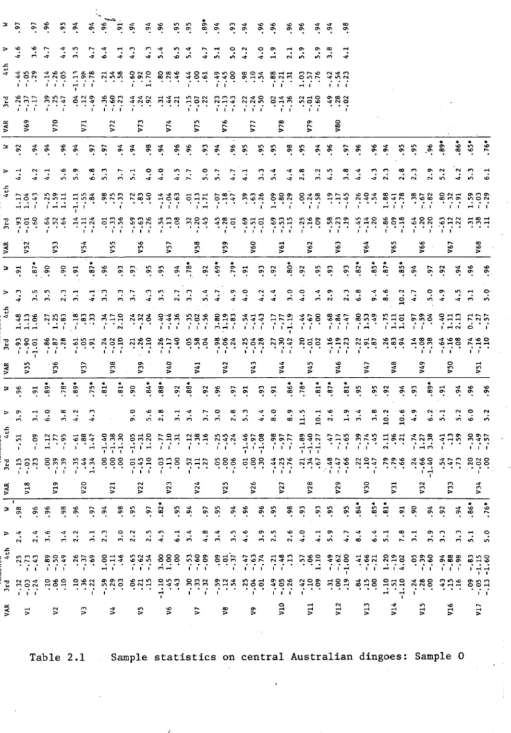
The precise forms these morphological elements take in individual dingoes will be picked up in later chapters. Certainly some traits are more diagnostic of dingo than others. One way of showing this is through a simple inspection of percentage occurrences of traits. Because I am not proposing to make multivariate analyses of this discontinuous data set, Table 1.1 has been drawn up using a central Australian sample of dingoes (referred to in the next chapter as Sample 0).
Ultimately I make no strong claims for this data set because of the way it was constructed and the limited purpose for which it was designed. But to leaven the rather plain fare of a single list of figures I have included scores for a sample of Australian working dogs and also for a group of Indian sub-continent wolves.
Table 1.1 Percentage occurrence of cranial non-metrical traits
Dingo Dog Wolf Trait n=60 n=28 n=30
Dingo Dog Wolf Trait n=60 n=28 n=30 N1 N2 N3 N4 N5 N6 N7 N8 N9 N10 Nil N12 N13 N14 N15 N16 90 75 88 71 43 6 98 91 93 40 88 25 85 50 36 55
10 16 28 89 7 72 7 36 25 33 17 10 35 85 10 76 28 63 85 16 60 93 0 53 78 80
0 0
57 30 90 68
N17. 43
N18 48
N19 23
N20 70
N21 20
N22 38
N23 3
N24 2
N25 5
N26 6
N27 76
N28 0
N29 0
N30 5
N31 21
42 53 28 71 35 12 21 3 0 10 0 7 0 0 7 30 30 10 36 20 23 10 0 6 10 3 6 0 3 0
Nevertheless the single most effective dingo diagnostic is undoubtedly N27 (bifurcated premaxilla). Whether this is ultimately more informative than other more variable traits could well form the substance of a future project. Indeed I am inclined to believe that until the unevenness of this data is improved, interpretative comment must be minimised. The main problem is, of course, that it has agglomerated what seem to be
truly dichotomous epigenetic traits (e.g. N14 to N31) with either
partially developmental (e.g. Nil to N13) or merely subjectively dicho tomised morphological features with underlying continuous variation (e.g. N1 to N9).
PHOTO RECORD
A considerable amount of time and energy has been spent by me in developing a satisfactory photo record of all important cranial material handled. The terms of reference I set for this record were that:
1. it should be standardised to a degree previously available only in anthropometric studies;
2. that the equipment should balance the needs of accuracy against a reasonably high throughput;
that the camera, frame and lighting be portable, rapidly assemblable, and weigh less than 5 kg.
There are obvious rationales for standardised procedures in photo graphing skulls. The very minimum is that the scale be transferable from one view of a skull to another. Also desirable is some control over the orthogonality of the three most commonly shown views of canid skulls:
dorsal^ ventral and lateral. With these basic design requirements a prototype photographic frame was made by Mr Jim Neale (ANU), which per formed reasonably well on some early m o d e m dingo collections and a major dingo fossil series (Western Australian Museum). The field experience with the first design suggested however that damaged skulls and/or fragile skulls were very difficult to handle in the suspension system. The basic model was modified to produce a final version, a brief description of which is provided in Appendix 14.
The skull rotation in this revised system is controlled from an initial lateral view with a machined rächet which produces 90° (±3°) turns. I
found in practice that manual fine tuning of the skull orientation is necessary when working at speed. Mandibles are held in a separate and not entirely satisfactory suspension arm. The orientations here are lateral and dorsal but there is no mechanical control over these. In the lateral view the mandibles are generally arranged so that the 'whole skull’ median plane of the mandible (not the mandible body) is parallel to the shutter plane of the camera.
The total number of frames taken by me using this system has been about 4000. The investment of time in setting up the equipment and mounting skulls has been about one-third of the time taken in examining any specimen. Whether the return has justified the effort is hard to judge, but as Dr T. Brown (1975) has shown, photogrammetric-based data can be very productive. This thesis project has not been able to develop that aspect of morphometries although using photographic prints to give areas and cranial angles has been tried for a number of analyses.
Plates 1.1, 1.2 and 1.3 are three modern dingoes with ages less than 10 months, between 10 months and 18 months, and greater than 18 months. The scale is approximately 2/3 life,
MODERN DINGO MATERIAL
The modern specimens used in this thesis have been drawn from
Overseas collections have yielded variable amounts of material, with the main holdings that I have looked at -hglng: British Museum (Natural History); American Museum of Natural History; Museum für Naturkunde, Berlin, DDR. This material does not amount to more than about 30 speci mens, often with uncertain provenance and unstated sex. Some of the limited number of specimens I have been able to use I have included for their historical value because of their use by the early taxonomists and osteologists.
When plotted onto a map of Australia (see Fig.1.4) the available material presents a mosaic distribution with occasional heavy representa tions amongst a spread of isolated specimens. The main series, provided by Dr A. Newsome (Division of Wildlife, CSIRO), was collected in the Alice Springs district, Northern Territory. Other reasonable groups come
from Edjndina and L o m a Glen (the Macintosh collection). Because the series became available sequentially over about three years (1976-78) their incorporation into the analysis in the thesis has also been sequential. Initially I used the CSIRO series to set up a basic dingo morphology. The subsequent additions allowed a simple-minded extension of interest into regional variability. Because the material came
sporadically, its distributional properties have been unplanned and in this sense the data have dictated the sorts of regional variability that could be examined.
The basic division I have made in the total Australian sample is between the dingoes from arid and non-arid environments. The non-arid dingoes (hereafter called Sample NA) are drawn widely from eastern Australia.
Perhaps more significant is a group of sexed individuals provided by CSIRO (Division of Wildlife) from the Barkley Tablelands which is an area with highly seasonal rainfall, falling between the
criteria for division by rainfall regions; but my inclination is to include this group with non-arid dingoes. It has not been possible to isolate the montane dingo (I have only two examples from Dargo and Mt Hotham), nor to treat separately the interesting and freely available south-central Queensland dingoes.
has the mnemonic AC (arid central). The remainder of the arid sample
I label AP (arid peripheral).
The structuring of the arid country samples into geographically
defined groups rather than biogeographic or climatic regions, is a pragmatic
response to the absence of previous regional studies on dingoes. At this
early stage in the ecological studies of dingoes, none of the bio-physical
parameters that might affect dingo morphology has been'elucidated. There
fore to superimpose regional sampling on the basis of parameters that have
as yet no demonstrable effect would be merely to introduce an arbitrary and
possibly misleading structure to the problem. In the exploratory work that
this thesis represents, the sampling division within the arid zone populations
is a stratified sample of the arid zone populations. The problem to which
this sampling procedure is directed, namely the investigation of sexual
dimorphism in regionally disparate populations, will be spelt out in more
A dingo's sex is one of the major determinants of its skeletal size and shape. Another is where it chooses to live, and by extension, what it finds to eat. Until recently however, very little has been done to investigate systematically these commonplace observations. When dingoes found their way into European museums and private collections early in the nineteenth century they entered that cephalocentric environment dis embodied, and therefore without accompanying postcranial sexing evidence
(e.g. os penis). The early taxonomists developed the field of comparative morphometries, but sexual dimorphism in canids was not an area of prime
concern. While dingo appears to have been selected as a suitable case for study very early (Mivart 1890), a search of the European literature up to 1970 shows that dingo has almost always been presented as a sexually undifferentiated population (Mivart 1890; Studer 1906; Duerst 1908; Noack 1907, 1915; Tichota 1937; Dahr 1937, 1941-2; Degerb^l 1961; Schultz 1968). Stockhaus (1965) used a minimal sexed dingo sample (69, Id), along with a much larger sample of both wolves and European-breed dogs, in a morphological study of variation within the species C. lupus.1 While it has been obvious to Australian observers that there is considerable sexual dimorphism in dingo, very little has been done to quantify it in morphological studies in this country.
Any morphometric data base is improved by information concerning the sexual distribution of the sample. Where there is evidence of morphological heterogeneity in the population due to sexual dimorphism, the rationale for developing a sexing procedure is very strong. Certainly, human studies have a lively record in this regard; canid studies are distinguished by
the absence of any such interest. In fact except for a reference to sexing domestic dogs (The and Trouth 1976), the post-war literature is silent on the matter. For this reason I have used physical anthropo logical approaches to skeletal sexing as possible analytic models for canid sexing.
There has been a traditional distinction between metrical and non-metrical methods of sex determination. The latter methods can be either by direct subjective assessments of sex on defined morphological traits
(e.g. Hrdlicka 1928) or by scoring morphological features on categorical scales of measurements which are then usually analysed using distribution-free non-parametric statistics (e.g. Larnach and Freedman 1964). Both these approaches are empirically based; they depend on extensive
familiarity with large series from which sexually dimorphic features are selected, rejected, combined, and on what is known as 'the art',
manipulated until a sexing criterion is produced. The key is not simply the discovery of 'hallmarks’ (traits) which partition a given sample by sex; the method depends considerably on the researcher's professional authority which is taken to define the confidence limits of sex assess ments by that person. Hence 'ninety percenters', like Hrdlicka (Stewart
1954) .
While there is certainly no lack of authority amongst the generations of canid anatomists, none appear to have directed it to this particular question of sex determination. There are consequently no recognised guides to sexually dimorphic traits in canids. A single feature, the tuberculum pharingicum, is reported to be dimorphic in West Indian dogs
(The and Trouth 1975); it is unfortunately not so in dingo.
I have recognised during the course of the research project, that sex assessment of new material is affected by the length of time away from handling the known-sex series. In other words, one 'forgets' the allowable variation within the sexed dingo samples. Given what was said earlier about confidence limits, I have no doubt therefore that something more than subjective sexing is necessary, so I will pick up the historical
thread again and look at alternative approaches worked out in the field of physical anthropology.
In discussions about the relative merits of 'guess' and 'measure', the question of replicability and transferability of subjective assessment was raised. This problem led to another, not strictly relevant: the
distinction between objective and subjective procedures (e.g. Hanna and Washburn 1953). The supposed identity between metrical methods and objectivity is, of course, misleading. Sex assessment based on measure ment alone is.merely replicable. It is no less dependent on judgment about what measures are appropriate or biologically meaningful. Indeed it is probably the case that the most successful metrical sexing methods are no more than the most successful mathematical models of existing subjective assessment procedures. However, replicability is a worthy goal; it became one in human studies in the 1950s (cf. Hanna et a l . 1953).
The first attempts to use measurement to model morphology depended on univariate and bivariate descriptions, usually of cranial and skeletal indices. This approach returned results with accuracy comparable to subjective techniques. But also, at this period of intense development of multivariate statistical techniques for data analysis, it became obvious that those with the fortitude to use multivariate analyses got results as good or better.
The use of multivariate criteria is a fundamental break from the anatomically oriented statistical models used previously. The difficulty that osteologists had in making that break is illustrated nicely in an important early study of sexual dimorphism in American negroes (Thieme and Schull 1957). The authors' discussion is still a set piece of honest concern about the underlying assumptions of multivariate models.
Thieme and Schull's method was to partition a population of 200 known-sex individuals on the ischium-pubis index and then to analyse the 40 individuals misidentified in this partition. They chose to use
ischium-pubis index is a simple mathematical description of pelvic proportions, the discriminant function on eight variables is an anatomically opaque model of the combined operation of eight sexually dimorphic features.
To accept an eight-feature composite one has to leave behind anatomical clarity for the overriding purpose of achieving a result. As they stress,
'Our -problem is to sex the individual and not the measurement' (p.250). Their results were good: 98% efficiency on their sample. In the discussion they question the validity of applying functions derived for one population to discrimination of individuals in different populations. They conclude that their statistical model is legitimate only because
'...the expression of sexual dimorphism is relatively similar for all varieties of man' (p.268). Whether this is true in the multivariate measurement space they used is of course an open question. Perhaps the desire to answer that question was the driving force behind the rash of similar analyses of different racial groups that appeared in the years around Thieme and Schull (e.g. Mukherjee et al. 1955; Pons 1955; Hanihara
1959) .
Certainly these early studies have highlighted the point that
discriminant analysis is a powerful method for allocating individuals to a priori groups. Where the differences between the groups are largely to do with mean vector differences, and where between-group covariance is similar to within-group covariance the procedure is highly successful. The transference, however, of discriminant functions constructed for one population to the discrimination of, say, sex in populations with
radically different mean vectors and covariance structures is not necessarily so successful.
This has also been my experience in an investigatory discriminant analysis of regionally diverse dingo populations, in which a function for cranial variables generated for a sample of central Australian dingoes has unsatisfactorily sexed dingo samples from non-arid areas.
Various techniques have been used to reduce the critical dependence of discriminant functions on the gross size characteristics of the groups under analysis. Transformation of data sets into ratios has been one response (see Sokal (1965) for discussion).