Laboratory for Papillomavirus Biology, Institute of Molecular and Cell Biology, National University of Singapore,
Singapore 1192601; Tulane Regional Primate Research Center, Covington, Louisiana 704332; Department of
Comparative Medicine, University of Washington, Seattle, Washington 981953; and Institute of
Human Genetics, University of Minnesota Medical School, Minneapolis, Minnesota 554554
Received 18 February 1997/Accepted 11 April 1997
We are studying the diversity of and relationships among papillomaviruses (PVs) to understand the modes and timescales of PV evolution and in the hope of finding animal PVs that may serve as model systems for disease caused by human PVs (HPVs). Toward this goal, we have examined 326 genital samples from rhesus monkeys and long-tailed macaques with a PCR protocol optimized for detecting genital HPV types. In 28 of the rhesus monkey samples, we found amplicons derived from 12 different and novel PV genomes, RhPV-a to RhPV-m, with the likely taxonomic status of “type.” The frequency with which novel RhPVs were detected suggests that rhesus monkeys may play host to PVs with a diversity similar to that of humans. In phylogenetic trees, all 12 of the different RhPVs and the previously described type RhPV-1 were members of the genital HPV supergroup and formed three minor branches distinct from the 11 branches formed by genital HPVs. We also identified a novel PV amplicon, MfPV-a, from a long-tailed macaque, a species belonging to the same genus as rhesus monkeys. MfPV-a turned out to be a close relative of five RhPVs. It appears that the evolution of primate lineages leading to the genus Macaca and to humans created transmission barriers for PVs, resulting in viral evolution closely linked to the host. Additional support for the linked-evolution hypothesis comes from considering the phylogenetic association of two other ape and monkey PVs with the genital HPVs, the supergroup formed by at least seven ungulate PVs, and the isolated phylogenetic position of the only known bird PV.
Papillomaviruses (PVs) are the most thoroughly sampled family of DNA viruses and therefore are an important model system for the study of virus evolution. They have genomes with sizes of 7.3 to 8 kb, which encode eight genes. Comparison of different PVs has shown that these genes are homologous, although there are functional and sequence differences. Align-ment and functional studies of these homologies have become the foundation of PV nomenclature, taxonomy, and phylogeny (5, 6, 8, 25, 39).
PVs are described as “types.” A human PV (HPV) type is defined as a genome whose L1 gene nucleotide sequence dif-fers from the homologous nucleotide sequence of every other HPV type by at least 10% (39). Most pairwise distances be-tween HPV types exceed this percentage by a wide margin. Similar distance magnitudes are found among all known ani-mal PV types, which suggests that the definition could apply to typing them as well. For HPVs, we have determined that the MY09 to MY11 segment is more conserved and that distances of 9.2% in this region correspond to the 10% criterion (6). Although the term “type” was established by definition, PV types turned out to be natural taxonomic units in the sense that no PV genomes obviously intermediate between two existing PV types have been found (5, 6). Independent isolates of the same PV types are referred to as “variants” or “subtypes,” and their nucleotide sequences are found to differ from one an-other by up to 5%, depending on the genomic region under investigation (5, 12, 13, 17, 27, 37, 41). More than 70 different
types of PVs have been described for humans (8, 19, 25), while only 20 types have been confirmed for animals. With the ex-ception of certain bovine PVs (BPVs), each PV type has been found only in a single host species. In some host species, multiple PV types have been found. The available data suggest some correlation between each PV type, pathology, and the preferred target tissue (skin, mucosal stratified epithelia, cer-tain simple epithelia, and mesenchymal cells of the dermis). New PV types can be recognized and studied after the cloning of partial genomic sequences; however, there is agreement that for the formal taxonomic establishment of a new PV type, the full-size genome should be cloned. Partial genomes are desig-nated by a code peculiar to each isolating laboratory.
PVs possibly evolve up to 6 orders of magnitude more slowly than RNA viruses because their genomes are replicated by the host DNA polymerase with its proofreading ability (38). Hy-potheses about the speed of HPV evolution have been pro-posed by linking phylogenetically informative nucleotide dif-ferences with the geographic distribution of these isolates, which reflects some features of the evolution and spread of their human host (13, 27). Many questions about the mode of PV evolution are unclear, in particular (i) whether some mo-lecular properties link each PV type to particular hosts and tissues, (ii) how multiple PV types that have similar target tissues and produce similar pathologies evolved in the same host, and (iii) whether the evolution of PV types is due to host speciation and whether interhost transmissions occur. This sur-vey of PVs in monkeys addresses the third question and also the question of whether the large number of HPVs in compar-ison to the small number of animal PVs detected so far reflects a bias in research efforts or a true difference in viral diversity. * Corresponding author. Mailing address: Institute of Molecular
and Cell Biology, National University of Singapore, Singapore 119260, Singapore. Fax: 65-779-1117. E-mail: [email protected].
4938
on November 9, 2019 by guest
http://jvi.asm.org/
MATERIALS AND METHODS
Sources and preparation of veterinary material.Cellular DNA was extracted from genital tissue samples from monkeys kept in three primate holding units. In total, we studied 326 monkey samples. Of these, 319 samples were from 286 rhesus monkeys (Macaca mulatta) and 7 samples were from long-tailed ma-caques (Macaca fascicularis). The samples included 7 fixed biopsies, 13 fresh biopsies, 7 fresh cervical smears, and 1 cervical polyp from rhesus monkeys of the Harlow Primate Laboratory, Wisconsin Regional Primate Center. These DNA extracts had been previously examined for rhesus monkey PV-1 (RhPV-1) DNA, and three were found not only to contain RhPV-1 DNA, but were derived from animals that exhibited pathological evidence of PV infections (28–30). Our study included an additional 291 rhesus monkey fresh cervical smears from the Tulane Regional Primate Research Center (TRPRC). All animals at TRPRC were housed in facilities following guidelines by the Association for Assessment and Accreditation of Laboratory Animal Care International and were cared for according to standard procedures (15). Forty-six of these samples had been previously examined for RhPV-1 DNA (4 positive), serological response to RhPV-1 proteins (25 positive), and pathological atypia (6 positive) (28–30). We also examined seven vaginal samples from long-tailed macaques that exhibited abnormal genital pathology. These animals were kept at the Washington Re-gional Primate Center. Gross lesions have included increased incidence of post-coital bleeding, vaginal leukoplakia, and vaginal polypoid masses that physically obstructed breeding. Histologic lesions included vaginal epithelial hyperplasia, mild-to-moderate lymphocytic vaginitis, and one case of severe vaginal epithelial dysplasia.
Formalin-fixed samples were washed with ethanol and dried prior to DNA extraction. Tissue samples were treated with 1% sodium dodecyl sulfate–100 mg of proteinase K per ml at 56°C and extracted with phenol-chloroform (1:1) and chloroform and then were precipitated with ammonium acetate and isopropanol. Following treatment with 100 mg of RNase per ml at 37°C for 30 min, the extractions and the precipitations were repeated.
Sources of cloned PV DNA.Plasmid clones of chaffinch PV (FPV) and rein-deer PV (RPV) (22, 23) were made available by J. Moreno-Lopez and U. Pettersson, University of Uppsala, Uppsala, Sweden, and have been deposited with E. M. deVilliers in the Papillomavirus Reference Collection, Heidelberg, Germany.
PCR and sequencing.To identify and clone part of the L1 gene of genital PVs, we used the MY09-MY11 primer pair (21). PCR amplicons with a size of approximately 450 bp were gel purified and directly sequenced with the PCR primers in order to screen out false positives. Amplicons whose preliminary nucleotide sequence suggested that they were derived from PV genomes were blunt-end ligated into PCR-Script (Stratagene, San Diego, Calif.) (RhPV-c, -d, -f, -g, -h, -i, -k, and -m) or, after amplification with MY09-MY11 primers con-taining BamHI and HindIII sites, were cleaved with BamHI and HindIII and cloned into pUC19 (the other five amplicons). The novelty of each sequence was determined by searching against the databases of all known PVs (7, 25). The sequences of the 13 monkey PV MY09-MY11 clones have been deposited in GenBank under the entry numbers U89656 to U89668, and the clones have been deposited with E. M. deVilliers in the Papillomavirus Reference Collection.
The original publications of the cloning of FPV and RPV (22, 23) included genomic sequences of the L1 region. We extended these sequences by primer walking until we had completely determined both strands of the L1 gene homol-ogous to the MY09-MY11 segment. As a result, we found that the original GenBank entry for FPV (accession no. K02020) had a sequencing error—an 18-bp in-frame deletion (59-CTTTCCCGGGCGCAGGGC-30) between posi-tions 129 and 130. These sequences have GenBank entry numbers U89669 and U89670.
Evaluation of nucleotide sequences.Phylogenetic analyses were based on multiple alignments (PILEUP in the Wisconsin Package, version 8.1-UNIX) (10) of the phylogenetically informative 291-bp L1 segments against all other avail-able PV sequences done with the Phylogeny Inference Package, version 3.5 (PHYLIP) (9) with modifications as published previously (7).
RESULTS
Detection of 12 novel PV genomes in cervical smears of rhesus monkeys. Numerous segments of PV genomes can be well aligned for the purpose of phylogenetic comparisons (25). The most comprehensive database exists for a 291-bp segment of the L1 gene, which has the advantage of being included in a PCR amplicon that is amplifiable by the MY09-MY11 consen-sus primers (6, 7). This database included 94 PV types prior to the beginning of this study and was expanded by our current research to 109 PV genomes.
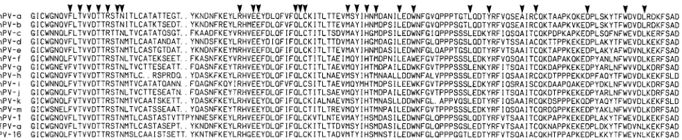
The HPV types that infect both the female and male genital tracts are frequently referred to as genital HPVs. Previous research has shown that all genital PVs are phylogenetically related. We have placed these viruses into a taxonomic super-group, which, however, includes a few HPV types without a predilection for genital target sites (6, 25). Fifty-one different HPV types belong to this supergroup, 40 of which are primarily found at genital sites. It is this supergroup of genital HPVs which are preferentially detected by the MY09-MY11 primers. To examine whether another primate species could also be infected with a similar diversity of genital PVs, we extracted DNA from 319 genital samples from 286 female rhesus mon-keys and performed PCR amplification with MY09-MY11 primers. Agarose gel electrophoresis of the products led to the identification and isolation of 52 amplicons with the expected size of approximately 450 bp. Direct partial sequencing con-firmed that 28 of these bands were derived from PV genomes, as judged by the presence of conserved amino acid residues (Fig. 1) (3). The other 24 bands were nonspecific amplicons. It was apparent after a database search that we had amplified 12 different novel PV genomes, the other 16 sequences being identical or very similar to any of these 12 amplicons. These 12 amplicons were cloned into plasmid vectors, and both strands were completely sequenced. Figure 1 shows the amino acid sequence between the primer sites of these amplicons aligned to HPV-16 and RhPV-1. Highly conserved amino acid residues are indicated which confirm that these amplicons have been derived from PV genomes. We used the abbreviations “RhPV-a” to “RhPV-m” to name these PV genomes; the letter “l” was excluded from this provisional nomenclature in order to avoid confusion between it and the number 1.
[image:2.612.64.555.69.170.2]Molecular distances among RhPV-1 and the novel RhPV clones suggest the type status of each of the 12 amplicons.To examine whether the clones RhPV-a to RhPV-m were derived from RhPV genomes that would qualify as separate RhPV types, we examined the simple distances between these clones and the distances to HPV-16 and RhPV-1, the only PV type previously detected in rhesus monkeys (18). The 12 novel RhPV clones differed from one another by distances ranging from 15.3 to 53.7% at the nucleotide level. Each of these 12
FIG. 1. Amino acid sequences of the MY09-MY11 segments of HPV-16, RhPV-1, MfPV-a, and 12 RhPV clones derived from likely novel RhPV types. The arrowheads indicate highly conserved amino acid residues that are diagnostic of PV genomes. Residues corresponding to the primer sequences are not listed.
on November 9, 2019 by guest
http://jvi.asm.org/
three RhPVs were each found in two animals, two RhPVs were each found in three animals, one RhPV was found in four animals, and one RhPV was found in six animals. An effort was made to retrace all contacts of female rhesus monkeys that carried the same RhPV genome, with the result that most likely none of these animals ever had direct or indirect (through common males) contact with another carrier of the same virus. Thus, it appears that these PV genomes entered the colony through independent animals at the time of capture. RhPV-1 was not detected in this sampling by the MY09-MY11 primers, although several animals were previously found positive by the specific RhPV-1 E6 and E7 primers (28, 30). RhPV-1 is a highly infectious virus, because it was found in the majority of all animals of an animal holding unit with sexual contact with one another. It may be rare, though, be-cause it had not been found in the same study in animals without contact with this particular group (18, 28). Even de-spite the obvious limits of our study, we have evidence that in rhesus monkeys, one can detect a diversity of genital PVs similar to what one would find in a similar-sized cohort of asymptomatic human patients (20, 40). None of these RhPVs reaches a frequency comparable to that of HPV-16 in humans. This may be due to the decreased sensitivity of the consensus priming approach. Further studies with primers more specific for each of the three clades and analysis of tumor samples may reveal a different picture.
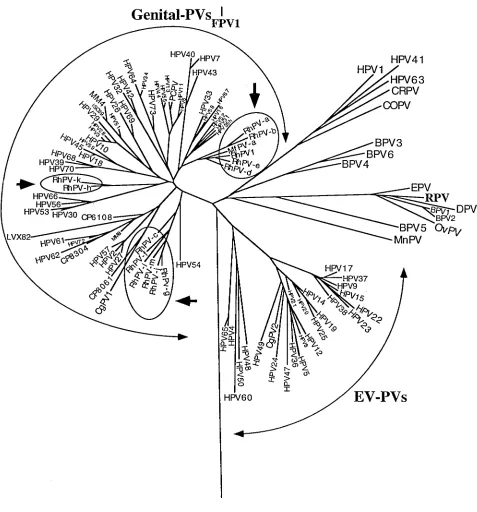
The phylogenetic relationship between rhesus monkey PVs and HPVs. To examine the evolutionary relationship among the RhPV clones and other PVs, we constructed phylogenies by several approaches. Figure 2 shows a neighbor-joining phy-logeny of 109 animal PVs and HPVs, including RhPV-1 and the 12 new RhPV clones. In all trees, all 13 RhPVs are part of supergroup A, the genital PVs. It is significant that they are not associated with any of the 11 minor branches formed by HPVs, but cluster in three new and separate minor clades. We note that the only HPV type remotely related to either of these branches is HPV-54, an HPV type that our previous analysis did not find to be included in any of the other 11 genital HPV branches (6, 25).
The phylogenetic positions of a PV from an additional mon-key species, ungulate PVs, and a bird PV.In the course of this study, we also examined a small collection of genital samples from a different monkey species, the long-tailed macaque, which belongs to the same genus as rhesus monkeys. One of seven samples from this species gave a positive PCR signal with the MY09-MY11 system, and the cloned amplicon was shown to be derived from a novel PV type (Fig. 1). We named this clone MfPV-a. Interestingly, our analysis included this se-quence in one of the three clades formed by RhPVs.
In addition to the 16 published animal PV genomic se-quences, two previously unavailable animal PV genomes, RPV (22) and FPV (23), were included in this study. We suspected that the phylogenetic analysis of these PVs would shed more light on the questions of the PV-host evolutionary linkage and interspecies transmission. Figure 2 includes these two viruses. As can be seen, RPV is a member of a branch formed by six
that this imbalance is probably due to biased sampling efforts and that it may be possible to identify numerous PV types in other primates or even in many nonprimate mammals. Con-tinued sampling with the aid of PCR may vastly expand the present database and further strengthen the status of this virus group as a paradigm for understanding virus-host evolution. Specifically, we are interested in estimates of the timescale of PV evolution, whether evolutionary diversification of PVs oc-curs as a consequence of host speciation, and whether or not interspecies transmission plays a role in generating PV type diversity.
Timescale of PV evolution.Several lines of evidence suggest that the speed of PV evolution is slow. (i) PVs are DNA viruses and use the same high-fidelity enzyme machinery for replica-tion as their eukaryotic host. (ii) They also replicate slowly, because their multiplication is linked to the division of the infected epithelial host cell. The rate of these divisions is sim-ilar to that of the male germ line, which drives the evolution of chromosomal genes (36), and one can hypothesize that PV genomes and genes of the host’s germ line may undergo a similar number of replications and consequently a similar amount of diversification over large timescales. (iii) Molecular epidemiology of variants of several HPV types suggests that these HPV types already existed at the time humans originated as a species, a few hundred thousand years ago (2, 13, 17, 27). These data permitted estimates that diversification of the most variable parts of HPV genomes occurs maximally at a rate of 0.25% over a period of 10,000 or 20,000 years. (iv) Serial detections of genital HPV types in the same patient and se-quencing of multiple subclones of HPVs in a single patient at a single point in time do not show the quasispecies spectrum of mutants characteristic of rapidly mutating RNA viruses (14).
Interspecies transmission. Presently no PV is known that uses both humans and any animal as a host. Interspecies trans-mission seems to occur so rarely in the case of some DNA viruses that it has been proposed to be restricted by molecular interactions between virus and host regulatory proteins (35). Such an extreme restriction is probably not exclusively opera-tive in all PVs, because some ungulate PVs can amplify in heterologous hosts under natural or experimental conditions (4, 31, 32). However, in spite of these exceptions, there is presently no report of any HPV type having been detected in an animal or of any animal PV type having been detected in a human. Although interspecies transmissibilities have not been systematically studied in experimental systems, the lack of an-ecdotal reports is perhaps telling, given the frequent contact of humans with domestic and wild animals.
An alternative view interprets the relationship between some HPVs and PVs of nonhuman primates as being the result of an active human-monkey interspecies transmission network. With particular reference to the pygmy chimpanzee PV (PCPV), HPV-6, HPV-11, and HPV-13 group, it has been proposed that interspecies transmission between nonhuman and human primates may be likely (24). If this were true, we would have expected to see in our survey of RhPVs many examples of RhPVs that had HPVs as their closest neighbor. Instead, the 13 RhPVs formed three new clades which
on November 9, 2019 by guest
http://jvi.asm.org/
cluded any HPV type. This shows that within another primate species, genital PV type evolution occurs with a pattern (types and comparable distances) similar to that in humans. The nearest neighbor to all RhPVs found was another RhPV and not an HPV. In consideration of the slow speed of PV evolu-tion, one should conclude that if interspecies transmission did occur, it didn’t happen between humans and rhesus monkeys
[image:4.612.66.543.64.576.2]but between ancestral species. The fact that MfPV-a is within an RhPV clade could also be interpreted in the same fashion. Specific instances of interspecies transmission as the impetus for type diversification are always possible; however, the orig-inal conclusion as to the generality of interspecies transmission is premature given the lack of information of PV diversity in pygmy chimpanzees and other apes.
FIG. 2. Neighbor-joining phylogeny tree of 110 HPV and animal PV types, including HPV and RhPV MY09-MY11 clones with a type-like status. The three novel RhPV clades are indicated by arrows. Bootstrap (100 replicates) percentages for the clades are as follows: RhPV-a and RhPV-b, 94%; RhPV-e and RhPV-1, 80%; RhPV-a, RhPV-b, RhPV-d, RhPV-e, RhPV-1, and MfPV-a, 67%; RhPV-h and RhPV-k, 100%; and RhPV-f, RhPV-g, RhPV-i, RhPV-j, and RhPV-m, 82%. RhPV-c is the closest type to the final clade, but the bootstrap percentage is very low (45%). Abbreviations follow those used in references 3 and 6. OvPV, ovine PV; MnPV, Mastomys natalensis PV; COPV, canine oral PV; DPV, deer PV; CRPV, cottontail rabbit PV; CP, MM, and LVX, laboratory abbreviations of untyped PVs (3).
on November 9, 2019 by guest
http://jvi.asm.org/
PVs are widely distributed among mammals, and this may also be true for other vertebrates. Large evolutionary distances generally separate PVs of hosts belonging to different mam-malian orders. There is also a very great distance between mammalian PVs and a PV (FPV) from a different class of host—birds. Given the slow rate of evolution, the insignifi-cance of interspecies transmission, and the host specificity of PVs, this distribution is best explained by a hypothesis of an ancient virus-host association. The evolutionary dynamics of this virus-host relationship are based on the general scenario of many well-adapted parasite types living in a single host species. In such cases, the selection pressure exerted by the host on the parasites is by far the dominant force in the relationship. This has been termed “sequential evolution” (34), but a more de-scriptive term might be “host-linked evolution.” We prefer these terms over the term “coevolution,” because this term is defined to describe mutual selective pressure between host and parasite (11, 16), which likely does not exist in the case of PVs and their hosts.
Speciation events of the hosts are likely to isolate virus populations, because host populations may become separated and because the close contacts required for PV infection may not take place between animals of different species. These bottlenecks of virus spread can be major occasions for virus diversification. The phylogenetic tree confirms the prediction that we should be able to detect some correlation between virus type phylogeny and host phylogeny. The observation that all RhPV genomes detected here form three specific clades excluding all HPV types is best explained by this model.
Diversification of PVs can occur independently from specia-tion events of the host.We can expect that any PV type infect-ing any particular host will undergo slow genomic changes due to genetic drift as well as in response to selection pressure by the host’s environment. Selection pressure, e.g., by immunore-sponses, may be specifically directed against the virus. In ad-dition, there is a continuous selection of mutants with proper-ties beneficial for virus strategies, e.g., mutants with improved amplification, regulation, and infectivity. For example, there is indirect evidence from the common saturation frequency of nonsynonymous mutations in the L1 gene of many PVs that strong selection has been acting at this locus (24).
The initial consequence of this will be the evolution of re-lated variants of HPV types that may or may not show pheno-typic differences. Remotely related variants will eventually evolve into closely related but distinct PV types that occupy similar niches. Examples are the pairs HPV-2 and HPV-27, HPV-6 and HPV-11, HPV-18 and HPV-45, and BPV-1 and BPV-2. We do not yet know enough about the biology of these PVs to answer the question of whether they really have nearly identical biologies, fill identical niches, and were driven apart only by genetic drift, or whether selection was also a major factor.
It is obvious that the timing of these branching events is independent of speciation at the level of the host. Our identi-fication of two colobus monkey PVs, CgPV-1 and CgPV-2 (7, 26, 33), as typical representatives of genital as well as epider-modysplasia verruciformis PVs (EV-PVs) showed that
branch-tion. Superimposed on this are mechanisms of drift and selec-tion unrelated to host speciaselec-tion resulting in the intrahost species diversity we see today. We crudely estimate that dis-tances exceeding the 10% type criterion have evolved over periods longer than 200,000 to one million years (2). Because the hominoid and monkey lineages separated about 22 million years ago (1), the genomic organization typical of genital PVs and some branching events leading to different HPV lineages must have existed before that time, while the split between groups of genital HPVs and groups of RhPVs must have oc-curred subsequently.
Are monkey PVs useful as animal models for medical re-search?It is not known whether or not the RhPV genomes detected in this study may induce lesions and which type of lesions they may be. Genital HPVs are associated with a wide variety of lesions, including cervical cancer, genital warts, and common cutaneous warts, but within this diverse supergroup, related viruses that form common branches are often associ-ated with lesions of similar pathology. Examples are relatives of HPV-16, like HPV-31, HPV-33, and HPV-35, which are found in cervical carcinomas. Because RhPV-1 has been iso-lated from a metastasis of a penile squamous cell carcinoma (18) and because this virus is related to four other RhPV types, we may have identified a group of viruses that may serve as a model system for malignant lesions and that may be useful in immunological and drug research.
ACKNOWLEDGMENTS
This research was supported in part by NIH grant RO1CA25462 (R.S.O. and A.J.F.), NIH grant P51RR/AG00165-35, FDA contract 223-94-1100 (M.R.), and NIH grant NCRR RR00166 (T.B.).
REFERENCES
1. Andrews, P. 1992. Evolution and environment in the Hominoidea. Nature
360:641–646.
2. Bernard, H. U., S. Y. Chan, and H. Delius. 1994. Evolution of papillomavi-ruses. Curr. Top. Microbiol. Immunol. 186:33–54.
3. Bernard, H. U., S. Y. Chan, M. M. Manos, C. K. Ong, L. L. Villa, H. Delius,
H. M. Bauer, C. Peyton, and C. M. Wheeler.1994. Assessment of known and novel human papillomaviruses by polymerase chain reaction, restriction di-gest, nucleotide sequence, and phylogenetic algorithms. J. Infect. Dis. 170: 1077–1085.
4. Bloch, N., M. Breen, and P. B. Spradbrow. 1994. Genomic sequences of bovine papillomavirus in formalin-fixed sarcoids from Australian horses re-vealed by polymerase chain reaction. Vet. Microbiol. 41:163–172. 5. Chan, S.-Y., H.-U. Bernard, C.-K. Ong, S.-P. Chan, B. Hofmann, and H.
Delius.1992. Phylogenetic analysis of 48 papillomavirus types and 28 sub-types and variants: a showcase for the molecular evolution of DNA viruses. J. Virol. 66:5714–5725.
6. Chan, S.-Y., H. Delius, A. L. Halpern, and H.-U. Bernard. 1995. Analysis of genomic sequences of 95 papillomavirus types: uniting typing, phylogeny, and taxonomy. J. Virol. 69:3074–3083.
7. Chan, S. Y., R. S. Ostrow, A. J. Faras, and H. U. Bernard. 1997. Genital papillomaviruses (PVs) and epidermodysplasia verruciformis PVs occur in the same monkey species: implications for PV evolution. Virology 228:213– 217.
8. deVilliers, E. M. 1994. Human pathogenic papillomavirus types: an update. Curr. Top. Microbiol. Immunol. 186:1–12.
9. Felsenstein, J. 1982. Numerical methods for inferring evolutionary trees. Q. Rev. Biol. 57:379–404.
10. Genetics Computer Group. 1994. Program manual for the Wisconsin pack-age, version 8, September 1994. Genetics Computer Group, University of Wisconsin, Madison.
on November 9, 2019 by guest
http://jvi.asm.org/
11. Hafner, M. S., and S. A. Nadler. 1988. Phylogenetic trees support the co-evolution of parasites and hosts. Nature (London) 332:258–259. 12. Heinzel, P. A., S.-Y. Chan, L. Ho, M. O’Connor, P. Balaram, M. S. Campo,
K. Fujinaga, N. Kiviat, J. Kuypers, H. Pfister, B. M. Steinberg, S.-K. Tay, L. L. Villa, and H.-U. Bernard.1995. Variation of human papillomavirus type 6 (HPV-6) and HPV-11 genomes sampled throughout the world. J. Clin. Microbiol. 33:1746–1754.
13. Ho, L., S.-Y. Chan, R. D. Burk, B. C. Das, K. Fujinaga, J. P. Icenogle, T.
Kahn, N. Kiviat, W. Lancaster, P. Mavromara-Nazos, V. Labropoulou, S. Mitrani-Rosenbaum, B. Norrild, M. R. Pillai, J. Stoerker, K. Syrjaenen, S. Syrjaenen, S.-K. Tay, L. L. Villa, C. M. Wheeler, A.-L. Williamson, and H.-U. Bernard.1993. The genetic drift of human papillomavirus type 16 is a means of reconstructing prehistoric viral spread and the movement of ancient human populations. J. Virol. 67:6413–6423.
14. Ho, L., S.-Y. Chan, V. Chow, T. Chong, S.-K. Tay, L. L. Villa, and H.-U.
Bernard.1991. Sequence variants of human papillomavirus type 16 in clinical samples permit verification and extension of epidemiological studies and construction of a phylogenetic tree. J. Clin. Microbiol. 29:1765–1772. 15. Institute of Laboratory Animal Resources, Commission on Life Sciences,
National Research Council.1996. Guide for the care and use of laboratory animals. National Academy Press, Washington, D.C.
16. Janzen, D. H. 1980. When is it coevolution? Evolution 34:611–612. 17. Kawase, M., G. Orth, S. Jablonska, C. Blanchet-Bardon, L A. Rueda, and M.
Favre.1996. Variability and phylogeny of the L1 capsid protein gene of human papillomavirus type 5: contribution of clusters of nonsynonymous mutations and of a 30-nucleotide duplication. Virology 221:189–198. 18. Kloster, B. E., D. A. Manias, R. S. Ostrow, M. K. Shaver, S. W. McPherson,
S. R. S. Rangen, H. Uno, and A. J. Faras.1988. Molecular cloning and characterization of the DNA of two papillomaviruses from monkeys. Virol-ogy 166:30–40.
19. Longuet, M., P. Cassonet, and G. Orth. 1996. A novel genital human pap-illomavirus (HPV), HPV type 74, found in immunosuppressed patients. J. Clin. Microbiol. 34:1859–1862.
20. Lorincz, A., R. Reid, A. B. Jenson, M. D. Greenberg, W. Lancaster, and R. J.
Kurman.1992. Human papillomavirus infection of the cervix: relative risk association of 15 common anogenital types. Obstet. Gynecol. 79:328–337. 21. Manos, M. M., Y. Ting, D. K. Wright, A. J. Lewis, T. R. Broker, and S. M.
Wolinsky.1989. The use of polymerase chain reaction amplification for the detection of genital human papillomaviruses. Cancer Cells 7:209–214. 22. Moreno-Lopez, J., H. Ahola, A. Eriksson, P. Bergman, and U. Pettersson.
1987. Reindeer papillomavirus transforming properties correlate with a highly conserved E5 region. J. Virol. 61:3394–3400.
23. Moreno-Lopez, J., H. Ahola, A. Stenlund, A. Osterhaus, and U. Pettersson. 1984. Genome of an avian papillomavirus. J. Virol. 51:872–875.
24. Myers, G., H. U. Bernard, H. Delius, M. Favre, J. Icenogle, M. van Ranst,
and C. Wheeler (ed.).1994. Human papillomaviruses 1994. A compilation and analysis of nucleic acids and amino acid sequences. Los Alamos National Laboratory, Los Alamos, N.Mex.
25. Myers, G., H. U. Bernard, H. Delius, C. Baker, J. Icenogle, A. Halpern, and
C. Wheeler (ed.).1995. Human papillomaviruses 1995. A compilation and analysis of nucleic acids and amino acid sequences. Los Alamos National Laboratory, Los Alamos, N.Mex.
26. O’Banion, M., J. Sundberg, A. Shima, and M. Reichmann. 1987. Venereal papilloma and papillomavirus in a colobus monkey (Colobus guereza). Inter-virology 28:232–237.
27. Ong, C.-K., S.-Y. Chan, M. S. Campo, K. Fujinaga, P. Mavromara-Nazos, V.
Labropoulou, H. Pfister, S. K. Tay, J. ter Meulen, L. L. Villa, and H.-U. Bernard.Evolution of human papillomavirus type 18: an ancient phyloge-netic root in Africa and intratype diversity reflect coevolution with human ethnic groups. J. Virol. 67:6424–6431.
28. Ostrow, R. S., S. M. Coughlin, R. C. McGlennen, A. N. Johnson, M. S.
Ratterree, J. Scheffler, N. Yaegashi, D. A. Galloway, and A. J. Faras.1995. Serological and molecular evidence of rhesus papillomavirus type 1 infec-tions in tissues from geographically distinct instituinfec-tions. J. Gen. Virol. 76: 293–299.
29. Ostrow, R. S., R. McGlennen, M. Shaver, B. Kloster, D. Houser, and A. J.
Faras.1990. A rhesus monkey model for sexual transmission of a PV isolated from a squamous cell carcinoma. Proc. Natl. Acad. Sci. USA 87:8170–8174. 30. Ostrow, R. S. Unpublished observation.
31. Otten, N., C. von Tscharner, S. Lazary, D. F. Antczak, and H. Gerber. 1993. DNA of bovine papillomavirus type 1 and 2 in equine sarcoids: PCR detec-tion and direct sequencing. Arch. Virol. 132:121–131.
32. Pfister, H., B. Fink, and C. Thomas. 1981. Extrachromosomal bovine pap-illomavirus type 1 DNA in hamster fibromas and fibrosarcomas. Virology
115:414–418.
33. Reszka, A. A., J. P. Sundberg, and M. E. Reichmann. 1991. In vitro trans-formation and molecular characterization of colobus monkey venereal pap-illomavirus DNA. Virology 181:787–792.
34. Ridley, M. 1993. Evolution. Blackwell Scientific, Cambridge, Mass. 35. Shadan, F. F., and L. P. Villareal. 1993. Coevolution of persistently infecting
small DNA viruses and their hosts linked to host-interactive regulatory domains. Proc. Natl. Acad. Sci. USA 90:4117–4121.
36. Shimmin, L. C., B. H. Chang, and W. H. Li. 1993. Male driven evolution of DNA sequences. Nature 362:745–747.
37. Stewart, A.-C., A. M. Eriksson, M. M. Manos, N. Mun˜oz, F. X. Bosch, J.
Peto, and C. M. Wheeler.1996. Intratype variation in 12 human papilloma-virus types: a worldwide perspective. J. Virol. 70:3127–3136.
38. Ustav, M., and A. Stenlund. 1991. Transient replication of BPV-1 requires two viral polypeptides encoded by the E1 and E2 open reading frames. EMBO J. 10:449–457.
39. van Ranst, M., R. Tachezy, and R. D. Burk. 1996. Human papillomaviruses: a neverending story? p. 1–19. In C. Lacey (ed.), Papillomavirus reviews: current research on papillomaviruses. Leeds University Press, Leeds, United Kingdom.
40. Wheeler, C. M. 1996. Human papillomavirus type-specific prevalence, part III, p. 112–124. In G. Myers, A. Halpern, C. Baker, A. McBride, C. Wheeler, and J. Doorbar (ed.), Human papillomaviruses 1996. A compilation and analysis of nucleic acids and amino acid sequences. Los Alamos National Laboratory, Los Alamos, N.Mex.
41. Yamada, T., C. M. Wheeler, A. L. Halpern, A.-C. Stewart, A. Hildesheim,
and S. A. Jenison.1995. Human papillomavirus type 16 variant lineages in United States populations characterized by nucleotide sequence analysis of the E6, L2, and L1 coding segments. J. Virol. 69:7743–7753.