0022-538X/96/$04.0010
Copyrightq1996, American Society for Microbiology
Transgenic Mice Expressing Human Measles Virus (MV) Receptor
CD46 Provide Cells Exhibiting Different Permissivities
to MV Infection
BRANKA HORVAT,1* PIERRE RIVAILLER,1GAYATHRI VARIOR-KRISHNAN,1ALICIA CARDOSO,1
DENIS GERLIER,2ANDCHANTAL RABOURDIN-COMBE1
Laboratoire d’Immunobiologie Mole´culaire, Ecole Normale Supe´rieure de Lyon, UMR 49, CNRS, 69364 Lyon,1
and Immunite´ et Infections Virales, IVMC, CNRS-UCBL UMR 5537, Faculte´ de Me´decine Lyon-Lae¨nnec,
69372 Lyon,2France
Received 19 April 1996/Accepted 11 July 1996
We have generated transgenic mice ubiquitously expressing the human receptor for measles virus (MV), CD46 (membrane cofactor protein). Various cell types were isolated from these transgenic mice and analyzed for their ability to support MV replication in vitro. Although MV could enter into all CD46-expressing cells, differential susceptibilities to MV infection were detected depending on the cell type. Cell cultures obtained from transgenic lungs and kidneys were found to be permissive of MV infection, since RNA specific for MV genes was detected and viral particles were released, although at a low level. Similarly to human lymphocytes, activated T and B lymphocytes isolated from transgenic mice could support MV replication; virus could enter, transcribe viral RNA, and produce new infectious particles. When expressing viral proteins, lymphocytes down-regulated CD46 from the surface. Interestingly, while activated T lymphocytes from nontransgenic mice did not support MV infection, activated nontransgenic murine B lymphocytes replicated MV as well as transgenic B lymphocytes, suggesting the use of an alternative virus receptor for entry. In contrast to the previous cell types, murine peritoneal and bone marrow-derived macrophages, regardless of whether they were activated, could not support MV replication. Furthermore, although MV entered into macrophages and virus-specific RNA transcription occurred, no virus protein or infectious virus particles could be detected. These results show the importance of the particular cell-type-specific host factors for MV replication in murine cells which may be responsible for the differential permissivity of MV infection.
Measles virus (MV), the causative agent of measles in
hu-mans, belongs to theParamyxoviridaefamily and the
Morbilli-virus genus. MV is an enveloped, negative, nonsegmented
strand RNA virus. Two virus-encoded glycoproteins are in-serted into the viral envelope: the hemagglutinin (H) protein, which mediates virus attachment to susceptible cells and hem-agglutination of certain simian erythrocytes, and the fusion (F) protein, which, together with H, is responsible for fusion with the cell membrane and virus entry (48).
MV is responsible for the acute childhood disease which is among the primary causes of infant death in developing coun-tries, and sporadic outbreaks of acute measles still occur in industrialized countries despite vaccination (47). Symptoms induced by MV range from respiratory infection, fever, and rash to less common infections of the nervous system, includ-ing acute encephalitis and subacute sclerosinclud-ing panencephalitis (SSPE). Patients with measles may develop immunosuppres-sion, which increases their susceptibility to secondary infec-tions and is largely responsible for the high incidence of MV-induced mortality (3, 27). During the incubation period, the virus replicates in the respiratory tract and then spreads to local lymphoid tissue. Amplification of the virus in lymph nodes produces a primary viremia that results in the spread of virus to multiple lymphoid tissues and other organs, including the skin, kidney, gastrointestinal tract, and liver, where it rep-licates in epithelial cells, endothelial cells, and monocytes-macrophages (29). Most of the infected cells in peripheral
blood are monocytes (9), although T and B lymphocytes sup-port viral replication after stimulation in vitro (15, 46).
MV infection is initiated by attachment of the virus to the host cell via a specific receptor which is followed by a virus-cell fusion and release of the nucleocapsid into the cytoplasm. Human CD46, which was initially described as the membrane cofactor protein (MCP), acts as a receptor for MV. CD46 is a widely distributed C3b/C4b-binding cell surface glycoprotein that serves as an inhibitor of complement activation on host cells (21). CD46 mediates MV binding and MV-induced cell-cell fusion and is down-regulated in MV-infected cell-cells (7, 33, 35). Transfection of nonpermissive rodent cells with different isoforms of CD46 renders some of these cells susceptible to MV (11, 25). In addition, another molecule, the membrane-organizing external spike protein moesin, has been described to be functionally associated with MV infectivity of the cells (8). This molecule has been found in all tissue culture cells investigated so far and was proposed to form with CD46 the complex functionally involved in the uptake of MV into cells (42).
The mechanisms of MV-induced immunopathology are not well understood, mainly because of the lack of convenient animal models. Humans are the only natural hosts for MV. Some other primates may be experimentally infected, but mice are usually resistant to MV infection, except under special experimental conditions such as intracranial inoculation of newborn mice (20). Identification of CD46 as a cellular recep-tor for MV has opened new perspectives in developing trans-genic animal models to study MV pathogenesis. We have es-tablished transgenic mice ubiquitously expressing the human CD46 molecule. Since these mice were rather resistant to MV
* Corresponding author. Phone: 33 72 72 81 26. Fax: 33 72 72 86 86.
6673
on November 9, 2019 by guest
http://jvi.asm.org/
infection in vivo, we have characterized the ability of cells harvested from different tissues of these mice to support MV infection in vitro. Here, we describe that MV could enter into the cell and proceed with the transcription of MV genes in all analyzed cell types obtained from CD46 transgenic mice. How-ever, complete virus replication occurs only in some CD46-expressing murine cell types but not in others, such as macro-phages, demonstrating the necessity of a particular intra-cellular environment for successful MV replication.
MATERIALS AND METHODS
Production of transgenic mice.Human CD46 cDNA of the C-CYT2 isoform, containing exons 1 to 6, 9 to 12, and 14 (5), was put under the control of the gene promoter of the ubiquitously expressed hydroxymethyl-glutaryl coenzyme A re-ductase (HMGCR) (10). This construct was microinjected into the pronuclei of B6DBA mouse ovocytes, and transgenic mice were generated by a previously described procedure (14). Two founding transgenic mice (MCP-3 and MCP-7) and their initial offspring were identified by dot blot analysis of tail DNA as described elsewhere (19). In both transgenic lines, the number of integrated transgenic copies was estimated to be 30 copies per genome by comparing the intensities of hybridizing DNA fragments of the mouse HMGCR gene contained in the transgene with the intensities of the same fragments present in the germ line endogenous HMGCR gene. The animals were backcrossed for several gen-erations with BALB/c mice until homogeneity for the major histocompatibility complexH-2dbackground and were further crossed among themselves to gen-erate a homozygous line.
Southern and Northern (RNA) blot analysis.Genomic DNA (10mg) prepared from the P815.CD46 cell line or from the tails of transgenic and BALB/c mice was digested withKpnI andSacI. DNA digests were separated by gel electro-phoresis on an 0.8% agarose gel and were analyzed by Southern blot hybridiza-tion according to standard procedures (41).
Total RNA was isolated from cell lines and various animal tissues (thymus, spleen, kidney, lungs, and brain) by using the RNA-now kit (Biogentex). A 20-mg amount of RNA was separated by electrophoresis on a formaldehyde agarose gel and was analyzed by Northern blot hybridization as described elsewhere (41).
Reverse transcription-PCR (RT-PCR) assaying.RNA was prepared from 53 106cells of noninfected or MV-infected primary cell cultures or cell lines as
described above. The cDNA was synthesized from 5mg of total RNA with avian myeloblastosis virus reverse transcriptase (GIBCO-BRL) and oligo(dT)15 primer (Boehringer Mannheim). Samples were amplified by PCR with primers specific for the MV genes NP, F, and H. The quality and quantity of RNA and cDNA were monitored by PCR amplifying the G3PDH gene. The oligonucleo-tide primers were as follows: NP1, 59-ATCCGCAGGACAGTCGAAGGT-39; NP2, 59-AGGGTAGGCGGATGTTGTTCT-39; F1, 59-GGCAATTGAGGCAA TCAGACA-39; F2, 59-CTTGAGAGCCTATGTTGTACG-39; H1, 59-AGTCAG TAATGATCTCAGCAACT-39; H2, 59-ATCCTTCAATGGTGCCCACTC-39; G3PDH-1, 59-CTCAGTGTAGCCCAGGATGC-39; and G3PDH-2, 59-ACCAC CATGGAGAAGGCTGG-39.
Amplification involved 30 cycles of denaturation at 958C for 45 s, annealing at 638C for 1 min, and extension at 728C for 1 min. The PCR products were analyzed by electrophoresis on a 1.5% agarose gel, transferred to the membrane Hybond N1, and hybridized with [g-32P]ATP-labeled internal oligonucleotides
specific for each of the following amplified gene segments: NP, 59-GCCATGG CAGGAATCTCGGAAGAACAAGGCTCAGA-39; F, 59-CCCGGATAACTC ACGTCGACACAGAGTCCT-39; H, 59-TACCTCTCATCTCACAGAGGTGT TATCGCTG-39; and G3PDH, 59-GTGGAAGGACTCA-TGACCACAGTCCA TGCC-39.
Cell culture.All cell lines were grown in Dulbecco’s modified Eagle’s medium (DMEM) supplemented with 6% fetal calf serum (FCS), 10 mMN -2-hydroxy-ethylpiperazine-N9-2-ethanesulfonic acid (HEPES), 2 mM glutamine, 531025
M 2b-mercaptoethanol, and 50mg of gentamicin per ml. The cell lines used in the experiments were human T-cell lymphoma Jurkat cells, Vero fibroblasts from African green monkey kidney, and two murine cell lines transfected with CD46 cDNA (the B-cell lymphoma cell line M12.CD46 [33] and the mastocytoma cell line P815.CD46 [2]).
T lymphocytes were prepared from the spleens of transgenic and control mice and were stimulated in mixed lymphocyte response culture with splenocytes obtained from C57BL/6 mice as described elsewhere (28). Briefly, 3 3106
responder spleen cells per ml were cultured with the same number of mitomycin-treated stimulator cells (C57BL/6) in DMEM (supplemented as described above with 10% FCS and 2% interleukin-2-rich supernatant from the phorbol myristate acetate-stimulated EL4 cell line) for 3 days at 378C and 7% CO2. At the end of
the incubation, the cells were washed, counted, and infected with MV in the same type of medium. Alternatively, T cells were purified by removing the macrophages adherent to gelatin-coated plastic petri dishes for 1 h and by sorting the B cells bound to immunoglobulin (Ig)-coupled beads (Dynabeads). The resulting cell population contained 97% T cells as determined after immunola-beling and cytofluorometry as described below.
B cells were purified from mouse spleen by standard procedures (6). Briefly,
adherent cells were removed by culturing the splenocytes for 1 h on gelatin-coated plastic petri dishes, and T cells were depleted by cytotoxic elimination with 30H12 anti-Thy-1.2 monoclonal antibody (MAb), GK1.5 anti-CD4 MAb, and YTS.169 anti-CD8 MAb in the presence of rabbit complement (Pasteur Merieux). The cells were further centrifuged over a Ficoll gradient (Lym-pholyte-M; Cedarlane). The purified cell population contained 97% B cells as determined by immunolabeling and cytofluorometry as described below. B cells were activated with either 10mg of lipopolysaccharide (LPS) (Difco Laborato-ries) per ml or with 10mg of anti-IgM F(ab9)2(Cappel) in the presence of the 2%
interleukin-2-supplemented DMEM for 48 h at 378C and 7% CO2.
Macrophages were prepared either from the peritoneal cavities or from the bone marrow of transgenic and control mice. Peritoneal macrophages were obtained as described previously (28). Briefly, the mice were injected intraperi-toneally with 2 ml of 3% thioglycolate medium. Four days later, the peritoneal cavities were opened and washed with phosphate-buffered saline, and the peri-toneal cells were harvested. The cells were washed twice and were infected with MV. Bone marrow-derived macrophages were obtained by stimulating bone marrow cells with granulocyte-macrophage colony-stimulatory factor (GM-CSF) as described previously (30). Briefly, femur-derived bone marrow cells were plated at 23105/ml in the DMEM culture supplemented with 10% FCS and
20%L-conditioned medium as a source of GM-CSF. After 6 days of culture, the cells either were infected with MV or were cultured for an additional 48 h in the presence of recombinant gamma interferon (25 U/ml) or LPS (5mg/ml) and infected afterwards.
Primary cell cultures were prepared from the lungs and kidneys of transgenic and BALB/c mice in the following way. Cell tissue was cut into small pieces, washed twice with the culture medium, and digested with 0.1% trypsin for 15 min at 378C under agitation. The cell suspension was filtered through gauze, centri-fuged for 10 min at 1,500 rpm, resuspended in the DMEM culture supplemented with 20% FCS, and transferred into the tissue culture flasks at 378C and under 7% CO2. As soon as enough cells were obtained, they were tested for MV
infection and analyzed by cytofluorometry.
Human peripheral blood mononuclear cells were isolated from the blood of healthy volunteers (CTS, Lyon, France) by Ficoll-Isopaque (Ficoll-Paque; Phar-macia Fine Chemicals, Uppsala, Sweden) gradients as described elsewhere (6). Infected and control cells (106/ml) were stimulated with phytohemagglutinin
(PHA) (10mg/ml; Pharmacia) and were incubated for 1 to 4 days at 378C and with 7% CO2.
Virus infection and titration.The Halle´ strain of MV was used in most of the experiments. Two other strains, LEC-Ki (SSPE strain) (1) and TT (wild-type strain of MV) (44), were sometimes used for comparative studies. Halle´ and LEC-Ki were grown on Vero fibroblasts, while TT was grown on Jurkat cells. Virus was harvested from the culture supernatant of infected cells when a strong cytopathic effect was developed. The suspensions containing MV were clarified by centrifugation and stored at2708C until use. Virus titers were determined by PFU assaying on Vero cell monolayers.
To determine the amounts of virus production in the different cell lines, adherent cells and nonadherent cells were plated in six-well tissue culture plates (Costar) at 106cells per well and in 24-well plates (Costar) at 23106cells per
well, respectively. They were infected with MV at 0.1 or 1 PFU per well. After 3 h of infection, the cells were washed three times with the medium and were further incubated for 1 to 5 days in the DMEM culture supplemented accordingly.
Virus production from infected cells was measured by an infection center assay. This assay, which is based on the coculture of infected cells with highly permissive Vero cells, was chosen because it is the most sensitive assay for the detection of infectious virus, since it is known that virions produced from MV-infected cells are often cell associated and poorly released in the supernatant (26, 49). Briefly, 10-fold dilutions of cells taken 1 to 5 days after MV infection were overlaid on a Vero cell monolayer (53105cells plated in six-well plates). Four
days later, the cell monolayers were fixed in 10% formalin and stained in a methylene blue solution and the lytic plaques were counted. In some of the experiments the presence of cell-free virus particles was measured as well, after freezing and thawing of infected cell cultures. Although 10-fold-lower PFU titers were obtained, the results were in good correlation with those measured by the infectious center assay.
Cytofluorometry analysis.For detection of cell surface CD46, 23105cells
were incubated for 30 min at 48C with biotinylated anti-CD46 MAb MCI20.6 (34), washed and incubated for 30 min at 48C with avidin-phycoerythrin, washed thoroughly, and analyzed afterwards. All incubations were carried out in DMEM containing 6% FCS. H and F MV proteins were detected by incubating the cells with anti-H MAb cl.55 (13) or anti-MV fusion protein antibody (23) and labeling with anti-mouse Ig-fluorescein isothiocyanate (FITC) conjugate (Jackson Labo-ratory). Intracellular antigens were detected after treatment of cells with 0.3% paraformaldehyde at room temperature for 15 min, which was followed by incubation with 0.5% Tween 20 for 15 min at 378C. Permeabilized cells were incubated with either cl.55, cl.27, or anti-MV nucleoprotein antibody cl.105 and were labeled with anti-mouse Ig-FITC conjugate. As a positive control for in-tracellular labeling, the anti-invariant chain MAb IN-1 and then anti-rat Ig-FITC conjugate (Caltag) were used. The purity of the B- and T-cell populations was determined by staining cells with mouse Ig-FITC conjugate or with anti-CD3 MAb (2C11), respectively, and anti-hamster Ig-FITC conjugate (Jackson Immunotech). Purification of macrophages from bone marrow and peritoneal
on November 9, 2019 by guest
http://jvi.asm.org/
cavities was confirmed with anti-MacI MAb (M170). When Fc receptor-express-ing cell populations were analyzed, the cells were preincubated with a rat anti-FcgRII MAb (24G2) for 30 min at 48C. MAbs with irrelevant specificities but with identical isotypes were used as a negative control. Flow cytometry analyses were carried out on a FACScan (Becton Dickinson).
RESULTS
Mice transgenic for human CD46. Transgenic mice were
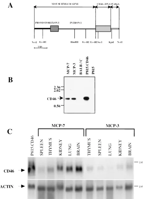
generated by using a construct in which CD46 cDNA of the C-CYT2 isotype (5) was linked to the mouse HMGCR pro-moter region containing 1.35 kb of upstream sequence, the first (noncoding) exon, and the first intron (Fig. 1A) (10). The
transgenic vector was linearized (by cutting at the NotI site
[Fig. 1A]), and the 7.4-kb fragment was microinjected into fertilized mouse eggs. Descendants of two transgenic founders, MCP-3 and MCP-7, were analyzed for the presence of the transgene in the genome by Southern blotting (Fig. 1B). Bands with the expected size were found after restriction map analysis in both transgenic lines, while no band was detected in DNA digests from nontransgenic mice, suggesting that the whole human CD46 cDNA had been integrated into the genomes of
MCP-3 and MCP-7 transgenic mice. Bands with the same size which were obtained by transfection with the same expression vector could be detected in the P815.CD46 cell line. Then, Northern blot analysis was performed to investigate the ex-pression of human CD46 in various tissues of transgenic mice (Fig. 1C). The blots were hybridized with a CD46-specific probe, stripped, and rehybridized with an actin-specific probe to monitor the quantity and quality of RNA. The density of CD46-specific bands was much stronger in MCP-7 mice than that in MCP-3 mice, suggesting a better CD46 expression in this line. This result was further confirmed by cell surface analysis of CD46 protein expression in the spleens and thy-muses of these two transgenic lines (data not shown). The MCP-7 line was obtained in the homozygous stage for the CD46 transgene and used for further analyses.
The expression of CD46 in different cell populations har-vested from the CD46 transgenic line MCP-7 was analyzed by cytofluorometry with the CD46-specific MAb MCI20.6 (Fig. 2). CD46 protein was expressed on activated T and B lympho-cytes, on macrophages, and on cell cultures from the lungs and kidneys of transgenic mice. No CD46 protein was detected on the same cell types harvested from nontransgenic mice, which is consistent with previous reports that murine cells do not express any protein reacting with the antibodies directed against human CD46 (33, 49).
Infection of nonlymphoid CD46 transgenic murine cells.
Cell cultures derived from CD46 transgenic murine lungs and
kidneys were infected with the Halle´ strain, and the production
of MV-specific gene transcription and infectious virus particles was determined. Figure 3A demonstrates the presence of NP-, F-, and H-specific mRNA in the primary cultures obtained from CD46 transgenic murine lungs, which were analyzed 3 days after MV infection, while no message was detected in noninfected cells. The same results were obtained with the cell cultures prepared from CD46 transgenic murine kidneys (data not shown), indicating that virus could enter in these cell types and actively transcribe MV-specific genes.
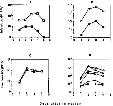
The cells were then analyzed for the production of infectious virus particles. The results shown in Fig. 4A indicate that infected lung cells isolated from transgenic mice produced some virus, with the maximal production at day 4. However, the level of virus production was low (maximally 300 PFU per culture). Equivalent results were obtained when these cells were infected with other MV strains such as Lec-Ki and TT and when kidney cell culture was used (data not shown). When lung and kidney cells from nontransgenic mice were infected, only a limited amount of virus progeny could be detected, and it was at least 10 times lower, confirming the importance of CD46 expression for virus infection.
Infection of CD46 transgenic lymphocytes. Since MV is
known to be lymphotropic in humans, the ability of MV to infect murine lymphocytes in vitro was tested. We were not able to detect any viral production from murine splenocytes unless they were activated (Table 1). Consequently, all of the analyses of sensibility to MV infection were done by using activated lymphocytes. Figure 3A demonstrates that murine CD46 transgenic T and B lymphocytes actively transcribe NP, F, and H genes after MV infection. Transcription was detect-able 24 h after infection, appeared to be more abundant on day 3 of infection, and was not detected unless lymphocytes were infected.
Furthermore, the production of infectious virus was tested. Figure 4C shows the kinetics of MV production by murine splenocytes activated by a mixed lymphocyte reaction (MLR)
for 3 days before being infected with the MV Halle´ strain. MV
production from transgenic splenocytes peaked on day 3 after
FIG. 1. (A) Structure of the transgenic construct used to generate MCP-3 and MCP-7 transgenic lines. Length scale is shown at bottom. SV40, simian virus 40. (B) Southern blot analysis of the transgenic lines. A 10-mg amount of genomic DNA was digested withKpnI andSacI restriction enzymes, was sepa-rated by agarose gel electrophoresis, and was analyzed by Southern blot hybrid-ization with the probe containing the fragment of the CD46 cDNA (prepared by
StuI digestion). (C) Northern blot analysis of the transgenic lines. A 20-mg amount of total RNA was electrophoresed on formaldehyde agarose gels and analyzed by Northern blot hybridization with the probe described above. The blots were stripped and subsequently rehybridized with the actin-specific probe. The positions of the 18S RNA and the expected sizes of CD46 and actin RNA are indicated on the right and left, respectively.
on November 9, 2019 by guest
http://jvi.asm.org/
[image:3.612.64.295.68.388.2]infection and was at least 100 times higher than that in acti-vated nontransgenic splenocytes. Similar results were obtained when MV strains Lec-Ki and TT were used for infection or when cells from another transgenic founder, MCP-3, express-ing a lower level of CD46 were tested (data not shown). Since during an MLR most activated cells are T cells, we verified whether purified activated T cells can sustain MV replication. MLR-activated T lymphocytes were purified to a level of purity of as much as 97% prior to their infection by MV. The level of
MV production in purified T lymphocytes (53103PFU per
culture) (Table 1) was similar to the titer achieved with
MLR-activated splenocytes (6 3103PFU per culture), confirming
the ability of transgenic murine T lymphocytes to support MV infection. In a parallel set of experiments, T lymphocytes were activated in other ways (anti-CD3 antibodies and interleukin-2 or phorbol myristate acetate and ionomycin). However, the efficiency of MV replication was lower than that observed after MLR activation (data not shown), demonstrating the ability of MLR cultures to provide activated murine T lymphocytes highly sensitive to MV infection.
The ability of murine B lymphocytes to produce infectious MV in vitro after purification and activation was then tested. B lymphocytes were purified from the spleens of transgenic and control mice, infected with one of the three strains of MV
(Halle´, Lec-Ki, or TT), washed, and activated either with the
F(ab9)2fragment of anti-Ig (Fig. 4C) or with LPS (Fig. 4D). A
virus production equivalent to or higher than that detected with T cells was observed and reached its maximum on day 2 after infection (Fig. 4C and D; Table 1). However, in contrast to the other cell types analyzed, activated nontransgenic mu-rine B lymphocytes were as potent as transgenic B lymphocytes in sustaining MV replication, indicating that MV can enter into activated murine B cells in the absence of CD46. This was observed regardless of the agent used for B-cell activation
(LPS or anti-Ig) or the virus strain used for infection (Halle´,
[image:4.612.102.513.68.274.2]Lec-Ki, or TT) (Fig. 4C and D). However, the level of MV production by activated B cells differed with the MV strain, since a lower level of MV replication was observed with the wild-type TT MV strain.
FIG. 2. CD46 expression in cell types obtained from homozygote CD46 transgenic mice (——) and control BALB/c mice (. . . .). (A) T lymphocytes; (B) B lymphocytes; (C) peritoneal macrophages; (D) primary cell culture from murine lungs; (E) primary cell culture from murine kidneys; (F) bone marrow-derived macrophages. The cells were labeled with biotinylated anti-CD46 MAb MCI 20.1 and then with phycoerythrin. The negative control, second step avidin-phycoerythrin labeling, was completely superimposable to labeling obtained with BALB/c cells.
FIG. 3. Expression of MV-specific RNA in different types of infected CD46 transgenic murine cells. RNA was prepared from different murine cell types and was analyzed by RT-PCR for the expression of MV-specific transcripts for NP, F, and H and control transcript (G3PDH). B and T lymphocytes were activated by LPS and MLR, respectively, before infection, and all cells were infected with 1.0 PFU of MV (Halle´ strain) per cell. (A) The kinetics of infection was monitored 1, 3, or 5 days after infection, depending on the cell type. NI, noninfected cells. (B) Semiquantitative analysis of F-specific RNA expression in MV-infected transgenic macrophages and activated transgenic B lymphocytes, tested on day 3 after infection. PCR amplifications of F- and G3PDH-specific sequences were performed with serial dilutions of cDNA.
on November 9, 2019 by guest
http://jvi.asm.org/
[image:4.612.344.523.361.635.2]To confirm the ability of transgenic murine lymphocytes to support MV infection in vitro, we analyzed the ability of in-fected cells to produce MV-specific envelope glycoprotein H and consequently down-regulate CD46 from the cell surface (Fig. 5). T and B lymphocytes were isolated from the spleens of transgenic and control mice, stimulated as described above,
and infected with MV (Halle´ strain). Cells were taken 72 h
later and were analyzed by cytofluorometry with biotinylated MAb specific for either MV H protein or CD46. The presence of H protein was detected on activated T- and B-cell-infected CD46 transgenic lymphocytes (Fig. 5A and C), although the latter expressed H much better than the former. When non-transgenic infected lymphocytes were analyzed, H was ex-pressed on B lymphocytes but was not detectable on T
lym-phocytes, in agreement with the observation that
nontransgenic B but not T lymphocytes are permissive for MV. During MV infection, viral H acts as a ligand for CD46 (12, 22). It was reported that during MV infection, concomitantly with the appearance of MV H protein on the cell surface, CD46 was down-regulated by internalization (35). We investi-gated whether CD46 down-regulation could occur in infected murine lymphocytes expressing transgenic CD46 glycoprotein. Transgenic murine lymphocytes were analyzed for CD46 ex-pression 72 h after MV infection and were compared with the same type of noninfected lymphocytes cultured under the same conditions (Fig. 5B and D). Reduction in CD46 expression was easily detectable in both types of murine lymphocytes, confirm-ing H expression on T cells and beconfirm-ing more pronounced in B cells, corresponding to the higher level of H expression in these cell populations. These results demonstrate that in MV-in-fected transgenic murine T and B lymphocytes, the CD46 mol-ecule is susceptible to the same phenomenon of down-regula-tion that has been described for human lymphocytes.
To further evaluate the permissivity of murine lymphocytes for MV in vitro, the rates of MV production in different CD46-expressing murine cell types was compared with those of pro-duction from activated human lymphocytes and two cell lines (murine lymphoma cells transfected with CD46 [M12.CD46] and the human lymphoma cell line Jurkat [Table 1]). Under our experimental conditions, we detected MV production from the Jurkat cell line that was 20 times higher than that from activated human lymphocytes. A similar ratio was observed between the M12.CD46 cell line and transgenic murine T lym-phocytes, although transgenic murine B lymphocytes produced MV at a level close to that of the murine B-cell line. In addi-tion, these results demonstrate that PHA-activated human lymphocytes produce six times more infectious MV particles than activated transgenic murine T lymphocytes and only three times more than activated transgenic murine B lymphocytes, indicating the similarities between human and murine lympho-cytes in the ability to replicate MV.
MV-infected macrophages derived from CD46 transgenic mice transcribe MV genes but do not produce viral proteins or
infectious virus. Since monocytes and macrophages were
shown to be one of the first targets of MV infection in humans (9, 29), we tested the ability of transgenic murine macrophages to support MV infection. Two populations of murine macro-phages were analyzed, peritoneal macromacro-phages and bone mar-row-derived macrophages cultured in the presence of GM-CSF. When obtained from transgenic mice, both macrophage populations expressed CD46 (Fig. 2C and F). We first analyzed the ability of infected macrophages to transcribe MV genes.
Macrophages were infected with MV (Halle´ strain) and
[image:5.612.60.300.70.296.2]washed, and at various time intervals, RNA was extracted and analyzed by RT-PCR with an oligo(dT) primer for RT, as described above. Active primary transcription of MV-specific NP, F, and H genes was detected in CD46-expressing macro-phages only when they were infected with MV and was present in infected bone marrow-derived macrophages (Fig. 3A) as well as in peritoneal macrophages and bone marrow-derived macrophages activated by gamma interferon or LPS before infection (data not shown). In contrast to infected lympho-cytes, the most abundant message for all three MV genes was
FIG. 4. Kinetics of MV infection. Cells were derived from CD46 transgenic (open symbols) or BALB/c (filled symbols) mice and infected with MV, and the kinetics of the production of infectious MV particles was monitored by infection center assaying. All cell types were initially infected with either 1 or 0.1 PFU per cell; since similar results were obtained, only data for 0.1 PFU per cell are shown. (A) Primary cell culture from murine lungs; (B) splenocytes activated in MLR; (C) B lymphocytes activated by the anti-Ig F(ab9)2fragment and interleukin-2;
(D) B lymphocytes activated by LPS and infected with three different strains of MV (squares, Halle´; triangles, Lec-Ki; circles, TT). The results are expressed as numbers of PFU derived from 23106infected cells.
TABLE 1. Replication of MV in CD46 transgenic murine cells and human cellsa
Cell type MV production
(PFU/well)b
M12.CD46... 23104 Nonactivated murine T lymphocytes... 0 Activated murine T lymphocytesc...53103 Nonactivated murine B lymphocytes... 0 Activated murine B lymphocytesc... 104 Murine macrophages... 0 Activated murine macrophagesc... 0
Jurkat... 73105 Activated human peripheral blood lymphocytesc...33104
aCells were infected with 0.1 PFU per cell for 3 h, washed thoroughly, and added into culture. When the same quantity of MV was cultured in the absence of cells under the same conditions for 24 h, no infectious MV remained (fewer than 100 PFU).
bInfected CD46 transgenic murine and human cells (23106) were taken at
different time points and added into the culture with Vero cells. The kinetics of virus production was monitored for each cell type, and the values shown corre-spond to the peaks of virus production (48 h for Jurkat cells and 72 h for all other cells).
cPurified murine T lymphocytes were activated by MLR, purified murine B lymphocytes were activated by LPS, bone marrow-derived and peritoneal mac-rophages were activated by LPS, and human lymphocytes were activated by PHA as described in Materials and Methods.
on November 9, 2019 by guest
http://jvi.asm.org/
[image:5.612.316.559.512.621.2]detected in macrophages soon after infection (day 1) and de-creased gradually with time (days 3 and 5). The intensity of the MV-specific message in infected macrophages was compared 3 days after infection with that in infected B lymphocytes by semiquantitative RT-PCR, and transcription of the F gene was detected in both cell populations (Fig. 3B). In addition, the ability of infected macrophages to produce MV-specific RNA was confirmed by Northern blot analysis (data not shown).
We then tested whether NP, F, and/or H proteins were synthesized in infected transgenic murine macrophages. In-fected cells were analyzed by cytofluorometry after immuno-labeling for the intracellular and membrane expression of these three proteins with specific MAb. Neither NP, F, nor H protein was detected intracellularly or on the cell membranes of transgenic mouse macrophages infected with MV (Fig. 6A and C). As controls, MacI and invariant chain antigen could be readily detected on the cell surface and intracellularly, respec-tively, and MV antigens were detected in P815.CD46 cells infected with MV in the same experiment (Fig. 6B and D). Synthesis of MV proteins N, H, and M was also analyzed by immunoprecipitation in metabolically labeled infected macro-phages and P815.CD46 cells. Although a strong level of pro-duction of MV proteins was observed in infected P815.CD46 cells, we failed to detect any significant MV protein synthesis in infected macrophages (data not shown). These results indicate that a block of MV replication in murine macrophages lies between virus transcription and protein synthesis.
Nonactivated and gamma interferon- or LPS-activated mac-rophages were infected with the three different strains of MV
(Halle´, Lec-Ki, and TT). The ability to produce MV was
mon-itored from 1 to 7 days after infection for either peritoneal macrophages or bone marrow-derived macrophages. However, no virus production was observed under any of the
experimen-tal conditions tested (Table 1), demonstrating that CD46 trans-genic murine macrophages are not capable of supporting the full cycle of MV replication, of which the other transgenic murine cell types are capable.
DISCUSSION
Transgenic mice expressing the human receptor for MV were generated to develop a murine model of MV infection. Since a small animal model susceptible to MV infection does not currently exist, CD46 transgenic mice might be very useful in the analysis of the immunopathology of MV infection. In-deed, it has been demonstrated that transgenic mice expressing a human poliovirus receptor become sensitive to infection by this virus and could serve as a new model for poliomyelitis (18, 39). We developed transgenic mice ubiquitously expressing CD46. The tissue-specific expression of the transgene differs slightly, at least quantitatively, from that reported for human tissues (16, 17), and such an altered specificity of receptor expression may influence the progress of MV infection in vivo. Indeed, our initial attempts to infect CD46 transgenic mice were unsuccessful (14a). However, we could demonstrate that MV can penetrate into all cell types isolated from these trans-genic mice and start virus transcription. In addition, similarly to what has been observed with human cells (35), MV infection induced the down-regulation of the transgenic CD46 in murine lymphocytes. Unexpectedly, we found a heterogeneity in the permissivity of MV infection in vitro among CD46 transgenic murine cells which can contribute to the apparent resistance of transgenic mice to MV infection in vivo.
The initial target cells in human infection are thought to be lung epithelial cells. In our experiments, lung and kidney cells from CD46 transgenic mice were found to sustain MV
repli-FIG. 5. Expression of MV glycoprotein H and down-regulation of the CD46 molecule on MV-infected transgenic murine lymphocytes. T lymphocytes (A and B) and B lymphocytes (C and D) were taken on day 3 after MV infection (Halle´ strain), washed thoroughly, and stained with either biotinylated anti-H MAb cl.55 (A and C) or anti-CD46 MAb MCI20.1 (B and D) and then were labeled with avidin-phycoerythrin. (A and C) . . . ., avidin-phycoerythrin labeling only; ——, infected transgenic lymphocytes; – – –, infected nontransgenic lymphocytes. (B and D) . . . ., avidin-phycoerythrin labeling only; ——, infected transgenic lymphocytes; – – –, noninfected transgenic lymphocytes.
on November 9, 2019 by guest
http://jvi.asm.org/
[image:6.612.123.499.71.330.2]cation in vitro, although with a modest efficiency. In humans, it has been reported that the second target cells responsible for dissemination of MV throughout the body are peripheral blood mononuclear cells (9, 29, 32). Activated CD46 trans-genic murine T and B lymphocytes supported MV replication; they allowed transcription of MV genes, expression of MV proteins, and production of infectious virions. Similarly to hu-man lymphocytes, MV replication occurred only when lympho-cytes were activated. In the various attempts to infect human T lymphocytes published so far, the polyclonal T-cell activator plant lectin PHA was used. In addition, this lectin was shown to enhance cell fusion induced by some viruses (38). In our experiments, we did not use concanavalin A, the corresponding polyclonal activator for mouse T cells, since it was shown to interfere with MV infection (24) and to inhibit release of some other viruses (37). Mouse T lymphocytes were activated by an MLR, whereby only a fraction of the T cells was activated, which could explain the limited amount of virus release that we observed.
In contrast to human monocytes (40) and murine transgenic lymphocytes which could replicate MV, murine transgenic CD46 macrophages are resistant to MV infection. In freshly isolated or activated infected transgenic CD46 murine macro-phages (either peritoneal or bone marrow derived), we were unable to detect any infectious virus progeny or MV protein expression under any of the conditions that we tried, although the presence of MV-specific mRNA production showed that CD46-mediated virus entry was efficient. However, expression of MV proteins (H, F, or N) was very limited in infected macrophages. Defects in polyadenylation of MV RNAs would not be expected, since cDNA was prepared from the polyade-nylated fraction of total cellular RNA. Our results indicated that some host factors important for the intermediate step of MV replication, possibly associated with early phases of the
translation of MV proteins, could be restricted in some specific tissues. Translation inhibition of MV-specific mRNAs has al-ready been demonstrated as a consequence of temperature shifting of persistently infected rat glioma cells, and inhibition of F protein synthesis was related to a cessation of elongation of the nascent polypeptide chain (36). A similar observation of a translational inhibition affecting MV protein synthesis, which was preceded by an unaffected synthesis of corresponding mRNA, has been described for human glioma cells as a con-sequence of their differentiation, while overall protein synthe-sis of infected cells was not inhibited (43). Certain host cell proteins have been suggested to be important for MV
replica-tion:b-tubulin (31) and actin (4, 31, 45). Nevertheless, their
roles have been associated with RNA synthesis (b-tubulin) or
the budding process during virus maturation (actin). All of these findings suggest that murine macrophages lack some intracellular factor(s) critical for MV replication, which re-mains unidentified.
We demonstrated that expression of the CD46 receptor is required for MV replication in activated T lymphocytes and organ cell cultures, since in its absence the production of MV particles was negligibly low. However, this was not the case for the activated B lymphocytes; wild-type murine B lymphocytes, which did not express CD46 receptor, replicated MV as well. It has been demonstrated that some murine cell lines could be infected by MV, although virus replication was severely re-stricted in the absence of CD46 (49). In our experiments, nontransgenic murine B cells produced infectious virions as efficiently as transgenic cells, indicating the existence of an-other way of virus entry that is particularly associated with B cells and apparently as effective as the CD46-mediated path-way. This was not due to the increased level of moesin in activated B cells, since we did not detect any difference either in intracellular or in extracellular expression of moesin
be-FIG. 6. Expression of MV-specific proteins in transgenic murine macrophages (A and C) and the murine mastocytoma cell line P815.CD46 (B and D). Cells were infected with MV (Halle´ strain) and 48 h later were analyzed for membrane (A and B) or intracellular (C and D) expression of MV proteins, with anti-H MAb cl.55 (——), anti-F MAb cl.27 (– – –), or anti-NP MAb cl.105 (. . . .). To saturate the Fc receptor, the cells were preincubated with anti-FcgRII MAb (24G2). For the positive control, the following MAbs were used for staining: anti-Mac-1 (A1) and anti-invariant chain MAb IN-1 (C1); MAbs with irrelevant specificities and with isotypes identical to the isotypes of the MAbs used for staining were used as a negative control (——).
on November 9, 2019 by guest
http://jvi.asm.org/
[image:7.612.131.487.68.318.2]tween B and T cells, either resting or activated (data not shown). We are currently trying to elucidate the nature of this other way of MV entry into cells.
Our results demonstrate the utility of CD46 transgenic mice as a source of different cell types for the analysis of factors of permissivity for MV infection and elucidation of the mecha-nisms of MV entry and replication. A better understanding of the nature of host factors critical for MV replication could be very important for further development of a murine model of MV infection. CD46 transgenic mice provide the opportunity to compare sensitivities to MV infection at least in vitro, among different tissues and cell types, originating from the same species and the same genotype. In addition, similarly to human lymphocytes, MV-infected transgenic murine lympho-cytes may be sensitive to MV-induced immunosuppression, and the present transgenic murine model may allow further functional in vitro dissection of this phenomenon.
ACKNOWLEDGMENTS
B. Horvat and P. Rivailler contributed equally to this work. We thank R. Buckland and P. Beauverger for the construction of pHMG-CD46 used in transgenesis. We thank F. Wild, A. Bernard, and M. Blixemkrone-Moller for helpful discussions during realization of this work; D. Aubert for microinjection of the transgenic construct; N. Glaichenhaus for the generous gift of GM-CSF; W. Whitby for pro-viding us with the MV TT strain; Centre de Transfusion Sanguine, Lyon, France, for supplying us with blood; and F. Chatllet for the artwork. The useful comments of S. Krantic, M.-C. Trescol-Bie´mont, P. Bertolino, C. Gimenez, K. Ravanel, J. Maryanski, and J. Marvel are greatly appreciated. The technical help of P. Marduel is acknowledged. This work was supported in part by grants from the ARC (CRC 6108) and the World Health Organization, Global Program for Vac-cine and Immunization-VacVac-cine Research and Development.
REFERENCES
1.Barbanti-Brodano, G. S., G. S. Oyangi, M. Katz, and H. Koprowsky.1970. Presence of two viral agents in brain cells of patients with subacute sclerosing panencephalitis. Proc. Soc. Exp. Biol. Med.134:230–236.
2.Beauverger, P., R. Buckland, and T. F. Wild.1993. Measles virus antigens induce both type-specific and canine distemper cross-reactive CTLs in mice: localization of a common Ld-restricted nucleoprotein epitope. J. Gen. Virol. 74:2357–2363.
3.Beckford, A. P., R. O. C. Kaschufa, and C. Stephan.1985. Factors associated with fatal cases of measles: a retrospective autopsy study. S. Afr. Med. J. 68:858–863.
4.Bohn, W., G. Rutter, H. Hohenberg, K. Mannweiler, and P. Nobis.1986. Involvement of actin filaments in budding of measles virus: studies on cy-toskeletons of infected cells. Virology149:91–106.
5.Cervoni, F., P. Fenichel, C. Akhoundi, H. Bae-Li, and V. Rossi.1993. Char-acterization of a cDNA clone coding for human testis membrane cofactor protein (MCP, CD46). Mol. Reprod. Dev.34:107–113.
6.Coligan, E. J., A. M. Kruisbeek, D. H. Margulies, E. M. Schevach, and W. Strober.1994. Isolation and fractionation of mononuclear cell populations, p. 3.1–3.7.InR. Coico (ed.), Current protocols in immunology. John Wiley & Sons, Inc., New York.
7.Dorig, R. E., A. Marcil, A. Chopra, and C. D. Richardson.1993. The human CD46 molecule is a receptor for measles virus (Edmonston strain). Cell 75:295–305.
8.Dunster, L. M., J. Schneider-Schaulies, J. Loffler, W. Lankes, R. Schwartz-Albiez, F. Lottspeich, and V. ter Meulen.1994. Moesin: a cell membrane protein linked with susceptibility to measles virus infection. Virology198: 265–274.
9.Esolen, L. M., B. J. Ward, T. R. Moench, and E. D. Griffin.1993. Infection of monocytes during measles. J. Infect. Dis.168:47–52.
10. Gautier, C., M. Mehtali, and R. Lathe.1989. A ubiquitous mammalian expression vector, pHMG, based on a housekeeping promoter. Nucleic Ac-ids Res.17:8389.
11. Gerlier, D., B. Loveland, G. Varior-Krishnan, B. Thorley, I. McKenzie, and C. Rabourdin-Combe.1994. Measles virus receptor properties are shared by several CD46 isoforms differing in extracellular regions and cytoplasmic tails. J. Gen. Virol.75:2163–2171.
12. Gerlier, D., M. C. Trescol-Biemont, G. Varior-Krishnan, D. Naniche, I. Fugier-Vivier, and C. Rabourdin-Combe.1994. Efficient major histocompat-ibility complex class II-restricted presentation of measles virus relies on hemagglutinin-mediated targeting to its cellular receptor human CD46
ex-pressed by murine B cells. J. Exp. Med.179:353–358.
13. Giraudon, P., and F. T. Wild.1985. Correlation between epitopes on hem-agglutinin of measles virus and biological activities: passive protection by monoclonal antibodies is related to their hemagglutination inhibiting activ-ity. Virology144:46–58.
14. Hogan, B., F. Costantini, and E. Lacy.1986. Manipulation the mouse em-bryo: a laboratory manual. Cold Spring Harbor Laboratory, Cold Spring Harbor, N.Y.
14a.Horvat, B., et al.Unpublished data.
15. Hyypia, T., P. Korkiamaki, and R. Vainionpaa.1985. Replication of measles virus in human lymphocytes. J. Exp. Med.161:1261–1271.
16. Johnstone, R. W., B. E. Loveland, and I. F. C. McKenzie.1993. Identification and quantification of complement regulator CD46 on normal human tissues. Immunology79:341–347.
17. Johnstone, R. W., S. M. Russell, B. E. Loveland, and I. F. C. McKenzie.1993. Polymorphic expression of CD46 protein isoforms due to tissue-specific RNA splicing. Mol. Immunol.30:1231–1241.
18. Koike, S., C. Taya, T. Kurata, A. Shinobu, I. Ise, H. Yonekawa, and A. Nomoto.1991. Transgenic mice susceptible to poliovirus. Proc. Natl. Acad. Sci. USA88:951–955.
19. Laird, P. W., A. Zijderveld, K. Linders, M. A. Rudnicki, R. Jaenisch, and A. Berns.1991. Simplified mammalian DNA isolation procedure. Nucleic Acids Res.19:4293.
20. Liebert, U. G., and D. Finke.1995. Measles virus infections in rodents. Curr. Top. Microbiol. Immunol.191:149–166.
21. Liszewski, M. K., T. W. Post, and J. P. Atkinson.1991. Membrane cofactor protein (MCP or CD46): newest member of the regulators of complement activation gene cluster. Annu. Rev. Immunol.9:431–455.
22. Maisner, A., J. Schneider-Schaulies, M. K. Liszewski, J. P. Atkinson, and G. Herrier. 1994. Binding of measles virus to membrane cofactor protein (CD46): importance of the disulfide bonds andN-glycans for the receptor function. J. Virol.68:6299–6304.
23. Malvoisin, E., and F. Wild.1990. Contribution of measles virus fusion pro-tein in protective immunity: anti-F monoclonal antibodies neutralize virus infectivity and protect mice against challenge. J. Virol.64:5160–5162. 24. Malvoisin, E., and F. Wild.1994. The role of N-glycosylation in cell fusion
induced by a vaccinia recombinant virus expressing both measles virus gly-coproteins. Virology200:11–20.
25. Manchester, M., M. K. Liszewski, J. P. Atkinson, and M. B. A. Oldstone. 1994. Multiple isoforms of CD46 (membrane cofactor protein) serve as receptors for measles virus. Proc. Natl. Acad. Sci. USA91:2161–2165. 26. Matsumoto, M.1966. Multiplication of measles virus in cell cultures.
Bac-teriol. Rev.30:152–176.
27. Miller, D. L.1964. Frequency of complications of measles. Br. Med. J. 2:75–78.
28. Mishell, B. B., and S. M. Shiigi.1980. Preparation of mouse cell suspensions, p. 3–27.InSelected methods in cellular immunology. W. H. Freeman and Company, San Francisco.
29. Moench, T. R., D. E. Griffin, C. R. Obriecht, R. T. Vaisberg, and R. T. Johnson.1988. Acute measles in patients with and without neurological involvement: distribution of measles virus antigen and RNA. J. Infect. Dis. 158:433–442.
30. Mougneau, E., F. Altare, A. E. Wakil, S. Zheng, T. Coppola, Z. E. Wang, R. Waldmann, R. M. Locksley, and N. Glaichenhaus.1995. Expression cloning of protective Leishmania antigen. Science268:563–566.
31. Moyer, S. A., S. C. Baker, and S. M. Horikami.1990. Host cell proteins required for measles virus reproduction. J. Gen. Virol.71:775–783. 32. Nakayama, T., T. Mori, S. Yamaguchi, S. Sonoda, S. Asamura, R.
Ya-mashita, Y. Takeuchi, and T. Urano.1995. Detection of measles virus ge-nome directly from clinical samples by reverse transcriptase-polymerase chain reaction and genetic variability. Virus Res.35:1–16.
33. Naniche, D., G. Varior-Krishnan, F. Cervoni, T. F. Wild, B. Rossi, C. Ra-bourdin-Combe, and D. Gerlier.1993. Human membrane cofactor protein (CD46) acts as a cellular receptor for measles virus. J. Virol.67:6025–6032. 34. Naniche, D., T. F. Wild, C. Rabourdin-Combe, and D. Gerlier.1992. A monoclonal antibody recognizes a human cell surface glycoprotein involved in measles virus binding. J. Gen. Virol.73:2617–2624.
35. Naniche, D., T. F. Wild, C. Rabourdin-Combe, and D. Gerlier.1993. Measles virus haemagglutinin induces down-regulation of gp57/67, a molecule in-volved in virus binding. J. Gen. Virol.74:1073–1079.
36. Ogura, H., B. K. Rima, P. Tas, K. Baczko, and V. ter Meulen.1988. Re-stricted synthesis of the fusion protein of measles virus at elevated temper-atures. J. Gen. Virol.69:925–929.
37. Poste, G., D. J. Alexander, P. Reeve, and G. Hewlett.1974. Modification of Newcastle disease virus release and cytopathogenicity in cells treated with plant lectins. J. Gen. Virol.23:255–270.
38. Reeve, P., G. Hewlett, and H. Watkins.1974. Virus-induced cell fusion enhanced by phytohaemagglutinin. Nature (London)249:355–356. 39. Ren, R., F. Constantini, E. J. Gorgacz, J. J. Lee, and V. R. Racaniello.1990.
Transgenic mice expressing a human poliovirus receptor: a new model for poliomyelitis. Cell63:353–362.
40. Salonen, R., J. Ilonen, and A. Salmi.1988. Measles virus infection of
on November 9, 2019 by guest
http://jvi.asm.org/
stimulated blood mononuclear cells in vitro: antigen expression and virus production preferentially in monocytes. Clin. Exp. Immunol.71:224–228. 41. Sambrook, J., E. F. Fritsch, and T. Maniatis.1989. Molecular cloning: a
laboratory manual, 2nd ed. Cold Spring Harbor Laboratory Press, Cold Spring Harbor, N.Y.
42. Schneider-Schaulies, J., L. M. Dunster, R. Schwartz-Albiez, G. Krohne, and V. ter Meulen.1995. Physical association of moesin and CD46 as a receptor complex for measles virus. J. Virol.69:2248–2256.
43. Schneider-Schaulies, S., J. Schneider-Schaulies, M. Bayer, S. Loffler, and V. ter Meulen.1993. Spontaneous and differentiation-dependent regulation of measles virus gene expression in human glial cells. J. Virol.67:3375–3383. 44. Schulz, T. F., J. G. Hoad, D. Whitby, E. J. Tizard, M. J. Dillon, and R. A.
Weiss.1992. A measles virus isolate from child with Kawasaki disease:
sequence comparison with contemporaneous isolates from ‘classical’ cases. J. Gen. Virol.73:1581–1586.
45. Stallcup, K. C., C. S. Raine, and B. N. Fields.1983. Cytochalasin B inhibits the maturation of measles virus. Virology124:59–74.
46. Vydelingum, S., K. Suryanarayana, R. G. Marusyk, and A. A. Salmi.1995. Replication of measles virus in human monocytes and T cells. Can. J. Mi-crobiol.41:620–623.
47. Weiss, R.1992. Measles battle loses potent weapon. Science258:546–547. 48. Wild, T. F., and R. Buckland.1995. Functional aspects of
envelope-associ-ated measles virus proteins. Curr. Top. Microbiol. Immunol.191:51–64. 49. Yanagi, Y., H. L. Hu, T. Seya, and H. Yoshikura.1994. Measles virus infects
mouse fibroblast cell lines, but its multiplication is severely restricted in the absence of CD46. Arch. Virol.138:39–53.