Copyright © 2000, American Society for Microbiology. All Rights Reserved.
Human Herpesvirus 8 LANA Interacts with Proteins of the mSin3
Corepressor Complex and Negatively Regulates Epstein-Barr
Virus Gene Expression in Dually Infected PEL Cells
ANITA KRITHIVAS,
1DAVID B. YOUNG,
1GANGLING LIAO,
2DEBORAH GREENE,
1AND
S. DIANE HAYWARD
1,2*
Department of Pharmacology and Molecular Sciences
1and Oncology Center,
2Johns Hopkins
School of Medicine, Baltimore, Maryland 21231
Received 18 April 2000/Accepted 24 July 2000
The human herpesvirus 8 (HHV-8) latency-associated nuclear antigen (LANA) is expressed in all latently
HHV-8 infected cells and in HHV-8-associated tumors, including primary effusion lymphoma (PEL). To better
understand the contribution of LANA to tumorigenesis and to the PEL phenotype, we performed a yeast
two-hybrid screen which identified the corepressor protein SAP30 as a LANA binding protein. SAP30 is a
constituent of a large multicomponent complex that brings histone deacetylases to the promoter. Glutathione
S
-transferase affinity assays confirmed interaction between LANA and SAP30 and also demonstrated
interac-tions between LANA and two other members of the corepressor complex, mSin3A and CIR. The corepressors
bound to the amino-terminal 340-amino-acid domain of LANA. In transient expression assays, this same
domain of LANA mediated repression when targeted to a 5
ⴛ
Gal4tk-CAT reporter as a GAL4-LANA fusion.
PEL cells have the unusual feature that they are frequently dually infected with both HHV-8 and Epstein-Barr
virus (EBV). We found that EBV EBNA-1 expression is downregulated in PEL cells at both the RNA and
protein levels. In transient expression assays, LANA repressed activated expression from the EBV Qp and Cp
latency promoters. Reduction of endogenous Qp activity could also be demonstrated in EBV-infected Rael cells
transfected with a LANA expression plasmid. In contrast to the effect of LANA on EBV latency promoters,
LANA activated expression from its own promoter. The data indicate that LANA can mediate transcriptional
repression through recruitment of an mSin3 corepressor complex and further that LANA-mediated repression
is likely to contribute to the low level of EBV latency gene expression seen in dually infected PEL cells.
Human herpesvirus 8 (HHV-8) or Kaposi’s
sarcoma-associ-ated herpesvirus (KSHV), is a recently identified human
her-pesvirus that is associated with Kaposi’s sarcoma, a proportion
of multicenteric Castleman’s disease, and primary effusion
lymphoma (PEL) (7, 9, 11, 16, 17, 48, 64). PEL occurs
pre-dominantly in human immunodeficiency virus-infected
pa-tients and has distinctive clinical, immunophenotypic, and
ge-netic characteristics (6, 25, 50). PEL cells are genotypically B
cells with plasmacytic features, and analyses of the
immuno-globulin locus have identified somatic mutations indicating
that most PEL cells are of germinal-center or
post-germinal-center origin (42, 43, 50). PEL cells lack c-myc rearrangements
and have the unusual feature that the tumor cells are
fre-quently dually infected with both HHV-8 and Epstein-Barr
virus (EBV) (10, 21, 50). In PEL cell lines, both HHV-8 and
EBV genomes exist as episomes, and the viruses maintain a
predominantly latent infection in the absence of chemical
ma-nipulation (10, 44, 49, 58, 61).
The HHV-8 latency protein LANA (latency-associated
nu-clear antigen) is a marker for HHV-8 infection (20, 30). LANA
is a 222- to 234-kDa protein that is encoded within open
reading frame (ORF) 73 (19, 29, 57). In a screen of a cDNA
expression library derived from a PEL cell line with Kaposi’s
sarcoma patient sera, the immunodominant epitope was
mapped to the C-terminal domain of ORF 73 (31). The
gen-eration of polyclonal and monoclonal antibodies against ORF
73 has allowed definitive demonstration that LANA is
consis-tently expressed in Kaposi’s sarcoma, multicentric Castleman’s
disease, and PEL tumor cells (17, 28, 32, 54). Two transcripts
encoding LANA have been described. These transcripts, which
initiate from the same promoter and are coterminal, differ only
in that one is unspliced and the other is spliced to remove 5
⬘
noncoding information (15, 62, 67). A third mRNA, which
encodes ORF K13 (vFLIP) and ORF 72 (vCYC) but splices
out LANA, is expressed from the same promoter and is also 3
⬘
coterminal. In HHV-8-infected cells, LANA has a punctate
nuclear distribution and associates with mitotic chromosomes
(29, 57, 65). LANA and HHV-8 DNAs have been shown to
colocalize in the punctate spots, and LANA mediates the
per-sistence of episomes containing a segment of the HHV-8 DNA
from the left-hand terminus of the HHV-8 genome (3, 14). The
punctate structures containing LANA and HHV-8 DNA do
not colocalize with other recognized nuclear bodies (66).
LANA is likely to be multifunctional. LANA interacts with a
protein, RING3, which may mediate phosphorylation of the
C-terminal domain of LANA (56) and has also been found to
interact with p53 (18). The interaction with p53 has been
shown to inhibit p53-mediated apoptosis, and thus LANA may
have a role in promoting the survival of HHV-8-infected cells.
In dually infected PEL cell lines, EBV gene expression is
limited. The latency promoter Qp is used to express EBNA-1
(22, 65). Expression of LMP-1 and LMP-2A transcripts have
also been detected, but the Wp and Cp promoters are silent
and the EBNA-2 and EBNA-3 family genes are not expressed
(8, 22, 45, 65). Expression of EBNA-1 in the absence of
* Corresponding author. Mailing address: Department of
Pharma-cology and Molecular Sciences, OnPharma-cology Center - Room CRB-308,
Johns Hopkins School of Medicine, 1650 Orleans St., Baltimore, MD
21231-1000. Phone: (410) 955-2548. Fax: (410) 502-6802. E-mail:
dhayward @jhmi.edu.
9637
on November 9, 2019 by guest
http://jvi.asm.org/
EBNA-2 and EBNA-3 is a mark of Qp promoter usage. The
dual infection of PEL cells with EBV and HHV-8 raises the
possibility of intervirus interactions that might modulate the
course of infection and potentially contribute to the unique
PEL phenotype. To gain further insight into the contribution
of LANA to HHV-8 pathogenesis, we performed a yeast
two-hybrid screen to identify LANA binding proteins. This screen
identified the mSin3-associated corepressor SAP30 as a
LANA-interacting protein. We demonstrate here that the N-terminal
domain of LANA mediates transcriptional repression through
tethering of an mSin3-containing corepressor complex. In
transfected cells, LANA reduced Qp-mediated EBNA-1
ex-pression and abolished activated Cp exex-pression, suggesting
that LANA may be responsible for the reduced levels of EBV
latency gene expression seen in dually infected PEL cells.
MATERIALS AND METHODS
Plasmids.The yeast expression plasmids Gal4ACT-SAP30, CIR-Gal4ACT, and Gal4ACT have been described previously (24). The LANA insert in Gal4DBD-LANA (pDY20) was present in aNarI-AccI fragment derived from a cloning intermediate, pDY15, which in turn was derived from the BCBL lambda clone 3-2b (52). For expression in mammalian cells, Gal4DBD fusion proteins were generated in the pGH250 vector, which has a simian virus 40 promoter. LANA fusions (with plasmid name in parentheses) were derived from the pDY15 derivative pDY22: Gal4DBD-LANA (pGL11), Gal4DBD-LANA(d341-939) (pDH338), Gal4DBD-LANA(1-340) (pDH339), and Gal4DBD-LANA (940-1177) (pDH341). Glutathione S-transferase (GST)-LANA fusions were constructed in the pGEX (Promega)-derived plasmid pGH413 with LANA se-quences derived from the corresponding GAL4-LANA fusions: GST-LANA (d341-939) (pGL9), GST-LANA(1-340) (pGL13), GST-LANA(940-1177) (pGL12), and GST-LANA(341-1177) (pMF8). GST-Zta (pDH237) was derived from pYNC76 (12) as aBamHI-EcoRI fragment. GST-EBNA2(1-58) (pPDL116) was derived as anEcoRI fragment from pPDL32A (40).
For eukaryotic expression, LANA was cloned into the SG5 (Stratagene)-based vectors pJH253 [Flag-LANA (pDY52) and Flag-LANA(1-275) (pMF23)] and pRTS2 (pDY17). LANA (pDY43) was generated in the vector pEBO-pLPP, which carries EBV oriP and the EBNA-1 coding region and is maintained as an episome in transfected cells (41). Myc-SAP30 (pAK1) was generated by moving Myc epitope-tagged SAP30 from pDY33 into a modified SG5 vector, pRTS2. Expression vectors have been described previously for Myc-mSin3A (36), CIR-Flag (pJH518) (24), EBNA-2 (pPDL151) (39), and JAK-1 (71) and for the reporter plasmids 5⫻GAL4DBDtk-CAT, tk-luciferase, Cp-CAT, and Qp-CAT (13, 23, 40). The LANAp-luciferase reporter was generated in pGL2basic (Promega) and contained HHV-8 sequences 127,981 to 127,126 (47, 60). The HHV-8 insert was generated by PCR using the primers described by Sarid et al. (62) and BC-2 genomic DNA.
Yeast assays.The yeast two-hybrid screen was performed usingSaccharomyces cerevisiaeHF7c transformed with Gal4DBD-LANA (pDY20) and a commercial B-cell Gal4ACT library (Clontech). A total of 3.7⫻106independent transfor-mants were screened for growth on His⫺medium in the presence of 50 mM
3-aminotriazole followed by selection for induction of beta-galactosidase activity. Subsequent interaction assays were performed inS. cerevisiaeY190. Beta-galac-tosidase activity was measured in two independent cotransformants using 2-ni-trophenol--D-galactopyranoside as a substrate. The amount of 2-nitrophenol liberated after 4 h of incubation was measured by absorbance at 420 nm.
GST affinity assay.GST and GST fusion proteins were induced by growth for 4 h at 37°C in medium containing 0.1 mM isopropyl--D-thiogalactopyranoside. Pelleted bacteria were resuspended in 50 mM Tris-HCl (pH 7.5)–0.5 mM EDTA–100 mM NaCl–5 mM MgCl–5% glycerol–0.1 mM phenylmethylsulfonyl fluoride (PMSF)–1g of aprotinin/ml–1g of pepstatin/ml–0.5% Nonidet P-40 and then sonicated. Cell debris was removed by centrifugation at 10,000⫻gfor 10 min. The supernatant was incubated at 4°C overnight with glutathione Sepha-rose 4B beads (Sigma, St. Louis, Mo.) and then washed three times in lysis buffer (50 mM Tris-HCl [pH 7.4], 1 mM EDTA, 200 mM NaCl, 0.5 mM MgCl2, 5% glycerol, 0.1 mM PMSF, 1g of aprotinin/ml, 1g of pepstatin/ml, 0.2% Nonidet P-40) without the protease inhibitors and Nonidet P-40. The amount of protein bound to the beads was determined by Coomassie blue staining of proteins separated by sodium dodecyl sulfate-polyacrylamide gel electrophoresis (SDS-PAGE). Equal amounts of each GST protein were used in the affinity assays.
Three 100-mm-diameter dishes of HeLa cells (1.5⫻106cells per dish) were transfected with 12g of DNA per dish, and the cells were harvested 48 h after transfection. The cell pellet was resuspended in 6 ml of lysis buffer and sonicated. Cell debris was removed by centrifugation at 10,000⫻gfor 10 min. The super-natant was incubated overnight at 4°C with the bead-bound GST fusion proteins, after which the complex was washed six times with lysis buffer. The complex was dissociated from the beads by boiling for 5 min in 6⫻SDS-PAGE loading buffer (2% SDS, 10% glycerol, 100 mM dithiothreitol, 60 mM Tris [pH 6.8], 0.2%
bromophenol blue), and the proteins were subjected to electrophoresis through an SDS–9% PAGE gel. The separated proteins were transferred to a nitrocel-lulose membrane (Bio-Rad, Hercules, Calif.), and the interacting proteins were detected by incubation with mouse anti-Myc (Santa Cruz) or rabbit anti-Flag (Sigma) antibody (1:200) and visualized using the enhanced chemiluminescence reaction (Amersham Life Sciences, Little Chalfont, Buckinghamshire, England).
Immunofluorescence assay.Vero cells were seeded at 8⫻104per well in two-well slide chambers (LabTek). Cells were transfected using the calcium phosphate procedure with Myc-SAP30 (1.0g), Flag-LANA (2.0g), or Myc-SAP30 plus Flag-LANA. The total amount of transfected DNA was equalized using SG5 vector DNA. The transfected cells were incubated in Dulbecco mod-ified Eagle medium plus 10% fetal bovine serum for 24 h at 35°C in 3% CO2, followed by a medium change and incubation for a further 24 h at 37°C in 5% CO2. Cells were washed in 1⫻ phosphate-buffered saline (PBS; 0.144 g of KH2PO4, 9.0 g of NaCl, and 0.795 g of Na2HPO4䡠7H2O per liter of H2O), fixed with 1% paraformaldehyde in PBS for 5 min at room temperature, washed in 1⫻
PBS, permeabilized for 20 min on ice in 2% Triton X-100 in PBS, and washed for 5 min on ice in 1⫻PBS. Cells were incubated with the primary antibody for 45 min at 37°C, followed by three washes for 15 min each on ice in 1⫻PBS. Incubation with the secondary antibody was carried out for 30 min at 37°C, followed by two washes for 10 min each on ice in 1⫻PBS. In single transfections, the primary antibodies were rabbit anti-Myc (Santa Cruz) and rabbit anti-Flag (Sigma) and the secondary antibody was donkey anti-rabbit IgG conjugated with rhodamine (Chemicon). In cotransfections, the primary antibodies were mouse anti-Myc (Santa Cruz) and rabbit anti-Flag and the secondary antibodies were donkey anti-mouse IgG conjugated with fluorescein isothiocyanate (FITC) (Chemicon) and donkey anti-rabbit IgG conjugated with rhodamine.
Chloramphenicol acetyltransferase (CAT) and luciferase assays.HeLa cells were plated in six-well dishes at 3⫻105cells per well 24 h before transfection, with a medium change 3 h prior to transfection. Cells were transfected by the calcium phosphate procedure with 5⫻Gal4BStk-CAT or tk-CAT reporters (1.5
g), a tk-luciferase control (1.0g), and the Gal4DBD-LANA fusion plasmid pDH338, pDH339, pDH341, or pGL11 (1.0g). Vector SG5 DNA was used to equalize the amount of DNA in each transfection. For EBV promoter studies, Cp-CAT (1.0g) was transfected into HeLa cells alone or in the presence of EBNA-2 (0.3g). LANA (2g), or EBNA-2 plus LANA. Qp-CAT (1.0g) was transfected into HeLa cells alone or in the presence of JAK-1 (2.0g). LANA (1.0g), or JAK-1 plus LANA. In each case the total amount of transfected DNA was equalized with vector DNA.
Transfected cells were incubated in Dulbecco modified Eagle medium plus 10% fetal bovine serum for 24 h at 35°C in 3% CO2and, after a medium change, for a further 24 h at 37°C in 5% CO2. CAT and luciferase activities were assayed as previously described (23, 40). CAT activity was quantified using an Instant Imager (Packard).
Reverse transcription-PCR (RT-PCR) for EBNA-1.mRNA was prepared from 107cells each of the EBV⫹B95-8 and Raji cell lines and the BC-2 and HBL-6
EBV⫹HHV-8⫹PEL cell lines using the Pharmacia Quickprep kit plus DNase
treatment. A 1-g portion of mRNA was reverse transcribed in a total volume of 28l using avian myeloblastosis virus (AMV) reverse transcriptase (Promega). Serial dilutions of 1:1, 1:10, 1:100, and 1:1,000 were made of each cDNA. The PCR was performed using the EBNA-1 primers LGH2643 and LGH2644, with denaturation at 95°C for 2 min and 29 cycles of 95°C for 15 s, 55°C for 15 s, and 72°C for 15 s. The PCR products were separated on a 2% agarose gel and photographed.
EBNA-1 protein expression.EBV⫹B95-8 cells, EBV⫹HHV-8⫹HBL-6 and
BC-2 cells, EBV⫺HHV-8⫹BCBL1 cells, and EBV⫺HHV-8⫺DG75 cells (4⫻
106) were pelleted at 800 rpm in a Beckman GS-6R centrifuge for 5 min and washed once with ice-cold 1⫻PBS. Cells were resuspended in 100l of 6⫻
loading buffer, sonicated, and boiled at 95°C for 5 min. Cell debris was removed by centrifugation for 2 min at 10,000 rpm in a Sorvall microcentrifuge. The supernatant (5l) was subjected to electrophoresis at 180 V for 3 h through an SDS–10% polyacrylamide gel, and the separated proteins were transferred to a nitrocellulose membrane. After being blocked for 1 h in 5% milk–1% Tween 20–PBS, the membrane was incubated with either a mouse anti-EBNA-1 mono-clonal antibody (EBNA.OT1x) at a 1:10,000 dilution or serum from a patient with nasopharyngeal carcinoma (NPC) (1:50) in 3% bovine serum albumin–0.1% Tween 20–PBS. After two washes with 0.1% Tween 20 in PBS, the membrane was incubated with horseradish peroxidase-conjugated secondary antibodies (1: 200) and the reactive bands were visualized using the enhanced chemilumines-cence system (Amersham).
Stably transfected LANA cell lines.EBV⫺DG75 cells or EBV⫹Rael cells
(3⫻107) were electroporated with 10g of pEBOpLPP vector or pEBO-LANA (pDY43), and transfected cells were selected by growth in medium containing 800g of hygromycin B/ml.
RESULTS
Identification of SAP30 as a LANA-interacting protein in
yeast.
Gal4DBD-LANA was cotransformed into yeast with a
B-cell cDNA library to seek cellular binding partners for
LANA. This screen identified four proteins involved in
on November 9, 2019 by guest
http://jvi.asm.org/
scriptional regulation, each of which interacted with LANA
with similar affinity. One of these proteins was SAP30, which is
a member of the mSin3-histone deacetylase (HDAC)
core-pressor complex (36, 73). Corecore-pressor complexes consist of a
multicomponent assembly of proteins that link HDACs to
spe-cific DNA-binding proteins. Interaction between
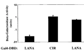
Gal4DBD-LANA and Gal4ACT-SAP30 in cotransformed yeast (Fig. 1)
was measured by induction of beta-galactosidase activity. A
known SAP30-associated cellular corepressor is CIR (24). The
SAP30-CIR interaction was used as a positive control in this
experiment.
LANA binds to members of the mSin3 corepressor complex.
The interaction between LANA and SAP30 was confirmed
using GST affinity assays. Extracts of HeLa cells transfected
with a vector expressing a Myc epitope-tagged SAP30 were
incubated with control GST protein and with four GST fusion
proteins containing the segments of LANA shown in Fig. 2A.
Expression of GST fusion proteins was examined by
SDS-PAGE and Coomassie staining, and equal amounts of protein
were used in each assay. As illustrated in Fig. 2B, the 30-kDa
Myc-SAP30 protein interacted with the GST fusion expressing
a LANA variant with the central region deleted (LANA
d341-939) (lane 1) and with that expressing the N-terminal domain,
LANA(1-340) (lane 3). No interaction was observed with GST
fusions expressing the carboxy-terminal regions of LANA from
amino acid (aa) 940 to 1177 or from aa 341 to 1177 (lanes 2 and
4). Similarly, there was no interaction with the control GST
protein (lanes 5 and 6).
SAP30 was originally identified as a partner of the
corepres-sor mSin3A (36), and an interaction between SAP30 and the
CBF1-associated corepressor CIR has also been reported (24).
Since SAP30 is closely associated with mSin3A and CIR,
evi-dence for a functional interaction between LANA and a
core-pressor complex would be strengthened by a demonstration
that LANA also bound, either directly or indirectly through
SAP30, to these other members of the complex. Binding of
LANA to mSin3A and to CIR was examined using
GST-LANA fusion proteins and extracts of HeLa cells transfected
with vectors expressing either Myc-mSin3A or CIR-Flag (Fig.
2C). The amino-terminal domain (aa 1 to 340) of LANA
bound both the 133-kDa Myc-mSin3A and the 50-kDa
CIR-Flag protein (lane 2). There was no interaction between the
corepressors and the GST fusions expressing LANA
carboxy-terminal aa 940 to 1177 (lane 1) or the extended carboxy
terminus, aa 341 to 1177 (lane 3). Minimal or no interaction
was seen with the control protein GST (lane 4), GST-Zta (lane
5), or GST-EBNA2(1-58) (lane 6). Taken together, these
re-sults indicate that LANA, and specifically the N-terminal
340-aa domain, interacts with recognized components of the
mSin3-HDAC corepressor complex.
Colocalization of LANA and SAP30 in cotransfected cells.
To investigate whether the localization of LANA and SAP30 in
mammalian cells was compatible with interaction between
them, immunofluorescence assays were performed on Vero
cells transfected with Flag-LANA and Myc-SAP30 (Fig. 3). In
individually transfected cells, Myc-SAP30 was detected using a
rabbit anti-Myc antibody and rhodamine-conjugated
anti-rab-bit secondary antibody, and Flag-LANA was detected using a
rabbit anti-Flag antibody and a rhodamine-conjugated
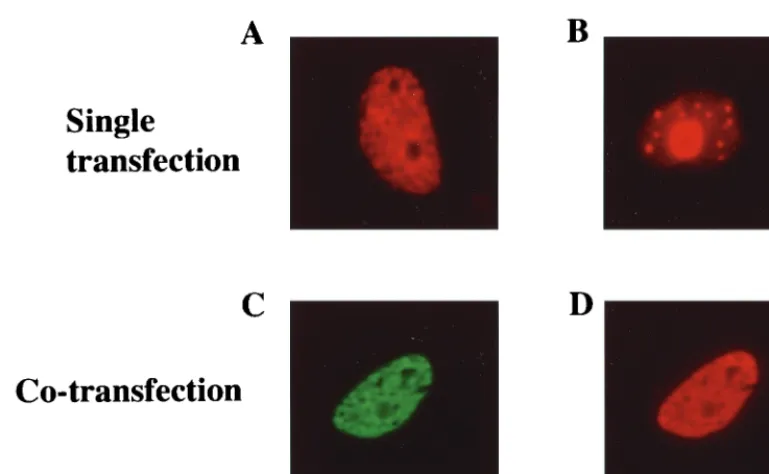
second-ary antibody. Flag-LANA gave patchy nuclear staining with
nucleolar sparing (Fig. 3A). (This staining pattern is typical of
LANA in the absence of HHV-8 genomes.) Myc-SAP30
[image:3.612.87.260.71.179.2]local-FIG. 1. LANA binds to SAP30. Shown are results of a yeast two-hybrid assay in which interaction is measured by induction of beta-galactosidase activity. Yeast cells were cotransformed with Gal4DBD-LANA and Gal4ACT-SAP30, Gal4DBD-LANA plus Gal4ACT vector (negative control), or Gal4DBD-CIR plus Gal4ACT-SAP30 (positive control). Data are averages from two experi-ments, with the range of values indicated.
FIG. 2. LANA interacts with the corepressors SAP30, mSin3A, and CIR in GST affinity assays. (A) Diagram of the GST-LANA constructs used in the affinity assay. (B) Extracts from HeLa cells transfected with Myc-SAP30 were incubated with the indicated GST-LANA fusion proteins (lanes 1 to 4) or with control GST protein (lanes 5 and 6). Transfected cell extract (15l) was loaded in lane 7. A longer exposure (lanes 6 and 7) was needed to detect Myc-SAP30 in the cell extract. (C) Extracts from HeLa cells transfected with Myc-mSin3A or CIR-Flag were incubated with the indicated GST-LANA fusion proteins (lanes 1 to 3) or with control GST protein, control GST-Zta, or GST-EBNA2 protein (lanes 4 to 6). Transfected cell extract (15l) was loaded in lane 7. Bound protein was separated by SDS-PAGE and subjected to Western blot analysis with either an anti-Myc or an anti-Flag antibody. Reactive proteins were visualized by chemiluminescence.
on November 9, 2019 by guest
http://jvi.asm.org/
[image:3.612.62.546.506.675.2]ized as discrete spots within the nuclei of transfected cells and
was also present in the nucleoli (Fig. 3B). In cotransfected
cells, LANA retained the staining pattern seen in the
individ-ually transfected cells (Fig. 3D). Myc-SAP30, which was
de-tected using a mouse anti-Myc antibody and a
fluorescein-conjugated anti-mouse secondary antibody, was redistributed
into the LANA staining pattern in the cotransfected cells (Fig.
3C). The colocalization of SAP30 with LANA in the dually
transfected cells is consistent with their participating in the
same functional complex.
LANA functions as a transcriptional repressor.
The mSin3
corepressor complex is part of a larger complex that recruits
HDAC to DNA-bound factors to mediate transcriptional
re-pression (34). To investigate whether LANA could mediate
transcriptional repression when tethered to DNA through the
Gal4 DNA binding domain (DBD), intact LANA (aa 1 to
1177) and three truncated LANA variants were generated as
Gal4DBD-LANA fusions in a mammalian expression vector
(Fig. 4A). These constructions were then cotransfected into
[image:4.612.110.495.68.305.2]HeLa cells with a 5
⫻
Gal4BS-tkCAT reporter and a
tk-lucif-erase control plasmid. Cotransfection of
Gal4DBD-LANA(1-1177) markedly repressed expression of the CAT reporter (Fig.
4B). The carboxy-terminal domain in
LANA(940-1177) had minimal repressive activity. In contrast,
Gal4DBD-LANA(1-340) was as effective a repressor as intact LANA.
Deletion of the central region of LANA resulted in a fusion
protein, Gal4DBD-LANA(d341-939), that was less effective at
repressing CAT expression than either
Gal4DBD-LANA(1-1177) or Gal4DBD-LANA(1-340). It is possible that the large
internal deletion affected protein folding. Cotransfection of
the Gal4DBD-LANA fusions with a tk-CAT reporter did not
result in repression of CAT expression, indicating that
repres-sion required targeting of LANA to the promoter (data not
shown). The localization of a transcriptional repression
do-main within aa 1 to 340 of LANA is consistent with the results
of the protein-protein interaction experiments, which
demon-strated that the amino terminus of LANA interacted with
corepressor proteins.
FIG. 3. SAP30 colocalizes with LANA in transfected cells. An indirect immunofluorescence assay was performed on Vero cells transfected with Flag-LANA (A), Myc-SAP30 (B), or Myc-SAP30 plus Flag-LANA (C and D). In singly transfected cells, Flag-LANA (A) exhibited micropunctate staining with nucleolar sparing and Myc-SAP30 (B) exhibited nuclear punctate plus diffuse nucleolar staining. The doubly transfected cells were stained for Myc-SAP30 (C) (green) or Flag-LANA (D) (red). In cotransfected cells, Myc-SAP30 (C) exhibited the same micropunctate distribution as Flag-LANA (D). Myc-SAP30 (green) was detected using a rabbit anti-Myc primary antibody and FITC-conjugated donkey anti-rabbit immunoglobulin secondary antibody. Flag-LANA (red) was detected using a mouse monoclonal anti-Flag primary antibody and a rhodamine-conjugated donkey anti-mouse secondary antibody.
FIG. 4. LANA acts as a repressor of gene expression. (A) Diagram of the Gal4-LANA fusion constructs used in the reporter assay. (B) Transient expression assay in which HeLa cells were transfected with the tk-luciferase control plasmid (1.0g) and a 5⫻Gal4BS-tkCAT reporter (1.5g) alone or in the presence of the indicated Gal4-LANA fusion plasmids (1g). Fusion proteins containing LANA aa 1 to 340 repressed reporter CAT expression as efficiently as full-length LANA(1-1177). The assay was repeated three times with similar results.
on November 9, 2019 by guest
http://jvi.asm.org/
EBV EBNA-1 expression in dually infected PEL cell lines.
In
PEL cell lines dually infected with HHV-8 and EBV, the EBV
Qp is used to express EBNA-1 and Cp is not active (22, 65).
The expression of EBV EBNA-1 also appears to be reduced
compared to the levels seen in latently EBV infected B-cell
lines (8). For example, when extracts from the EBV B95-8
B-cell line and from three dually infected PEL cell lines were
probed in an immunoblot with an anti-EBNA-1 monoclonal
antibody, EBNA-1 was detected only in the B95-8 extract (Fig.
5A). The sensitivity of detection in this case may be biased by
the derivation of the monoclonal antibody, which was raised
against EBNA-1 from B95-8. Western blot analysis using NPC
serum as the probe detected weak EBNA-1 expression in the
dually infected BC-2 PEL cells but not in HBL-6 cells (Fig.
5B). The EBNA-1 expression in BC-2 cells was much reduced
compared to that in the latently EBV infected Raji B-cell line
or in the Qp-using Akata cell line. Note that polymorphisms in
the Gly-Gly-Ala repeat region of EBNA-1 lead to
character-istic differences in the migration of EBNA-1 from the different
cell lines.
The low levels of EBNA-1 protein in BC-2 and HBL-6 PEL
cells correlated with a reduction in the amount of EBNA-1
mRNA detectable in these cells compared to that in EBV
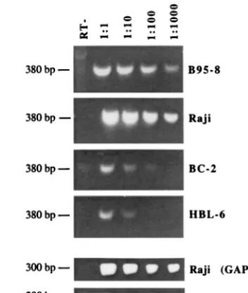
⫹B-cell lines. A semiquantitative RT-PCR analysis of EBNA-1
transcripts revealed significantly reduced levels of EBNA-1
mRNA in BC-2 PEL cells and severely reduced levels of
EBNA-1 mRNA in HBL-6 cells (Fig. 6). RT-PCR for cellular
glyceraldehyde-3-phosphate dehydrogenase (GAPDH) mRNA
was performed on all samples to ensure equal RNA quality and
quantity. Results of the control reactions for Raji and HBL-6
cells are presented in Fig. 6. The relative amounts of EBNA-1
mRNA detected in the BC-2 and HBL-6 cells also correlated
with the EBNA-1 protein levels in these cells, with EBNA-1
protein being detectable in BC-2 cells but below the detection
level in HBL-6 cells (Fig. 5B). The RNA analysis suggests that
the reduced levels of EBNA-1 in PEL cells derive from
repres-sion at the transcriptional level.
To determine whether LANA had a role in the low-level
expression of EBNA-1 from Qp in PEL cells, two approaches
were taken. First, LANA was cotransfected with a Qp-CAT
reporter into HeLa cells (Fig. 7A). Qp is positively regulated
by the JAK/STAT pathway (13), and CAT expression from
Qp-CAT was upregulated in the presence of transfected
JAK-1. The addition of LANA had no effect on basal Qp-CAT
expression but blocked JAK-1 upregulation of Qp.
To examine whether the negative modulatory effect of
LANA seen in the reporter assays was likely to be relevant to
EBNA-1 expression levels in established cell lines, a pair of
stably transfected cell lines was created. Rael is an
EBV-pos-itive B-cell line that utilizes Qp for EBNA-1 expression. Rael
cells were transfected with the pEBO vector or with
pEBO-LANA, and stably transfected cell lines were selected by
growth in hygromycin B. The pEBO vector expressed an
EBNA-1 of a different size from the endogenous Rael
EBNA-1, and the two proteins can be distinguished by their
migration in denaturing gels. The pEBO EBNA-1 migrates
more slowly through the gel than the endogenous Rael
EBNA-1. After 3, 7, and 11 weeks of selection, the amount of
EBNA-1 in the transfected cells was examined by Western
analysis using NPC serum (Fig. 7B). pEBO uses a heterologous
promoter to express EBNA-1, and we could therefore compare
the ratio of vector-expressed EBNA-1 to that of Qp-driven
Rael EBNA-1 as a measure of the effect of LANA on Qp
activity. After 11 weeks of selection, the Rael cells transfected
with the pEBO vector were expressing approximately twofold
more Rael EBNA-1 than pEBO EBNA (Fig. 7B, lane 2).
However, expression of Rael EBNA-1 decreased relative to
that of pEBO EBNA-1 with increased passaging in the
pres-ence of coexpressed LANA. After 11 weeks of selection in the
presence of LANA, Rael cells exhibited a reversed ratio, with
Qp-driven EBNA-1 being approximately fourfold less
abun-dantly than pEBO EBNA-1 (Fig. 7B, lane 6). It is noteworthy
that the total amounts of EBNA-1 expressed in the parental
Rael cells and in the Rael pEBO-LANA cells are comparable.
Expression of LANA in the Rael cells after 11 weeks of
selec-tion is shown in Fig. 7C. These data suggest that LANA may
have a role in the reduced EBNA-1 expression seen in dually
infected PEL cell lines.
LANA represses EBV Cp activity.
The EBV Cp, which drives
expression of the EBNA-1, EBNA-2, and EBNA-3 family
pro-teins during primary infection and in the majority of
EBV-positive lymphoblastoid cell lines, is not used in dually infected
PEL cells. To address whether LANA might contribute to the
downregulation of Cp, transient expression assays were
per-FIG. 5. EBV EBNA-1 protein expression is reduced in dually infected PEL cells. Shown are Western blot analyses comparing the expression of EBNA-1 in EBV-positive cell lines and HHV-8 and EBV dually infected cell lines. B95-8, Raji, and Akata are EBV⫹B-cell lines. HBL-6 and BC-2 are HHV-8⫹EBV⫹
PEL cell lines. BCBL-1 is an HHV-8⫹EBV⫺ PEL cell line, and DG75 is
[image:5.612.341.518.74.282.2]negative for both viruses. EBNA-1 was detected by using the anti-EBNA-1 monoclonal antibody EBNA.OT1x (A) or NPC serum (B). Reactive proteins were visualized by using chemiluminescence.
FIG. 6. EBNA-1 mRNA expression is also reduced in PEL cells. A semi-quantitative RT-PCR assay for EBNA-1 transcripts was performed using the indicated serial 10-fold dilutions of the template cDNA. RT-PCR products were separated by agarose gel electrophoresis, stained with ethidium bromide, and photographed. B95-8 and Raji are EBV⫹B-cell lines; HBL-6 and BC-2 are
EBV⫹HHV-8⫹PEL cell lines. Control RT-PCR results for cellular GAPDH
mRNA are shown for the Raji and HBL-6 cell lines.
on November 9, 2019 by guest
http://jvi.asm.org/
[image:5.612.72.278.74.157.2]formed in which the effect of cotransfected LANA on basal
and EBNA-2-activated expression from a Cp-CAT reporter was
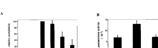
examined (Fig. 8A). As expected, cotransfection of EBNA-2
strongly activated expression of Cp-CAT. LANA had no effect
on basal expression of the Cp-CAT reporter but ablated
EBNA-2 activation in a dose-responsive manner.
To provide some context for the negative regulation of the
EBV Qp and Cp latency promoters, the effect of cotransfected
LANA on an HHV-8 latency promoter was also examined. A
LANAp-luciferase reporter containing 924 bp of the LANA
promoter (LANAp) from
⫺
773 to
⫹
30 was generated (62).
Cotransfection of full-length LANA resulted in approximately
twofold activation of the LANAp (Fig. 8B). A truncated
ami-no-terminal derivative of LANA did not mediate this effect.
Thus, LANA appears to differentially regulate EBV and
HHV-8 latency genes, with the EBV latency Qp and Cp
pro-moters being repressed while LANA potentially stimulates its
own expression.
DISCUSSION
The consistent detection of LANA in all HHV-8-infected
cell lines and tumor specimens examined strongly suggests that
LANA contributes either to HHV-8 latency or to growth
pro-liferation. The demonstration that LANA colocalizes with
HHV-8 genomes on metaphase chromosomes and is sufficient
to maintain HHV-8 episomal DNA in dividing cells (3)
pro-vides experimental evidence for a role for LANA in the
seg-regation of HHV-8 genomes and in the maintenance of
HHV-8 latent infection. LANA may also contribute to tumor
development, and the ability of LANA to interfere with
p53-mediated apoptosis by negatively regulating p53
transcrip-tional activity may represent one such function (18). Our
dem-onstration that LANA binds to members of the mSin3
corepressor complex suggests that LANA may have a more
global effect on cellular gene expression.
[image:6.612.107.501.73.184.2]HDACs are believed to mediate transcriptional repression
in part by modifying chromatin structure. HDACs generally
associate with multiprotein corepressor assemblies that are
brought to DNA through interactions with DNA binding
pro-teins such as the nuclear hormone receptors or CBF1 (24, 27,
51). Two distinct vertebrate corepressor complexes have been
described, an mSin3-containing complex and a NuRD complex
that does not contain mSin3 (34). The ability to detect mSin3A
binding to LANA in GST affinity assays indicates that the
mSin3 corepressor complex is the one that is likely to be
involved in LANA-mediated transcriptional repression. While
we have demonstrated that LANA can function as a
transcrip-tional repressor when artificially targeted to DNA through the
Gal4 DBD, the natural targeting mechanism remains unclear.
In transient assays, LANA repressed activated expression from
the EBV Cp and Qp but had relatively little effect on basal
reporter expression. These two EBV latency promoters are
FIG. 7. LANA attenuates Qp-driven expression of EBNA-1. (A) Transient expression assay in which HeLa cells were transfected with a Qp-CAT reporter (1g) alone or with JAK-1 (2g) and in the presence or absence of cotransfected LANA (1g). The results shown are averages from three experiments, with the standard deviations indicated. (B) LANA reduces Qp-driven EBNA-1 protein expression in Rael cells. Shown is a Western blot examining EBNA-1 expression in EBV-negative DG75 cells carrying the pEBO vector, which expresses EBNA-1 (lane 1), EBV-positive Rael cells carrying the pEBO vector and grown for 11 weeks in selection medium containing hygromycin B (lane 2), Rael cells (lane 3), Rael pEBO-LANA cells grown for 3 weeks in selection medium (lane 4), Rael pEBO-LANA cells after 7 weeks of selection (lane 5), and Rael pEBO-LANA cells after 11 weeks of selection (lane 6). The pEBO-encoded EBNA-1 differs in mobility from the endogenous Rael EBNA-1. EBNA-1 was detected using NPC serum and a chemiluminescence visualization protocol. (C) LANA expression in transfected Rael cells. Shown is a Western blot in which LANA was detected using a rat anti-LANA monoclonal antibody (ABI). Lane 1, Rael pEBO cells after 11 weeks of selection; lane 2, Rael cells; lane 3, Rael pEBO-LANA cells after 11 weeks of selection. Rael pEBO-LANA cells express LANA, unlike the parental Rael or control Rael pEBO cells.
FIG. 8. In transient assays LANA represses EBV Cp-driven expression but activates the HHV-8 LANAp. CAT and luciferase reporter assays were performed in transfected HeLa cells. Results shown are averages from three experiments, with the standard deviations indicated. (A) Cells were transfected with the Cp-CAT reporter (1.0g), EBNA-2 (0.3g), LANA (2.0g), or EBNA-2 (0.3g) plus increasing amounts of LANA (0.5, 1.0, or 2.0g) as indicated. LANA did not affect basal Cp activity but abolished EBNA-2-induced expression. (B) Cells were transfected with the LANAp-luciferase reporter (1.0g), alone or together with LANA (2.0g) or LANA(1-275) (2.0g) as indicated. LANA positively regulated its own promoter.
on November 9, 2019 by guest
http://jvi.asm.org/
[image:6.612.147.467.584.682.2]responsive to different transactivators: Cp is activated by viral
EBNA-2, and Qp is activated by cellular STATs. A
common-ality to EBNA-2 and STAT transactivation is their dependence
on the p300, CBP, and P/CAF coactivators (4, 26, 35, 70).
Adenovirus E1A binds to p300 and interferes with p300
func-tion (2, 38), and it is possible that LANA may similarly target
one of the coactivators. Such a mechanism would be consistent
with the reported inhibition of p53 activity and with the
dif-ferential effect of LANA on basal versus activated
transcrip-tion that we observed.
The majority of PEL cells are dually infected with HHV-8
and EBV. Although PEL cell lines singly infected with HHV-8
have been established from PEL tumors, HHV-8 alone has not
been shown to transform B cells. Incubation of primary B cells
with supernatant from dually infected PEL cells led to the
outgrowth of B-cell lines that were dually infected or EBV
infected, but no singly HHV-8 infected lines were obtained (1,
33). The specific outgrowth of dually infected B cells in one set
of experimental conditions further implies that dual infection
may provide a growth advantage over EBV infection alone
(33). In our analyses, EBV EBNA-1 expression was reduced at
both the protein and mRNA levels in HBL-6 and BC-2 PEL
cells compared to expression in EBV
⫹lymphoblastoid cell
lines. In both transient transfections and established cell lines,
LANA downregulated Qp-driven EBNA-1 expression,
sug-gesting that LANA may be responsible for the EBNA-1
phe-notype. An example of an HHV-8
⫹EBV
⫹PEL tumor giving
rise to an HHV-8
⫹EBV
⫺cell line has been described
else-where (28). EBNA-1 is required for maintenance of the EBV
genome (37), and it is possible that in culture EBNA-1 may
become sufficiently downregulated in PEL cells to lead to loss
of EBV genomes and outgrowth of an HHV-8
⫹EBV
⫺cell
population.
Cp is the promoter used to express the EBV EBNA proteins,
including EBNA-1, during primary infection and is also
fre-quently active in lesions developing in immunosuppressed
pa-tients with lymphoproliferative disease (53, 69, 72). Promoter
switching from Cp to Qp has the effect of eliminating
expres-sion of the immunogenic EBNA-2 and EBNA-3 latency
pro-teins and hence rendering the infected cell less susceptible to
immune surveillance. Cp repression is maintained through
methylation (46, 55, 59, 63, 68), but the regulation of Cp
methylation repression is not fully understood. Whether
LANA-mediated Cp downregulation might predispose Cp to
methylation inactivation in dually infected PELs is an
interest-ing question. One of the methyl CpG bindinterest-ing proteins,
MeCP2, interacts with mSin3A (5), as does LANA, and it is
possible that LANA-mediated tethering of mSin3 might
stim-ulate recruitment of MeCP2.
While LANA repressed expression from the EBV Cp and
Qp promoters, the HHV-8 latency promoter that regulates
LANA expression was activated by LANA. The implication is
that LANA autoregulates its own expression and apparently
has the capacity to modulate transcription either positively or
negatively. Our experiments specifically link LANA-mediated
repression to the binding of LANA to the mSin3 corepressor
complex and implicate LANA in the modification of EBV
latency gene expression in dually infected PELs.
ACKNOWLEDGMENTS
We thank C. Laherty for the mSin3A plasmid, D. Alcindor for
HHV-8 lambda 3-2 DNA, Mabel Chiu for technical assistance, and
Feng Chang for help with manuscript preparation.
This work was supported by National Institutes of Health (NIH)
grant RO1 CA85151 to S.D.H. A.K. and D.G. received support from
NIH Anti-Cancer Drug Development training grant 5 T32 CA09243.
REFERENCES
1.Aguirre, A. J., and E. S. Robertson.1999. Characterization of intertypic recombinants of the Epstein-Barr virus from the body-cavity-based lym-phoma cell lines BC-1 and BC-2. Virology264:359–369.
2.Arany, Z., D. Newsome, E. Oldread, D. M. Livingston, and R. Eckner.1995. A family of transcriptional adaptor proteins targeted by the E1A oncopro-tein. Nature374:81–84.
3.Ballestas, M. E., P. A. Chatis, and K. M. Kaye.1999. Efficient persistence of extrachromosomal KSHV DNA mediated by latency-associated nuclear an-tigen. Science284:641–644.
4.Bhattacharya, S., R. Eckner, S. Grossman, E. Oldread, Z. Arany, A. D’Andrea, and D. M. Livingston.1996. Cooperation of Stat2 and p300/CBP in signalling induced by interferon-␣. Nature383:344–347.
5.Bird, A. P., and A. P. Wolffe.1999. Methylation-induced repression-belts, braces, and chromatin. Cell99:451–454.
6.Boshoff, C., S.-J. Gao, L. E. Healy, S. Matthews, A. J. Thomas, L. Coignet, R. A. Warnke, J. A. Strauchen, E. Matutes, O. W. Kamel, P. S. Moore, R. A. Weiss, and Y. Chang.1998. Establishing a KSHV negative cell line (BCP-1) from peripheral blood and characterizing its growth in Nod/SCID mice. Blood91:1671–1679.
7.Boshoff, C., T. F. Schulz, M. M. Kennedy, A. K. Graham, C. Fisher, A. Thomas, J. O. McGee, R. A. Weiss, and J. J. O’Leary.1995. Kaposi’s sarco-ma-associated herpesvirus infected endothelial and spindle cells. Nat. Med.
1:1274–1278.
8.Callahan, J., S. Pai, M. Cotter, and E. S. Robertson.1999. Distinct patterns of viral antigen expression in Epstein-Barr virus and Kaposi’s sarcoma-associated herpesvirus coinfected body-cavity-based lymphoma cell lines: potential switches in latent gene expression due to coinfection. Virology
262:18–30.
9.Cesarman, E., Y. Chang, P. S. Moore, J. W. Said, and D. M. Knowles.1995. Kaposi’s sarcoma-associated herpesvirus-like DNA sequences in AIDS-re-lated body-cavity-based lymphomas. N. Engl. J. Med.332:1186–1191. 10. Cesarman, E., P. S. Moore, P. H. Rao, G. Inghirami, D. M. Knowles, and Y.
Chang. 1995. In vitro establishment and characterization of two AIDS-related lymphoma cell lines containing Kaposi’s sarcoma-associated herpes-virus-like (KSHV) DNA sequences. Blood86:2708–2714.
11. Chang, Y., E. Cesarman, M. S. Pessin, F. Lee, J. Culpepper, D. M. Knowlex, and P. S. Moore.1994. Identification of herpesvirus-like DNA sequences in AIDS-associated Kaposi’s sarcoma. Science266:1865–1869.
12. Chang, Y.-N., D. L.-Y. Dong, G. S. Hayward, and S. D. Hayward.1990. The Epstein-Barr virus Zta transactivator: a member of the bZIP family with unique DNA-binding specificity and a dimerization domain that lacks the characteristic heptad leucine zipper motif. J. Virol.64:3358–3369. 13. Chen, H., J. M. Lee, Y. Wang, D. P. Huang, R. F. Ambinder, and S. D.
Hayward.1999. The Epstein-Barr virus latency Qp promoter is positively regulated by STATs and Zta interference with JAK-STAT activation leads to loss of Qp activity. Proc. Natl. Acad. Sci. USA96:9339–9344.
14. Cotter, M. A., II, and E. S. Robertson.1999. The latency-associated nuclear antigen tethers the Kaposi’s sarcoma-associated herpesvirus genome to host chromosomes in body cavity-based lymphoma cells. Virology264:254–264. 15. Dittmer, D., M. Lagunoff, R. Renne, K. Staskus, A. Haase, and D. Ganem.
1998. A cluster of latently expressed genes in Kaposi’s sarcoma-associated herpesvirus. J. Virol.72:8309–8315.
16. Dupin, N., T. L. Diss, P. Kellam, M. Tulliez, M. Q. Du, D. Sicard, R. A. Weiss, P. G. Isaacson, and C. Boshoff.2000. HHV-8 is associated with a plasmablastic variant of Castleman disease that is linked to HHV-8-positive plasmablastic lymphoma. Blood95:1406–1412.
17. Dupin, N., C. Fisher, P. Kellam, S. Arid, M. Tulliez, N. Franck, E. van Marck, D. Salmon, I. Gorin, J. P. Escande, R. A. Weiss, K. Alitalo, and C. Boshoff.1999. Distribution of human herpesvirus-8 latently infected cells in Kaposi’s sarcoma, multicentric Castleman’s disease, and primary effusion lymphoma. Proc. Natl. Acad. Sci. USA96:4546–4551.
18. Friborg, J., Jr., W. Kong, M. O. Hottiger, and G. J. Nabel.1999. p53 inhibition by the LANA protein of KSHV protects against cell death. Nature
402:889–894.
19. Gao, S. J., L. Kingsley, D. R. Hoover, T. J. Spira, C. R. Rinaldo, A. Saah, J. Phair, R. Detels, P. Parry, Y. Chang, and P. S. Moore.1996. Seroconversion to antibodies against Kaposi’s sarcoma-associated herpesvirus-related latent nuclear antigens before the development of Kaposi’s sarcoma. N. Engl. J. Med.335:233–241.
20. Gao, S.-J., L. Kingsley, M. Li, W. Zheng, C. Parravicini, J. Ziegler, R. Newton, C. R. Rinaldo, A. Saah, J. Phair, R. Detels, Y. Chang, and P. S. Moore.1996. KSHV antibodies among Americans, Italians and Ugandans with and without Kaposi’s sarcoma. Nat. Med.2:925–928.
21. Gessain, A., J. Briere, C. Angelin-Duclos, F. Valensi, H. M. Beral, F. Davi, M. A. Nicola, A. Sudaka, N. Fouchard, J. Gabarre, X. Troussard, E. Dulmet, J. Audouin, J. Diebold, and G. de The.1997. Human herpesvirus 8 (Kaposi’s sarcoma herpesvirus) and malignant lymphoproliferations in France: a mo-lecular study of 250 cases including two AIDS-associated body cavity based lymphomas. Leukemia11:266–272.
22. Horenstein, M. G., R. G. Nador, A. Chadburn, E. M. Hyjek, G. Inghirami, D. M. Knowles, and E. Cesarman. 1997. Epstein-Barr virus latent gene
on November 9, 2019 by guest
http://jvi.asm.org/
expression in primary effusion lymphomas containing Kaposi’s sarcoma-associated herpesvirus/human herpesvirus-8. Blood90:1186–1191. 23. Hsieh, J. J.-D., T. Henkel, P. Salmon, E. Robey, M. G. Peterson, and S. D.
Hayward.1996. Truncated mammalian Notch1 activates CBF1/RBPJk-re-pressed genes by a mechanism resembling that of Epstein-Barr virus EBNA2. Mol. Cell. Biol.16:952–959.
24. Hsieh, J. J.-D., S. Zhou, L. Chen, D. B. Young, and S. D. Hayward.1999. CIR, a corepressor linking the DNA binding factor CBF1 to the histone deacetylase complex. Proc. Natl. Acad. Sci. USA96:23–28.
25. Jaffe, E. S.1996. Primary body cavity-based AIDS-related lymphomas. Evo-lution of a new disease entity. Am. J. Clin. Pathol.105:141–143.
26. Jayachandra, S., K. G. Low, A. E. Thlick, J. Yu, P. D. Ling, Y. Chang, and P. S. Moore. 1999. Three unrelated viral transforming proteins (vIRF, EBNA2, and E1A) induce the MYC oncogene through the interferon-re-sponsive PRF element by using different transcription coadaptors. Proc. Natl. Acad. Sci. USA96:11566–11571.
27. Kao, H.-Y., P. Ordentlich, N. Koyano-Nakagawa, Z. Tang, M. Downes, C. R. Kintner, R. M. Evans, and T. Kadesch.1998. A histone deacetylase core-pressor complex regulates the Notch signal transduction pathway. Genes Dev.12:2269–2277.
28. Katano, H., Y. Hoshino, Y. Morishita, T. Nakamura, H. Satoh, A. Iwamoto, B. Herndier, and S. Mori.1999. Establishing and characterizing a CD30-positive cell line harboring HHV-8 from a primary effusion lymphoma. J. Med. Virol.58:394–401.
29. Kedes, D. H., M. Lagunoff, R. Renne, and D. Ganem.1997. Identification of the gene encoding the major latency-associated nuclear antigen of the Ka-posi’s sarcoma-associated herpesvirus. J. Clin. Investig.100:2606–2610. 30. Kedes, D. H., E. Oberskalski, M. Busch, R. Kohn, J. Flood, and D. Ganem.
1996. The seroepidemiology of human herpesvirus 8 (Kaposi’s sarcoma-associated herpesvirus): distribution of infection in KS risk groups and evi-dence for sexual transmission. Nat. Med.2:918–924.
31. Kellam, P., C. Boshoff, D. Whitby, S. Matthews, R. A. Weiss, and S. J. Talbot.
1997. Identification of a major latent nuclear antigen, LNA-1, in the human herpesvirus 8 genome. J. Hum. Virol.1:19–29.
32. Kellam, P., D. Bourboulia, N. Dupin, C. Shotton, C. Fisher, S. Talbot, C. Boshoff, and R. A. Weiss.1999. Characterization of monoclonal antibodies raised against the latent nuclear antigen of human herpesvirus 8. J. Virol.
73:5149–5155.
33. Kliche, S., E. Kremmer, W. Hammerschmidt, U. Koszinowski, and J. Haas.
1998. Persistent infection of Epstein-Barr virus-positive B lymphocytes by human herpesvirus 8. J. Virol.72:8143–8149.
34. Knoepfler, P. S., and R. N. Eisenman.1999. Sin meets NuRD and other tails of repression. Cell99:447–450.
35. Korzus, E., J. Torchia, D. W. Rose, L. Xu, R Kurokawa, E. M. McInerney, T. M. Mullen, C. K. Glass, and M. G. Rosenfeld.1998. Transcription factor-specific requirements for coactivators and their acetyltransferase functions. Science279:703–707.
36. Laherty, C. D., W. M. Yang, J. M. Sun, J. R. Davie, E. Seto, and R. N. Eisenman.1997. Histone deacetylases associated with the mSin3 corepressor mediate Mad transcriptional repression. Cell89:349–356.
37. Lee, M. A., M. E. Diamond, and J. L. Yates.1999. Genetic evidence that EBNA-1 is needed for efficient, stable latent infection by Epstein-Barr virus. J. Virol.73:2974–2982.
38. Li, Q., A. Imhof, T. N. Collingwood, F. D. Urnov, and A. P. Wolffe.1999. p300 stimulates transcription instigated by ligand-bound thyroid hormone recep-tor at a step subsequent to chromatin disruption. EMBO J.18:5634–5652. 39. Ling, P. D., and S. D. Hayward.1995. Contribution of conserved amino acids
in mediating interaction between EBNA2 and CBF1/RBPJk. J. Virol.69:
1944–1950.
40. Ling, P. D., J. J. Ryon, and S. D. Hayward.1993. EBNA-2 of herpesvirus papio diverges significantly from the type A and type B EBNA-2 proteins of Epstein-Barr virus but retains an efficient transactivation domain with a conserved hydrophobic motif. J. Virol.67:2990–3003.
41. Margolskee, R. F., P. Kavathas, and P. Berg.1988. Epstein-Barr virus shuttle vector for stable episomal replication of cDNA expression libraries in human cells. Mol. Cell. Biol.8:2837–2847.
42. Matolcsy, A., R. G. Nador, E. Cesarman, and D. M. Knowles.1998. Immu-noglobulin VH gene mutational analysis suggests that primary effusion lym-phomas derive from different stages of B cell maturation. Am. J. Pathol.
153:1609–1614.
43. Matsushima, A. Y., J. A. Strauchen, G. Lee, E. Scigliano, E. E. Hale, M. T. Weisse, D. Burstein, O. Kamel, P. S. Moore, and Y. Chang.1999. Posttrans-plantation plasmacytic proliferations related to Kaposi’s sarcoma-associated herpesvirus. Am. J. Surg. Pathol.23:1393–1400.
44. Miller, G., L. Heston, E. Grogan, L. Gradoville, M. Rigsby, R. Sun, D. Shedd, V. M. Kushnaryov, S. Grossberg, and Y. Chang.1997. Selective switch between latency and lytic replication of Kaposi’s sarcoma herpesvirus and Epstein-Barr virus in dually infected body cavity lymphoma cells. J. Virol.
71:314–324.
45. Miller, G., M. O. Rigsby, L. Heston, E. Grocan, R. Sun, C. Metroka, J. A. Levy, S.-J. Gao, Y. Chang, and P. Moore.1996. Antibodies to butyrate-inducible antigens of Kaposi’s sarcoma-associated herpesvirus in patients
with HIV-1 infection. N. Engl. J. Med.334:1292–1297.
46. Minarovits, J., L. F. Hu, S. Imai, Y. Harabuchi, A. Kataura, S. Minarovits-Kormuta, T. Osato, and G. Klein.1994. Clonality, expression and methyl-ation patterns of the Epstein-Barr virus genomes in lethal midline granulo-mas classified as peripheral angiocentric T cell lymphogranulo-mas. J. Gen. Virol.
75:77–84.
47. Moore, P. S., C. Boshoff, R. A. Weiss, and Y. Chang.1996. Molecular mimicry of human cytokine and cytokine response pathway genes by KSHV. Science274:1739–1744.
48. Moore, P. S., and Y. Chang.1995. Detection of herpesvirus-like DNA se-quences in Kaposi’s sarcoma in patients with and without HIV infection. N. Engl. J. Med.332:1181–1185.
49. Moore, P. S., S.-J. Gao, G. Dominguez, E. Cesarman, O. Lungu, D. Knowles, R. Garber, P. E. Pellett, D. J. McGeoch, and Y. Chang.1996. Primary characterization of a herpesvirus agent associated with Kaposi’s sarcoma. J. Virol.70:549–558.
50. Nador, R. G., E. Cesarman, A. Chadburn, D. B. Dawson, M. Q. Ansari, J. Sald, and D. M. Knowles.1996. Primary effusion lymphoma: a distinct clinicopathologic entity associated with the Kaposi’s sarcoma-associated her-pesvirus. Blood88:645–656.
51. Nagy, L., H. Y. Kao, D. Chakravarti, R. J. Lin, C. A. Hassig, D. E. Ayer, S. L. Schreiber, and R. M. Evans.1997. Nuclear receptor repression mediated by a complex containing SMRT, mSin3A, and histone deacetylase. Cell89:373– 380.
52. Nicholas, J., J.-C. Zong, D. J. Alcendor, D. M. Ciufo, L. J. Poole, R. T. Sarisky, C. J. Chiou, X. Zhang, X. Wan, H.-G. Guo, M. S. Reitz, and G. S. Hayward.1998. Novel organization features, captured cellular genes and strain variability within the genome of KSHV/HHV8. J. Natl. Cancer Inst. Monogr.23:79–88.
53. Oudejans, J. J., M. Jiwa, A. J. C. Vandenbrule, F. A. Grasser, A. Horstman, W. Vos, P. M. Kluin, P. Vandervalk, J. M. M. Walboomers, and C. J. L. M. Meijer.1995. Detection of heterogenous Epstein-Barr virus gene expression patterns with individual post-transplantation lymphoproliferative disorders Am. J. Pathol.147:923–933.
54. Parravicini, C., B. Chandran, M. Corbellino, E. Berti, M. Paulli, P. S. Moore, and Y. Chang.2000. Differential viral protein expression in Kaposi’s sarcoma-associated herpesvirus-infected diseases: Kaposi’s sarcoma, primary effusion lymphoma, and multicentric Castleman’s disease. Am. J. Pathol.
156:743–749.
55. Paulson, E. J., and S. H. Speck.1999. Differential methylation of Epstein-Barr virus latency promoters facilitates viral persistence in healthy seropos-itive individuals. J. Virol.73:9959–9968.
56. Platt, G. M., G. R. Simpson, S. Mittnacht, and T. F. Schulz.1999. Latent nuclear antigen of Kaposi’s sarcoma-associated herpesvirus interacts with RING3, a homolog of theDrosophilafemale sterile homeotic (fsh) gene. J. Virol.73:9789–9795.
57. Rainbow, L., G. M. Platt, G. R. Simpson, R. Sarid, S.-J. Gao, H. Stoiber, C. S. Herrington, P. S. Moore, and T. F. Schulz.1997. The 222- to 234-kilodalton latent nuclear protein (LNA) of Kaposi’s sarcoma-associated her-pesvirus (human herher-pesvirus 8) is encoded byorf73and is a component of the latency-associated nuclear antigen. J. Virol.71:5915–5921.
58. Renne, R., M. Lagunoff, W. Zhong, and D. Ganem.1996. The size and conformation of Kaposi’s sarcoma-associated herpesvirus (human herpes-virus 8) DNA in infected cells and virions. J. Virol.70:8151–8154. 59. Robertson, K. D., and R. F. Ambinder.1997. Methylation of the
Epstein-Barr virus genome in normal lymphocytes. Blood90:4480–4484.
60. Russo, J. J., R. A. Bohenzky, M.-C. Chein, J. Chen, M. Yan, D. Maddalena, J. P. Parry, D. Peruzzi, I. S. Edelman, Y. Chang, and P. S. Moore.1996. Nucleotide sequence of the Kaposi’s sarcoma-associated herpesvirus (HHV8). Proc. Natl. Acad. Sci. USA93:14862–14867.
61. Sarid, R., O. Flore, R. A. Bohenzky, Y. Chang, and P. S. Moore.1998. Transcription mapping of the Kaposi’s sarcoma-associated herpesvirus (hu-man herpesvirus 8) genome in a body cavity-based lymphoma cell line (BC-1). J. Virol.72:1005–1012.
62. Sarid, R., J. S. Wiezorek, P. S. Moore, and Y. Chang.1999. Characterization and cell cycle regulation of the major Kaposi’s sarcoma-associated herpes-virus (human herpesherpes-virus 8) latent genes and their promoter. J. Virol.73:
1438–1446.
63. Schaefer, B. C., J. L. Strominger, and S. H. Speck.1997. Host cell-deter-mined methylation of specific Epstein-Barr virus promoters regulates the choice between distinct viral latency programs. Mol. Cell. Biol.17:364–377. 64. Soulier, J., L. Grollet, E. Oksenhendler, P. Cacoub, D. Cazals-Hatem, P. Babinet, M. F. d’Agay, J. P. Clauvel, M. Raphael, L. Degos, et al.1995. Kaposi’s sarcoma-associated herpesvirus-like DNA sequences in multicen-tric Castleman’s disease. Blood86:1276–1280.
65. Szekely, L., F. Chen, N. Teramoto, B. Ehlin-Henriksson, K. Pokrovskaja, A. Szeles, A. Manneborg-Sandlund, M. Lowbeer, E. T. Lennette, and G. Klein.
1998. Restricted expression of Epstein-Barr virus (EBV)-encoded, growth transformation-associated antigens in an EBV- and human herpesvirus type 8-carrying body cavity lymphoma line. J. Gen. Virol.79:1445–1452. 66. Szekely, L., C. Kiss, K. Mattsson, E. Kashuba, K. Pokrovskaja, A. Juhasz, P.
Holmvall, and G. Klein.1999. Human herpesvirus-8-encoded LNA-1
on November 9, 2019 by guest
http://jvi.asm.org/
mulates in heterochromatin-associated nuclear bodies. J. Gen. Virol.80:
2889–2900.
67.Talbot, S. J., R. A. Weiss, P. Kellam, and C. Boshoff.1999. Transcriptional analysis of human herpesvirus-8 open reading frames 71, 72, 73, K14, and 74 in a primary effusion lymphoma cell line. Virology257:84–94.
68.Tao, Q., K. D. Robertson, A. Manns, A. Hildesheim, and R. F. Ambinder.
1998. Epstein-Barr virus (EBV) in endemic Burkitt’s lymphoma: molecular analysis of primary tumor tissue. Blood91:1373–1381.
69.Tao, Q., L. J. Swinnen, J. Yang, G. Srivastava, K. D. Robertson, and R. F. Ambinder.1999. Methylation status of the Epstein-Barr virus major latent promoter C in iatrogenic B cell lymphoproliferative disease. Am. J. Pathol.
155:619–625.
70.Wang, L., S. R. Grossman, and E. Kieff.2000. Epstein-Barr virus nuclear
protein 2 interacts with p300, CBP, and PCAF histone acetyltransferases in activation of the LMP1 promoter. Proc. Natl. Acad. Sci. USA97:430–435. 71. Xu, X., Y.-L. Sun, and T. Hoey.1996. Cooperative DNA binding and
se-quence-selective recognition conferred by the STAT amino-terminal do-main. Science273:794–797.
72. Young, L., C. Alfieri, K. Hennessy, H. Evans, C. O’Hare, K. C. Anderson, J. Ritz, R. S. Shapiro, A. Rickinson, and E. Kieff.1989. Expression of Epstein-Barr virus transformation-associated genes in tissues of patients with EBV lymphoproliferative disease. N. Engl. J. Med.321:1080–1085.
73. Zhang, Y., Z.-W. Sun, R. Iratni, H. Erdjument-Bromage, P. Tempst, M. Hampsey, and D. Reinberg.1998. SAP30, a novel protein conserved between human and yeast, is a component of a histone deacetylase complex. Mol. Cell
1:1021–1031.
on November 9, 2019 by guest
http://jvi.asm.org/