Copyright © 1998, American Society for Microbiology. All Rights Reserved.
Regions of Human Immunodeficiency Virus Type 1 nef
Required for Function In Vivo
GRACE M. ALDROVANDI,
1LIANYING GAO,
2GREGORY BRISTOL,
2ANDJEROME A. ZACK
3*
University of Alabama at Birmingham AIDS Center, Birmingham, Alabama 35294,
1and Division of
Hematology-Oncology, Department of Medicine,
2and Department of Microbiology and Molecular
Genetics,
3University of California—Los Angeles School of Medicine and University of
California Los Angeles AIDS Institute, Los Angeles, California 90095-1678
Received 9 September 1997/Accepted 19 May 1998
In vivo studies in monkeys and humans have indicated that immunodeficiency viruses with Nef deleted are
nonpathogenic in immunocompetent hosts, and this has motivated a search for live attenuated vaccine
candidates. However, the mechanisms of action of Nef remain elusive. To define the regions of human
immunodeficiency virus type 1 (HIV-1) Nef which mediate in vivo pathogenicity, a series of mutated isogenic
viruses were inoculated into human thymic implants in SCID-hu mice. Mutation of several regions, including
the myristoylation site at the second glycine and a region encompassing amino acids 41 through 49 of Nef,
profoundly affected pathogenicity. Surprisingly, mutations of prolines in either of the two distant PXXP SH3
binding domains did not affect pathogenicity, indicating that these regions are not required for Nef activity in
developing T-lineage cells. These data suggest that some functions of Nef described in vitro may not be relevant
for in vivo pathogenicity.
The nef open reading frame of human immunodeficiency
virus type 1 (HIV-1) is located at the 3
9
end of the virus,
partially overlapping the U3 region of the 3
9
long terminal
repeat. nef mRNA represents more than 80% of the multiply
spliced transcripts expressed during early viral transcription,
and encodes a 27- to 29-kDa cytoplasmic protein which is
membrane localized by an N-myristyl group. Nef-deficient
sim-ian immunodeficiency virus (SIV) fails to produce AIDS in
infected adult macaques (25). Some humans infected with
Nef-deleted HIV have remained disease free, with normal CD4
counts 10 to 14 years after infection (9, 26), although deletion
of Nef is not a universal finding in nonprogressors (21). In
vitro, Nef also confers a growth advantage (8, 34, 48), whose
magnitude depends upon the activation state of the cell.
Bio-chemically, Nef associates with various cellular kinases (5, 41,
44, 45). The Nef of one SIV strain has been implicated in
lymphocyte activation (10, 13). Nef expression also results in
posttranslational down-regulation of the CD4 protein (15, 19);
however, the significance of this down-regulation in HIV-1
pathogenesis is unclear. The effect of Nef on CD4
down-reg-ulation appears to be distinct from its effect on infectivity (17,
43, 46, 52).
Unlike in vitro systems, the SCID-hu mouse model involves
infection of a functional human hematopoietic organ (Thy/Liv)
that resembles a human thymus and directs normal T-cell
development from hematopoietic precursor cells (33, 36). This
system allows for the simultaneous assay of infection,
replica-tion, and pathogenicity. This dynamic in vivo model has been
used to detect differences in pathogenicity not observed in
traditional in vitro systems. Infection of this organ with HIV-1
results in depletion of CD4-bearing cells and a histologic
pic-ture reminiscent of that observed in human infection (1, 2, 6,
22, 28, 49). However, HIV strains lacking a functional nef gene
have a decreased ability to deplete CD4-bearing cells in this
system (2, 22). With this system, we have now used a series of
isogenic mutated viruses to localize regions of the nef gene that
confer increased pathogenicity. Our data indicate that the
myr-istic acid site at position 2 and a region encompassing amino
acids (aa) 41 to 49 of Nef are required for a fully pathogenic
phenotype. Interestingly, neither of the two proline repeat
regions previously shown to associate with multiple cellular
kinases are required for virus replication and pathogenesis.
MATERIALS AND METHODS
Virus and cells.HIV nef deletion mutantsDnef,DNRE, andDnef/NRE were
obtained from Ron Desrosiers and are described elsewhere (16). All the mutants were constructed with pNL101, which contains the same proviral sequence as HIV-1NL4-3, with the entire 59-flanking region and no 39-flanking region recloned
into plasmid pUCBM21 (40). This plasmid has a unique XbaI site in the 39 -flanking region. The BamHI-XbaI fragment was cloned into pBluescript KS1
(Stratagene) for oligonucleotide-directed mutagenesis, by using the Amersham in vitro mutagenesis system, v.2, as specified by the manufacturer. The primers used to construct deletion mutants are as follows: deletion 1, 59-ctcagctcgtctcat tctttcttttgaccacttgcc-3 (deletion of nucleotides [nt] 22 to 51 of nef inclusive); deletion 2, 59-atgtttttctaggtcctcagctcgtctcattctttc-39(deletion of nt 73 to 105 of nef inclusive); deletion 3, 59-ggcacaagcagcattgttagcatgtttttctaggtc-39(deletion of nt 118 to 147 of nef inclusive); deletion 4, 59-aggtgtgactggaaaaccggcacaagcagcattgt tagc-39(deletion of nt 169 to 198 of nef inclusive); deletion 5, 59-gctaagatctacag ctgcaggtgtgactggaaaacc-39(deletion of nt 217 to 246 of nef inclusive); and dele-tion 6, 59-ggagtgaattagcccctctaagatctacagctgc-39(deletion of nt 265 to 294 of nef inclusive). All the deletion mutations were in frame, and all were confirmed both before and after being cloned into the full-length plasmid. Point mutations were introduced into single-stranded DNA by the method of Kunkel et al. (30). The substrate for mutagenesis was a plasmid carrying the BamHI (nt 8466)-XbaI (nt 9711) fragment of pNL101, prepared in Escherichia coli CJ236. The primer for the myristoylation-deficient mutant (MYR2) (59-ccacttgccaGccatcttat-39)
di-rected a G-to-C point mutation at position 8791 (resulting in a Gly-to-Ala change). The primer for the point mutation, which abrogated the second start (ATG 2) (59-agctcgtctaattctttccc-39), directed an A-to-T mutation at position 8823 (resulting in a Met-to-Leu change. Primer SH334 for the PXXP4 mutant (antisense, 59-cattgctcttaagctacctgagctgtgactgcaaaaccc-39) directed C-to-G muta-tions at posimuta-tions 214, 223, and 232 of nef. The primer for the PXXP2 mutant (59-atctgCctcaaactgCtactag-39) directed C-to-G mutations at positions 439 and 448. Primer R105L (59-caaggatatcttctaataattgggagtgaattag-39) directed A-to-T mutations at positions 313 and 316 and G-to-T mutations at positions 314 and 317, which resulted in the arginines at aa 105 and 106 being converted to leucines to construct the ARGX2 mutant.
The sequence of each altered DNA fragment was confirmed by DNA sequenc-ing (Sequenase 2.0 kit; United States Biochemical Corp., Cleveland, Ohio) both before and after being cloned into the full-length plasmid. Plasmid preparations
* Corresponding author. Mailing address: Department of
Microbi-ology and Molecular Genetics, UCLA School of Medicine and UCLA
AIDS Institute, 10833 Le Conte Ave., Los Angeles, CA 90095-1678.
Phone: (310) 794-7765. Fax: (310) 825-6192. E-mail: [email protected].
7032
on November 9, 2019 by guest
http://jvi.asm.org/
of wild-type and mutant viruses were grown on a large scale and purified with QIAgen maxipreps (Qiagen, Chatsworth, Calif.).
Virus stocks of the deletion mutants were prepared by transfection of the DNA into CEMx174 cells, while constructs containing point mutations were transfected into COS cells, as previously described (2, 7). Viral stocks were collected, filtered, and assayed for p24 content by enzyme-linked immunosorbent assay (Coulter, Hialeah, Fla.). Aliquots of the viral stocks were stored at270°C. The virus titers were determined in parallel by fivefold limiting dilution in duplicate on human peripheral blood mononuclear cells (PBMC), from a single donor, that had been stimulated for 3 days with phytohemagglutinin (PHA). Infectious units were standardized to wild-type HIV-1NL4-3in which 2.5 ng of p24
was equivalent to 100 infectious units. Normal human PBMC were obtained from Leukopaks purchased from the American Red Cross. Peripheral blood lymphocytes were isolated by centrifugation over Ficoll-Hypaque and depleted of macrophages by adherence to plastic for 72 h. Growth kinetics of the different viral isolates were determined by infection with equal infectious units on human PHA-stimulated PBMC followed by quantitation by ELISA specific for the viral p24 Gag protein. Western blot analyses for mutant Nef protein expression were performed on cell lysates of infected C8166 cells, using a polyclonal anti-Nef antiserum, and are also reported elsewhere (50). After infection of the Thy/Liv grafts, an aliquot of virus from the same vial used to infect the tissue was used to infect PHA-stimulated PBMC to confirm virus viability.
Construction, infection, and biopsies of SCID-hu mice.C.B.-17 scid/scid mice with severe combined immunodeficiency (SCID mice) were originally obtained from K. Dorshkin and subsequently bred at the University of California Los Angeles (UCLA). All experimental animals were housed in a Biosafety Level 3 facility at UCLA and handled in accordance with institutional guidelines. All the animals were anesthetized by intraperitoneal injection of a ketamine HCl-xyla-zine mixture (1 mg/10 g of body weight) before any invasive manipulation. When the mice were 6 to 8 weeks of age,;1-mm3pieces of human fetal thymus and
liver (Thy/Liv) were surgically implanted under the murine kidney capsule, as previously described (1). Fetal tissue (Advanced Bioscience Resources, Ala-meda, Calif.) was obtained from donors ranging in gestational age from 16 to 24 weeks. At 4 to 6 months postimplantation, the grafts were infected with 100 or 500 infectious units (IU), as indicated in the text, in approximately 50-ml volumes by direct injection. Mock-infected implants were injected with medium.
Since it is possible to obtain only limited numbers of reconstituted SCID-hu mice from a single donor, multiple donors were used for these experiments. To control for any variation in genetic backgrounds that might have had a bearing on the susceptibility of the target cells to HIV infection, mice transplanted with tissues from different donors were distributed randomly among the experimental groups that were infected with the various HIV-1 mutants. In addition, wild-type and “mock” virus and at least two isogenic accessory gene mutants were inocu-lated into tissue from a single donor. In these experiments, we did not detect any obvious differences attributable to donor variation; however, the numbers of animals and donors were too small to perform statistical analysis.
Wedge biopsy specimens of Thy/Liv tissue were obtained at 3-week intervals after infection. Approximately one-fourth to one-third of the implant was re-moved at each biopsy. Human thymocytes were teased from the stromal ele-ments, filtered through a screen (Cell strainer; Falcon, Franklin Lakes, N.J.), washed in phosphate-buffered saline, counted, and then aliquoted for subsequent PCR and flow cytometric analyses.
Quantitative PCR amplification.DNA was isolated from single cells obtained from the biopsied implants by using the QIAamp blood kit (Qiagen) as specified by the manufacturer. Purified DNA was then subjected to quantitative PCR as previously described (1, 53, 54). Briefly, PCR amplifications were carried out for 25 cycles with32P-end-labeled primers. The M667-AA55 primer pair, which is
specific for the R/U5 region of the viral long terminal repeat, was used to detect HIV-1 sequences (53, 54). The amount of human cellular DNA in each sample was quantified by PCR amplification with primers specific for the humanb -glo-bin gene (nt 14 to 33 and 123 to 104). Standard curves for HIV-1 DNA consisted of linearized HIV-1JR-CSFin normal human PBMC DNA (10mg/ml) as carrier.
Standard curves for humanb-globin were derived from 10-fold dilutions of normal PBMC DNA. Both the HIV-1 andb-globin standard curves were am-plified in parallel with Thy/Liv samples. The PCR amplifications were carried out in 15ml of low-salt PCR buffer (25 mM Tris [pH 8.0], 2 mM MgCl2, 30 mM NaCl,
0.1 mg of bovine serum albumin per ml, 0.25 mM deoxynucleoside triphosphate). S/P high-purity water (Baxter Healthcare Corp., McGaw Park, Ill.) was used to bring the reaction volume to 25ml. Following amplification the PCR products were resolved on a 6% polyacrylamide gel. Quantitation was achieved by extrap-olation to the standard curves by radioanalytic image analysis (Ambis, San Diego, Calif.). This method of DNA PCR can detect ten proviral copies permg of genomic DNA. Values obtained from this assay never varied above 30% of the actual values in controlled experiments.
Confirmation of infection.To confirm infection of each implant with the appropriate deletion mutant, primer pairs flanking each deletion were used to amplify proviral sequences by PCR. Analysis was performed on DNA obtained 6 weeks postinfection, and no contamination with wild-type sequences was ever detected. DNA from implants infected with viruses containing point mutations was sequenced to determine whether introduced mutations were maintained. No reversion of point mutations was observed at the 6-week time point in any of the analyzed implants.
Flow cytometric analysis of Thy/Liv cells.Thymocytes were stained with phy-coerythrin-conjugated mouse monoclonal antibody to human CD4 (Becton Dickinson, Mountain View, Calif.) and fluorescein isothiocyanate-conjugated mouse monoclonal antibody to human CD8 (Becton Dickinson) as specified by the manufacturer. Thymocytes were also stained with PE- and/or FITC-conju-gated anti-mouse immunoglobulin G1 as isotype controls. Data were acquired on a FACScan flow cytometer and analyzed with the Cell Quest program (Becton Dickinson). The live cell population was determined by gating on the forward-versus-side-scatter plot of thymocytes derived from mock-infected implants. A total of 5,000 to 10,000 events were acquired, except in the case of severely depleted implants.
Statistical analyses.Comparisons of proportions between groups were made by Fisher’s exact test. This test is more appropriate than a chi-square test for small samples and is asymptotically equivalent to a chi-square test for large samples. To test whether two distributions were the same, the Wilcoxon rank sum test was used. This nonparametric test is valid over a wide range of distri-butional assumptions and is invariant with respect to monotonic transformations such as taking logarithms. All comparisons were for two-sided alternatives.
Infectivity was measured by determining the proportion of implants showing detectable HIV at the 3- and/or 6-week time points. Animals that had undetect-able HIV 3 weeks postinfection and that died before the 6-week biopsy were excluded from the analysis, since infection could not be confirmed. Calculations for demonstrating viral replication and differences in pathogenicity were made by using only the animals that were positive for HIV DNA sequences at the three-and/or 6-week time points. Pathogenicity was measured by determining the percentage of CD41CD81double-positive thymocytes at both time points. Viral
replication was calculated as the number of proviral copies per 100,000 cells.
RESULTS
Effects of Nef deletions on virus replication.
To determine
the regions of the nef gene responsible for mediating this
pathogenic phenotype, a series of deletion and point mutants
were constructed, as depicted in Fig. 1 (also see Fig. 3). When
100 IU of viruses containing large deletions in either the 5
9
or
3
9
region of nef were introduced into Thy/Liv implants, many
did not become infected (reference 2 and data not shown). To
overcome the effects of decreased infectivity of viruses with
Nef deleted, all experiments reported here were performed at
a higher multiplicity of infection (500 IU). The effects of these
deletions on HIV-1-induced depletion of CD4-bearing
thymo-cytes and proviral load are depicted in Fig. 2. Attenuated viral
pathogenicity was seen following deletion of the 5
9
half (Fig. 2)
or the 3
9
half (
D
NRE, not shown) of nef or a combination of
the two deletions (
D
nef/NRE, not shown). Consequently,
vi-ruses with smaller nested deletions (approximately 10 aa) in
the 5
9
half of nef were inoculated into the Thy/Liv implants of
SCID-hu mice.
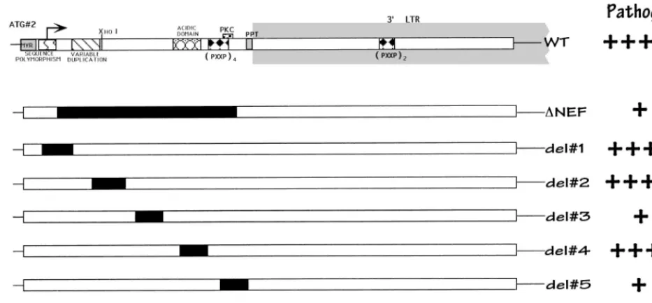
Deletions were made in regions of the gene which had been
suggested to be important for protein function. Deletion 1 (aa
8 to 17 inclusive) includes a region of sequence polymorphism
(47) that has also been postulated to be a nuclear targeting
sequence (35). Deletions 2 (aa 25 to 35 inclusive) and 3 (aa 41
to 49 inclusive) are relatively conserved regions of
indetermi-nate function. The region excised in deletion 4 (aa 57 to 66
inclusive) has been predicted to lie on the outer surface of the
protein because it is charged and acidic. This region also
en-codes a putative viral protease cleavage site in the protein (12,
14, 39, 51). Deletion 5 (aa 73 to 82 inclusive) encompasses the
two terminal proline residues of the 5
9
PXXP SH3 binding
motif, as well as a potential protein kinase C phosphorylation
site (threonine 80). In addition, this region appears to be
im-portant for the association of Nef with the cellular cytoskeleton
(23). Deletion 6 (aa 89 to 98) removes the polypurine tract,
which is important in initiating plus-strand DNA synthesis
dur-ing reverse transcription. Followdur-ing infection, all nested
dele-tion and point mutant viruses, except the one lacking the initial
ATG and deletion 6 (see Fig. 3), produced Nef proteins
de-tectable by immunoblot analysis with Nef antiserum (50).
Pro-tein levels shown by Western blotting in cells infected with
most mutants were similar, with the level PXXP4 mutant being
V
OL. 72, 1998
REGIONS OF HIV-1 nef REQUIRED FOR FUNCTION IN VIVO
7033
on November 9, 2019 by guest
http://jvi.asm.org/
only slightly lower than that in the wild-type-infected cells.
However, Nef protein levels in cells infected with PXXP4
1
2,
ARGX2, or deletion 5 were significantly lower than in the
wild-type-infected cells, so we cannot exclude that loss of
pro-tein stability may have contributed to the observed phenotypes
of these mutants (see below).
After 6 weeks postinoculation, all the implants injected with
viral strains containing deletions 1, 2 or 4 were productively
infected, achieving viral loads and thymocyte depletion
equiv-alent to those due to wild-type virus (Fig. 2). In our system,
deletion of the putative nuclear localization signal (deletion 1)
did not alter the infectivity, replication, or pathogenicity of the
virus. In contrast, viruses bearing deletions 3 or 5 did not infect
all animals tested, in that the proviral DNA of one of five
implants injected with deletion 3 and the proviral DNA of two
of nine implants infected with deletion 5 were undetectable by
PCR (Fig. 2B). Implants productively infected with these
strains showed a marked decrease in depletion of CD4-bearing
thymocytes, despite levels of proviral DNA that were similar to
those of the wild-type virus. This is similar to what we had
previously reported with Nef-minus strains (2, 22). Nef protein
levels in cells infected with deletion 3 were identical to those of
cells infected with wild-type virus or with deletion mutants 1, 2
and 4 (which had no altered phenotype), suggesting that this
region is truly important for in vivo pathogenicity. However,
since the levels of Nef protein were significantly lower in cells
infected with deletion 5, the apparent effect of this deletion is
likely to be due to decreased protein stability (see above).
Viruses with a deletion of the polypurine tract (deletion 6)
could not be detected in the implants as late as 9 weeks
postin-fection, presumably because of impaired reverse transcription.
Nuclear magnetic resonance, combined with proteolytic
ex-periments, has suggested that Nef consists of two main
do-mains: an anchor domain located at the N terminus (aa 2 to
65), which is probably located at the surface of the protein, and
a more compactly folded core C-terminal domain (12). In
addition, it has been reported that Nef is cleaved by proteases
between Trp 57 and Leu 58, suggesting that cleavage is crucial
for correct biologic function (14, 39, 51). Interestingly, deletion
of several regions (deletions 1, 2, and 4) of this putative anchor
domain had no effect on viral replication or pathogenicity.
However, deletion 3 (aa 41 to 49 inclusive) attenuated viral
replication and pathogenicity, presumably by disrupting overall
protein structure. The area encompassed by deletion 4 (aa 57
to 66) included the putative protease cleavage domain (14, 39,
51), as well as a conserved glutamic acid-rich segment of the
protein (47). Deletion of this region resulted in a virus with a
pathogenicity phenotype identical to that of wild-type virus.
This finding was somewhat surprising, since an overlapping
deletion (aa 60 to 71) was found by others to lower infectivity
and abrogate CD4 down-regulation in vitro (17). These results
are not necessarily discordant, since the mutations were not
identical; thus, we cannot comment on the role of CD4 in
down-regulation in Nef-mediated pathogenicity in this system.
Effect of point mutations on pathogenicity.
To further define
regions responsible for Nef function, a series of viruses
con-taining point mutations in regions associated with various in
vitro Nef functions were introduced into Thy/Liv implants (Fig.
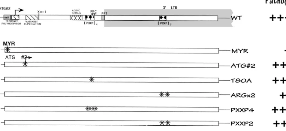
3). The myristoylation signal in Nef is conserved in both
lab-oratory and viral isolates, suggesting a critical role in the life
cycle. Myristoylation has been demonstrated to be critical for
the subcellular targeting of Nef to the cytoplasmic membranes
(3, 11, 20), and localization has been proposed to regulate both
viral replication and T-cell activation (4). Myristoylation has
also been noted to be important for maximal Nef cytoskeletal
binding (37), and this has led some investigators to suggest a
parallel between Nef and other myristoylated proteins such as
Marcks (myristoylated alanine-rich C-kinase substrate) and Src
(37). Some traces of Nef have also been detected in the nuclei
(27, 29, 38, 42). Loss of the myristoylation signal in Nef mutant
G2A produced a virus with decreased infectivity and
pathoge-nicity in the SCID-hu model (Fig. 4). This finding confirms the
importance of myristoylation for all of the putative functions of
Nef, as suggested previously (17, 55).
[image:3.612.61.528.72.289.2]Neither loss of the second ATG (mutation M20L; Fig. 3) nor
mutation of the potential PKC phosphorylation site (T80A)
FIG. 1. Schematic of the nef gene and isogenic deletion mutant forms. Putative functional regions of Nef are indicated at the top. At the far right is a summary of relative Nef activity, as determined by our studies.1111, wild-type growth;1, attenuated growth;2, no detectable virus. This interpretation was based on our statistical analyses (see Fig. 2).
on November 9, 2019 by guest
http://jvi.asm.org/
affected infectivity, pathogenicity, or viral replication (Fig. 4).
All the implants injected with these mutants were productively
infected and exhibited marked thymocyte depletion by 6 weeks
postinfection. The results seen with the T80A mutant indicated
that the attenuated phenotype seen with deletion mutant 5
(Fig. 2) was not due to loss of the protein kinase C
phosphor-ylation site. Mutation of two arginine residues at positions 105
and 106 to leucines (the ARGX2 mutant), which have been
reported to interact with cellular serine kinases and have been
implicated in SIV pathogenesis (46), did not appear to affect
infectivity or viral replication in the SCID-hu system.
Never-theless, this virus was less able to deplete CD4-bearing
thymo-cytes than was the wild-type virus (Fig. 4A). Sequence analysis
of the nef region of proviral DNA obtained from Thy/Liv
implants infected with the ARGX2 mutant revealed that no
reversion to wild-type sequences occurred (data not shown).
However, Nef protein levels with this mutant were somewhat
decreased (approximately one-third) compared to wild-type
infection in vitro. Thus, while it is attractive to speculate that
this region appears to make a modest but significant
contribu-tion to Nef funccontribu-tion in this system, we cannot rule out protein
stability effects contributing to the altered phenotype.
We also explored whether inactivation of previously
identi-fied SH3 recognition sequences would have an effect on
patho-genicity. Mutation of four prolines (PXXP4 mutant) which
constitute a putative SH3 binding domain in the 5
9
region
(P69A, P72A, P75A, and P78A) or two prolines (PXXP2
mu-tant) in the 3
9
region of Nef (P147A and P150A) did not affect
infectivity, pathogenicity, or viral load (Fig. 4). However,
si-multaneous mutation of all six prolines in both regions
(PXXP4
1
2 mutant) had a significant effect on pathogenicity
and viral replication but not on infectivity. All five implants
inoculated with this viral strain were productively infected;
however, only one of the five implants exhibited depletion of
FIG. 2. Thymocyte depletion (A) and proviral load (B) in Thy/Liv implants 6 weeks postinfection. Each symbol represents a different implant infected with the corresponding viral strain, as described in Fig. 1. (A) Percentage of CD4-bearing cells (including both CD41CD81double-positive and CD41single-positive subsets), as determined by flow cytometry (1, 2). An implant was considered depleted if it was infected and had fewer than 55% CD4-bearing cells. Nef-minus viruses (Dnef) and strains with deletions 3 or 5 were less capable of depleting CD4-bearing cells than was the wild-type virus (NL). This difference was statistically significant (Fisher’s exact test, two tailed), with P50.0002, 0.0004, and 0.00002 forDnef and deletions 3 and 5, respectively. Deletion 6 was not included in these calculations, because the virus was not infectious (B). Other viruses yielded P.0.05. (B) Number of HIV-1 genomes in 105human thymocytes, as determined by quantitative DNA PCR.
Symbols on the x axis indicate implants with undetectable HIV DNA. The minimum amount of DNA assayed was the equivalent of 104thymocytes.
VOL. 72, 1998
REGIONS OF HIV-1 nef REQUIRED FOR FUNCTION IN VIVO
7035
on November 9, 2019 by guest
http://jvi.asm.org/
CD4-bearing thymocytes (P
5
0.0002). This phenotype,
how-ever, may be attributable to decreased Nef protein levels, since
cells infected with this virus contained lower levels of Nef
protein (approximately one-third, similar to the ARGX2
mu-tant) than did cells infected with wild-type virus.
DISCUSSION
We set out to define regions of HIV-1 Nef responsible for
pathogenicity in vivo. Similar to our previous study (2), we
were unable to find a statistically significant difference in viral
loads at 6 weeks postinfection between wild-type virus and any
of the mutants, with the exception of deletion 6, which was not
infectious. We did, however, identify several regions that
af-fected pathogenicity and others that appeared unimportant.
Perhaps the most interesting mutants in this latter category
were those that encompassed the proline repeat regions, which
contain a minimal PXXP consensus SH3 binding domain
mo-tif, which can interact in vitro with high specificity and affinity
with the SH3 domains of the Src kinases, Hck and Lyn, but not
other kinases in the same family (43). Recently, the binding
surface of Nef, which interacts with the SH3 domain of Hck
tyrosine protein kinase, has been mapped and was found to be
noncontiguous, a feature not previously observed for
SH3-target interactions (18). However, the interaction of Hck and
the other tyrosine protein kinases mapped only to the region
surrounding the N-terminal tetraproline repeat and did not
include the C-terminal PXXP domain located more than 70
residues away. While we did not perform protein binding
stud-ies with these mutants, we were unable to detect Hck or Lyn
expression by Northern blot analysis in either thymocytes or
peripheral blood lymphocytes (data not shown). Thus, it is
unlikely that the interaction of these particular kinases can
account for our findings.
In our studies, mutation of either proline repeat domain
singly yielded a wild-type phenotype, suggesting that
confor-mational changes in these regions following mutation do not
alter function. Simultaneous alteration of both SH3 binding
domains severely attenuated the virus; however, this could be
attributable to an effect on protein stability, since low levels of
Nef were detected in cells infected with this mutant in vitro.
Initially, it was reported that both these regions were required
for high-affinity binding of Hck (43); however, the same group
later reinterpreted this data and attributed it to an artifact of
refolding of denatured Nef after transfer to a filter (32). The
present data would suggest that interaction of cellular factors
with these regions is not necessary in vivo. This may be because
these particular factors (Hck and Lyn) are not expressed in
thymocytes. Interestingly, it has been reported that point
mu-tations of the N-terminal tetraproline repeat motif alone could
decrease infectivity but not CD4 down-regulation in vitro (17,
32, 52). Recently, Wiskerchen and Cheng-Meyer (52) reported
that the tetraproline repeat region also associates with a
cel-lular serine kinase. However, the C-terminal PXXP domain
was not required for this association. Our data indicate that
alteration of the tetraproline repeat or the double proline
repeat regions is not sufficient to mediate the Nef-minus
phe-notype in vivo. Recent studies in the SIV system, involving
mutation of the single SH3 binding domain in nef, failed to
show an effect on pathogenicity (31). Thus, our conclusions
obtained with HIV are similar to those reported for SIV.
In the SIV system, mutation of a pair of arginines in Nef
known to interact with PAK-related serine kinases lowered the
initial burst of virus replication in vivo, but rapid reversion
occurred at these positions, and clinical progression to AIDS
occurred (46). This suggests that the arginines and the
associ-ated activation of PAK are strongly selected for in vivo and are
linked to the pathogenicity of SIV. However, our studies of
HIV Nef showed that the attenuation conferred by mutations
of the paired arginines is not as severe as that in Nef-deleted
virus and that reversion of these mutations to wild-type
se-quences did not occur. We also cannot rule out nonspecific
effects on protein levels with this mutant.
[image:5.612.58.535.80.299.2]Recent work with the hu-SCID model reconstituted with
peripheral blood lymphocytes has shown that a mutant bearing
a Nef lacking aa 72 to 75 is attenuated in this system (24). This
FIG. 3. Schematic of the nef gene and isogenic point mutant forms. As in Fig. 1, a summary of relative pathogenic potential is given at the right.1111, wild-type growth;1, attenuated growth;11, somewhat attenuated growth. The statistical data used to make these interpretations are given in the legend to Fig. 4.
on November 9, 2019 by guest
http://jvi.asm.org/
region is encompassed in our deletion mutant 5. However, we
cannot conclude that this region is critical for Nef function in
our in vivo model, due to the low protein expression of our
mutant.
In conclusion, we have mapped HIV in vivo pathogenicity to
two regions of Nef, including the myristoylation site and the
region encompassed by deletion 3 (aa 41 to 49 of Nef). The
importance of the myristoylation site probably relates to the
need for appropriate subcellular localization of the protein. It
is unclear why deletion 3 conveyed an attenuated phenotype;
however, this was not due to nonspecific alterations in the
levels of Nef protein, as infected cells contained comparable
levels to cells infected with wild-type virus. Further studies are
required to determine how this region affects pathogenicity.
Importantly, we have eliminated the role of several regions of
Nef in in vivo pathogenicity. Notably, the second ATG site, the
putative PKC phosphorylation site (at aa 80), and the protease
cleavage site, in addition to the above-mentioned proline
re-peat regions, were not required for HIV to replicate and
de-plete thymocytes in the SCID-hu mouse. Additional studies
with this system will be important to fully understand how Nef
is involved in HIV pathogenesis in the thymus.
ACKNOWLEDGMENTS
We thank H.-G. Krausslich and R. Welker for sharing their data
regarding in vitro protein expression of mutant viruses. We also thank
B. D. Jamieson for critical review of the manuscript, A. Kacena and N.
Negoitas for technical assistance, and W. Aft for editorial and
word-processing assistance. We thank M. Liu and J. Taylor for statistical
analysis.
This work was supported by grants from the NIH (AI36059) and the
UCLA CFAR (NIH AI28697). G.M.A. is a Pediatric AIDS Scholar,
and J.A.Z. is an Elizabeth Glaser Scientist supported by the Pediatric
AIDS Foundation.
REFERENCES
1. Aldrovandi, G. M., G. Feuer, L. Gao, M. Kristeva, I. S. Y. Chen, B. Jamieson,
and J. A. Zack.1993. HIV-1 infection of the SCID-hu mouse: an animal
FIG. 4. Thymocyte depletion (A) and proviral load (B) in implants 6 weeks postinfection with point mutated viruses. Each symbol represents a different implant infected with the corresponding viral strain, as described in Fig. 3. (A) Percentage of CD4-bearing cells (both CD41CD81double-positive and CD41single-positive subsets), as determined by flow cytometry (Fig. 2). An implant was considered depleted if it was infected and had fewer than 55% CD4-bearing cells. MYR2virus and the strains containing the PXXP412 point mutation and the ARGX2 mutations were significantly less capable of depleting CD4-bearing cells (Fisher’s exact test, two tailed), with P50.001, 0.0002, and 0.015 for MYR2, PXXP412, and ARGX2, respectively. Other viruses yielded P.0.05. (B) Number of HIV-1 genomes in 105
human thymocytes, as determined by quantitative DNA PCR (Fig. 2B).
VOL. 72, 1998
REGIONS OF HIV-1 nef REQUIRED FOR FUNCTION IN VIVO
7037
on November 9, 2019 by guest
http://jvi.asm.org/
model for virus pathogenesis. Nature 363:732–736.
2. Aldrovandi, G. M., and J. A. Zack. 1996. Replication and pathogenicity of human immunodeficiency virus type 1 accessory gene mutants in SCID-hu mice. J. Virol. 70:1505–1511.
3. Bachelerie, F., J. Alcami, U. Hazan, N. Israel, B. Goud, F.
Arenzana-Seis-dedos, and J.-L. Virelizier.1990. Constitutive expression of human immu-nodeficiency virus (HIV) nef protein in human astrocytes does not influence basal or induced HIV long terminal repeat activity. J. Virol. 64:3059–3062. 4. Baur, A. S., E. T. Sawai, P. Dazin, W. J. Fantl, C. Cheng-Mayer, and B. M.
Peterlin.1994. HIV-1 Nef leads to inhibition or activation of T cells, de-pending on its intracellular localization. Immunity 1:373–384.
5. Bodeus, M., A. Marie-Cardine, C. Bougeret, F. Ramos-Morales, and R.
Benarous.1995. In vitro binding and phosphorylation of human immunode-ficiency virus type 1 Nef protein by serine/threonine protein kinase. J. Gen. Virol. 76:1337–1344.
6. Bonyhadi, M. L., L. Rabin, S. Salimi, D. A. Brown, J. Kosek, J. M. McCune,
and H. Kaneshima.1993. HIV induces thymus depletion in vivo. Nature
363:728–736.
7. Cann, A. J., Y. Koyanagi, and I. S. Y. Chen. 1988. High efficiency transfection of primary human lymphocytes and studies of gene expression. Oncogene
3:123–128.
8. Chowers, M. Y., C. A. Spina, T. J. Kwoh, N. J. S. Fitch, D. D. Richman, and
J. C. Guatelli.1994. Optimal infectivity in vitro of human immunodeficiency virus type 1 requires an intact nef gene. J. Virol. 68:2906–2914.
9. Deacon, N. J., A. Tsykin, A. Solomon, K. Smith, M. Ludford-Menting, D. J.
Hooker, D. A. McPhee, A. L. Greenway, A. Ellett, C. Chatfield, V. A. Lawson, S. Crowe, A. Maerz, S. Sonza, J. Learmont, J. S. Sullivan, A. Cunningham, D. Dwyer, D. Dowton, and J. Mills.1995. Genomic structure of an attenuated quasi species of HIV-1 from a blood transfusion donor and recipients. Science 270:988–991.
10. Du, Z., S. M. Lang, V. G. Sasseville, A. A. Lackner, P. O. Ilyinskii, M. D.
Daniel, J. U. Jung, and R. C. Desrosiers.1995. Identification of a nef allele that causes lymphocyte activation and acute disease in macaque monkeys. Cell 82:665–674.
11. Franchini, G., M. Robert-Guroff, J. Ghrayeb, N. T. Chang, and F.
Wong-Staal. 1986. Cytoplasmic localization of the HTLV-III 39 orf protein in cultured T cells. Virology 155:593–599.
12. Freund, J., R. Kellner, T. Houthaeve, and H. R. Kalbitzer. 1994. Stability and proteolytic domains of Nef protein from human immunodeficiency virus (HIV) type 1. Eur. J. Biochem. 221:811–819.
13. Fultz, P. N. 1991. Replication of an acutely lethal simian immunodeficiency virus activates and induces proliferation of lymphocytes. J. Virol. 65:4902– 4909.
14. Gaedigk-Nitschko, K., A. Schon, G. Wachinger, V. Erfle, and B. Kohleisen. 1995. Cleavage of recombinant and cell derived human immunodeficiency virus 1 (HIV-1) Nef protein by HIV-1 protease. FEBS Lett. 357:275–278. 15. Garcia, J. V., and A. D. Miller. 1992. Downregulation of cell surface CD4 by
nef. Res. Virol. 143:52–55.
16. Gibbs, J. S., D. A. Regier, and R. C. Desrosiers. 1994. Construction and in vitro properties of HIV-1 mutants with deletions in “nonessential” genes. AIDS Res. Human Retroviruses 10:343–350.
17. Goldsmith, M. A., M. T. Warmerdam, R. E. Atchison, M. D. Miller, and
W. C. Greene. 1995. Dissociation of the CD4 downregulation and viral infectivity enhancement functions of human immunodeficiency virus type 1 Nef. J. Virol. 69:4112–4121.
18. Grzesiek, S., A. Bax, G. M. Clore, A. Groneborn, J.-S. Hu, J. Kaufman, I.
Palmer, S. Stahl, and P. Wingfield.1996. The solution structure of HIV-1 Nef reveals an unexpected fold and permits delineation of the binding surface for the SH3 domain of Hck tyrosine protein kinase. Nat. Struct. Biol.
3:340–344.
19. Guy, B., M. P. Kieny, Y. Riviere, C. Le Peuch, K. Dott, M. Girard, L.
Montagnie, and J.-P. Lecocq.1987. HIV F/39orf encodes a phosphorylated
GTP-binding protein resembling an oncogene product. Nature 330:266–269. 20. Hammes, S. R., E. P. Dixon, M. H. Malim, B. R. Cullen, and W. C. Greene. 1989. Nef protein of human immunodeficiency virus type 1: evidence against its role as a transcriptional inhibitor. Proc. Natl. Acad. Sci. USA 86:9549– 9553.
21. Huang, Y., L. Zhang, and D. D. Ho. 1995. Characterization of nef sequences in long-term survivors of human immunodeficiency virus type 1 infection. J. Virol. 69:93–100.
22. Jamieson, B. D., G. M. Aldrovandi, V. Planelles, J. B. M. Jowett, L. Gao,
L. M. Bloch, I. S. Y. Chen, and J. A. Zack.1994. Requirement of HIV-1 nef for in vivo replication and pathogenesis. J. Virol. 68:3478–3485.
23. Kaminchik, J., R. Margalit, S. Yaish, H. Drummer, B. Amit, N. Sarver, M.
Gorecki, and A. Panet.1994. Cellular distribution of HIV type 1 Nef protein: identification of domains in Nef required for association with membrane and detergent-insoluble cellular matrix. AIDS Res. Human Retroviruses 10:1003– 1010.
24. Kawano, Y., Y. Tanaka, N. Misawa, R. Tanaka, J.-I. Kira, T. Kimura, M.
Fukushi, K. Sano, T. Goto, M. Nakai, T. Kobayashi, N. Yamamoto, and Y. Koyanagi.1997. Mutational analysis of human immunodeficiency virus type 1 (HIV-1) accessory genes: requirement of a site in the nef gene for HIV-1
replication in activated CD41T cells in vitro and in vivo. J. Virol. 71:8456–
8466.
25. Kestler, H. W., III, D. J. Ringler, M. Kazuyasu, D. L. Panicali, P. K. Sehgal,
M. D. Daniel, and R. C. Desrosiers.1991. Importance of the nef gene for maintenance of high virus loads and for development of AIDS. Cell 65:651– 662.
26. Kirchoff, F., T. C. Greenough, D. B. Brettler, J. L. Sullivan, and R. C.
Desrosiers.1995. Absence of intact nef sequences in a long-term survivor with nonprogressive HIV-1 infection. N. Engl. J. Med. 332:228–232. 27. Kohleisen, B., M. Neumann, R. Herrmann, R. Brack-Werner, K. J. Krohn,
V. Ovod, A. Ranki, and V. Erfle.1992. Cellular localization of Nef expressed in persistently HIV-1-infected low-producer astrocytes. AIDS 6:1427–1436. 28. Kollmann, T. R., M. Pettoello-Mantovani, X. Zhuang, A. Kim, M.
Hachamo-vitch, P. Smarnworawong, A. Rubinstein, and H. Goldstein.1994. Dissemi-nated human immunodeficiency virus type 1 (HIV-1) infection in SCID-hu mice after peripheral inoculation with HIV-1. J. Exp. Med. 179:513–522. 29. Krohn, K. J. E., V. Ovod, A. Langerstedt, U. Saarialho-Kere, A. Ranki, F. O.
Gombert, G. Jong, and K. Molling.1991. Expression kinetics and cellular localization of HIV tat and nef proteins in relation to expression of viral structural mRNA and viral particles. Vaccines 91:97–101.
30. Kunkel, T. A., J. D. Roberts, and R. A. Zakour. 1987. Rapid and efficient site-specific mutagenesis without phenotypic selection. Methods Enzymol.
154:367–382.
31. Lang, S. M., A. J. Iafrate, C. Stahl-Hennig, E. M. Kuhn, T. Nisslein, F.-J.
Kaup, M. Haupt, G. Hunsmann, J. Skowronski, and F. Kirchhoff.1997. Association of simian immunodeficiency virus Nef with cellular serine/thre-onine kinases is dispensable for the development of AIDS in rhesus ma-caques. Nat. Med. 3:860–865.
32. Lee, C., B. Leung, M. A. Lemmon, J. Zheng, D. Cowburn, J. Kuriyan, and K.
Saksela.1995. A single amino acid in the SH3 domain of Hck determines its high affinity and specificity in binding to HIV-1 Nef protein. EMBO J.
14:5006–5015.
33. McCune, J. M., R. Namikawa, H. Kaneshima, L. D. Schultz, M. Lieberman,
and I. L. Weissman.1988. The SCID-hu mouse: murine model for the analysis of human hematolymphoid differentiation and function. Science
241:1632–1639.
34. Miller, M. D., M. T. Warmerdam, I. Gaston, W. C. Greene, and M. B.
Feinberg.1994. The human immunodeficiency virus-1 nef gene product: a positive factor for viral infection and replication in primary lymphocytes and macrophages. J. Exp. Med. 179:101–113.
35. Murti, K. G., P. S. Brown, L. Ratner, and J. V. Garcia. 1993. Highly localized tracks of human immunodeficiency virus type 1 Nef in the nucleus of cells of a human CD41T-cell line. Proc. Natl. Acad. Sci. USA 90:11895–11899. 36. Namikawa, R., K. N. Weilbaecher, H. Kaneshima, E. J. Yee, and J. M.
McCune.1990. Long-term human hematopoiesis in the SCID-hu mouse. J. Exp. Med. 172:1055–1063.
37. Niederman, T. M., W. R. Hastings, and L. Ratner. 1993. Myristoylation-enhanced binding of the HIV-1 Nef protein to T cell skeletal matrix. Virol-ogy 197:420–425.
38. Ovod, V., A. Lagerstedt, A. Ranki, F. O. Gombert, R. Spohn, M. Tahtinen, G.
Jung, and K. J. Krohn.1992. Immunological variation and immunohisto-chemical localization of HIV-1 Nef demonstrated with monoclonal antibod-ies. AIDS 6:25–34.
39. Pandori, M. W., N. J. S. Fitch, H. M. Craig, D. D. Richman, C. A. Spina, and
J. C. Guatelli.1996. Producer-cell modification of human immunodeficiency virus type 1: Nef is a virion protein. J. Virol. 70:4283–4290.
40. Planelles, V., A. Haislip, E. S. Withers-Ward, S. A. Stewart, Y. Xie, N. P.
Shah, and I. S. Y. Chen.1995. A new reporter system for detection of viral infection. Gene Ther. 2:369–376.
41. Poulin, L., and J. A. Levy. 1992. The HIV-1 nef gene product is associated with phosphorylation of a 46 kD cellular protein. AIDS 6:787–791. 42. Ranki, A., A. Lagerstedt, V. Ovod, E. Aavik, and K. J. Krohn. 1994.
Expres-sion kinetics and subcellular localization of HIV-1 regulatory proteins Nef, Tat and Rev in acutely and chronically infected lymphoid cell lines. Arch. Virol. 139:365–378.
43. Saksela, K., G. Cheng, and D. Baltimore. 1995. Proline-rich (PxxP) motifs in HIV-1 Nef bind to SH3 domains of a subset of Src kinases and are required for the enhanced growth of Nef1viruses but not for down-regulation of CD4. EMBO J. 14:484–491.
44. Sawai, E. T., A. S. Baur, B. M. Peterlin, J. A. Levy, and C. Cheng-Mayer. 1995. A conserved domain and membrane targeting of Nef from HIV and SIV are required for association with a cellular serine kinase activity. J. Biol. Chem. 270:15307–15314.
45. Sawai, E. T., A. Baur, H. Struble, B. M. Peterlin, J. A. Levy, and C.
Cheng-Mayer.1994. Human immunodeficiency virus type 1 Nef associates with a cellular serine kinase in T lymphocytes. Proc. Natl. Acad. Sci. USA 91:1539– 1543.
46. Sawai, E. T., E. H. Khan, P. M. Montbriand, B. J. Peterlin, C. Cheng-Mayer,
and P. A. Luciw.1996. Activation of PAK by HIV and SIV Nef: importance for AIDS in rhesus macaques. Curr. Biol. 6:1519–1527.
47. Shugars, D. C., M. S. Smith, D. H. Glueck, P. V. Nantermet, F.
Seillier-Moiseiwitsch, and R. Swanstrom. 1993. Analysis of human
on November 9, 2019 by guest
http://jvi.asm.org/
ciency virus type 1 nef gene sequences present in vivo. J. Virol. 67:4639–4650. 48. Spina, C. A., T. J. Kwoh, M. Y. Chowers, J. C. Guatelli, and D. D. Richmond. 1994. The importance of nef in the induction of human immunodeficiency virus type 1 replication from primary quiescent CD4 lymphocytes. J. Exp. Med. 179:115–123.
49. Stanley, S. K., J. M. McCune, H. Kaneshima, J. S. Justement, M. Sullivan,
E. Boone, M. Baseler, J. Adelsberger, M. Bonyhadi, J. Orenstein, C. H. Fox, and A. S. Fauci.1993. Human immunodeficiency virus infection of the human thymus and disruption of the thymic microenvironment in the SCID-hu mouse. J. Exp. Med. 178:1151–1163.
50. Welker, R., M. Harris, B. Cardel, and H.-G. Krausslich. Virion incorpora-tion of human immunodeficiency virus type 1 Nef is mediated by a bipartite membrane targeting signal and correlates with enhancement of viral infec-tivity. Submitted for publication.
51. Welker, R., H. Kottler, H. R. Kalbitzer, and H. G. Krausslich. 1996. Human
immunodeficiency virus type 1 Nef protein is incorporated into virus parti-cles and specifically cleaved by the viral proteinase. Virology 219:228–236. 52. Wiskerchen, M., and C. Cheng-Mayer. 1996. HIV-1 Nef association with
cellular serine kinase correlates with enhanced virion infectivity and efficient proviral DNA synthesis. Virology 224:292–301.
53. Zack, J. A., S. J. Arrigo, S. R. Weitsman, A. S. Go, A. Haislip, and I. S. Y.
Chen.1990. HIV-1 entry into quiescent primary lymphocytes: molecular analysis reveals a labile, latent viral structure. Cell 61:213–222.
54. Zack, J. A., A. Haislip, P. Krogstad, and I. S. Y. Chen. 1992. Incompletely reverse transcribed human immunodeficiency virus type 1 genomes in qui-escent cells can function as intermediates in the retrovirus life cycle. J. Virol.
66:1717–1725.
55. Zazopoulos, E., and W. A. Haseltine. 1992. Mutational analysis of the human immunodeficiency virus type 1 Nef function. Proc. Natl. Acad. Sci. USA
89:6634–6638.