E
E
U
U
S
S
T
T
A
A
C
C
H
H
I
I
A
A
N
N
T
T
U
U
B
B
E
E
F
F
U
U
N
N
C
C
T
T
I
I
O
O
N
N
B
B
E
E
F
F
O
O
R
R
E
E
A
A
N
N
D
D
A
A
F
F
T
T
E
E
R
R
S
S
U
U
R
R
G
G
E
E
R
R
Y
Y
F
F
O
O
R
R
M
M
U
U
C
C
O
O
S
S
A
A
L
L
C
C
H
H
R
R
O
O
N
N
I
I
C
C
S
S
U
U
P
P
P
P
U
U
R
R
A
A
T
T
I
I
V
V
E
E
O
O
T
T
I
I
T
T
I
I
S
S
M
M
E
E
D
D
I
I
A
A
SUBMITTED BY
EUSTACHIAN TUBE FUNCTION BEFORE
AND AFTER SURGERY FOR MUCOSAL
CHRONIC SUPPURATIVE OTITIS MEDIA
A dissertation submitted in partial fulfillment of MS Branch IV, ENT
examination of the Tamil Nadu Dr. MGR Medical University, To be held in
Christian Medical College, Vellore
Certificate
This is to certify that the dissertation entitled
‘Eustachian tube function
before and after surgery for mucosal chronic suppurative otitis media’
is the bonafide original work of Dr. Lisa Mary Cherian carried out under my
guidance, in fulfillment of the rules and regulations for the MS Branch IV,
ENT examination of the Tamil Nadu Dr. MGR Medical University, to be
held in April 2012.
Dr. RupaVedantam
Professor and Head
Department of ENT Unit III
Christian Medical College
Acknowledgements
I express my deep sense of gratitude and respect to my esteemed professor and
guide Dr. RupaVedantam for taking me on as a thesis candidate and mentoring and
guiding me through the process. I am deeply indebted to her support and patience
throughout this process and all her encouragement and guidance at every stage of the
work on this dissertation.
I would like to thank Dr. Suma Susan Mathew and Dr. Reji Kurien for help and
support in carrying out this study
I am grateful to the staff in biostatistics department Ms.Thenmozhi and Mr.
Sathiya Murthi and also Ms. Jayasree for patiently analyzing the data and formatting it.
I am extremely thankful to all the audiology lab staff for their constant support
I am indebted to all the patients who consented to be a part of this study and each
and every ENT surgeon for the meticulous recording of the findings.
My sincere thanks to all my professors, seniors, colleagues and friends for all their
encouragement and help.
A special thanks to my family, especially my husband Dr. Viju Daniel and
daughter Tanvi, for supporting me throughout the work on this study.
Table of Contents
S. No.
Content
Page No.
1
Aims and objectives
1
2
Present knowledge and Review of Literature
2
3
Materials and Methods
24
4
Results and Analysis 30
5
Discussion
49
6
Conclusion
55
7
Bibliography
56
8
Appendix
A – Proforma used for Data Collection
B – Form for Informed Consent
C – Data Sheet
1
AIMS AND OBJECTIVES
1. To assess preoperative Eustachian Tube (ET) function using the inflation deflation
technique and Valsalva maneuver in patients with mucosal chronic suppurative otitis
media undergoing surgery for perforation closure.
2. To investigate if there is any change in ET function following tympanoplasty in these
patients. The null hypothesis is that there is no change in ET function following
tympanoplasty
ABSTRACT
Eustachian tube function before and after surgery in patients with mucosal
chronic suppurative otitis media
Introduction
The Eustachian tube (ET), also called the pharyngotympanic tube, is a narrow passage
made of bony and cartilaginous parts connecting the middle ear cleft to the nasopharynx.
Normal ET function is necessary for ventilation of the middle ear (ME), maintaining the ME
space and drainage of ME secretions. Any structural or functional abnormalities of ET can
affect these functions and lead to negative ME pressure, fluid accumulation and, eventually, a
perforation of the tympanic membrane. The pathogenesis of mucosal chronic suppurative
otitis media (CSOM) is thus believed to be linked to abnormal ET function.
Materials and methods
This study was performed in the ENT outpatient department at Christian Medical College
Vellore, between May 2010 and July 2011. All patients aged 16 years and above with
mucosal chronic suppurative otitis media (CSOM), who were scheduled to undergo surgery,
were included in the study. A brief history of their problems and clinical findings, were
entered into the proforma .Preoperatively otomicroscopic examination, pure tone audiogram,
Valsalva maneuver and inflation-deflation test were done. Post operatively after 3 months,
otomicroscopic examination, pure tone audiogram, Valsalva maneuver and tympanometry
were done. Of a calculated sample size of 104 (180 patients with 40% loss to follow up), 106
Results
Preoperative Valsalva showed that the ET was patent in (54.9%). (62.6%)have
blocked ET by the preoperative inflation-deflation test. 72.1% of the patients with a patent
ET on Valsalva maneuver showed patent ET on the inflation deflation test also. The
association between the 2 tests of ET function preoperatively was high and this was found to
be statistically significant (p<0.01). When assessed by Valsalva maneuver and
inflation-deflation test preoperatively and tympanometry postoperatively, if a patient had a blocked ET
preoperatively there was a higher chance of it changing than if a patient had a patent tube
(p<0.001). The success rate of tympanic membrane closure was 78.4%. Although the
patients with non-patent ET had a higher rate of residual perforation, when assessed with
Valsalva maneuver and inflation deflation test, there was no statistical correlation between
preoperative eustachian tube status and postoperative graft status.
Conclusion
We arrived at 3 important conclusions in this study. Firstly, the ET appeared to be
blocked in most patients with mucosal CSOM when assessed preoperatively with the
inflation deflation test, whereas most appeared tube patent with the Valsalva maneuver.
Secondly, there was a change in the ET function after surgery which was significant
(p<0.001) when assessed with both Valsalva and inflation deflation preoperatively and
tympanometry post operatively. The function of the ET tends to change from blocked
towards patency, possibly due to the removal of the disease and return of normal
Finally, there was an increased tendency of a patient with blocked ET to develop a
residual perforation, although this did not reach statistical significance.
KEY WORDS
2
PRESENT KNOWLEDGE AND REVIEW OF LITERATURE
Introduction
The Eustachian tube (ET), also called the pharyngotympanic tube, is a narrow
passage made of bony and cartilaginous parts connecting the middle ear cleft to the
nasopharynx. Normal ET function is necessary for ventilation of the middle ear (ME),
maintaining the ME space and drainage of ME secretions. Any structural or functional
abnormalities of ET can affect these functions and lead to negative ME pressure, fluid
accumulation and, eventually, a perforation of the tympanic membrane.
The pathogenesis of mucosal chronic suppurative otitis media (CSOM) is believed
to be linked to abnormal ET function.1 Good preoperative ET function has been
associated with high rates of success with regard to tympanic membrane closure.2 Higher
failure rates are seen in patients with poor ET function.3 Closure of the tympanic
membrane perforation may reduce chronic irritation of ME and thus encourage return of
normal ET function .4
Historical aspects of Eustachian tube anatomy and function
Alcmaeon of Sparta was the first to introduce the term Eustachian tube (ET) in
400 BC.5 It was Eustachius Bartholomeus (1510 – 1574), who described the ET in his
thesis Epistola de auditus organis.6 Antonio Maria Valsalva(1666 – 1723) described the
osseous and cartilaginous part of the ET and identified the „dilator tubae‟ part of the
tensor veli palati muscle. The Valsalva maneuver which is used to assess the patency of
the ET is named after him .5 Joseph Toynbee (1819 – 1856), a pioneer in aural pathology,
3
introduced the method of Politzerisation, by which air is inflated into the middle ear
through the ET which decreases the tension in the middle ear and partly or completely
remove secretions thus improving the conductive hearing apparatus.8
Embryology
The ET develops from the lateral extension of the endoderm of the first
pharyngeal pouch (Figure 1). By the 10th week of intrauterine life the epithelium
differentiates, by 12 to 14 weeks the levator veli palati and tensor veli palate develop and
the cartilage starts differentiating and epithelial rugae and glandular tissue develops.9 The
length increases from 1mm at 10 weeks to 13mm at birth. The lumen height increases
and also the angle between the tensor veli palati and the cartilaginous ET become more
acute.10 As the skull base grows and increases in size, the distance between it and the
cartilaginous ET increases. The ET reaches adult size by 7 years of age.11 The angle
between the ET and the Frankfurt horizontal plane changes from 10 degree in infants to
45 degree in adults.12
4
Adult Eustachian tube
The ET has an osseous and a cartilaginous part. The length ranges from 31 to 38
mm.13 The posterior one third (11 to 14 mm) of the adult ET is osseous and the anterior
two third (20 to 25 mm) is cartilaginous. The osseous part starts from the middle ear
(ME) and travels anteriorly and medially through the petrous temporal bone. The medial
wall of this part of ET lies close to the carotid canal and the labyrinth.14 The osseous ET
is lined with epithelium similar to that of the ME and the cartilaginous part with mucosa
similar to that of nasopharynx, consisting of ciliated cells and mucous glands.15 The ET
takes a slowly curving inverted „S‟ curve from the ME to the nasopharynx.9
The
nasopharyngeal end has an elevation called the torus.
The cartilaginous part has two arms, a short lateral lamina and a longer medial
lamina. The lateral arm has a constant height. The medial arm is initially short, and
increases to a height of 13mm just behind the attachment of the cartilage to the medial
pterygoid plate. Posteriorly the height decreases to 9mm and enters the petrous temporal
bone. Thus the dome shaped cartilage continues into the bone between the lumen and the
tensor tympani muscle superiorly.10, 16 The nasal orifice is 8.5mm in height and it
gradually decreases posteriorly to 3.5mm. The nasopharyngeal end of the ET is attached
to the medial pterygoid and the posterior part to the skull base. The narrowest part of the
ET is called the isthmus and it lies in the cartilaginous part. It is around 20mm from the
pharyngeal orifice and 0.65mm2 in cross section. The tensor veli palate tendon is
attached to this part in the lateral lamina suggesting that its contraction opens the isthmus
5
The ET is lined with pseudo stratified ciliated columnar epithelium which is
continuous with that of the ME posteriorly and that of the nasopharynx anteriorly. It also
contains goblet cells rich in secretory granules, lysozymes and immunoglobulins, A, G, E,
and M which contributes to the local immunodefence of the ET.18 The submucosa
contains mucoserous glands which decreases in density with increasing age.19
The lateral membranous portion of the ET is defined in the middle portion of the
cartilaginous ET. Its medial boundary is submucosa of the lumen. It is anchored
superiorly to the inferior curvature of the lateral lamina.10 The tensor veli palate (TVP)
takes its origin from the lateral lamina and the lateral membranous wall.20
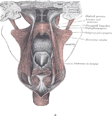
The muscles associated with ET are: Tensor veli palati, Levator veli palati,
salpingopharyngeus, and tensor tympani (Figure-2). The bony part remains open and the
cartilaginous ET is closed at rest. It opens during swallowing, sneezing and yawning thus
equalising the middle ear pressure to that of the atmospheric pressure. Closure of the tube
is achieved by the extrinsic forces of the surrounding deformed tissues and /or by the
recoil of the elastic fibres or the tubal wall and cartilage.10 The TVP consists of two
muscle bundles. The more lateral bundle originates from the scaphoid fossa and the
greater wing of sphenoid bone superior to the ET cartilage. The muscle descends
anteriorly, laterally and inferiorly to converge into a tendon that winds around the
hammular process of the medial pterygoid and inserts into the posterior border of
horizontal process of the palatine bone and palatine aponeurosis. The more postero-
superior muscle fibres extend into the semi canal of tensor tympani muscle and receive a
second muscle slip from the tubal cartilage and the sphenoid bone. The fibres converge
to a tendon and winds around the processes cochleariformis and inserts into the neck of
6
from the posterior half of lateral membranous wall of ET. The fibres descend and blend
with the lateral bundle. This also called the dilator tubae is the primary dilator of the
ET.21 LVP arises from the inferior aspect of the petrous temporal bone. It lies
inferolateral to the inferior margin of the medial lamina of the anterior ET and inferior to
the medial lamina of the posterior ET suggesting that the LVP opens the anterior
cartilaginous portion of the ET by rotating the medial lamina with increased dimension of
the muscle body when it contracts.23 The Salpingopharyngeus muscle arises from the
medial and inferior border of tubal cartilage and passes inferoposteriorly to blend with the
Palatopharyngeal muscle. The fibres are very thin and lack ability for any physiological
[image:14.595.128.489.416.789.2]function.10
7
The middle ear together with the ET, aditus, antrum, and the mastoid air cells is
called the middle ear cleft. The ME has a roof, a floor, medial, lateral, anterior and
posterior walls. The anterior wall is narrow as the medial and lateral walls converge. The
lower portion of the anterior wall consists of a thin plate of bone covering the carotid
artery. The smaller upper part has two parallel tunnels placed one above the other. The
lower one leads to the bony ET while the upper, separated from the bony ET contains the
tensor tympani muscle.
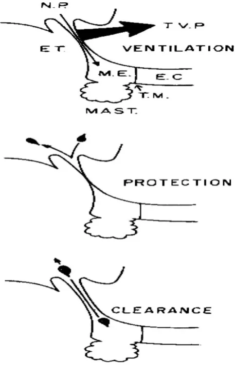
Function of ET
Three physiological functions have been described for the ET.24 (Figure.3).
1) Ventilation of the middle ear to equilibrate the middle ear pressure with that of the
atmosphere. 2) To transport the middle ear secretions to the nasopharyx by mucociliary
clearance. 3) To protect the middle ear from nasopharyngeal sound pressure and
secretions.
Ventilatory function:
The transducer function (necessary for the normal hearing mechanism) of middle
ear is optimal when the ambient and middle ear pressures are equal. This is facilitated by
the opening of the ET. The ME is considered as a rigid non-collapsible gas pocket
surrounded by vascular mucosa. Gases are exchanged between the middle ear spaces and
the mucosa. Partial pressure of gases in the mucosa approximates that of microcirculation
of middle ear mucosa. Middle ear O2 is slightly higher than in microcirculation, whereas
CO2 is in equilibrium.25 The diffusion gradient is created by N2 partial pressures and
8
deglutition and other maneuvers, the pressure difference between the environment and the
ME are equalised by the inflow and outflow of gases. The ventilator function maintains
near equilibrium between internal and external pressures, thus maintaining the near
optimal transducer function of the ME and preventing pathological consequences that
result from unabated ME to mucosa gas exchange. In an attempt to describe normal ET
function using micro flow technique in a pressure chamber, Elner et al26 studied 102
adults with intact tympanic membrane and no ear disease. The patients were divided into
four groups according to their ability to equilibrate static relative positive and negative
pressures of 100mm of water in the ME. Group 1 equilibrated the pressure completely;
group 2 equilibrated positive pressure, but had a small residual negative pressure in the
ME. Group 3 were capable of equilibrating only relative positive pressure with a small
residual remaining, but not negative pressure; those in group 4 were unable to equilibrate
any pressures. This indicated decreased stiffness of ET in group 2 to 4 when compared to
group 1. This study also showed that 95% of normal adults could equilibrate an applied
positive pressure and 93% could equilibrate applied negative pressures to some extent by
active swallowing. However 28% of the subjects could not completely equilibrate either
applied positive or negative pressure or both.
Protection, drainage and clearance
The ET, ME and mastoid air cells can be considered as a flask with a long narrow
neck.27 The nasopharyngeal end can be considered as the mouth of the flask, the narrow
neck, the isthmus of the ET and the bulbous portion the ME and the mastoid air cells.
Fluid flow through the neck will depend upon the pressures at both ends, the radius and
length of the neck and the viscosity of the fluid. When a small amount of fluid is instilled
9
capillarity at the neck and positive pressure that develops in the chamber of the flask. If
there is a perforation of the tympanic membrane, secretions can move from the
nasopharynx to the ME as there is no positive pressure from the mastoid to prevent the
reflux. If a high negative pressure develops in the ME and the mastoid, fluid gets
aspirated into the ME and mastoid from the nasopharynx. Similarly if a high positive
pressure is applied at the nasopharynx, fluid insufflates into the ME. The other factors
which affect the flow of gas and fluid through the ET are 1) mucociliary clearance.28, 29,
[image:17.595.186.428.354.746.2]2) active tubal opening and closure, 3) Surface tension characteristics.30
10
Pathophysiology of middle ear disease
There are various factors associated with the etiology and pathogenesis of ME
disease. These include ET dysfunction, other host related factors like genetic
predisposition, age, prematurity, allery, immunodeficiency, craniofacial abnormalities,
environmental factors like upper respiratory tract infections, attendance to day care,
number of siblings, absence of breast feeding, and socioeconomic factors. ET dysfunction
is one of the most common causes of ME diseases. There are many studies which show
sufficient evidence to support the „hydrops ex vacuo‟ theory of Adam Politzer, which
says that when the ET is blocked and there is edema of the ME mucosa, ME under
pressure develops and there is transudation of serous fluid into the ME.31Anatomic or
functional obstruction of the ET is the primary cause of ME negative pressure. Anatomic
obstruction is frequently due to inflammation mostly due to viral infection. Others are
inflammation due to allergy or obstruction at the nasopharyngeal end due to adenoids or
tumour and the middle ear due to granulations. Functional causes are floppy cartilage,
dysfunction of the TVP muscle, constriction of the ET while swallowing and otic
barotrauma. Secondary cause of ME negative pressure is inflammation in the ME. This
can lead to obstruction of the bony ET with secretions, thus impairing the pressure
regulatory and clearance functions of the ET. ME under pressure leads to acute otitis
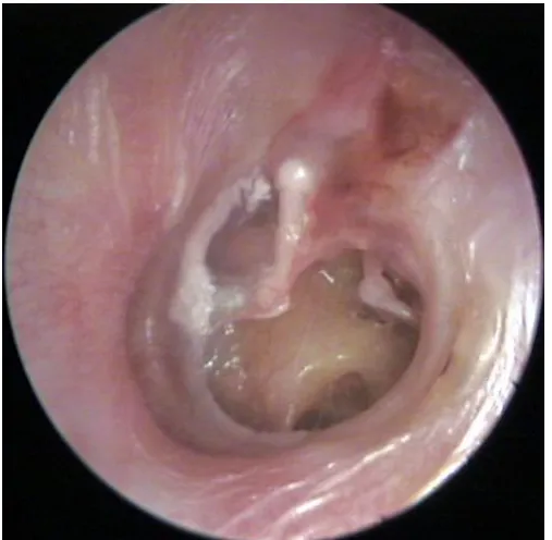
media (Figure.4) and otitis media with effusion (Figure.5). Acute otitis media with
otorrhea occurs when there is rupture of tympanic membrane. If acute otitis media with
tympanic membrane rupture does not heal spontaneously or following treatment it can
lead to chronic otitis media (Figure.6). Negative ME pressure also enhances reflux and
11
dysfunction of the ET due to reflux or insufflation of nasopharyngeal secretions into ME,
which can occur in condition like patulous ET or cleft palate. In chronic suppurative
otitis media the ME and mastoid get infected from the nasopharyngeal secretions and the
contaminants from the external auditory canal (Figure.7). One of the most important
factors associated with otitis media in infants and young children is immaturity of the
structure and function of the ET and immaturity of the immune system.32
The antecedent event in an acute otitis media is a viral upper respiratory tract
infection. This leads to congestion and inflammation of the mucosa of the nasopharynx
and ET and thus obstruction of the ET. Further development of ME under pressure
occurs especially in mastoids, where the mastoid air cell system (MACS) volume is less.
Larger MACS will partially protect the ME from development of a pathological under
pressures by allowing for a decreased frequency of ET openings and/or less efficient ET
openings when compared to a smaller MACS volume.33 Negative ME pressure is
followed by aspiration of pathogens (viral and bacterial) into the ME from the
nasopharynx. This along with the secretions from the middle ear mucosa gets collected,
allowing the pathogens to proliferate that eventually leads to suppuration. In a study by
Buchman34 and colleagues where 32 healthy adults were subjected to a nasal inoculation
of respiratory syncytial virus, 18 (56%) of them were infected with the virus and by the
6th day only 46% of the infected subject had a normal ME pressure (> 100mmof H2O),
showing the relationship between viral upper respiratory tract infection and otitis media.
Otitis media with effusion has a similar sequence of events as that of acute otitis
media. Swarts35 and associates were also able to produce ME effusion in the monkey
shortly after inducing negative middle-ear pressure by flushing the middle ear with CO2.
This study confirm the „hydrops ex vacuo‟ theory of the pathogenesis of ME effusion,
12
into the microcirculation of the mucous membrane causes a middle-ear under pressure,
followed by transudation of effusion.
Tympanic membrane perforations that are acute are usually secondary to AOM
which may also occur during the course of chronic otitis media with effusion .36 Chronic
perforation of the tympanic membrane usually occurs when an acute perforation fails to
heal. When there is acute drainage through perforated tympanic membrane that persists
for 2 weeks to 3 months or longer the infection is chronic suppurative otitis media. There
is no consensus on the duration of otorrhea to be termed chronic; however, as per the
WHO definition.37, otorrhoea persisting through a perforated tympanic membrane for > 2
weeks is believed to be due to CSOM. It has been shown that in patients with chronic
otitis media with effusion there is degeneration of lamina propria of the tympanic
membrane with decrease in thickness of fibrous layers.38 This weakness of tympanic
membrane predispose to perforation with less chance of spontaneous healing. Thus the
pathogenesis of CSOM is linked to abnormal ET function.32 In inactive mucosal CSOM,
the structure and often the hearing is impaired by the presence of a permanent tympanic
membrane defect but there is no active infection or mucus discharge. Such an ear may
remain inactive, become active or may even occasionally heal. Active mucosal CSOM is
associated with resorption of parts or all of the ossicular chain (resoptive osteitis).39
13
Figure.4: Acute suppurative otitis media
[image:21.595.194.418.496.735.2]14
[image:22.595.181.435.150.398.2]Figure.6: Chronic suppurative otitis media - tubotympanic disease
15
Figure.8:Resorptive osteitis
Surgical treatment of CSOM tubotympanic disease
The main objectives of surgery in patients with CSOM tubotympanic disease
(TTD) are
1) To remove the active disease and promote healing
2) To restore an air filled ME cavity and prevent recurrence.
3) To prevent complications
4) To restore hearing function
The surgical procedures often done are myringoplasty, tympanoplasty and cortical
16
edges of the perforation and placement of a graft of temporalis fascia or perichondrium by
an underlay or overlay technique to close the perforation. Tympanoplasty involves
reconstruction of tympanic membrane and/or the ossicular chain. Studies on outcomes in
terms of tympanic membrane uptake in these surgeries have been reported to be 78% and
95% in the hands of trainees and senior staff respectively.40 Many authors suggest that a
cortical mastoidectomy (Figure.10) should be carried out at the same time as
myringoplasty in active ears and inactive ears.41 In a study by Mishiro et al42 in patients
with mucosal CSOM, 251 ears were operated and followed up for one year. It was
divided into two groups; group A with 147 patients underwent tympanoplasty with
mastoidectomy and group B with 104 tympanoplasty without mastoidectomy. Graft
success rates were 90.5% in group A and 93.3% in group B. There was no statistically
significant difference in graft success rate. Graft success rates of discharging ears were
90.0% in group A and 85.7% in group B. Graft success rates of dry ears were 90.7% in
group A and 94.4% in group B. There was no statistically significant difference between
discharging ears and dry ears. The rates of the postoperative air-bone gap within 20dB
were 81.6% in group A and 90.4% in group B, without a statistically significant
difference. This study shows that mastotoidectomy need not be combined with
17
18
Figure.10: Cortical mastoidectomy
Eustachian tube function tests
There are various tests to assess the function of the ET. These include pneumatic
otoscopy.43, nasopharyngoscopy and ET endoscopy44 and tympanometry. The latter is an
objective way of determining ME pressure and thus the function. It is used to test the
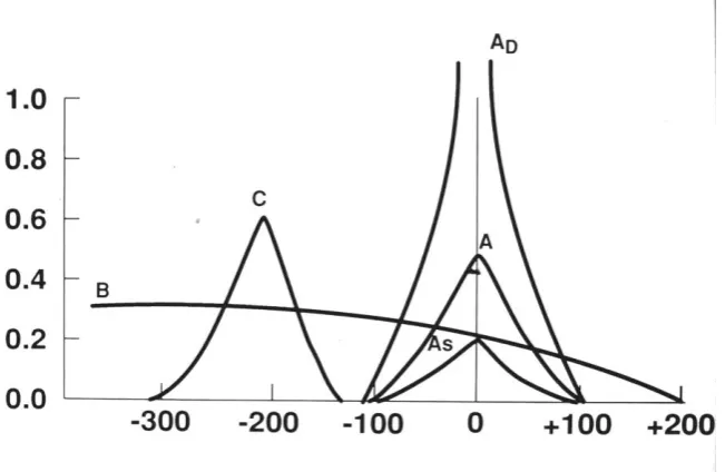
condition of the middle ear and mobility of the tympanic membrane by creating variations
of air pressure in the ear canal. A compliance peak on a tympanogram indicates that the
ear canal and middle ear pressures are equal, and that acoustic transmission through the
middle ear is maximal. Tympanograms (Figure.11) are classified with respect to this
peak.45 Some of the classical tests of tubal patency include Valsalva maneuver, Toynbee
19
the ME inflates following Valsalva or Politserisation, then the tube is not totally
obstructed. Similarly if the tympanic membrane is not intact, the passage of air into the
ME indicates ET patency. Elner et al26 reported that 86% of otologically normal adults
could perform Valsalva. The other tests include dye studies with fluroscien.46,
[image:27.595.146.472.274.486.2]saccharine.47 and sonotubometry.48
Figure.11: Tympanogram
Drawbacks of the ET function tests:
Otoscopy cannot differentiate between a functional impairment and mechanical
obstruction of the ET. Valsalva maneuver is an objective method of assessment of ET
function and is difficult to perform in the paediatric age group. Catheterization can
traumatise the ET while inserting the catheter. Tympanometry which is done in an intact
20
21
Inflation-deflation test was first reported by Flisberg et al.49 It can be done in ears with
chronic suppurative otitis media with a perforated drum, in patients who have undergone
myringotomy and grommet insertion for otitis media with effusion and also in an intact
tympanic membrane. The test is easy to perform, non-invasive and reproducible. It is
possible by this method to determine at which level of negative or positive pressure
applied within the ear space air passes through the tube during swallowing. (Figure.13,
[image:29.595.91.521.329.755.2]14)
22
Figure.14: Deflation test of Eustachian tube function
Takahashi et al50 reviewed 78 patients with non cholesteatomatous chronic otitis
media, without ossicular damage. ET pressure-regulation functions were examined using
an inflation-deflation test, and postoperative hearing levels were measured. Patients with
poor pressure regulation showed a significantly higher incidence of poor outcomes, poor
hearing recovery, spontaneous perforation of the tympanic membrane, or persistent wet
condition. Uzun51 reported 61 ears with pars tensa cholesteatoma who underwent
tympanoplasty with cartilage palisades or fascia. The authors evaluated preoperative ET
function using the Valsalva maneuver and found that late hearing results were better in
patients with preoperative positive Valsalva maneuver than they were in those with
23
significantly better in those treated with cartilage palisade tympanoplasty than they were
in those treated with fascia reconstruction. The authors suggested that cartilage palisade
tympanoplasty might be a better reconstruction option for patients with poor ET function.
Choi et al52 in their study of in their study of 137 patients with non cholesteatomatous
ears found that a successful type 1 tympanoplasty and restoration of well aerated ME and
good ET function permit better compliance of tympanic membrane and predictive of
improved postoperative hearing. According to a study done by Kurien et al53, the ET
inflation deflation test is a sensitive test for predicting aditus patency in patients with dry
24
MATERIALS AND METHODS
This study was performed in the ENT outpatient department at Christian Medical
College Vellore, between May 2010 and July 2011. All patients aged 16 years and above
with mucosal chronic suppurative otitis media (CSOM), who were scheduled to undergo
surgery, were included in the study. A brief history of their problems and clinical
findings, were entered into the proforma. Otomicroscopic examination was performed to
assess the size and site of the tympanic membrane perforation. The presence of
tympanosclerosis, granulations, discharge and exposure of round window or ossicles was
noted. Details were entered into the proforma.
Exclusion criteria: Presence of any of the following conditions:
Profusely discharging ear
Cholesteatoma
Immunosuppression
Poorly controlled diabetes mellitus
Methods:
a) Audiological assessment
Hearing assessment was performed by pure tone audiometry to note the mean value of the
air-bone gap at 500, 1000, 2000 and 3000 Hz. Hearing loss was quantified as per
25
Mild - 26-40 dB
Moderate - 41-55 dB
Moderately severe - 56-70 dB
Severe - 71-90 dB
Profound - > 90 dB
b) ET patency was assessed on the day prior to surgery by the following method:
1. Valsalva maneuver
In this maneuver positive pressure is built in the nasopharynx by sealing it at the
nasal and pharyngeal openings so that air enters the ET. This was done by asking the
patient to pinch his nose and close his mouth and blow air into his ears. ET was deemed
to be patent if the patient got a subjective sensation of opening of the ear or air passing
through the ear. This test was done both pre and postoperatively.
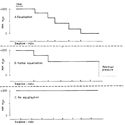
Inflation-deflation test
The external auditory canal of the patient was fitted with a rubber ear tip of
corresponding size so as to maintain an air tight seal. The otoadmittance probe was
connected to a standard otoadmittance meter (GSI Ltd). The middle ear was then inflated
with a constant flow of air until the ET is opened passively. This was marked by a sudden
26
The middle ear pressure was then equilibrated by performing 5 consecutive
swallows with an interval of ten seconds in between to prevent strain on the pharyngeal
muscles (active tubal function). The residual pressure after 5 swallows if any was
recorded. The deflation phase of the test was performed by giving a pressure of - 400 mm
to the middle ear and asking the patient to swallow again. The pressure remaining in the
middle ear after 5 consecutive swallows was recorded. The ET was considered to be
patent if there was no or minimal residual pressure during both inflation and deflation.
The results of the inflation deflation test were categorised based on whether the
patients could equalise pressure or have a residual pressure of 100mmof H2O or less as
being patent. Those patients who could not equalise pressures on swallowing either with
positive or negative pressures to <100mm of H2O were deemed to have a non-patent or
blocked ET. This group included patients in whom there was no change with either
positive or negative pressure.54 The participants of the study underwent myringoplasty or
tympanoplasty with cortical mastoidectomy on the following day. The tympanic
membrane perforation was closed using a temporalis fascia graft.
c) Postoperative assessment
1. Otomicroscopy was performed to note the integrity of the tympanic membrane.
The following specific features were observed with respect to the tympanic
membrane:
i. 1 – Normal
27
iii. 3 – Retraction
iv. 4 - Tympanosclerosis
v. 5 – Granulation over graft
2. Postoperative pure tone audiometry was performed at the same frequencies as
preoperatively.
3. Postoperative middle ear aeration was assessed in those patients with intact
tympanic membranes using tympanometry. An automated otoadmittance meter
was used to record a tympanogram. The results of tympanometry were expressed
as follows:
A-normal
As-Reduced compliance at ambient pressure
Ad-Increased compliance at ambient pressure
B-flat or dome shaped
C-Maximum compliance at pressures more than -100 mm H2O (Blocked
28
Sample size calculation:
Sample size for this study was calculated using the following formula
2 2 1
2 1 2 / 1
) (
* 2 * ) (
p p
PQ Z
Z n
Where,
n = sample size
p1 = incidence of blocked ET postoperatively in patients with patent ET preoperatively
p2 = incidence of blocked ET postoperatively in patients with blocked ET preoperatively
P = (p1 +p2) / 2
Q = 1 – P
Alfa = 5%
Beta = 80%
This calculation was derived based on the study by Choi HS et al52 in which poor
ET function was seen postoperatively in 40% of those patients with CSOM who had
minimal or no residual pressure (good ET function) preoperatively and 67% of those who
had moderate to large amounts of residual pressure ( poor ET function) preoperatively.
Using these figures we estimated that in order to detect this difference at 5% level of
significance, with a power of 80% the required sample size is 57 in each group. With a
speculated loss to follow up of 40%, the number required in each group will be 90
29
Statistical analysis
Descriptive analysis was used for frequency calculation. This was used to analyse
the preoperative and postoperative ET function. Pearson Chi-square test and Fisher‟s
exact test was used for categorical data to check the association. The change in ET
function and the association of the ET function with graft uptake were analysed using
30
RESULTS AND ANALYSIS
Of the 182 patients in our study who underwent surgical closure of tympanic
membrane, 106 of them were reviewed postoperatively over a period of 3 to 15 months.
Table 1: Distribution of age of study patients (n = 182)
Age (yrs) Number (%)
<40 133 (74.1)
>40 49 (26.9)
Total 182
Figure 15: Age distribution of study patients
133 (73%) 49 (27%)
< 40 years
> 40 years
The age range was 16-59 years with a mean age of 32.6 years. Most patients
(74.1%) of them were below the age of 40 years or less. This was probably because it was
31
Table 2: Distribution of sex (n = 182)
Sex Number (%)
Male 105 (57.6)
Female 77 (42.3)
Total 182
Figure.16: Sex distribution of study patients
[image:39.595.89.453.449.672.2]32
Table 3: Laterality of disease in affected patients (n = 182)
Side Number (%)
Unilateral 102 (56)
Bilateral 80 (44)
Total 182
[image:40.595.142.473.145.383.2]Unilateral disease (56%) was slightly more common than bilateral disease (44%).
Figure 17: Distribution of laterality of ear disease:
102 (56%)
80 (44%)
0 20 40 60 80 100 120
Unilateral disease Bilateral disease
33
Table 4: Distribution of duration of ear discharge (n=182)
The duration of the ear discharge ranged from (0.08 yrs to 50 yrs). While most
patients had ear discharge for less than 10 years, a significant number (41.2%) had
[image:41.595.127.462.125.359.2]discharge of > 10 year‟s duration. Of these, only a small proportion had sought surgery.
Figure 18: Distribution of duration of ear discharge
107 (59%) 75 (41%)
< 10 years
> 10 years
Duration of ear discharge Number (%)
< 10yrs 107 (58.8)
>10yrs 75 (41.2)
34
Table 5: Distribution of revision surgery (n = 182)
Previous surgery Number (%)
Revision surgery 14 (7.6)
First surgery 168 (92.3)
Total 182
Most of the ears (92.3%) were operated for the first time and only 7.6% had
[image:42.595.137.474.77.334.2]undergone revision surgery.
Figure 19: Distribution of revision surgery
14 (8%)
168 (92%)
Revision surgery
35
Table 6: Distribution of co morbidities (n = 182)
Comorbid conditions Number (%)
Present 21 (11.5)
Absent 161 (88.5)
Total 182
11.5% of the patients had comorbid conditions like diabetes mellitus, hypertension,
[image:43.595.84.419.76.334.2]hypothyroidism or bronchial asthma.
Figure 20: Distribution of comorbid conditions
21 (12%)
161 (88%)
Comorbid conditiond present
36
Table 7: Distribution of perforation size (n = 182)
Size of perforation Number (%)
Small / Moderate 36 (19.8)
Large / Subtotal 146 (80.2)
Total 182
Most (79.6%) of the patients had a large or subtotal sized perforation.
Table 8: Distribution of state of middle ear mucosa (n = 182)
Middle ear mucosa Number (%)
Dry 92 (50.5)
Moist 90 (49.5)
Total 182
[image:44.595.140.473.479.715.2]37
Table 9: Distribution of preoperative Valsalva maneuver test results (n = 182)
Preoperative Valsalva maneuver test results Number (%)
Patent 100 (54.9)
Not patent 82 (45)
Figure.21: Distribution of preoperative Valsalva maneuver
100 (55%)
82 (45%) Valsalva patent
Valsalva nonpatent
Preoperative Valsalva showed that the ET was patent in a little over half of the
patients. Thus, this test of ET function showed that ET dysfunction was not an important
38
Table 10: Distribution of preoperative inflation and deflation test results (n = 182)
Preoperative inflation & deflation test results Number (%)
Patent 68 (37.4)
Not patent 114 (62.6)
Total 182
Figure.22: Distribution of preoperative inflation deflation test
68 (37%)
114 (63%)
Inflation deflation patent
Inflation deflation nonpatent
The other test of ET functioned employed in this study was the inflation-deflation
test. The test was done in patients with mucosal chronic suppurative otitis media
preoperatively. Most patients appeared to have blocked ET by the preoperative
inflation-deflation test. The results of this test are quite different from that of the Valsalva test
39
Table 11: Distribution of antral contents (n =114)
Antral mucosa Number (%)
Normal mucosa 57 (50)
edematous mucosa 57 (50)
Total 114
Almost equal numbers of patients had either edematous or normal mucosa.
Table 12: Distribution of aditus status (n =114)
Aditus status Number
Patent 57(50)
Blocked 57(50)
Total 114
Equal numbers of patients had blocked and patent aditus intra operatively. This is
probably related to the status of the antral mucosa. Thus patients with edematous mucosa
40
Table 13: Association between preoperative Inflation deflation test result and
preoperative Valsalva result
Preoperative Valsalva test
result
I-D patent I-D not patent Test used for significance
P value
Patent 49 (72.1%) 51 (44.7%) Pearson Chi-
square without Yates correction
0.000*
Not patent 19 27.9%) 63(55.3%)
72.1% of the patients with a patent ET on Valsalva maneuver showed patent ET
on the inflation deflation test also. The association between the 2 tests of ET function
[image:48.595.85.514.229.403.2]41
Postoperative follow up was obtained in 106 patients, all of whom underwent
[image:49.595.87.417.264.543.2]tympanometry and otoscopy.
Table 14: Distribution of postoperative results (n = 106)
Postoperative
tympanic membrane status
Number (%)
Intact tympanic membrane 83 (78.4)
Residual perforation 23 (21.6)
Total 106
There were a total of 106 patients who came back for follow up. The success rate of
42
Table 15: Distribution of postoperative tympanometry test results (n =83)
Postoperative tympanometry was done to assess the middle ear status and the ET
function in all those with intact tympanic membranes. 72.2% had a normal postoperative
tympanometry and 27.7% had an abnormal tympanometry. The presence of B curves
suggests either the presence of middle ear fluid or thickened tympanic membrane, those
with C curves probably had ET block.
Postoperative tympanometry test results Number (%)
A, As, Ad 60 (72.2)
C, Cs, B 23 (27.7)
43
Table 16: Distribution of postoperative Valsalva test results (n = 106)
Postoperative Valsalva maneuver test results Number (%)
Patent 61 (57.5)
Not patent 45 (42.5)
Total 106
Figure 23: Distribution of postoperative Valsalva maneuver
61 (58%) 45 (42%)
Valsalva patent
Valsalva nonpatent
There was an increase in the number of patients with postoperatively patent ET
[image:51.595.144.461.442.645.2]44
Table 17: Comparing the change in ET function assessed by Valsalva maneuver
preoperatively and Valsalva maneuver postoperatively
Preoperative Valsalva
maneuver test result
Change present in ET function
assessed by Valsalva
maneuver (%)
No change in ET function
assessed by Valsalva
maneuver (%)
Test used for significance
P value
Patent 12(48) 48(59.2) Pearson Chi-
square without Yates correction
0.320
Not patent 13(52) 33(40.7)
Thus, several patients with preoperatively blocked ET on Valsalva maneuver had
a change in ET function postoperatively. A greater number of patients with blocked ET
showed change when assessment was done by Valsalva maneuver. However, the
difference between the groups was not found to be statistically significant (p=0.32).
When ET function was assessed by Valsalva maneuver preoperatively and by
tympanometry postoperatively, the change in ET function was more apparent (Table 18),
particularly for patients who had a blocked ET preoperatively. An „A‟ tympanogram was
[image:52.595.97.518.181.405.2]45
Table 18: Comparing the change in ET function assessed by Valsalva maneuver
preoperatively and tympanometry postoperatively
Preoperative Valsalva maneuver test
result
Change present in ET function assessed by
tympanometry
(%)
No change in ET function
assessed by tympanometry
(%)
Test used for significance
P value
Patent 9(30.0%) 39(73.5%) Pearson Chi-
square without Yates
correction
< 0.001
Not patent 21(70.0%) 14(26.4%)
When assessed by Valsalva maneuver preoperatively and tympanometry
postoperatively, if a patient had a blocked ET preoperatively there was a higher chance of
it changing than if a patient had a patent tube (p<0.001). Conversely, if a patient had a
patent ET preoperatively, there was not much change in the ET function postoperatively.
The difference between the two groups those with patent and non-patent ET was
statistically significant (p<0.001).
The analysis was repeated using the inflation deflation test preoperatively and
tympanometry postoperatively (Table 19). Here again, an A tympanogram with an intact
[image:53.595.76.538.138.350.2]46
Table 19: Comparing the change in ET function assessed by inflation deflation test
preoperatively and tympanometry in the postoperative period
Preoperativ e inflation
deflation test result
Change present in ET function assessed
by
tympanometry(%)
No change in ET function assessed by tympanometry
(%)
Test used for significance
P value
Patent 10 (22.2) 25 (65.8) Pearson Chi-
square without Yates correction
< 0.001
Not patent 35 (77.8) 13(34.2)
Change in ET function following surgery was also assessed by the inflation
deflation test. Here again, patients with preoperatively diagnosed ET block by the
inflation deflation test were more likely to show a change in ET function than those with
preoperatively patent ET. This difference between those with preoperatively patent and
blocked ET was statistically significant (p< 0. 001).
We then analysed the data to see if the preoperative ET function test result was
predictive of closure of the perforation. The analysis was done first with the results of the
47
Table 20: Association between preoperative inflation-deflation test result and
successful closure of perforation
Preoperative inflation deflation
test result
Intact tympanic membrane
Residual perforation
Test used for significance
p-value
Patent 35 (42.2%) 5 (21.7%) Pearson
Chi- square without
Yates correction
0.074
Not patent 48 (57.8%) 18 (78.3%)
Although the patients with non-patent ET had a higher rate of residual perforation
[image:55.595.94.518.261.454.2]48
Table 21: Association between preoperative Valsalva test result and successful
closure of perforation
The same association was looked at with preoperative Valsalva maneuver and a similar
result was seen. So the ET function does not impact on successful closure of perforation.
This could be because a number of patients had a change in ET function following
closure of the perforation.
Pre OP Valsalva maneuver test
result
Intact tympanic membrane.
Residual perforation
Test used for significance
p-value
Patent 48 (57.8%) 12 (52.2%) Pearson
Chi- square without
Yates correction
0.628
49
DISCUSSION
The etiopathology of middle ear infection can be attributed to many causes of
which ET dysfunction is considered to be a very important factor. Middle ear infection
leads to further edema and obstruction of the ET.5 Whether ET dysfunction is a cause or a
result of chronic otitis media has been a matter of much debate. Several methods like
Valsalva maneuver, Toynbee manoeuvre, ET catheterization, Politzerisation, fluroscein
dye method, saccharine test sonotubometry, inflation deflation method, and
tympanometry have been described for the assessment of the ET function and different
results have been obtained. Among all these tests, the Valsalva test and the inflation
deflation test have proved to be the most reliable and popular.55‟56
ET block is usually transient in many patients with mucosal CSOM. In normals,
too, there is diurnal variation in ET patency. Brattmo et al 57 study showed that middle ear
pressure regulation is a dynamic process and that at various periods during the day and
the night, the ET could be blocked or patent. He also found that there was a significant
difference in the diurnal changes in the middle ear pressure in patients with central
perforation compared to those with normal healthy ears.
The results of different tests of ET function often show disparate results. This is a
big drawback when comparison of results of ET function using different techniques is
performed. MacKinnon58 found that a number of patients who had good ET function on
Valsalva maneuver had a poorly functioning ET on the aspiration deflation study. In the
present study we found, however, that there was a good correlation between the results of
50
tympanometry and Valsalva test were quite disparate. One of the reasons for this could be
that the manoeuvre was not performed properly. There could have been a number of
patients who despite adequate training could not perform a proper Valsalva and were
labelled as having blocked ET. Another reason could be that the ET function is variable
and can give different results at different points of time.
The Valsalva maneuver is simple to perform and can be performed at the clinic or
bedside and does not require much time to evaluate. Many authors have utilised this
technique to evaluate ET function. It is an easily performed test not requiring any extra
equipment and hence could be routinely performed for all patients undergoing middle ear
surgery. Tos55 studied both squamous and mucosal chronic otitis media using the
Valsalva maneuver and found that the poorest function was in those with squamous
disease and active mucosal chronic otitis media. The dysfunction was least in those with
inactive chronic mucosal otitis media. Andreasson and Harris56 in their study felt,
however, that training is needed to perform a Valsalva maneuver. The Valsalva maneuver
does not reflect on the normal physiological function of the ET. The present study is
comparable with results of other studies. When Valsalva maneuver was used to assess the
ET patency, many authors found that the ET was patent in the majority 55, 56, 57, 58, 59. The
exact amount of pressure generated while doing Valsalva is not measured. The pressures
generated are high and may possibly force open the ET. The major inference that we can
make from a positive Valsalva is that there is no organic stenosis of ET.
ET function assessed by aspiration deflation method is considered to be a more
physiological test. Choi et al 52 showed that in patients with mucosal chronic otitis media
the ET was more likely to be blocked on modified inflation deflation test. He also showed
51
MacKinnon58 in his study using this test found that with a negative middle ear pressure
test, more patients with mucosal chronic otitis media showed a blocked ET.
Virtanen60 in his study using this test, showed that most of the patients with
mucosal chronic otitis media had a blocked ET with the inflation deflation method. In the
present study, too, we found that most patients had a blocked ET on the inflation-
deflation test. The block was more evident when negative pressure was applied. In
contrast, Andreasson and Harris56 in their study found most patients to have a patent ET
on aspiration deflation test. This could be due to the short duration for doing the test,
which do not pick up fluctuations in the ET function during a longer period and also other
methods like yawning, moving the jaw or autoinflation which equilibrate the
underpressure.
Although our null hypothesis for this study was that there was no change in ET
function with surgery, it was found on analysis of the results (when preoperative
Valsalvamaneuver and inflation deflation were compared with postoperative
tympanometry) that many of the non-patent ET became patent following surgery. This
was quite different from those patients whose ET was patent preoperatively and this
difference was statistically significant (p<0.001). This disproved our null hypothesis.
However, when the results of preoperative and postoperative Valsalva were compared,
the changes were not significant. Similarly, Andreasson and Harris56 found that there was
no change in a patient‟s ability to perform Valsalva maneuver and hence there was no
change in ET function when assessed by Valsalva maneuver. However there was a
significant change in ET function when assessed with the aspiration deflation test. Since
none of their patients with a preoperative blocked ET had granulations or polyps blocking
52
environment in the closed middle ear cavity resulting in a change in the middle ear
mucosal lining. Also, a pressure difference occurs across the ET in a closed middle ear
cavity which might act as a trigger for the ET to regain function. Our study clearly
showed that ET function changed after surgical closure of the tympanic membrane
perforation. The change was seen in a far greater number of patients than that showed by
other studies.
Tos55 in his study also showed that ET function changed for a number of patients
after surgery. In a long term follow up, patients who persisted to have poor postoperative
ET function had a higher chance of developing retractions and adhesions of the tympanic
membrane. Choi et al52 in their study of patients with mucosal CSOM who underwent
myringoplasty or tympanoplasty also showed that ET function changed after surgery. It
was suggested that removal of mucosal edema and granulations blocking the ET probably
resulted in the change in ET function. In contrast, the number of patients with change in
ET function in Virtanen‟s60
study comparing preoperative aspiration deflation with
postoperative tympanometry, were not many. His study does not support the hypothesis
that ET function changes after surgery. He suggested that this could not occur without
intervention at the tubal orifice during surgery. He also suggested that the small pressure
range used in tympanometric studies may be insufficient to move a tympanic membrane
and hence may be inaccurate in assessing the normal aeration of middle ear.
The success rate of perforation closure in various studies in which ET function
tests have been done varies from 74.5 %to 94.8% 59‟ 52. In the present study the rate of
tympanic membrane closure was 78.4%. The relatively high rate of residual perforation
(21.6%) may be related to the fact that surgery was performed by surgeons with varying
53
resistant organisms and, of course, poor ET function. The importance of poor ET function
as an etiological factor for residual perforation has been stressed by several authors.50, 54,
59
Some authors have evaluated ET function in relation to successful closure of
perforation in mucosal chronic suppurative otitis media also. Depending on the test used
to assess ET function, variable results were obtained.
The effect of ET function as assessed by the Valsalva maneuver on successful
closure of tympanic membrane perforation was assessed in the present study. There
appeared to be no correlation between preoperative Valsalva test result and the presence
of either an intact tympanic membrane or residual perforation. Similar results were
obtained by other authors.56 In the study by Reimer et al59, however, a patent ET on
Valsalva was more likely to be associated with an intact tympanic membrane. His study
also showed that ears with a combination of positive Valsalva and a functional volume of
more than 2 ml had a better healing rate than those with a negative Valsalva and volume
of less than 2 ml. There was no association between a blocked ET preoperatively and
residual perforation.
When the inflation deflation technique was used, some authors MacKinnon58
found that healing was much more in patients with a good preoperative ET function than
those who had bad ET function. A dry middle ear without any signs of infection and with
a good or moderate preoperative ET function had more chance of healing after surgery.
However on longer follow up in spite of a good ET function, the authors found that there
was a chance of developing recurrent otitis media and tympanic membrane perforation in
some individuals (10%). Choi et al52 showed that a good ET function was related to well
aerated middle ear with an „A‟ type tympanogram and a good tympanic membrane
54
also block the ET. Re-perforations in his study were seen in patients with diseased middle
ear mucosa. Holmquist‟s54 study also showed a good correlation of preoperative ET
function with results of myringoplasty. However Andreasson and Harris56 showed that
there was no correlation between healing and preoperative ET function when assessed
with the aspiration deflation method. According to the authors, the available ET function
tests are of no value for selecting patients suitable for middle ear surgery. In the present
study, we found an increased percentage of residual perforation in those with blocked ET
when assessed by the inflation deflation test. However, this did not reach statistical
55
CONCLUSION
In conclusion, the results of our study on preoperative ET function in patients with
mucosal CSOM show that the results of evaluation of ET function vary with the
technique used. There appears to be no single test of ET function which may be
consistently relied upon. However, the Valsalva tests and inflation deflation test are the
easily performed and most popular.
We arrived at 3 important conclusions in this study.
Firstly, the ET appeared to be blocked in most (62.6%) patients with mucosal
CSOM when assessed preoperatively with the inflation deflation test, whereas most
appeared tube patent (54.9%) with the Valsalva maneuver.
Secondly, there was a change in the ET function after surgery which was
significant (p<0.001) when assessed with both Valsalva and inflation deflation
preoperatively and tympanometry post operatively. The function of the ET tends to
change from blocked towards patency, possibly due to the removal of the disease and
return of normal physiological state after surgery.
Finally, there was an increased tendency of a patient with blocked ET to develop a
56
BIBLIOGRAPHY
1. Bluestone CD. Epidemiology and pathogenesis of chronic suppurative otitis media:
implications for prevention and treatment. Int. J. Pediatr. Otorhinolaryngol.
1998;42:207–23.
2. Holmquist J. Middle ear ventilation in chronic otitis media. Arch Otolaryngol.
1970;92:617–23.
3. Vartiainen E, Nuutinen J. Success and pitfalls in myringoplasty: follow-up study of
404 cases. Am J Otol. 1993 ;14:301–5.
4. Jonathan D. The predictive value of eustachian tube function (measured with
sonotubometry) in the successful outcome of myringoplasty. Clin Otolaryngol Allied
Sci. 1990 ;15:431–4.
5. Bluestone CD. Historical Perspective. In: Larissa Byj. Editor. Eustachian Tube
Structure, Function, Role in Otitis Media. 2nd ed. Vol 2. Hamilton. London BC
Decker Inc. 2005.p 4-9.
6. Eustachius B. Epistola de auditusorganis. Arch Otolaryngol 1944. (40):123.
7. Toynbee J. On Disease of the Internal Ear.Lond J Med. 1852 ;4:724–730.
8. Politzer A. Methods of Examination of Middle ear. In: Sir William Dalby.
Editor.Politzer's Text Book of the Diseases of the Ear and adjascent organs. 3rd ed.
Philadelphia and New york. Lea Brothers & Co.1894. p. 113-124
9. Swarts JD, Rood SR, Doyle WJ. Fetal development of the auditory tube and paratubal
57
10. Bluestone CD. Anatomy and Physiology of the Eustachian Tube. In: Cummings
Otolaryngology Head and Neck Surgery. 3rd ed. Vol 4.p 3003-3012
11. Sadler-Kimes D, Siegel MI, Todhunter JS. Age-related morphologic differences in the
components of the eustachian tube/middle ear system. Ann. Otol. Rhinol.
Laryngol.1989 ;98:854–858.
12. Proctor B. Embryology and anatomy of the eustachian tube. Arch Otolaryngol.
1967;86:503–514.
13. Macbeth R. Some thoughts on the eustachian tube. Proc. R. Soc. Med. 1960 ;53:151–
61.
14. Hasebe S, Sando I, Orita Y. Proximity of carotid canal wall to tympanic membrane: a
human temporal bone study. Laryngoscope.2003 ;113:802–7.
15. Prades JM, Dumollard JM, Calloc‟h F, Merzougui N, Veyret C, Martin C. Descriptive
anatomy of the human auditory tube. SurgRadiol Anat. 1998;20:335–40.
16. Luntz M, Pitashny R, SadJ. Cartilage in the bony portion of the eustachiantube.Eur
Arch Otorhinolaryngol.1990 ;247:56-59.
17. Sudo M, Sando I, Ikui A, Suzuki C. Narrowest (isthmus) portion of eustachian tube: a
computer-aided three-dimensional reconstruction and measurement study. Ann. Otol.
Rhinol. Laryngol.1997 ;106:583–8.
18. Lim DJ. Functional morphology of the mucosa of the middle ear and Eustachian tube.
Ann. Otol. Rhinol. Laryngol.1976 ;85:36–43.
19. Tomoda K, Morii S, Yamashita T, Kumazawa T. Deviation with increasing age in
histologic appearance of submucosal glands in human eustachian tubes.