0022-538X/94/$04.00+0
Copyright
©
1994, AmericanSociety forMicrobiologyHigh-Efficiency Identification
of Genes by Functional Analysis
from a Retroviral cDNA Expression
Library
BRYAN Y.
WONG, HONG CHEN, SIU-WAH CHUNG, AND PETER M. C. WONG*
Morse Institute
for Molecular
Genetics, Department of Microbiology and Immunology, State Universityof
New YorkBrooklyn,
NewYork 11203, and Fels Institute for
CancerResearch
and
Molecular Biology,
Departmentsof
Biochemistry, Microbiology, and Immunology, Temple
University,Philadelphia, Pennsylvania
19140 Received 23 March1994/Accepted
1June 1994Retroviral
genetransfer
efficiently
delivers genes of interest stably into target cells, andexpression cDNA
cloning
has beenshown
tobe
highly successful.
Considering
these twoadvantages,
we nowreport amethod
by which one can identify genes stimulating cell growth throughfunctional
analysis. The first step requires theconstruction of
a retroviral cDNA expressionlibrary
and theoptimization
of transfection of vector DNA into virus packaging cells. The second step involves the cocultivation of target cells with libraries ofretrovirus-producing
cells,resulting in the amplification of target cellstransduced
witha gene(s) stimulating cell growth.Under
standardized
conditionsof
transfection,
wedetected
an averageof 4,000 independent clones
perdish,
among which expression of a retroviral13-galactosidase
geneat an abundance of 0.2% could be detected. Next, wedemonstrated
theaugmentation of thesensitivity
ofthe assay by retroviralinfection and functionalanalysis.
We
did
thisby cocultivating
factor-dependent (FD)
cellswith dishes
ofGP/E
cells
transfected
withplasmids
containing various
molarratios of
pN2-IL3
DNAand retroviral
library
cDNAand by determining
thehighest
dilution
of
pN2-1L3
which still
resulted in the conversion of FD cells to factorindependence.
Theretroviral
interleukin-3
gene at anabundance
aslow as0.001%
could bedetected. Indeed,
wewere able to detectfrom FD cells thedevelopment
offactor-independent colonies with different phenotypes after retroviral
transfer ofcDNAs from
animmortalized hemopoietic
stem cellline. Thus,
the combination of astandardized
high-efficiency
DNAtransfection and
retrovirus-mediated
genetransfer should
facilitate the identification of
genescapable of
conferring
totarget FDcells adetectable
newfunction
orphenotype.
By
scaling
up thesize of
theexperiment realistically during
screening,
the assay candetect
cDNA at an abundance of lower than0.0001%.
Among
the
different methods of gene transfer, the use of
retrovirus vectors
has been shown to be highly
desirable.
Foreign
DNAof up to 10 kb may be efficiently delivered and
stably
incorporated
into target cells
(2,
10, 14-17,
21, 28, 34, 39,
58,
59). Packaging
cell lines which allow the generation of
heler-free recombinant retrovirus vectors at titers as high as
10
infectious particles per ml are available. The system itself
is well characterized and considered
tobe
relatively
safe for
usein gene
therapy,
despite
the
infrequent
occurrence
of
insertional mutagenesis.
Expression cloning
of genes without much
knowledge
of the
gene
products has been reported
by
anumber of
investigators
(45).
This
is usually achieved by assays in which
specific
biological
activities
canbe measured.
Size-selected
mRNAs
arethen converted
into cDNAs in plasmid vectors, which
arethen transfected into COS cells for
high
levels of
transient
expression of the gene
products
(1).
Inthis way, cDNAs of
interleukin-3
(IL-3) (69), granulocyte-macrophage
colony-stimulating
factor
(GM-CSF) (35),
IL-2
(68),
B-cell
stimula-tory
factor
1(36),
IL-4
(33),
receptor
of human GM-CSF
(19),
receptor
of
granulocyte
colony-stimulating
factor
(18),
and
IL-5
(60)
have been
isolated.
Hemopoiesis is regulated
by
alarge
family
of
molecules
known
asinterleukins, cytokines,
lymphokines,
orhemopoietic
growth factors. Many of these molecules
arepleiotropic
and
redundant in function
(49).
Because
of the
biological
proper-ties of these
molecules,
several
cell lines whose
growth
and
*Correspondingauthor. Mailingaddress: Fels Institute for Cancer
Research andMolecularBiology, Temple University, AHB,Rm.552, 3307 N. Broad St., Philadelphia, PA 19140. Phone: (215) 707-8361. Fax:(215)707-8351.
proliferation
wereeach
initially established
tobe
dependent
on aparticular known
growth factor
weresubsequently
found
tobe
responsive
toseveral
other
factors. For
example, FDC-P1
and
32D cells have been shown
torespond
toIL-3
(6,
7, 13,
65),
GM-CSF
(26, 30, 31),
macrophage
colony-stimulating
factor
(55),
granulocyte
colony-stimulating factor (61), IL-2 (37, 48),
anderythropoietin
(44).
By
introduction and expression of
anumber
of
receptor genesinto these
cells,
agrowth
responseof
FDC-P1
or32D
tothe
corresponding ligands
is observed.
These molecules
arediverse in that
they stimulate growth
through
different
signal
transduction
pathways.
They
include
colony-stimulating
factor-1
(27),
insulin-like
growth
factor-1
(42),
IL-5
(60),
erythropoietin
(52),
fibroblast growth factor
(38), epidermal growth
factor
(51),
and
platelet-derived growth
factor
(41). Transduction into FDC-P1 and 32D cells with
genes
such
asmyc
(3,
12, 53),
myb (20), myb-ets (57),
c-cbl
(32),
v-fins
(25),
v-fps
(43),
c-erb-2
(67),
abl
(8,
9, 11,
24,
29,
47,
50),
and
ras(4, 54)
also
results in cellular
proliferation without
an exogenoussupply
of the
original growth
factor.
Thus,
it is clear
that
multiple signal
transduction
pathways
areoperative
in
these cells.
Itis therefore conceivable that FDC-P1 cell
proliferation
canbe used
as afunctional
assay to screenfor
novel
factor(s) capable
of
replacing
the
original
growth factor
requirement.
Recently,
weestablished
animmortalized
hemopoietic
cell
line,
BL3, that retains certain
properties
of
hemopoietic
stemcells
(66).
BL3
cells
werederived from
arecipient
of
mousefetal liver
stemcells transduced with
aretrovirus
vector(64,
65)
and werenon-tumorigenic
when
injected
into nude mice.
Wehypothesized that
asingle
mutational
event hadtaken
place in BL3
cells,
resulting
in the
activation of
anendogenous
gene
whose
expression
led
tostimulation of
BL3 cell
growth
5523
on November 9, 2019 by guest
http://jvi.asm.org/
mRNA AAA
) Annealing
cDNASynthesis Oligo-dCTails
00,
pLl Linker Pstl Digestion
Oligo-dG Tails
SV40
Hind3Digestion- H P
_JGGG
Hind3Digestion Annealing ofpLi Linker
Cyclization byLigase
A,kA
&Replacement of L
RNA by DNAwith: T
MMW- R RNaseH
DNAPolymerase1
DNALigase
FIG. 1. ConstructionofpRN-BL3 retroviral cDNA library. LTR, longterminalrepeat;SV40, simian virus 40; neo,neomycin.
andimmortalization. At that time, wedecided to establisha
method to identify this genewithout much knowledge of its
geneproduct. Itisobvious thatthegeneof interestcanencode
asecretorygrowth factoror acytoplasmicornuclearmolecule.
Despite its unknown nature, this molecule should participate inthesignal transduction pathway affecting cell growth.
In thisreport,we establish amethod with which a
growth-stimulating gene can be detected from a library of plasmid
DNAconsisting ofat least 100,000 independent clones. With thisefficiency,it becomes feasibletoscreenforevenregulatory
genes,whose mRNAs areusually present in low abundance.
This method entails the construction of a retrovirus vector
cDNAlibrary (pRN), transfection of the pRN library plasmid DNA into packaging cells, cocultivation of FDC-P1 with packaging cells to produce a library of recombinant retrovi-ruses,andfactor-independent (FI) growth of retrovirus-trans-duced FDC cells as an assay to screen for the geneproduct
with growth-stimulating activity.
(Part of this workwaspresentedatthe 1993 annual meeting of theAmerican Society of Hematologyin St.Louis, Mo.)
MATERIALS AND METHODS
Construction of retroviral vector pRN. To accommodate future cDNAinserts,we constructed a smallretroviralvector
plasmid, pRNT, by ligating the 4-kb
KpnI
fragment from pXT1 and the 3.8-kb KpnI fragment from pUC-ABL. BamHI and HindIII sites located outside the proviral sequence ofpRNT were digested and blunt-end ligated, respectively. TocreateaHindIll cloning site,alinkerDNA,CAAGCTTG,wasinserted
into the Sall site blunted by Klenow, generating the 7.8-kb pRN plasmid shown in Fig. 1.
Construction ofretroviralvectorprimer. Toprepare
retro-viralvectorprimer DNA,wedigested pRN with XhoI. Thiswas
followed by a Klenow fill-in reaction. Next, we performed
oligo(dT) tailingofpRN/XhoIDNA (46).Asmall-scale
reac-tion produced an estimate that the addition of 40 to 60 dT residues per tail would take 30
min.
A large-scale reaction based on this estimate was then set up. To remove the 5' oligo(dT) end,we digested theDNAwithBglII.Asmall-scale BglII digestion ofpRN/XhoI/dTwasdone in a20-,u reactionvolume, and the minimal incubation time and the amountof
enzyme required for complete digestion ofDNAwere
deter-mined. This was followed by a large-scale BglII digestion of
DNA, whichwasthenpurified bysucrosegradient
centrifuga-tion.
To select poly(dT)-vector primer DNA, a 5-ml pipette column containing 0.16 g of oligo(dA)-cellulose was
equili-bratedwith lx loading buffer (1 M NaCl, 10 mMTris-Cl, 1 mM EDTA [pH 7.5]). pRN/XhoI/dTIBglII DNAwas loaded
p
on November 9, 2019 by guest
http://jvi.asm.org/
[image:2.612.98.527.68.434.2]onto the column twice, eluted with diethyl pyrocarbonate (DEPC)-treated H20, and collected in 0.5-mlfractions; those fractions containing DNAwerecombined, purified,and stored in Tris-EDTA until used.
Purification of poly(A) RNA. BL3 cells (1.2 x
109)
wereharvested and lysed in GTC solution, and poly(A) RNAwas
isolated asdescribed previously (46). Oligo(dT)-cellulose (0.1
g)wassuspended in 10ml of 0.1 N NaOH andpackedina5-ml pipette used as a column. The columnwaswashedwith 10 ml
of DEPC-H20 and then 7 ml of lx binding buffer (0.5 M
NaCl, 10 mM Tris-Cl [pH 7.5], 1 mM EDTA [pH 7.0]). Total RNA (1.8 mg) in a
300-plI
solution was mixed with 350 of DEPC-H20 and 650 of 2x binding buffer, heated at 65°C for 5 min, and then loaded onto the column. The eluatewascollected, heated, and reloaded. The columnwaswashed with 7 ml ofIx binding buffer. Poly(A) RNAwas eluted with 3 ml
ofDEPC-H20.The eluatewasmixed withanequalvolume of
2x binding buffer, heated, and reapplied to the column
equilibrated with lx binding buffer. DEPC-H20 eluate was
collected. The amount ofpoly(A) RNAwas determined, and
the samplewas stored at -70°C until used.
cDNA cloning. For first-strand synthesis, we compared the
efficiencyof cDNA synthesis in small-scale reactions by using (i) no primer, (ii) oligo(dT) primer, or (iii) retroviral vector
primer. Reactionswerecarried out under conditionssimilar to
those described previously (46). Results indicated that in the
absence of a primer, there was no incorporation of the
radioactive isotope and that an incorporationof 20 and 14% occurred in reactions containing oligo(dT) and retroviral
vec-tor primers, respectively. Following that, a large-scale cDNA
synthesiswas performed with a
45-plI
reaction. At the end ofthe reaction, cDNA synthesis was monitored as in the
small-scale reaction.
Purified cDNAwassubjectedtodC tailing,ahalf-scalepilot reactioncontaining 11 of DNA from the previous step,4 .I1 of 5x TdT buffer (Boehringer Mannheim Biochemicals
[BMB]), 1.5 of 10 mM CoCl2, and 1 p.l each of 1 mM dithiothreitol,0.3 p.gofpoly(A) per p.l,and 1.2 mM [32P]dCTP
(5 pL. of 2 mM dCTP mixed with 3.3 of[32P]dCTP),andwas
incubated at 37°C for 15
min.
A 0.6-p.l aliquot was removedand precipitated in 10% trichloroacetic acid as described
earlier. dCtailingwas initiatedby adding 1 p.l of25-U/,ul TdT (BMB) and incubating at 37°C for 10 min. Another 0.6-,ul aliquotwastaken andprecipitatedin 10% trichloroaceticacid.
The radioactivity of each aliquotwas measured fora
calcula-tion of the number ofdC residues per end. dC-tailedDNAwas
thenpurified and dissolved in 26 p.l ofH20.Three microliters
of 10 x buffer B (BMB)and 1 ofHindlll (10 U/p.l) (BMB)
wereadded. The mixturewasincubated at 37°Cfor 60minand
endedby adding stopbuffer.Thesamplewasthenpurifiedand
dissolved in 12 pI. of
H20-To circularize the mRNA-cDNA-vectorhybrid molecule,we
performed the following reactions. Six microliters of
HindIII-digested DNA from the last reaction, 2 p.l of 0.05-pmol/,ul oligo(dG)-tailed linker DNA (Pharmacia), and 2 of 5x hybridization buffer (50 mM Tris-Cl [pH 7.5], 5 mM EDTA, 500 mM NaCl)were mixed, incubated at 65°Cfor 5 min and 43°C for 30
min,
and put on ice. Eighteen microliters of 5x ligase buffer [100mM Tris-Cl(pH 7.5),20 mMMgCl2,50 mM(NH4)2SO4,
500mMKCI,
250 p.g of bovineserumalbuminper ml], 70 pL. ofH20, and 1 p.l of 10 mM ,-NADwereadded tothe reaction tube.The mixturewasincubatedonice for10min, and then 3 pL. of Escherichia
coli
DNAligase (2 U/p.l) (BMB)
wasadded.The reaction tubewasincubatedat 12°C overnight. Next, the following were added to the tube: 4 p.l of 1 mM deoxynucleoside triphosphate,0.5 pL. of10 mM 3-NAD,3 p.l of
E.
coli
DNAligase,
2p.l
of
DNApolymerase
(BMB),
and 2[L1
of
RNase H(1
U/,ul)
(BMB).
The
mixture
was incubated at12°C
for
I hand then
at roomtemperature
for
1 h.This
product
wasstored
at-20°C
until used.
Competent
DH5a. cells weremade
according
to Hanahan'smethod
(23)
withefficiencies
of 1 x10⁸
to 10 x108
colonies
per ,ugofpBR322.
Theligation
mixture
wasdividedinto
aliquots
of
2to4,ul,
eachof which
wasused
totransform
0.2 p.1of
competent
cells
(23).
Tubes
werepooled,
and six
sublibraries
wereconstructed,
with
sizes
ranging
from
2.3 x 10⁴to 1.3 x10⁵
persublibrary.
Cell culture.
GP/E
retroviral
packaging
cells
(40)
wereobtained from Arthur Bank
(Columbia
University)
and
pas-saged
in Dulbecco
modified
Eagle
medium
supplemented
with
10%
fetal bovine
serum.FDC-P1 cells
weremaintained in
RPMI
medium
supplemented
with10%
fetal bovine
serumand 10%
WEHI-conditioned medium. Coculture infection
wasperformed
inalpha
medium
supplemented
with
10%
fetalbovine
serum(ot1OF),
10% WEHI-conditioned
medium,
and 5p.g
of
Polybrene
perml.Transfection.
GP/E
cells
weretrypsinized,
replated
at0.5 x106to 2 x 10⁶
cells
per100-mm-diameter
dish,
and used the
next
day.
Approximately
1 hprior
tothe
addition of
transfec-tion
reaction mixtures
tocell
cultures,
dishes
of
GP/E
wererefed with
5ml of fresh medium.
Reaction mixtures
wereprepared by
mixing
upto50p.g
of
plasmid
DNAwith
250 p.1of
2x HEPES
(N-2-hydroxyethylpiperazine-N'-2-ethanesulfonic
acid)-buffered
saline and
water to atotal volume of
500pl.
Thirty
microliters of
2 MCaCl2
wassubsequently
added
dropwise
for
afinal volume of
530p.l
of reaction mixture per
dish.
Reaction
mixtures wereallowed
tostand
at roomtem-perature
for
0.5 hand
were thenadded
dropwise
tothe
appropriate
dishes. Disheswereincubated
at37°C
for
6to 12h,
atwhichtime transfection
wasended
by
replacement
with
fresh medium. Plasmid
DNAs usedincluded
pJRgal (62),
pLTK
(5),
pN2-IL3
(65),
and
pRN-BL3,
a BL3retroviral
vector
cDNA
library.
Allvectorscontained
aneomycin
resis-tance
(Neor)
gene,facilitating
the selection of transfectants.
Assay
for transfectants.Neor
wasassayed
by
G418
selection.
One
day
after
transfection,
medium
containing
1 mg of G418 perml
wasadded
tocell
cultures;
it
wasreplaced
whenever it
became
acidic,
usually
within
4 to5
days.
Resistant colonies
were
counted after
approximately
2weeks.
P-Galactosidase-positive
(3-Gal')
cells
wereassayed by
X-Gal
(5-bromo-4-chloro-3-indolyl-,3-D-galactopyranoside)
staining
(62)
2 to 4days
after
transfection. Genomic
DNAsamples
from pN2-IL3-andpRN-BL3-infected
FDC-P1 cells
weresubjected
toSouth-ern
blot
analysis
with
IL3-
and
neomycin-specific probes.
RESULTS
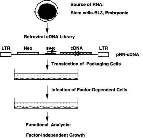
Experimental
design.
Figure
2displays
the structuresof theparental pRN
retrovirus
vectorand the
pRN-cDNA
vector. In thepRN
vector,
athymidine
kinase
promoter
is
present
downstream of the Neor
gene,which is driven
by
thelong
terminal
repeat
promoter.
InpRN-cDNA,
the Neor geneis
also driven
by
thelong
terminal
repeat promoter,
but the cDNA insert istranscribed off the simian
virus 40promoter
derived
from thepLl
plasmid
(Fig.
1).
Thisdesign
is similartothat
reported
previously
(6, 65).
Figure
3displays
astrategy
forscreening
genes from aretroviral
cDNA expressionlibrary
on the basis of availablebiological
assays. Weconstructed
thepRN-BL3
retroviral
library
by
incorporating
theadvantages
of
theOkayama-Berg
system,
which includehigh-frequency
generation
offull-length
cDNA inserts and theirdirectional
cloning
intovectors(46).
Inthis
study,
mRNAfrom
immortalized
hemopoietic
stemcells,
on November 9, 2019 by guest
http://jvi.asm.org/
Sall Hind3Sall Bgi2 XholClal
_\|/T
K
'i
Ineo
rF
SallHind3 Clal
neo SV40 cDNA
,1
17
'7
'
I
pRN
s
o
0 4
-c 3
0
o 2
-i
?I 1
pRN-cDNA FIG. 2. Structures of the retroviral cDNA library vector, pRN-cDNA,and its parental vector, pRN. Notethatthethymidine kinase (TK) promoter in pRN was replaced by a simian virus 40 (SV40) promoter in pRN-cDNA during the construction of the retroviral cDNAlibrary. neo,neomycin.
BL3
(66), was reverse transcribed and cloned into the pRN
vector.
Six
sublibraries were made. Plasmid DNA from each
sublibrary
wastransfected into
GP/E packaging cells; medium
containing
1mg
of G418 per ml was added. Twelve days
later,
thousands of G418-resistant colonies per 100-mm-diameter
dish
wereable to grow
to nearconfluence.
Factor-dependent
(FD)
cells
werethen added
tothe dishes for 2 to 3
days and
assayed for
FT
growth.
Transfection
standardization.
Transfection was
standard-ized
by using the plasmid pJRgal under various conditions
(Fig. 4).
From
six
independent experiments under
standard-ized conditions,
anaverage
of about 4,000
independent
trans-fectants per dish
wasrecorded when
weused 50 ,ug of
plasmid
DNAfor a 12-h exposure to
calcium phosphate (Fig. 5).
To define the
minimum relative abundance of
mRNArequired for successful cDNA
expression
cloning,
variousratios of
pJRgal
and pLTK
weretransfected into GP/E cells.
Sourceof RNA:
Stemcells-BL3,Embryonic
Retroviral cDNALibrary
LTR Neo sV40 cDNA LTR
m
''
.i
,
I
pRN-cDNA
Transfectionof
Packaging
CellsL...
...*
Infection of Factor-Dependent CellsC
0 00
z
-U
i-?I
4
I.
o 3 6 9
Txf.time(hrs.)
2
12
4
10 20 30 40 Amountof DNA(pg)
50
1-
i
I 1 2 3 4 Timeaftertxf.(days)
|
I
5 10 15 20 No.ofcel splated (105) FIG. 4. Transfectionstandardization. GP/E cellsweretransfected with the plasmid pJRgal and X-Gal stained to assay for positive transfectants(seeMaterialsandMethods).Unlessotherwisespecified, transfection was performed asfollows: GP/E cellswere
prepared
asdescribed, platedat 106cells per 100-mm-diameterdish, transfected with 20 ,ug ofplasmidpJRgal per dish for9h,and thenassayed by X-Gal staining3daysposttransfection.Eachpanelshows the average oftwoexperimentswithduplicatesateachpoint,exceptforpanel C, whichgivesthe averageof threeexperiments.Thestandard deviation for eachpointisincluded. Results indicate that theoptimal conditions consist of
106
cells perdish,transfectionwith50pug
ofplasmidDNA perdish for12h,and assayby X-Galstaining4daysposttransfection. txf.,transfection; col., colonies.While both
pJRgal
and pLTK contain sequences
conferring
neomycin resistance,
only pJRgal
encodes the
n-Gal
gene.
Results showed that while the number of Neor colonies
remained
relatively constant, the number of ,-gal+ cells varied
directly with the ratio
of pJRgal to pLTK (Fig. 6) and that it
wasdetectable
only when the ratio of pJRgal
topLTK
was8
-6
-4.-Mean
0
t-m
ci. z
[image:4.612.325.560.67.287.2]Functional Analysis: Factor-independentGrowth
FIG. 3. Strategyofscreening forgenesby functional analysis witha
retroviralcDNAexpression library. LTR, long terminalrepeat;Neo,
neomycin; sv4O, simianvirus40.
ExperimentNo.
FIG. 5. Transfection underoptimal conditions.Data arethe results of six separateexperiments;the median value is3,948 transfectantsper dish.col.,colonies.
on November 9, 2019 by guest
http://jvi.asm.org/
[image:4.612.65.300.71.183.2] [image:4.612.66.314.444.684.2] [image:4.612.325.561.531.682.2]10000.0 r
cit
a)
0
ci
z
100r
1000.0
F
100.0 I
10.0 I 1.0
-u.I
0.0o
El
l
KElLl
1:0 1:1 1:10 1:100 1:1000 1:10000 1:100000
Ratio ofpJRgal:pLTK
FIG. 6. Sensitivity of theassaybasedonX-Galstaining. GP/E cells
weretransfected withmixturesof plasmids pJRgal andpLTK;ratiosof the plasmids varied from 1:0to1:100,000. In each experiment, three
dishes of cells were transfected at each of the various ratios. Two disheswereassayed by X-Gal staining. The third dishwastrypsinized
immediately after transfection; cellswerereplatedatdilutions of 1/10 and1/100 and placed under G418 selection thenextday. Countswere
multiplied by the appropriate dilution factors, and the resulting
numberswereaveragedtoyieldanestimate of the number of colonies (col.) originally in the transfected dishes. Resultsare theaveragesof three separate experiments at each indicated ratio, except for the ratios 1:10 and 1:100,000, which were performed twice and once,
respectively. Solid bars, ,B-Gal+;openbars,Neor.
higher than 1:500, which was equivalent to an mRNA
abun-dance of 0.2%.
The use of ,B-Gal might have caused the sensitivity in
detectingageneproduct capable of stimulating cellgrowthto
be underestimated. Detection of this enzyme was done by
X-Gal staining, a method in which a relatively high level of
geneexpression mightbenecessary.Indeed,wenoticed thatin dishes in which transfectants of pJRgal were used, ,B-Gal+
coloniesoftencontained no morethanfourpositive cells that
were richly stained with X-Gal after 4 days in culture.
NIH/
3T3-based cellsused for transfection haveadoublingrateof 12 to16h; therefore, each transfected cell should have undergoneanaverageof six divisionsnormally. That less than four cellsin
a pJRgal colony stained positively with X-Gal indicated the levelof 13-Galexpressionwas adetermining factor for
detec-tionbystaining.
Enhanceddetection based onbiological assay. It isknown
that genes encoding growth factors or their receptors are
usuallyexpressedatlowlevels andareoften detectableonly by sensitive biological assays. Therefore, to investigate whether
we could enhance the efficiency of this retroviral cDNA cloning,weemployed the biologicalassaysoutlined in Fig. 3. GP/E cells were transfected with mixtures of
pN2-IL3
and pRN-BL3at ratios ofpN2-IL3
topRN-BL3 ranging from 1:0to1:10,000. After infectionforaperiod of 3 days, FDC-P1 cells were collected and cultured in the absence of
WEHI-condi-tioned medium. Percent viability and total cell numberwere
evaluated every 2 days. Results in Fig. 7 indicate that in all
casesinfectedFDC-P1cellswereabletogive risetoFlgrowth
at rates significantly higher than that of the control culture. Furthermore, the growth rate was positively correlated with the amount of N2-IL3 used for transfection. Interestingly, althoughWEHI-conditioned mediumwasaddedduring infec-tion, the percentage of viability of FD cells was inversely
correlatedwiththe dilution of
pN2-IL3.
Weinterpretthedata
80p-,1:102
1:103
,1:104
601->1
4)
4J1
a)
a- 40
-20
-0
o--
O---0 2 4 6 8
[image:5.612.52.291.75.231.2]No.of days after nfecti on
FIG. 7. Sensitivity of the assay based on IL3 biological function,
FDC-P1
viability as afunction oftime and IL3 concentration. GP/E cells were transfected under optimal conditions with mixtures of plasmidspN2-IL3 andpRN-BL3atratiosranging from1:0 to1:10,000. Dishes were placed under G418 selection, and Neor colonies were allowed to expanduntil dishesapproached confluence.Atthispoint, 106 FDC-P1 cells were incubated in coculture with these virus-producingcells in 5 ml ofalOFmedium with10%WEHI-conditioned medium and 5 ,ug of Polybrene per ml. After 1 day of infection, another 5ml of medium wasadded;infectionwascontinued for 2 more days,after which FDC cells werecollected, washed,and thencultured in the absence of WEHI-conditioned medium. Percent viability and total cell number wereevaluated every 2 days by trypan blue exclusion. Individual curvescorrespond toFDC-P1
cellsderived from coculture with GP/E cells transfected with the indicated ratios of pN2IL3 to pRN-BL3. ControlFDC-P1
cells werecoculturedwith untransfected GP/E cells. Total cell number also increased withtime, correlatingin each case to percentviability (datanotshown).as
indicating that
during cocultivation,
veryhigh levels of
metabolic
activity of both virus-producing cells and
targetcells
occurred. The
amountof conditioned
medium added
wasoptimal for
maintaining
FDcells
during
passage, notfor
infection.
Therefore,
under this
experimental condition,
many FDcells
nottransduced with the
N2-1L3
vectorwould
notsurvive.
To
further confirm the
sensitivity
of this
assay, wedeter-mined by
Southern blot analysis the
highest
N2-1L3 dilution
atwhich
FDC-P1 could still
betransduced with the retroviral
vectorand result in
Neor and
Fl growth.
Toexamine whether
multiple retroviral infections had taken
place,
wefirst did
junction
fragment
analysis.
Genomic
DNAwasdigested
with
BglII,
electrophoresed,
andhybridized with either the IL3-
orneomycin-specific probe. Figure
8a
shows that FDC-P1 cells
cocultivated with libraries of these
virus-producing
cells
wereindependently
infected
with
manyretroviruses since
hybridiza-tion of their
genomic
DNAswith
probes
yielded
smears ormultiple
bands.
Todetermine whether transduced FDC-P1
cells contained
N2-1L3
proviral DNA,
wedigested their
genomic
DNAswith
Sacl,
thusreleasing
the intact viral
genome,and
hybridized
the blots
withIL3-
orneomycin-specific probes. Figure
8bindicates that all these
Fl cell
linescontained
the4.2-kb
proviral
IL3
sequence. DNAof
cellsfrom
culturesof
pN2-IL3
andpRN-BL3
at aratioof
1:10,000
hadaweak
signal
atthe 4.2-kb
bandwith the
neomycin probe
butnotwith
theIL-3
probe.
During
repeated
experiments
withplas-mids
derived
frombacteria of
thesamesublibrary,
the 4.2-kb
band
probed
with both
neomycin
and
IL-3wasclearly
present
atthat
ratio.
Wealso
transduced another
FDcell
line,
32D,
rl 1
on November 9, 2019 by guest
http://jvi.asm.org/
[image:5.612.311.546.77.236.2]5528 WONG ET AL.
IL3
a 000 Q0
0000 C
. a
b
0
0 0 a)
.C~~~~~~~~~~~.
CD~0CD .
S W
c
:n -i
z QL
neo
-0 0
0o 0
00C00
_) CD CD
..
r- _r _-
v-r----4.9kb
0CD0
n 00 0
-n 00C 0 0
CJ O
[image:6.612.68.306.88.508.2]..
...
44.2kb 4~..
CD)
z 00CD
0 0 o o0 o
CD 0D( 0 000CD
CD CD CD - m a CD CD C m
_E _ 00 EC>>
.-
iibi...
_- r O CL t - ...... ..
...4
,.,.p...
*'~ Wiw''~ ~ .k ''':W
511
151(;
1II
(I,- 11
I..
'II(F 11 SaI
Ne(10 I1L3
SV4() SticI
N._O
r½DNA4
Neocv N A
FIG. 8. Southern blot analysis. (a) Junction fragment
DNA samples extracted from transduced FDC-P1 cell lin digested with BglII. 1L3-specific (left) and
neomycin-specifi4
probeswereused.The 4.9-kbbands correspond to the endoger sequence. (b)GenomicDNAsamplesweredigested with Sa( the flanking retroviral long terminal repeats contain Sacl si releases theintegratedviral sequence.The DNA samples in tl
werederived from thecorresponding cell cultures in Fig.7.
TI
harvested after6(for 1:0, 1:1, and 1:10), 8 (for 1:100), 10(1:1, (1:10,000), and 22days(for control). (c) AnotherFDcell lir
wasusedfor infection and was rendered Fl by coculturingw
cells transduced with amixture ofN2-1L3 anda different su
plasmid DNA of pRN-BL3. Genomic DNA samples were with Sacl. CV301, DNA from
kj2
packaging cells transfect pN2-IL3, was used as apositive control; FDC, DNA from cells,wasusedasnegative control (uninfected). Other lanes cc DNAsfromFT
32D cells cocultured with various ratios ofp] pRN-BL3 plasmids. Fl cells at ratios of 1:1,000,000, 1:100,( 1:10,000were obtained after 70, 25, and 30 days in culture, tively.Inpanels b and c, the 4.2-kb bands correspond to the in N2-1L3viral sequence. Neo, neomycin; SV40, simian virus4(
with
GP/E
cells transfected withplasmid DNA from a differentpRN-BL3 sublibrary (pBL32),
andFig.
8c indicates that N2-IL3proviral
DNA was present at the1:100,000
dilution. Of note wasthe fact that we obtainedFT growth of 32D cells at the 1:1,000,000 dilution only after 70 days compared with less than 30days
for cultures withlower dilutions. Taken together, these data indicate that under near-optimal transfection conditions, thesensitivity
of our assay can be ashigh
as1:100,000, i.e.,
a BglII particular species of mRNArepresented in aretroviral cDNAexpression
library at an abundance level as low as 0.001% can be isolated.Conversion to Fl after transduction with retroviral cDNAs. To further confirm the effectiveness of this methodology, we infected target FDC-PI cells by cocultivating them with subli-braries of virus-producing cells. We observed that a subclone ofFDC-PI cells, FDE(Simon Jones, Genetic
Institute),
gave a lowbackground
of spontaneous Flgrowth; therefore,
weused FDE cells as target cells for this series of experiments.Spontaneous
mutation in FDE cells was estimated to be less Sac I than I in107
cells(7a). Five types ofretrovirus-producing
cells were used: 7GN2, GP/E cellsproducing
N2virus;
B2-22,B3-20,
B6-35,
andB6-45,
GP/E
cellsproducing
four different sublibraries ofpRN-cDNAs
fromBL3
cells. Theefficiency
ofretroviral
genetransfer
into FDE cellsranged
from27%,
thelowest,
for one dish of B3-20 to86%,
thehighest,
for one dish of7GN2 (Table 1).To allow for the observation of
reproducible independent
infection
events,duplicated dishes of
each typeof
virus-producing
cell were used forcocultivation
and cells harvested from each dish weretreated
and cultured in methylcellulose SacI separately. Table 1 indicates that dishes of bothB2-22
and B3-20, but notB6-35,
B6-45, or7GN2,
gave rise toFl colonies. It isimportant
to point outthat colonies
fromboth dishes
of B3-20 werequalitatively entirely different from those of
B2-22.They
weremacroscopic
insize, and
17days after
infection,
they
hadproliferated
to2 to 6million
cells, whereas macroscopic
colonies from dishes of othersublibraries cultured with growthcI
factors had
fewer than30,000 cells.
This type ofmacroscopic
colony
was notobserved in
anyof the dishes from infection
by
eight other
plates of
retrovirus-producing
cells. These
datasuggested
that it was a cDNA inB3-20 whoseproduct
coulduniquely stimulate transduced
FDEcells todevelop this
type IBo1 ofcolony.
Fl
colonies observed
incells transduced
with retroviruses
from
B2-22 weretypical colonies, appearing
tobe diffuse
and
analysis. with a centercore.They
did becomemacroscopic
insize, but es were theirrateofproliferation
wasfar lessrapid
than that of those c (right) transduced withB3-20.
Thenumber ofFl
colonies per culture nous IL3 transduced by B2-22 cells was much more significant than that cl. Since transduced by B3-20. These data suggested that the cDNA inites,
thisB2-22,
different from that
inB3-20,
was moreabundant than
Mspanel
that in B3-20 and
that its
expression also resulted in
stimulat-000),
18 ing FT growth. ne,32D,ith GPE iblibrary digested ted with FDC-P1 Dntained N2-IL3-)00, and respec-tegrated
I
DISCUSSION
We
have standardized the
conditions for
high-efficiency
transfection
ofplasmids of
aretroviral
cDNAlibrary stably
into
GP/E
packaging
cells. Byassaying
for Flgrowth
of FDcells after retrovirus-mediated
genetransfer,
we wereable
toisolate
IL-3 transduced cellswhen
the vectorcarrying
IL-3 cDNAwasrepresented
inthe
cDNAlibrary
atafrequency
as low as0.001%. Thisefficiency
wasbased
ontransfecting
vector DNAinto
two100-mm-diameter tissue
culture dishes so that an averageof
4,000
independent
clones of retroviral
library
transfectantswerepresent(Fig. 5).
Realistically,
one canscale J. VIROL.on November 9, 2019 by guest
http://jvi.asm.org/
TABLE 1. Infection ofFDEcells with sublibraries ofretrovirus-producingcellsa
Plasmid Cell yieldper dish No. of
colonies/1,000
cells No. of colonies/1.5 x 106cells6 Efficiencyretroviralofsublibrary
(106)
+GF, - G418 +GF, + G418 -GF,
- G418 -GF,
+ G418transduction
(%)'7GN2 5.4 620 528 0 0 85
3.5 399 344 0.9 (1) 0 86
B2-22 4.1 559 270 13.7 (18) 6.9 (9) 48
3.0 554 206 12.8(12) 7.5(7) 37
B3-20 8.2 650 221 2.3 (6)d 0 34
4.8 656 174 0.7 (1)d 0 27
B6-35 6.2 643 198 0 0 31
3.1 556 198 0 0 37
B6-45 5.5 668 268 0 0 40
4.0 413 160 0 0 39
aFDEcells werecocultivatedwithvirus-producing cells,and each rowrepresentsthe results of a separate infection.Nonadherentcellswere harvestedandplated
inmethylcellulose cultures. For each harvest, 1,000 cells wereplatedinto two dishes of cultures with (+)growth factor (GF) (WEHI-3 CM); remainingcells were dividedequallyandplatedat acellconcentrationof5x 105/ml in cultures without(-) GF and with (+) or without (-) 1 mg ofG418per ml. Totalnumberofdishes preparedfor cultures with no GF was afunctionof cell yield since allcells harvested wereplated intomethylcellulosecultures.Forcultures with noGF,number of
colonies per 1.5x 10⁶cellswasusedbecause nocoloniescould be observed when 1.5 x 10⁶cells were plated intothreedishes; all zerovaluesforotherpoints had
moredishessincemorecells were availablefor plating.
6Numbers inparentheses representactualtotal numbersofcoloniesobserved in dishes for eachculture condition. 7GN2is an N2 producer testednegativeforhelper
virus by XCplaqueassay (56).
c Calculated by dividing thenumbersofcoloniesincultureswith growthfactor (GF)and G418 by thenumbersofcoloniesin cultureswith GFand withoutG418
and then multiplying by100.
dMacroscopic coloniesqualitatively differentfromthosederived from FDEcocultivatedwithother retroviral cultures.
up
the
screening effort
easily by expanding the number of
dishes, for example, to 20. Accordingly, the sensitivity of the
method increases
10-fold; in other words,
mRNA at0.0001%
canbe isolated
by this method.
Amplification also took place during cocultivation between
target
cells and
virus-producing cells, which occurred for
aperiod of
3days.
Depending
onthe
experiment, there
werebetween 10,000 and
100,000 different types of retroviruses
produced in
adish
containing
anaverage
of 4,000 independent
transfectants, each of which had
toproduce
morethan one
type
of retrovirus
(Fig.
7and
8).
Asthe
exposure
time of target
cells
tovirus-producing
cells
increased, successful transduction
by
aretrovirus
vectorcarrying
acDNA with
growth-stimulating
function also increased. As
aresult,
this
particular
clone of
FDcells
transduced
with the
correctcDNA would have
adistinct
selective
growth
advantage
overthose transduced with genes
having
nogrowth-stimulating
function. This type of clonal
amplification should enhance the
probability
of
detection.
Despite the low frequency of recombination for
production
of
Moloney
murine leukemia virus
(40),
areplication-compe-tent
virus may still be present and be
aconcern. However, the
presence
of such
ahelper virus may actually
help
tospread
the
desired
constructinto
alarger
number of target cells and lead
topreferential expansion,
thus
providing
another
level of
amplification.
Genomic DNA from cells transduced with and
stimulated
by the
product
of the desired construct,
either
introduced
initially
oracquired through subsequent virus
spread, should reveal
aband of the
samesize upon Sacl
digestion,
consistent with the results in
Fig. 8, whereas
inde-pendent Fl
clones
generated
as aresult
of other mutational
eventswould
not.Also,
if the
helper
virus
is present in
large
quantities, prolonged
cocultivation would
notincrease the
frequency
of infection due
toviral interference.
In ourexper-iments,
aprolonged
time of infection resulted in increased
efficiency
of
retroviral gene transfer
(63). Therefore,
arepli-cation-competent
helper
virus
could be present but
only
atvery
low levels, if
atall; furthermore, its presence would
only
facilitate the
screening process.
In
the system
wetested,
onemay
identify genes capable of
converting
FDcells
to Flgrowth.
Inthis
regard, it should
facilitate
oureffort
toidentify
newgrowth-stimulating
genes
from
immortalized
BL3
cells, which
werecently established
retain
certain properties of hemopoietic stem cells (66).
Ret-roviral transduction of
FDcells with
sublibraries of pRN
cDNA
from
BL3
cells
resulted in the
development of different
types
of
FT
colonies
(Table 1).
The type of
Fl colonies observed
in
cultures of B3-20
werestriking in that they
were notobserved
incells from
eight other independent infection
cultures and that the cells in these
colonies
proliferated
at arate
significantly faster than, for example, those in
Fl
colonies
from
infection cultures of B2-22
(Table 1).
These data
strongly
suggested
that the
twocDNAs
separately
inserted into the
retroviral
vector weredistinct from each other. Molecular
isolation and
characterization of the
two cDNA arein
progress. We
wereencouraged
by
this result because the
results of RT-PCR and cell
proliferation
assay
indicate that
BL3
cells do
notproduce
orrespond
tomany
cytokines
(22),
including IL-1
toIL-7,
IL-9 toIL-11,
stemcell
factor, leukemia
inhibitory
factor, GM-CSF,
granulocyte
colony-stimulating
fac-tor,
macrophage
colony-stimulating factor, oncostatin-M,
gamma
interferon,
and
tumornecrosis factor
alpha.
However,
they
do
produce
anautocrine
growth
factor
(22).
The
effects of
the
proteins
of the isolated
cDNAs onhemopoietic
stemcells
will be
analyzed in
afuture
study.
In our
experimental
protocol, plasmid
DNAsof various
sublibraries of
pRN-BL3
werestably
incorporated
into the
genome
of
GP/E
transfectants
becausethey
wereselected with
G418.
Assuch,
cells from several
dishes,
each of which
contained
anaverage
of
2 to 4thousand
independent colonies,
were
expanded, pooled,
and
frozen
inalarge
number of vials
asstocks for each
retroviral
sublibrary.
When
needed,
cells of
asublibrary
of interest
werethawed
out toinfect target cells.
on November 9, 2019 by guest
http://jvi.asm.org/
One
suchexperiment
wasperformed,
and the results arepresented
inTable
1. Inthis
way, wecircumvented the need
for
extensive labor and yetprovided consistent stocks of
various
sublibraries
ofretrovirus-producing cells.
ACKNOWLEDGMENTS
GP/E cells,FDEcells,andplasmid pJRgalwereprovidedbyArthur Bank at Columbia University (New York, N.Y.), Simon Jones at
GeneticInstitute,andMichael Gould (University of Wisconsin, Mad-ison), respectively.Wealso thank XiaodongHanandTina Tubbsfor helpfulcomments and editorial assistance.
Thiswork was supported by NIH grantsDK41298 and HL46547. B.Y.W.isaDaniel Swern Fellow. P.M.C.W. is a SinsheimerScholar.
REFERENCES
1. Aruffo,A.,and B.Seed.1987. Molecular cloning ofaCD28 cDNA
byahigh-efficiency COS cellexpressionsystem. Proc.Natl.Acad. Sci. USA 84:8573-8577.
2. Belmont, J. W., J. Henkel-Tigges, S. M. W. Chang, K.
Wager-Smith,R. E.Kellems, J. E. Dick, M. C. Magli, R. A.Phillips, A.
Bernstein, and C. T. Caskey. 1986. Expression of human
adeno-sine deaminase in murine haematopoietic progenitorcells
follow-ingretroviral transfer. Nature (London) 322:385-387.
3. Blasi,E.,B.J. Matheson,L.Varesio,J. L.Cleveland,A.Borchert,
and U. R. Rapp. 1985. Selective immortalization of murine
macrophages from fresh bonemarrow by a raf/myc recombinant murine retrovirus. Nature (London) 318:667-670.
4. Boswell, H. S., T. S. Nahreini, G. S. Burgess, A.Srivastava,T.G.
Gabig,L.Inhorn,E. F.Srour,and M.A.Harrington. 1990. ARAS
oncogene imparts growth factor independence to myeloid cells
thatabnormally regulate protein kinase C: anonautocrine
trans-formationpathway. Exp. Hematol. 18:452-460.
5. Boutler, C. A., and E. F. Wagner. 1988. The effects of v-src
expression on the differentiation of embryonal carcinoma cells.
Oncogene2:207-214.
6. Browder,T.M., J.S.Abrams,P. M.C.Wong, and A. W. Nienhuis.
1989. Mechanism of autocrine stimulation in hematopoietic cells
producing interleukin-3 after retrovirus-mediated gene transfer.
Mol. Cell. Biol. 9:204-213.
7. Chang, J. M., D. Metcalf, R. A. Lang, T. J. Gonda, and G. R.
Johnson. 1989. Nonnioplastic hematopoietic myeloproliferative syndrome induced by dysregulated multi-CSF (IL-3) expression.
Blood 73:1487-1497.
7a.Chung,S.-W.Unpublisheddata.
8. Chung,S.W., P. M. C. Wong,H.Durkin,Y. Wu, and J.Petersen.
1991. Leukemia initiatedby hemopoieticstemcells expressing the
v-abl oncogene. Proc. Natl. Acad. Sci.USA88:1585-1589. 9. Chung, S. W., P. M. C. Wong, G. Shen-Ong, S. Ruscetti, T.
Ishizaka, and C. J. Eaves. 1986. Production of granulocyte-macrophage colony-stimulating factor by abelson virus-induced tumorigenic mastcell lines.Blood68:1074-1081.
10. Cone, R.D., E. B. Reilly, H. N.Eisen, and R. C. Mulligan. 1987.
Tissue-specific expression of functionally rearranged X1 Ig gene
througharetrovirusvector.Science236:954-957.
11. Cook, W. D., D. Metcalf, N. A. Nicola, A. W. Burgess, and F.
Walker. 1985. Malignant transformation of a growth
factor-dependent myeloidcell lineby Abelson virus withoutevidence of anautocrine mechanism.Cell41:677-683.
12. Dean, M., J.L.Cleveland,U. R. Rapp, and J. N.Ihle.1987. Role
ofmycin theabrogationof IL3 dependence ofmyeloidFDC-P1 cells. OncogeneRes. 1:279-287.
13. Dexter,T.M.,J. Garland,D.Scott, E. Scolnick, and D. Metcalf.
1980. Growth of factor-dependent hemopoietic precursor cell
lines. J. Exp. Med. 152:1036-1047.
14. Dick, J. E., M. C. Magli, D. Huszar, R. A. Phillips, and A.
Bernstein. 1985. Introduction ofaselectable gene into primitive stemcellscapableoflong-term reconstitutionof thehemopoietic
systemof
W/WV
mice. Cell 42:71-79.15. Dzierzak,F.A.,T.Papayannopoulou, and R. C.Mulligan. 1988.
Lineage-specific expression ofahuman
i-globin
gene inmurinebone marrowtransplant recipientsreconstituted with
retrovirus-transducedstemcells. Nature (London)331:35-41.
16. Eglitis, M. A., P. Kantoff, E. Gilboa, andW. F. Anderson. 1985. Gene expression in mice after high efficiency retroviral gene transfer. Science 230:1395-1398.
17. Friedman,R. L.1985. Expressionofhuman adenosinedeaminase using a transmissable murine retrovirus vector system. Proc. Natl. Acad. Sci. USA 82:703-707.
18. Funkunaga, R., E. Ishizaka-Ikeda,Y. Seto, and S. Nagata. 1990. Expression cloning of a receptor for murine granulocyte colony-stimulating factor. Cell 61:341-350.
19. Gearing, D. P., J. A. King, N. M. Gough, and N. A. Nicola. 1989. Expression cloning of a receptor for human granulocyte-macro-phage colony-stimulating factor. EMBO J. 8:3667-3676. 20. Gonda, T. J., D. K. Sheiness, and J. M. Bishop. 1989. Murine
myeloid cell lines derived by in vitro infection with recombinant c-myb retroviruses express myb from rearranged vector proviruses. EMBO J. 8:1767-1775.
21. Gruber, H. E., K. D. Finley, R. M. Hershberg, S. S. Katzman, P. K. Laikind, J. E.Seegmiller, T. Friedmann, J. K. Yee, andD.J.Jolly. 1985. Retroviral vector-mediated gene transfer into human hema-topoietic progenitor cells. Science 230:1057-1061.
22. Han, X. D., and P. M. C. Wong. Unpublished data.
23. Hanahan, D. Studies on transformation of Escherichia coli with plasmids. J. Mol. Biol. 166:557-580.
24. Hariharan, I. K., J. M. Adams, and S. Cory. 1988. bcr-abl oncogene renders myeloid cell line factor independent: potential autocrine mechanism in chronic myeloid leukemia. Oncogene Res. 3:387-399.
25. Heard, J. M., M. F. Roussel, C. W. Rettenmier, and C. J. Sherr. 1987. Multilineage hematopoietic disorders inducedby transplan-tation of bone marrow cells expressing the v-fms oncogene. Cell 51:663-673.
26. Johnson, G. R., T. J. Gonda, D. Metcalf,I.K. Hariharan, and S. Cory. 1989. A lethal myeloproliferative syndrome in mice trans-planted with bone marrow cells infected with a retrovirus express-ing granulocyte-macrophage colony stimulatexpress-ing factor. EMBO J. 8:441-448.
27. Kato, J., M. F. Roussel, R. A. Ashmun, and C. J. Sherr. 1989. Transduction of human colony-stimulating factor-1 (CSF-1) re-ceptor into interleukin-3-dependent mouse myeloid cells induces both CSF-1-dependent and factor-independent growth. Mol. Cell. Biol. 9:4069-4073.
28. Keller, G., C. Paige, E. Gilboa, and E. Wagner. 1985. Expression of a foreign gene in myeloid and lymphoid cells derived from multipotent haematopoietic precursors. Nature (London) 318: 149-154.
29. Keller, J. R., S. K. Ruscetti, and F. W. Ruscetti. 1990. Introduction of v-abl oncogene induces monocytic differentiation of an IL-3-dependent myeloid progenitor cell line. Oncogene 5:549-555. 30. Laker, C., C. Stocking, U. Bergholz,N. Hess, J. F. De Lamarter,
and W. Ostertag. 1987. Autocrine stimulation after transfer of the granulocyte/macrophage colony-stimulating factor gene and au-tonomous growth are distinct but interdependent steps in the oncogenic pathway. Proc.Natl. Acad. Sci. USA84:8458-8462. 31. Lang, R. A., D. Metcalf, N. M. Gough, A. R. Dunn, and T. I.
Gonda. 1985. Expression of a hemopoietic growth factor cDNA in a factor-dependent cell line results in autonomous growth and tumorigenicity. Cell 43:531-542.
32. Langdon, W. Y., J. W. Hartley, S. P. Klinken, S. K. Ruscetti, and H. C. Morse III. 1989. v-cbl, an oncogene from a dual-recombinant murine. Proc. Natl. Acad. Sci. USA 86:1168-1172.
33. Ledley, F. D., G. J. Darlington, T. Hahn, and S. L. C. Woo. 1987. Retroviral gene transfer into primary hepatocytes: implications for genetic therapy of liver-specific functions. Proc. Natl. Acad. Sci. USA 84:5335-5339.
34. Ledley, F. D., H. E. Grenett, M. McGinnis-Shelnutt, and S. L. C. Woo.1986. Retroviral-mediated gene transfer of human phenyl-alanine hydroxylase into NIH 3T3 and hepatoma cells. Proc. Natl. Acad.Sci.USA83:409-413.
35. Lee, F., T. Yokota, T. Otsuka, L. Gemmel, N. Larson, J. Luh, K. I. Arai, and D. Rennick. 1985. Isolation of cDNA for a human granulocyte-macrophage colony-stimulating factor by functional expression in mammalian cells. Proc. Natl. Acad. Sci. USA 82:4360-4364.
on November 9, 2019 by guest
http://jvi.asm.org/
36. Lee, F., T. Yokota, T. Otsuka, P. Meyerson, D. Villaret, R.
Coffman,
T. Mosmann, D. Rennick, N. Roehm, C. Smith, A. Zlotnik,and K.I. Arai. 1986. Isolation andcharacterization of amouse interleukin cDNA clone that expresses B-cell stimulatory factor 1 activities and T-cell- and mast-cell-stimulating activities. Proc. Natl.Acad. Sci. USA 83:2061-2065.
37. Le Gros, G., S. Gillis, andJ. Watson. 1985. Induction of IL 2
responsiveness
inamurine IL3-dependentcellline. J. Immunol. 135:4009-4014.38. Li, M.,and0.Bernard. 1992.FDC-P1myeloidcellsengineeredto expressfibroblastgrowthfactorreceptor 1 proliferateand differ-entiate in the presence of fibroblast growth factor and heparin. Proc. Natl.Acad.Sci. USA 89:3315-3319.
39.
Lim,
B., D. A. Williams, and S. H. Orkin. 1987. Retrovirus-mediated gene transfer of human adenosine deaminase: expres-sion of functional enzyme in murinehematopoietic stemcells in vivo.Mol. Cell. Biol. 7:3459-3465.40. Markowitz, D.,S.Goff,and A.Bank. 1988. A safepackagingline for gene transfer:
separating
viral geneson twodifferentplasmids. J.Virol.62:1120-1124.41. Matsui, T., J. H.Pierce,T. P.Fleming, J. S. Greenberger, W. J. LaRochelle,M.Ruggiero,andS.A.Aaronson.1989.Independent
expression
of humanalpha
orbetaplatelet-derived growthfactor receptorcDNAs inanaivehematopoietic
cell leadstofunctionalcoupling
withmitogenic
andchemotacticsignaling pathways.Proc. Natl. Acad. Sci. USA86:8314-8318.42. McCubrey, J. A., S.L.Steelman,M. W.Mayo,P. A.Algate,R.A.
Dellow,
andM.Kaleko.1991.Growth-promotingeffects of insulin-likegrowth factor-1 (IGF-1) onhematopoieticcells: overexpres-sion of introduced IGF-1 receptorabrogatesinterleukin-3depen-dency
of murinefactor-dependent
cells by a ligand-dependent mechanism. Blood 78:921-929.43.
Meckling-Gill,
K.A.,S.Yee, J.W.Schrader,and T.Pawson. 1992. A retrovirusencoding
thev-fps protein-tyrosine
kinase inducesfactor-dependent growth
andtumorigenicity
in FDC-P1 cells. Biochim.Biophys.
Acta1137:65-72.44.
Migliaccio,
A.R.,
G.Migliaccio,
A.D'Andrea,
M. Baiocchi, S.Crotta,
S.Nicolis,
S.Ottolenghi,
and J. W. Adamson. 1991.Response
toerythropoietin
in erythroidsubclones of thefactor-dependent
cell line 32D is determinedby
translocation of theerythropoietin
receptortothe cell surface. Proc. Natl. Acad. Sci. USA 88:11086-11090.45.
Noma, Y.,
P.Sideras,
T.Naito,
S.Bergstedt-Lindquist,C.Azuma,E.
Severinson,
T.Tanabe,T.Kinashi,F.Matsuda,Y.Yaoita,and T.Honjo.
1986.Cloning
of cDNAencoding
the murineIgGl
induction factorby
a novel strategyusing
SP6promoter. Nature(London)
319:640-646.46.
Okayama, H.,
and P.Berg.
1982.High-efficiency cloning
offull-length
cDNA. Mol.Cell. Biol. 2:161-170.47.
Oliff, A.,
0.Agranovsky,
M. D.McKinney,
V. V. V.S.Murty,
andR. Bauchwitz. 1985. Friend murine leukemia virus-immortalized
myeloid
cellsareconverted intotumorigenic
cell linesby
Abelson leukemiavirus. Proc. Natl. Acad. Sci. USA 82:3306-3310. 48.Otani, H.,
J.P.Siegel,
M.Erdos, J.R.Gnarra,
M. B.Toledano,M.Sharon,
H.Mostowski,
M. B.Feinberg,
J. H.Pierce,
andW.J. Leonard. 1992. Interleukin(IL)-2
and IL-3 induce distinct butoverlapping
responses inmurineIL-3-dependent
32D cellstrans-duced with human IL-2receptor ,B chain: involvement of
tyrosine
kinase(s)
other thanpS6
ck. Proc. Natl.Acad. Sci. USA 89:2789. 49.Paul,
W. E. 1989.Pleiotropy
andredundancy:
T cell-derivedlymphokines
in the immune response. Cell 57:521-524.50.
Pierce,
J.H.,
P. P.DiFiore,
S. A.Aaronson,
M.Potter,
J.Pumphrey,
A.Scott,
andJ.N.Ihle. 1985.Neoplastic
transforma-tion ofmastcells colonies. Cell41:685-693.51.
Pierce,
J.H.,
M.Ruggiero,
T. P.Fleming,
P. P.DiFiore,
J. S.Greenberger,
L.Varticovski,
J.Schlessinger,
J.Rovera,
andS. A.Aaronson. 1988.
Signal
transductionthrough
the EGF receptor transfected inIL-3-dependent hematopoietic
cells. Science 239: 628-631.52. Quelle, D. E., and D. M. Wojchowski. 1991. Localized cytosolic domainsof the erythropoietin receptor regulate growthsignaling and down-modulate responsiveness to granulocyte-macrophage colony-stimulating factor. Proc. Natl. Acad. Sci. USA 88:4801-4805.
53. Rapp, U. R., J. L. Cleveland, K. Brightman, A. Scott, and J. N. Ihle. 1985.Abrogation of IL-3 and IL-2 dependence by recombi-nant murine retroviruses expressing v-myc oncogenes. Nature (London) 317:434-438.
54. Rein, A., J. Keller, A. M. Schultz, K. L. Holmes, R. Medicus,and
J. N. Ihle.1985. Infection of immune mast cells by Harvey sarcoma virus: immortalization without loss of requirement for interleu-kin-3. Mol. Cell.Biol. 5:2257-2264.
55. Rohrschneider, I. R., and D. Metcalf. 1989. Induction of macro-phage colony-stimulating factor-dependent growth and differenti-ation after introduction of the murine
c-fins
gene into FDC-P1 cells. Mol.Cell. Biol. 9:5081-5092.56. Rowe, W.P., W. E. Pugh, and J.W.Hartley. 1970. Plaque assay techniques for murine leukemiaviruses. Virology42:1136-1139. 57. Ruscetti, S., R. Aurigemma, C.-C. Yuan, S. Sawyer, and D. G.
Blair.1992.Induction of erythropoietin responsiveness inmurine hematopoietic cells by the gag-myb-ets-containing ME26 virus. J. Virol. 66:20-26.
58. Sorge, J., W. Kuhl, C. West, and E. Beutler. 1987. Complete correction of the enzymatic defect of type I Gaucher disease fibroblasts by retroviral-mediated gene transfer. Proc. Natl. Acad. Sci. USA84:906-909.
59. Sorge, J., D. Wright, V. D. Erdman, and A. E. Cutting. 1984. Amphotropic retrovirus vector system for human cell gene
trans-fer. Mol.Cell. Biol. 4:1730-1737.
60. Takaki, S., A. Tominaga, Y. Hitosh, S. Mita, E. Sonoda, N. Yamaguchi,andK. Takatsu. 1990.Molecularcloning and expres-sion of the murineinterleukin-5 receptor. EMBO J. 9:4367-4374. 61. Valtieri, M., D. J. Tweardy, D. Caracciolo, K. Johnson, F. Mavilio, S.Altman, D. Santoli, and G. Rovera. 1987. Cytokine-dependent granulocytic differentiation: regulation of proliferative and differ-entiative responses in amurine progenitorcell line. J. Immunol. 138:3829-3835.
62. Wang, B.,W.S.Kennan, J. Yasukawa-Barnes,M.J.Lindstrom, and M. N. Gould. 1991. Overcoming the activity of mammary carcinoma suppressor gene inCopenhagenratsbyv-H-ras
onco-gene transfer into mammaryepithelialcells in situ. Cancer Res. 51:2642-2648.
63. Wong,B.Y.,and P.M.C.Wong.Unpublisheddata.
64. Wong,P.M.C.,S. W.Chung,C.E.Dunbar,D.M.Bodine,S. K. Ruscetti, and A. W. Nienhuis. 1989. Retrovirus-mediated transfer andexpressionof the interleukin-3 gene inmousehematopoietic cells result in a myeloproliferative disorder. Mol. Cell. Biol. 9:798-808.
65. Wong, P.M.C.,S.W.Chung,and A.W. Nienhuis. 1987. Retroviral transfer and expressionof the interleukin-3 gene inhemopoietic cells. Genes Dev. 1:358-365.
66. Wong, P. M. C., X. D. Han, F. Ruscetti, and S. W. Chung. Immortalizedhemopoietic cellswithstemcellproperties. Submit-tedforpublication.
67. Wongsasant, B., S. Matsuda, and T. Yamamoto. 1991. Active c-erbB-2 induces short-term growth of FDC-P2 cells after IL-3 depletion.Biochem.Biophys. Res. Commun.181:981-988. 68. Yokota, T., N.Arai,F.Lee,D.Rennick,T. Mosmann,andK.I.
Arai. 1985. Use of a cDNA expression vector for isolation of
mouse interleukin 2 cDNA clones: expression of T-cell growth-factoractivityaftertransfectionofmonkeycells. Proc. Natl. Acad. Sci. USA 82:68-72.
69. Yokota,T.,F.Lee,D.Rennick,C.Hall,N.Arai,T.Mosmann,G. Nabel,H.Cantor, and K. Arai. 1984. Isolation and characteriza-tion of a mouse cDNA clone that expresses mast-cell growth-factor activity in monkey cells. Proc. Natl. Acad. Sci. USA 81:1070-1074.