JOURNAL OF VIROLOGY, May1994,p.3193-3199 Vol. 68,No. 5 0022-538X/94/$04.00+0
CopyrightC) 1994,AmericanSocietyforMicrobiology
Hepatitis
B
Virus
RNA
Element That
Facilitates
Accumulation
of Surface Gene Transcripts in the
Cytoplasm
ZHI-MING
HUANG'
AND T. S. BENEDICTYEN'
2*Departmentof Pathology, University of Califomia School of Medicine,1 andAnatomic Pathology Service, Veterans
Affairs
MedicalCenter,2
SanFrancisco,
Califomia
Received 14 October 1993/Accepted 4 February 1994
Hepatitis B virus enhancers I and II are critical for high-level expression from the viral major surface gene promoter. These enhancers are in an unusual position, since both are entirely contained within the downstream transcribed region of the surface gene. In this report, we present data showing that a fragment of the viral genome encompassing enhancer II activates accumulation of surface gene transcripts at the posttranscrip-tional level. Specifically, the total steady-state amount of surface gene transcripts in the cell drops by more than fourfold when enhancerIIis displaced to a position downstream of the transcription termination site. There is a similar decrease in the amount of cytoplasmic surface gene transcripts but not of nuclear transcripts. These changes in steady-state transcript levels do not result from a decrease in the rate oftranscriptional initiation or from an increased rate of degradation in the cytoplasm. Reinsertion of enhancer II in the correct orientation into the surface gene transcribed region partially restores transcript levels. From these data, we concludethat ahepatitisBvirus RNAelementfunctionsincis toincrease thesteady-state levels of surface gene transcripts by facilitating cytoplasmic accumulation of these transcripts.
Hepatitis B virus (HBV) is an unusual DNA virus that replicates via reverse transcription of an RNA intermediate and is the only memberof the hepadnavirus family known to
infect human beings (reviewed in reference 7). As its name implies, HBV efficientlyreplicates in hepatocytes and causes hepatitis as the main symptomology (reviewed in reference
10). While acute hepatitis B is usually mild and can be even
asymptomatic, asignificant percentage of infected people fail to clear the virus and become chronic carriers. There is at presentnoeffectivetherapy for chronicHBVinfection. This is an important public health problem, as there are estimated to be wellover200millionpeople worldwide who are chronically infected with HBV and therefore at risk for liver failure, cirrhosis, and hepatocellular carcinoma. Therefore, we have beeninvestigating the regulation of HBV gene expression, in the hope that these studies will facilitate the formulation of strategies to down-regulate viral gene expression and hence preventHBV-associated diseases.
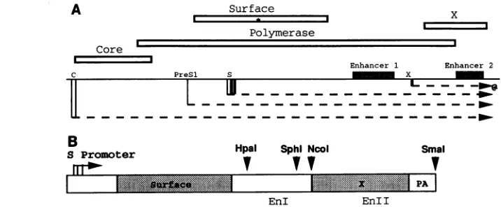
Fourclasses of HBVtranscripts,specifiedby four promoters ontheminus strand andterminatingat thesame
polyadenyl-ationsignal,are translated into the various viralproteins(Fig.
1A; reviewed in references 19, 22,and30).Themajorsurface
(virionenvelope) protein is synthesized from the major surface
(S)genetranscripts.Theamountof S genetranscriptsis known tobe up-regulated incis bythe viral enhancersI and II (EnI
and EnlI, respectively). However, both enhancers are in an unusualposition,being presentwithin the S gene transcribed region, downstream of the Scoding region (Fig. 1). We have previously demonstrated that when the enhancersweredeleted fromthispositionandreplacedupstreamof the S promoter, S genetranscriptlevelsdropped bymorethanfourfold(12).This result raised thepossibilitythatone orboth enhancersfunction
*Corresponding author. Mailing address: Anatomic Pathology
113B, VAMC,4150 Clement St., San Francisco, CA 94121. Phone: (415) 476-5334. Fax: (415) 750-6947. Electronic mail address: yen@ sanfrancisco.va.gov.
optimally only when present within the transcribed region of the S gene. We present data here showing that RNA tran-scribed from the region encompassing EnlI activates the accumulation of S gene transcripts by a posttranscriptional
mechanism, since removal of this RNA segment from the transcribed region does not have an effect on the transcrip-tional initiation rate of the S promoter. Instead, there is a cis-actingelement,overlapping EnII, that promotes the accu-mulation of S gene transcripts in the cytoplasm. Therefore,
theseobservationspointtoanadditionallayerofregulationof gene expression in HBV that hitherto has not been well
characterized.
MATERUILSAND METHODS
Plasmid constructions. HBV fragmentswere derived from pHBV2, which contains a head-to-tail dimer of the HBV genomicDNA, strain adw(25). The plasmid
pSAgAHin
con-tains the HincII-to-BglII fragment of HBV DNA (Fig.iB)
inserted into the plasmid pTZ19U (34); we have previously demonstrated that this subgenomic fragment contains the cis elements necessary forhigh-level expressionof the S gene in hepatoma cells (34). To generate plasmids giving rise to truncated S genetranscripts,aDNAfragmentcontainingthree
copiesof the simian virus 40(SV40)
polyadenylation signal
was excised from theplasmid pTAG-1(6)
with SalI and insertedseparatelyinto fourrestriction sites in
pSAgA&Hin:
HpaI,SphI,
NcoI, and SmaI (Fig. 1). The names of these plasmids are,
respectively, pSHpA-HpaI, pSHpA-SphI, pSHpA-NcoI, and
PSHpA-SmaI.
In addition, a fragment similar in size to the SV40 polyadenylation signal, excised from the vector se-quences in pTAG-1 bydigestion
with EcoRI andXbaI,
was inserted into theHpaI sitetogenerate pSHNS-HpaI.The plasmids
pSHpA-NcoI(160bpC)
andpSHpA-NcoI
(160bpR)wereconstructedby
synthesizing
afragment
ofEnlI (mappositions1600to1760,flankedbySphI
sites) by
PCRandinsertingthisfragmentinto the
SphI
site ofpSHpA-NcoI
inthe wild-type andreverseorientations,respectively.
Theplasmid pXGH5 contains the human
growth
hormone 3193on November 9, 2019 by guest
http://jvi.asm.org/
3194 HUANG AND YEN
A
SurfacePolymerase
x
=
Core r
Enhancer 1 Enhancer 2
C PreSl S _ X
_______---~-
--.~r
--- _ft~~~~~---
---0B
S Promoter
1mro
Hpal SphI Ncol
v Vtv
EnI
Smal
Sia EnI I
FIG. 1. (A)Mapof theHBVgenome(central solidline). Theopenreadingframesareshownasboxes, while thetranscriptsarerepresented asdashedarrows.Notethat themajor surfaceprotein is translated fromaninternal ATG codon (*) of the Sopenreading frame, since theS promoteris embeddedwithin this openreadingframe. (B)Map of theHincII-BglII fragmentof HBV present in theplasmidpSagAHin.The relevant restriction sites usedtogeneratethepAseriesofplasmids(Fig. 2)areindicated. Inthisdiagram, only the portion of the surfaceopen reading frame that istranslated into the majorSprotein isshaded.pA,HBVpolyadenylation signal.
gene driven by the mouse metallothionein II promoter (21). The plasmid pMT-S (also called p30A8) contains the HBV surface gene under the control of the mouse metallothioneinII promoter(32).
Cell culture and transfection. HuH-7 well-differentiated
human hepatoma cells (16), which are free of endogenous HBV sequences, were cultured in DME-H21 medium (Cell Culture Facility, University of California San Francisco) with 10% fetal bovine serum, at 37°Cunderan atmosphereof 7% carbon dioxide-93% air. Cellswere transfected at 50% con-fluency for 15 h by the calcium phosphate coprecipitation
method, as described previously (8, 34). Five to ten micro-gramsofplasmid DNAwasused per 100-mm-diameter dish, and the cells and media were harvested approximately 48 h
following transfection.
In experiments measuring RNA stability,
5,6-dichloro-1-p-D-ribofuranosylbenzimidazole (DRB; Sigma Chemicals) was addedtoculture mediato afinal concentration of 25 ,ug/mlat 40 h after transfection (20). The cellswere then harvested at various timepoints after the addition of the drug.RNAanalyses. Total RNA was extracted from transfected cells with RNAzolB (obtained from Biotecx)by the manufac-turer's suggested protocol. Nuclear and cytoplasmic RNA fractionswere obtainedby the protocol of Daar and Maquat
(5); this protocol was chosen because the extensive washing prevents significant contamination of the nuclear fraction by
cytoplasmic fragments. Briefly, cells on the second day after transfectionweretrypsinizedandlysed in Nonidet P-40. Nuclei were removed by centrifugation, and the supernatant was extracted with RNAzolB to produce the cytoplasmic RNA fraction. The crude nuclearpellet was then washed twice with buffercontainingNonidet P-40 and once with buffer containing
NonidetP-40,Tween80,and sodium deoxycholate. The rinsed nucleiwerethencentrifuged through a 2.1 M sucrose cushion and extracted with RNAzolB to produce the nuclear RNA fraction.
Forprimer extension analysis (18), 10 ,ug of total RNA, 1 ,ug ofcytoplasmicRNA,or1,ugof nuclear RNA was used in each reaction. Theprimer for S gene transcripts has been previously described(33)andyields five major products ranging from 105 to 136 bases long for the transcripts arising from the S promoter (33) but a major product of 203 bases for the
transcript arisingfrom the metallothionein promoter (32). The
primer for
P-actin
transcripts (5'GGAGTCCTTCTGGCCC ATGCCCACCAT) is designed to produce extension products -230 bases long (9).ForNorthern (RNA) blotting (18), 20 ,ug of RNA per lane waselectrophoresedon a1% agarose-formaldehyde gel, trans-ferred to a nylon membrane (obtained from Amersham), and probed with a 32P-labeled fragmentof HBV DNA extending from the EcoRI site to the BglII site and hence capable of detecting allportions of the S gene transcripts.
Nuclear run-on transcription. Run-on transcription was performed as described by Almendral et al. (1). Briefly, on the second day after transfection cells were lysed by shearing
throughaneedle inNonidet P-40 and the nucleiwerecollected through a sucrose cushion. Nascent transcripts within the nuclei wereextended in the presence of[ct-32P]UTP,and the labeled RNA was partially purified and used to probe a dot blotof variousplasmidDNAs(4 jigperdot).Prehybridization wasperformed in 1.5x SSPE(1x SSPE is 0.18 M NaCl, 10 mM NaPO4, and 1 mM EDTA [pH 7.7]), 5x Denhardt's reagent,0.1% sodium dodecyl sulfate, and 0.3 mg of salmon sperm DNA per ml at 42°C for 2 h (18). Hybridization was performed in thesamebufferat 65°C for 16to24 h.
RESULTS
Effect of enhancer position on S gene transcript levels. Previously, we had demonstrated that when the HBV enhanc-ers were removed from their usual position and replaced upstreamof the S promoter,theamountofS gene transcripts
droppedbyapproximately fourfold(12).This result raised the
possibilitythat the enhancersfunctioned optimally only when presentwithin thetranscribed region of the S gene. However, other explanations such as distance and/or position effects couldnotbeconclusivelyruled outwith those constructs used in previous experiments. Therefore, we constructed an addi-tional series ofplasmids to address this issue.
The parentplasmid, pSAgAHin,contains the entire S gene with its promoter, both enhancers, and the polyadenylation
signal (Fig. 1). The derivatives contain three copies of the SV40 polyadenylation signal inserted into various restriction sites (specifically, the NcoI and SphI sites between EnI and EnlI, andHpaI upstream of EnI) (Fig. 1). As aconsequence,
[image:2.612.113.471.76.224.2]---...
---.gg
;" PA... ....
J.VIROL.
on November 9, 2019 by guest
http://jvi.asm.org/
VOL.68, 1994
c
i
i
i.
.1
I
-114i
~ aa
[image:3.612.118.242.76.222.2]1 2 3 4 5 6
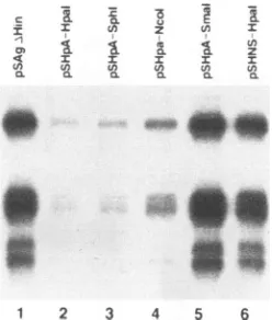
FIG. 2. Primer extension analysis of S gene transcripts synthesized in HuH-7 cells transfected with
pSAgA&Hin
or derivatives. The pA series ofplasmids containsthree tandem copies of the SV40 polyad-enylation signal insertedinto therestriction sitesindicated(see Fig.1). The pSHNS-HpaI plasmid contains a randomly selected plasmid sequenceinserted into theHpaIsite. Similar resultswereobtainedin three independenttransfections. The fivemajor bands represent the five major start sites of the S gene transcripts, which show 5' end microheterogeneity(34).the S gene transcripts synthesized from these plasmids are truncatedat those sites,while thetopographic relationshipof theenhancers to the S promoteris preserved. These plasmids wereindividually transfected into HuH-7 hepatoma cells, and the amount of S gene transcripts was measured by primer extension analysis. Indeed, the amount of S transcripts
dropped significantly when the polyadenylation signal was inserted into the Ncol site (compare lanes 1and4,Fig. 2). In several duplicate experiments, this decrease was four- to
fivefold, asquantitatedbyphosphor imaging. Afurther small drop(less than twofold)wasseen in the transcript levelswith insertion of the SV40polyadenylationsignal further upstream into theSphIsite (Fig.2, lane3),but nofurther decrease was seen with insertion into theHpaI site (Fig. 2, lane 2). This
decrease in transcript levels was not an artifact of primer
extension analysis, since Northern blotting confirmed that
insertion of theSV40polyadenylation signalinto the NcoI site markedly decreased the amountof S gene transcripts
synthe-sized by the transfected cells(Fig.3).
Toensure that thesechangeswerenotdue tothe inserted
polyadenylation signal increasing the distance between the enhancers and theS promoter,weinsertedarandomlychosen
plasmidsequenceof similar length into the HpaI site. Asseen inFig. 2, lane 6, this insertion did not result in any detectable
change in S genetranscriptlevels. Furthermore,we ruledout the possibility that the SV40 polyadenylation signal
fortu-itouslycontained atranscriptionalrepressorelement,as inser-tion of the SV40 sequences downstream of the HBV polyad-enylation signal did not decrease the amount of S gene
transcriptsandevenhadasmall
positive
effect(Fig.
2,lane5).
Insummary,truncation of the S genetranscripts byinsertion ofaheterologous
polyadenylation
signalresulted in decreasedsteady-state levels of these transcripts. This decrease cannot have been dueto theloss ofacis-actingDNAelement, since no HBV sequences were deleted or rearranged in these
plasmids.Rather, these resultssuggestedthatanRNAelement downstream of the Scodingregionfunctioned in cistoincrease accumulation of S genetranscripts. Fromthe results shown in
Fig. 2, this element appears to overlap largely with the X
POSTTRANSCRIPTIONAL REGULATION IN HEPATITIS B VIRUS 3195
H 0
U
Z
Cl)0.0CT)
28S-
[image:3.612.402.474.78.303.2]18s-1 2
FIG. 3. Northern blot
analysis
of S genetranscripts synthesized
in HuH-7cells transfected withpSAgAHin
orpSHpA-Ncol
(see Fig.
lB formap).
Similar results were obtained with threeindependent
transfections. The migration positions of the cellular 18S and 28S rRNAareindicated.
gene/EnII region
(Fig. 1).
Therefore,
it seemedprobable
thatpartof the
positive
effectofEnlI onSpromoter
strength
was dueto itsfunctioning
at the RNA level. To testthissupposi-tion,we insertedasecondcopy of
EnII,
in eitherorientation,
into
pSHpA-NcoI
upstream of the SV40polyadenylation
signal
(Fig. 4A).
As shown inFig.
4B,
the amountofsteady-state S gene
transcripts synthesized
from thisplasmid
was increased almosttowild-type
levelswhenEnIIwasinserted in the native orientation(compare
lane 3 with lane 1containing
the wild
type).
In contrast, insertion ofEnll in the reverse orientation did not increase S genetranscript
levels(Fig.
4B,lane
4).
Insertion of EnII in the nativeorientation,
but downstream of the SV4Opolyadenylation signal,
also did not have any effect on S genetranscript
levels(data
notshown).
Therefore,
Enllwasabletoup-regulate
thesteady-state
levels of the S genetranscripts
only
when itwaspresentinthecorrect orientation within the transcribedregion
of the S gene.The above
results,
takentogether,
show thataregion
of the HBVgenome(the
EnIIIXgeneregion;
seeFig.
1)
actsin cisat the RNA level to increase thesteady-state
levels of S genetranscripts.
Forconvenience,
hereafter we will refer to thisregion
astheposttranscriptional
regulatory
element(PRE).
PRE acts
posttranscriptionally.
Enhancers arecommonly
identifiedasDNAelements.However,there isrecentevidence that sometranscriptional
trans activators can function when tethered via RNA elements(24).
To determine whether the HBV PRE wasacting
as an RNA enhancer, weperformed
nuclear run-onanalysis
to measurethe rateoftranscriptional
initiation from the S promoter,
using
both theparentplasmid
(pSAgAHfin)
andpSHpA-Ncol,
theplasmid
with the SV40polyadenylation
signal
inserted in the Ncol site(Fig. iB)
whichgives
riseto truncated S genetranscripts
devoid of the PRE. For theseexperiments,
oneplate
of HuH-7 cells was trans-fected withpSAgAHin,
while aparallel plate
wastransfectedon November 9, 2019 by guest
http://jvi.asm.org/
3196 HUANG AND YEN
A
s Promoter Ncol-Site ofpolyAInsertion
[~~~~~~~~~~~~~~~~i
,--
P7A
|EnIIA
EnIISphl-Slteof EnilInsertion
B £ CL
8 a
~1
._
CD Il I
(A) (A
ak&
2 . _~~~~~~mddm
1 2 3 4
FIG. 4. (A) Map of theHBVfragment inpSAgAHin. Also indi-catedarethe NcoI site into which theSV40 polyadenylation signalis
inserted in the plasmids pSHpA-NcoI, pSHpA-NcoI(160bpC), and
pSHpA-NcoI(160bpR)and theSphIsite into whicha160-bp fragment
ofEnIlisinserted in theplasmidspSHp-NcoI(160bpC) and pSHpA-NcoI(160bpR),inthewild-typeandreverseorientations, respectively.
PA,HBVpolyadenylation signal. (B)Primer extension analysisof S
gene transcripts synthesized from the indicated plasmids. Similar
resultswereobtained in threeindependent transfections.
with pSHpA-NcoI. On the second dayaftertransfection, the cellswere permeabilized and nascent RNAtranscripts were
elongatedin thepresence of labeled UTP. The labeled RNA
was then used toprobe adot blot of HBV S gene DNA. As
TranscriptDetected
u pSAg%Hin
E pXGH5
E pSHpA-Nco a *pXGHS
Nn
&f None
uin
t~~~~
~~~~E
>
co
3: 3:D
*t*12
: .A0x4 C. A
*iL>
FIG. 5. Nuclearrun-onanalysis of HuH-7 cells transfectedwith the
plasmids indicated on the left. The three dots on the left contain immobilized HBVSgeneDNA, thethree dots inthemiddle contain
human growth hormone (HGH) DNA, while the three dots on the
right contain the plasmid pTZ19U. See the text for a detailed
descriptionofexperimental methods.Similar resultswereobtained in
threeindependent transfections.
A
Cyto. Nuc.
I
0
0 :0
-4 z
aa3 -C 0.
0
0 U
a z
a IC
0.
B
Cyto.
0
3:
.-4 z
Ol 0.
En U)
Q Q
Nuc.
0
o. u
.,M z
: x
a 3:
En En
~~~~~~~~~~~~~~~
_ _
,:I~r 0
[image:4.612.63.300.79.352.2]1 2 3 4
FIG. 6. (A) Primer extension analysis of cytoplasmic (Cyto.) and nuclear(Nuc.)RNAforS genetranscripts inHuH-7cellstransfected with the indicated plasmids. Similar results were obtained in three independent transfections.Notethat thefraction of Sgenetranscripts in the nucleus is small, since nuclear RNA from approximately an entireplate of cellswasused forprimer extensionanalysis,while less than20% ofcytoplasmicRNA from eachplatewasused.Therefore, theslightlyincreasedamountof nuclearSgenetranscriptsseenincells transfected withpSHpA-NcoIwas notsufficienttocompensatefor the lossoftranscriptsin thecytoplasm. Hence, theamountof total S gene transcriptswasdecreased incells transfected withpSHpA-NcoI(Fig. 2). (B) Primer extension analysis of 3-actin transcripts in the same
RNAsamples analyzedinpanelA.
seen in Fig. 5, there was no significant difference in the amounts of hybridization to the S gene between the RNA transcripts synthesizedfrom these twoplasmids (comparethe dots labeled HBV Surface in rows 1 and2). Thetransfection
efficiencies of the two plates of cells were similar, since the amountoftranscriptionfromacotransfectedplasmidwith the humangrowthhormone gene under the control of the
metal-lothionein promoter (pXGH5) was the same in both sets of cells (comparethe dots labeledHGH inFig. 5,rows 1and2).
Thehybridizationwasspecific for either Sorgrowth hormone genes,since therewasonlyalowbackground levelof
hybrid-ization to plasmid DNA sequences (dots labeled Plasmid in Fig.5).Furthermore,RNAfrom untransfected cells also failed to hybridizesignificantly toeither gene (Fig. 5,lane3). From theseresults,weconclude that theHBV PRE(thesequences downstream of the NcoIsite)couldnothave beenelevatingthe
steady-state levels of the S gene transcripts by increasingthe rate oftranscriptional initiation from the S promoter.
There-fore, it appears thatPREincreases S genetranscript accumu-lation by a posttranscriptional mechanism.
PREfacilitatesRNAaccumulation in the cytoplasm without
affectingRNAstabilityinthecytoplasm.Since the 3' untrans-latedregionsof manytranscripts contain determinants of RNA
stability (reviewedinreference14), it seemed possible that the HBV PRE might stabilize the S gene transcripts in the cytoplasm. To test this possibility, we transfected cells with either
pSAgAHin
or pSHpA-NcoI and separately examined the nuclear and cytoplasmic fractions for S gene transcript levels. Indeed, theamountof S transcriptsin thecytoplasmic fractionwasmuch smaller in cells transfected with theplasmidwith the displaced PRE (Fig.6A, lane 2) than in cells trans-fected with thewild-type plasmid (Fig. 6B, lane 1). However, J.VIROL.
n
on November 9, 2019 by guest
http://jvi.asm.org/
[image:4.612.315.561.82.257.2] [image:4.612.107.258.526.662.2]POSTTFRANSCRIPTIONAL REGULATION IN HEPATITIS B VIRUS 3197
pSAgAHin
pSHpA-NcoI
I a>
a-0'
>1 0as 0o Os a)
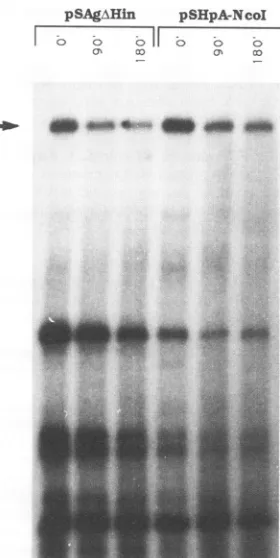
FIG. 7. Analysis ofS gene transcript stability in the cytoplasm.
HuH-7cellswere transfected with the plasmids indicated atthe top
and after 2daysweretreatedwith DRBtostoptranscription.Atthe indicated times afterdrugtreatment, RNAwasharvested andanalyzed
withprimerextension. Similar resultswereobtained in three
indepen-dent transfections. As an internal control for transfection efficiency
and RNArecovery, the cellswereallcotransfected withpMT-S,which contains the S gene under the control of themousemetallothionein
promoter. TheS gene transcripts synthesized from this plasmid give
rise toasingleextendedproductof ---203 bases (arrow).
contrarytoexpectations,theamountofnucleartranscriptswas
slightly but consistently larger in cells transfected with the
plasmidcontainingthedisplacedPRE(Fig.6A,lanes 3 and4).
In allcases, the amountof 13-actin RNAwas the same in the
two sets of RNA fractions (Fig. 6B), ruling out differential RNA recovery as a source of variability between different
samples. It is difficult to envision that destabilization of
tran-scripts in the cytoplasm could lead to a change in nuclear
transcriptlevels. Rather,these resultssuggested that thePRE
mayincrease S genetranscriptlevelsby facilitatingan intranu-clear event that subsequently leads to accumulation of the
transcripts in the cytoplasm. To measure directly the rate of
degradation of S gene transcripts in the cytoplasm,we
trans-fected cells with either pSAgAHin or pSHpA-NcoI and after
40 h addedDRB, aninhibitor ofRNApolymerase11(20).At
various times after drug treatment, cytoplasmic RNA was
extracted from the cells and the amount of remaining S
transcripts was quantitated by primer extension. The S
tran-scripts synthesized from both plasmids indeed had similar half-lives ofapproximately3 h(Fig. 7).Therefore,weconclude that thePREdoesnotpreventS genetranscriptdegradationin the cytoplasmbut must instead facilitate the accumulation of these transcripts in the cytoplasm, presumably by activating
theirtransport from the nucleus.
DISCUSSION
The region of the HBV genome downstream of the S open reading frame contains two DNA segments that can function as transcriptional enhancers when coupled to heterologous promoters(reviewed in references 19, 22, and 30).Previously, we had demonstrated that when this region was placed up-stream of the S promoter, it did not function optimally to increase the steady-state amount of HBV S gene transcripts (12). This result raised the possibility that this region also has an effecton gene expression at the posttranscriptional level. The datapresented here confirm that the X gene/EnII region of HBV acts in cis at the RNA level to increase S gene transcript levels. As we showed, there was a markedly de-creased amountof S gene transcripts when they were termi-nated before this region. Insertion of EnII back into the transcribed region largely restored the transcript levels in an orientation-dependentmanner.Since previous reportsclearly demonstrated transcriptional regulation by EnII (27, 29, 31,
33),theregion of the HBV genomeencompassing EnII must be abletoactivate gene expressionatboth the transcriptional and postranscriptional levels. We have named this region, when it functions at the RNA level, the postranscriptional
regulatoryelement (PRE). Interestingly, Vannice and Levin-son(26) havepreviouslyshown that inaheterologous context, EnIfunctions better when it is within the transcribed region.
Therefore, it is possible that there are other similar RNA elements within the HBV genome.
Changes in steady-state RNA levels induced by the PRE were correlated neither with any change in transcriptional initiationrate norwith achange intherateof RNA degrada-tion in thecytoplasm. Therefore,the PRE influencesastepin RNAmetabolism between initial synthesis and eventual deg-radation. This ispresumablyanintranuclear event,since RNA
processing takes place primarily in the nucleus. Indeed, re-moval of the PRE from the S gene transcripts led to the accumulation of thetranscriptsin the nucleus toalevel slightly
higherthan thewild-typelevel. The mostparsimonious
inter-pretationof these results is that in the absence of the PRE, a
largefraction of thesetranscriptsisnottransported out of the nucleus but is insteaddegraded within the nucleus at a rate
slightly slower than the normal rate of export out of the nucleus. Unfortunately, we could not directly test this infer-ence, since both actinomycin D and DRB appeared toblock the transport of S gene transcripts out ofthe cytoplasm and stabilize the intranucleartranscripts (datanotshown).
Never-theless,ourdata takentogetherareconsistentwith the model that the PRE functionstofacilitate thenuclear-to-cytoplasmic
transport of the S gene transcripts, although further experi-ments are needed toconfirm this model.
We note that the behavior of PRE-minus transcripts is similar to that of intron-containing transcripts that do not undergopropersplicing. Oneclass of suchtranscriptsappears to be retained in the nucleus because theyare recognized as being incompletely spliced.Aprominent exampleis the human
immunodeficiency
virus structuralprotein transcripts,
which containinefficiently
utilizedsplice
sites(2)
that leadtonuclear retention anddegradation (15).
This retention is antagonizedby
thecis-acting
RNAsequence known as the Rev response element(RRE),
which activates the transport ofRRE-contain-ing
transcripts
into thecytoplasm
(reviewed
inreferences4, 17,
and
28).
These characteristics are similar to those we have found for the PRE.Indeed,
the PREcanpartially
substitute for the RRE to effect thecytoplasmic
transport of humanimmunodeficiency
virustranscripts (11, 13). However,
while it has beenreported
that there arepotentially
functional splice VOL.68, 1994on November 9, 2019 by guest
http://jvi.asm.org/
[image:5.612.109.249.78.357.2]3198 HUANG AND YEN
acceptor sites in the S gene
transcripts
(3, 23), no spliced S genetranscripts
have beenreported. Furthermore,
removal of theseacceptorsites fromthesetranscripts
didnotrelievetherequirement
for the PRE(13). Therefore,
at this time wecannot ascertain the reason for the nuclear retention of PRE-minusS gene
transcripts.
In
addition,
wespeculate
thatatrans-acting
factorinteract-ing
withthe PREisinvolvedinreleasing
Sgenetranscripts
into thecytoplasm.
The RRE is known todepend
on the Revprotein
of the humanimmunodeficiency
virus to function.However,
theHBVXprotein,
theonly
knownregulatory
geneproduct
ofHBV,
doesnot appearto be thisfactor,
since the PRE functions in its absence(12). Therefore,
it islikely
that thefactor is of cellularorigin
and mayalso beresponsible
forposttranscriptional regulation
of cellular genes.In summary,we haveobtained data
showing
thataregion
in the HBV S genetranscripts encompassing
enhancerII facili-tatescytoplasmic
accumulation of thesetranscripts.
Further characterization of thiscis-acting region,
and thetrans-acting
factors that interact with
it,
may lead to novel means ofdown-regulating
HBVgeneexpression
ininfectedhepatocytes
andtonew
insights
into the control ofcytoplasmic
transportof cellular genes.During
the revision of this report,Huang
andLiang (11)
published
a paperdescribing findings
similar to ours.They,
too, came to the conclusion that an HBV RNA element is
important
inallowing
thenuclear-to-cytoplasmic
transport ofHBV
transcripts.
ACKNOWLEDGMENTS
We thank J. Chanfor
providing
the1-actinplasmid,J. Fridovich-Keilforproviding pTAG-1,
C. C. Lu forproviding
the 3-actinprimer, Y. W.Zheng
forproviding
theplasmid
pMT-S,N.Bakerfor oligonu-cleotidesynthesis,
R. Soto forgraphic assistance,
and E. Fodor and J. Ou for criticalreading
of themanuscript.
This workwas
supported by
NIHgrantRO1CA55578.Cell culture wasperformed
in afacility partially
supported bythe San Francisco VACenter forAIDSResearchandEducation.Oligonucleotidesweresynthesized
on ashared instrumentboughtwithfundsprovided bytheUCSF Academic Senate
Opportunity
Fund. ADDENDUMINPROOFA similar RNAelement in the Mason-Pfizer
monkey
virus genomehasrecently
been described(M.
Bray,
S.Prasad,
J. W.Dubay,
E.Hunter,
K. T.Jeang,
D.Rekosh,
and M.L.Ham-marskjold,
Proc. Natl. Acad. Sci. USA91:1256-1260,
1994).
REFERENCES
1. Almendral, J. M., D. Sommer, H. Macdonald-Bravo, J.
Burck-hardt, J. Perera, and R. Bravo. 1988. Complexity of the early
genetic
responsetogrowthfactorsinmousefibroblasts.Mol. Cell. Biol.8:2140-2148.2.
Chang,
D. D., and P. A. Sharp. 1989. Regulation byHIV Revdepends
uponrecognition ofsplicesites.Cell59:789-795. 3. Chen, P. J., C. R. Chen, J. L. Sung, and D. S. Chen. 1989.Identification of a doubly spliced viral transcript joining the
separated
domains forputativeprotease andreversetranscriptase ofhepatitis
Bvirus.J.Virol. 63:4165-4171.4.
Cullen,
B. R., andM. H. Malim. 1991.TheHIV-1 Revprotein: prototypeofanovelclass ofeukaryoticposttranscriptional regu-lators. Trends Biochem. Sci.16:346-350.5. Daar, I. O., and L. E. Maquat. 1988. Premature translation termination mediatestriosephosphateisomerasemRNA degrada-tion.Mol.Cell. Biol.8:802-813.
6. Fridovich-Keil,J. L.,J. M. Gudas, I. B.Bryan,and A. B. Pardee. 1991. Improved expression vectors for eucaryotic promoter/en-hancerstudies. BioTechniques11:572-579.
7. Ganem, D.,and H. E. Varmus. 1987. The molecularbiologyof the hepatitisBviruses. Annu. Rev. Biochem. 56:651-693.
8. Gorman, C. 1985. High efficiency gene transfer into mammalian cells, p. 143-190. In D. M. Glover (ed.), DNAcloning. IRLPress, Oxford.
9. Gunning, P., P. Ponte, H. Okayama, J. Engel, H. Blau, and L. Kedes. 1983. Isolation and characterizationoffull-lengthcDNA clones for human ao-, P-, and -y-actin mRNAs: skeletal but not cytoplasmicactins have an amino-terminalcysteinethat is subse-quently removed. Mol.Cell. Biol. 3:787-795.
10. Hollinger, F. B. 1990. Hepatitis B virus, p. 2171-2238. In B. N. Fieldsand D. M.Knipe (ed.), Virology.Raven Press,New York. 11. Huang, J.,and T.J. Liang. 1993. A novel hepatitis B virusgenetic element with Revresponseelement-like propertiesthat is essen-tial for expression of HBV gene products. Mol. Cell. Biol. 13:7476-7486.
12. Huang, Z. M., and T. S. B. Yen. 1993. Dys-regulated surface gene expression from disrupted hepatitis B virus genomes. J. Virol. 67:7032-7040.
13. Huang, Z. M.,andT. S. B. Yen.Unpublisheddata.
14. Jackson,R.J. 1993. Cytoplasmic regulation of mRNA function: theimportance of the 3' untranslated region. Cell74:9-14. 15. Malim, M. H., and B. R. Cullen. 1993. Rev and the fate of
pre-mRNA in the nucleus: implications for the regulationof RNA processing ineucaryotes.Mol. Cell. Biol.13:6180-6189. 16. Nakabayashi, H., K. Taketa, K. Miyano, T. Yamane,andJ.Sato.
1982. Growth of humanhepatoma cells lines with differentiated functions in chemically defined medium. Cancer Res. 42:3858-3863.
17. Rosen, C. A., and G. N. Pavlakis. 1990. Tat and Rev: positive regulators of HIV gene expression AIDS 4:499-509. (Erratum, 4(8), followingA48.)
18. Sambrook, J., E. F. Fritsch, and T. Maniatis. 1989. Molecular cloning:a laboratory manual, 2nd ed.ColdSpringHarbor Labo-ratory, ColdSpring Harbor, N.Y.
19. Schaller, H., and M. Fischer. 1991. Transcriptional control of hepadnaviral gene expression. Curr. Top. Microbiol. Immunol. 168:21-39.
20. Sehgal, P.B., J. J. Darnell, and I. Tamm. 1976. The inhibition by DRB (5,6-dichloro-1-beta-D-ribofuranosylbenzimidazole) of hnRNA and mRNAproductioninHeLa cells.Cell 9:473-480. 21. Selden,R.F.,K. B.Howie,M. E.Rowe,H. M.Goodman, and D. D.
Moore. 1986. Human growth hormone as a reporter gene in regulationstudiesemployingtransientgeneexpression. Mol. Cell. Biol. 6:3173-3179.
22. Shaul, Y. 1991. Regulation of hepadnavirus transcription, p. 193-211. In A. McLachlan(ed.),Molecularbiologyof thehepatitis Bvirus.CRCPress,Boca Raton,Fla.
23. Suzuki, T.,K.Kajino,N.Masui,I.Saito,and T.Miyamura.1991. Alternative splicing ofhepatitis B virus RNAs in HepG2 cells transfectedwith the viral DNA.Virology. 179:881-885.
24. Tiley,L.S.,S.J. Madore,M.H.Malim,and B. R.Cullen. 1992. The VP16 transcription activation domain is functional when targeted to a promoter-proximal RNA sequence. Genes Dev. 6:2077-2087.
25. Valenzuela, P.,M.Quiroga,J. Zaldivar, P. Gray, and W. Rutter. 1980.Thenucleotide sequence of thehepatitisB genomeand the identification ofthemajorviral genes, p. 57-70. In B. Fields, R. Jaenisch,andC.Fox(ed.),Animal virus genetics. AcademicPress, New York.
26. Vannice, J., and A. Levinson. 1988. Properties of the human hepatitisB virusenhancer:position effectsand cell-type nonspeci-ficity.J. Virol. 62:1305-1313.
27. Wang,Y., P.Chen,X.Wu,A.-L. Sun, H. Wang, Y.-A. Zhu, and Z.-P.Li.1990. Anewenhancerelement,EnIl, identifiedin the X geneofhepatitisBvirus.J.Virol.64:3977-3981.
28. Wong, S. F., and W. A. Haseltine. 1992. Regulatory genes of humanimmunodeficiencyviruses.Mol. Genet. Med. 2:189-219. 29. Yee,J.K. 1989. A liver-specific enhancer in thecore promoter
regionof humanhepatitisBvirus.Science246:658-661. J. VIROL.
on November 9, 2019 by guest
http://jvi.asm.org/
POSTTRANSCRIPTIONAL REGULATION IN HEPATITIS B VIRUS 3199 30. Yen, T. S. B. 1993. Regulation of hepatitis B virusgeneexpression.
Semin. Virol.4:33-42.
31. Yuh, C. H.,and L.P.Ting.1990. Thegenomeofhepatitis B virus
containsasecond enhancer: cooperation oftwoelements within this enhancer is required for its function. J. Virol.64:4281-4287. 32. Zheng, Y.W.,andT. S.B.Yen.Unpublished data.
33. Zhou, D. X., and T. S. B. Yen. 1990. Differential regulation ofthe hepatitis B viral surface gene promtoers bya second viral
en-hancer.J. Biol. Chem. 265:20731-20734.
34. Zhou, D. X., and T. S. B. Yen. 1991. The hepatitis B virus S
promotercomprisesaCCAAT motif andtwoinitiation regions. J. Biol.Chem.266:23416-23421.
VOL. 68, 1994