Copyright © 1994,American Society for Microbiology
Inhibition of
Vesicular Stomatitis
Virus RNA
Synthesis
by
Protein
Hyperphosphorylation
THERESA LI-YUN CHANG, CAROL S. REISS,ANDALICE S. HUANG* Departmentof Biology, NewYorkUniversity, NewYork, New York 10003-6688
Received 24 January1994/Accepted22April 1994
Vesicular stomatitis virus (VSV) RNA synthesis requires the template nucleocapsid, the polymerase (L) protein, and thecofactorphosphorylated (P/NS) protein.Todetermine whether the degree of
phosphorylation
regulated VSV RNA synthesis, infected Chinese hamsterovarycells weretreated with okadaic acid (OKA),a serine/threonine phosphatase inhibitor. OKA reduced viral penetration and uncoating but had littleor no effect on
primary
transcription or viral protein synthesis. However, approximately 80% of total viral RNA synthesis was inhibited when 2 ,uM or more OKA was added to infected cells after viral uncoating hadtaken place. Analysis of proteins and RNA species in infected cells labeled with 32p showed that OKA led tohyperphosphorylation of two viral phosphoproteins, the P/NS protein and matrix protein (M), resultingin inhibition offull-length RNA synthesis and subsequent
secondary
transcription. Pulse-chase experiments demonstrated that thehyperphosphorylated
P/NS specieswasconvertedrapidly
fromthe lessphosphorylatedform. Hyperphosphorylated P/NS as well as the less phosphorylated form, but not M, were found to be associated with nucleocapsids isolated fromcytoplasmicextracts. Theseresultssuggestthatphosphorylation played an important role in theregulation between viral transcriptionandviral RNAreplicationaswell as the turningof of RNAreplication. Thus,phosphatase inhibitors promise tobe avaluabletool for dissectingthe regulatory mechanisms involving phosphorylatedviral proteins.
Phosphorylated proteins are widely recognized to play a central role in theregulation of macromolecularsynthesis.To understand themechanismsandtodetermine how phosphor-ylation affects regulation, simple regulatory systems need to be studied. Viruses, particularly those that replicate only in the
cytoplasm, areideal becausethey code forafew well-defined phosphoproteins and these proteins can be readily detected by radiolabeling. This investigation combinesvesicularstomatitis virus(VSV)-infectedcells with theproteinphosphatase inhib-itor okadaic acid (OKA) to determinewhether alterations in
phosphoproteins can be correlated directly with important
regulatory steps during the synthesis of viral progeny. It is hoped that such studies will lead to an understanding of the detailed mechanismsunderlyingregulation.
VSV, an enveloped, negative-strand RNAvirus, codes for five structural proteins, two of which, P/NS (found
predomi-nantly in the cytoplasm and previously considered a nonstruc-tural [NS] protein) and M (matrix protein), are
phosphopro-teins. Theirmajorphosphoaminoacidsarephosphoserineand
phosphothreonine (9). Both proteins play a role in the regu-lation of VSV RNAsynthesis. Viral transcription and replica-tion require the participation of P/NS, complexed with the RNA-dependent RNA polymerase (13, 14, 31). Phosphoryla-tion of P/NS was found to be essential for transcription in cell-free assays(1,17,18). Within infected cells(in vivo),there aretwophosphorylated species,separable onurea-containing sodium dodecyl sulfate (SDS)-polyacrylamide gels; the less phosphorylated one binds to VSV nucleocapsid templates (6) which contain full-length RNA covered with nucleoprotein. Studies of RNA synthesis by temperature-sensitive (ts) mu-tants with lesions in the P/NS gene suggest that this protein
playsarole in the switch from transcription to replication (20,
*Corresponding author. Mailing address: Departmentof Biology,
Main 1009, New YorkUniversity,NewYork, NY 10003-6688. Phone: (212)998-3800. Fax:(212)995-4181. Electronic mail address: huanga @acfcluster.nyu.edu.
30).Ithas beenpostulatedthatthis switch is regulatedbythe dissociation of the lessphosphorylated proteinfromtemplates
and that this dissociation may be triggered by increased
phosphorylation(6). Mprotein,ontheotherhand,appearsto regulate only transcription. This effect of M was determined whentranscriptionwasgreatlyincreasedatthenonpermissive
temperaturewhencellswereinfected withtsmutantsofM (7)
and when in vitrotranscriptionwasinhibitedbytheaddition of wild-type Mproteinto a polymeraseassay (4).Inadditionto
phosphoserineandphosphothreonine,Mproteinalsocontains
phosphotyrosine (9); however,whenthephosphotyrosine con-tentofMisincreased20-fold,there isnodetectable effecton VSVtranscriptionorprogenyformation (8).
Although there isincreasingin vitroevidence that phosphor-ylation ofP/NS is essential for the regulation of VSV RNA
synthesis,it has beendifficulttocomparetheresults obtained by cell-free (in vitro) studies directlywith those obtained by
using infected cells. In part this is because gel systems for
separating the different species ofP/NS have not been used
consistently in different laboratories and because direct
com-parisons of the in vitro-synthesized P species have not been done with in vivo-synthesized NS species to ensure their identity. Until such comparisons have been made, we shall
continuetouse theterminologyof Clinton andcolleagues(6, 7)torefertothe differentspeciessynthesizedinVSV-infected cells asNS1 for the lessphosphorylated oneand NS2 for the more phosphorylated one. In distinction to P species
synthe-sized in vitro, the completely nonphosphorylated P is not detectable in infected cells.
OKA is aspecific inhibitor ofprotein phosphatases 1 and 2A, which dephosphorylate serine and threonine residues
(reviewed in reference 11). The approach taken herewas to add the inhibitor to VSV-infected cells and determine its
effect(s) on VSV macromolecular synthesis and progeny for-mation. Despite pleiotropic effects of the drug, a specific
correlationwasseenbetween hyperphosphorylationofNS1to NS2 and inhibition offull-lengthRNAreplication.Inaddition,
4980
on November 9, 2019 by guest
http://jvi.asm.org/
binding of NS proteins to RNA templates was reexamined, resulting in a model which helps us to understand the transi-tion fromtemplate function to progeny formation. This report contains in vivo data that support the essential regulatory role forphosphorylation of NS during VSV RNA synthesis. More-over, it shows that hyperphosphorylated NS2, rather than dissociating from templates, remained, and that it may be
responsible for inhibition of full-length RNA replication after exposure to OKA.
(This work was done in partial fulfillment of the require-ments for the Ph.D. degree by T.L.-Y.C.)
MATERIALS AND METHODS
Isotopes, chemicals, and media.
["4C]uridine
(>50mCi/
mmol), [35S]methionine (>1,000 Ci/mmol), and
32p;
(8,500 to9,120 Ci/mmol) were obtained from DuPont/New England Nuclear Corp. OMNISORB cells were from Calbiochem. Joklik's modified minimal essential medium (MEM) without
phosphate or methionine, penicillin G-streptomycin sulfate, andOKA were from GIBCO Laboratories. Endoglycosidase H
(endo H)was from Boehringer Mannheim Biochemica. Com-plete Joklik's modified MEM, fetal bovine serum (FBS; type
II), and other chemicals were from Sigma.
Cells and virus. In general, Chinese hamster ovary (CHO) cells and VSV were propagated as described by Stampfer et al.
(28). CHO cells were grown in continuous suspension culture at 37°C and maintained at 4 x 10⁵ cells per ml in MEM containing 2% FBS, nonessential amino acids, 100 U of
penicillinGsodium per ml, and 100 jig of streptomycin sulfate per ml. The Indiana serotype, San Juan strain, of VSV was used. A ts mutant of the Indiana serotype, tsG114 (comple-mentation group I), was obtained from Craig Pringle.
Radioactive labeling of cells and virus. For32p or 35S
labeling of proteins, MEM depleted of phosphate or methio-nine was used, and either 75,uCi of
32Pi
per ml or 110,uCi
of[35S]methionine
per ml was added to each medium. Cells wereplacedat2x 106/mlinmedium containing 5 jig of actinomycin D (ActD) per ml, 25 mM N-2-hydroxyethyl
piperazine-N'-2-ethanesulfonic acid (HEPES; pH 7.4), nonessential amino
acids, and 2% dialyzed FBS. For labelling RNA with32p, 300
,uCi of
32Pi
per ml was added to phosphate-depleted MEM, and all otheradditions remained the same.Cumulative uridine incorporation into VSV RNA. Experi-ments were carried out as described previously for pilot infections(15).CHO cells (4x 10⁶) were infected with VSV in 2 ml of MEM containing 2% FBS, nonessential amino acids, and 25 mM HEPES (pH 7.4). The multiplicity of infection
(MOI)for each experimentis indicated in the figure legends. ActD at5 ,ug/ml was added, and viral attachment and pene-tration was allowed to proceed for 40 min at
37°C
before the addition of 0.3 ,uCi of ['4C]uridine. To measure primarytranscription, infected cells were exposed to S,ugof ActD per
ml, 100 ,ug ofcycloheximide per ml, and 0.6,uCi of
[14C]uri-dine. Since primary transcription depends on input virions, MOIs had to be increased in order to conserve radioisotope usage. Asample ofuninfected, ActD-treated cells served as a
negativecontrol. When OKA was used, the amounts and time of addition are indicated in the figure legends. Incorporation ofradioactive uridine into RNA was determined by
withdraw-ing 0.1 ml from each sample and mixing it into 1 ml of 5% trichloroacetic acid at 4°C. The acid-precipitated RNA was concentratedon an HAfilter(Millipore HAWP), washed with 15 ml of 5% trichloroacetic acid, dissolved in 5 ml of liquid scintillation cocktail(Ready Protein; Beckman), and counted in aBeckman LS6000LL scintillation counter.
Analysis of viral RNA species. ActD-treated infected cells (4 x
106)
in the presence or absence of OKA (5,uM)
were labeled with 32p from 1.5 to 4 h postinfection(p.i.). Total RNA from VSV-infected cells was then extracted by a single-step procedure (5). Cells were resuspended in 400 pI of solutionD containing 4 M guanidinium thiocyanate, 25 mM sodium citrate (pH 7.0), 0.5% sarcosyl, and 0.1 M,3-mercaptoethanol
and mixed in 40,ul
of 2 M sodium acetate(pH 4.0). RNA was extracted with 400jl
of saturated phenol and 80[lI
of chloroform-isoamyl alcohol (49:1). After 15min
ofincubation on ice, RNA in the aqueous phase was collected by centrifu-gation at 14,000x g in amicrocentrifuge at4°C
for 20min
and precipitated with 1 volume of isopropanol at-20°C
overnight. Each sample was resuspended in diethyl pyrocarbonate-treated H20. Aliquots of each sample were analyzed on a0.7% agarose gel containing formaldehyde (25). rRNAs werestained with 30 jig of acridine orange per ml in 10 mM sodium phosphate buffer (pH 7.0). The gel was dried and then exposed to Kodak X-Omat AR film.Immunoprecipitation. Proteins from infected cells (4x
106)
labeled with[35S]methionine
were harvested and lysed in NEB buffer (0.01 M Tris [pH7.4],
0.01 M NaCl, 0.02 M EDTA) containing 1% Nonidet P-40, 1% sodium deoxycholate, and 2 mM phenylmethylsulfonyl fluoride. Samples were incubated with 3jil
of hyperimmune goat anti-VSV at4°C
for 1 h, and the complexes were precipitated with a 10% prewashed cell suspension of heat-inactivated protein G-bearing group G Streptococcus sp. (OMNISORB cells) for 1 h at4°C.
The complexes were then washed twice with radioimmunoprecipi-tation assay buffer (0.01 M Tris [pH7.2],
0.15 M NaCl, 1% Triton X-100, 0.1% SDS, 1% sodium deoxycholate, 5 mM EDTA) containing 0.5 M NaCl and three times with radioim-munoprecipitation assay buffer. Aliquots of each sample were boiled in Laemmli buffer (19) and analyzed in SDS-10% polyacrylamide gels.Endo H treatment. After immunoprecipitation, viral pro-teins from 4 x
106
cells were resuspended in endo H incuba-tion buffer (200 mM sodium citrate [pH5.8],
0.5 mM phenyl-methylsulfonyl fluoride 0.1% SDS, 0.1 M,-mercaptoethanol).
Half of each sample was boiled for 5min
and then treated with endo H (3 mU) overnight at37°C.
Samples were then boiled in Laemmli buffer and analyzed on SDS-10% polyacrylamide gels.Preparation of VSV ribonucleoprotein-associated viral pro-teins. VSV-infected cells (8 x
107)
were labeled with 0.3 jiCiof
['4C]uridine
per ml at 0.5 h p.i. OKA at 2 jiM was added tohalf of the cells at 1.5 h p.i., and 75
,uCi
of32p per ml was added at 2.5 h p.i. At 4.5 h p.i., infected cells were harvested, washed with cold (PBS-A), and lysed in NEB buffer containing 1% Nonidet P-40 and 1% sodium deoxycholate as described by Huang et al. (15). After removal of nuclei by centrifugation, the cytoplasmic extract was fractionated in a 15 to 30% sucrose gradient in NEB buffer. The pattern of optical density at 260 nm was recorded with a Beckman DU640 spectrophotometer. The polyribosomes were dissociated to50S
and 30S subunits in NEB buffer and used as markers for locating the ribonucleo-capsids (120S). The location of VSV ribonucleoribonucleo-capsids was determined by the pattern of optical density at 260 nm and the profile of['4C]uridine
incorporation as analyzed by acid precipitation of 1/10 of each fraction. The volume of each fraction was about 1.3 ml. Fractions containing cores were pelleted at 150,000 x g for 90min,
and the proteins were analyzed in SDS-10% polyacrylamide gels with 5 M urea and 0.19 MTris-HCl
(8). Nucleocapsid (N) proteins, but not M protein, wereobservedon the gel stained with Coomassie blue.on November 9, 2019 by guest
http://jvi.asm.org/
30
0
i
20
10
F
0
100
._
' 80
c
D
C _
° 2
,N2
0 1 2 3 4 5
hrp.i.
0 1 2 3 4
pMofokadaicacid
20
e14
0
U,
15
10
5 14C e g>
-0
0 l 2 3 4 5
hrp.i.
FIG. 2. Cumulative VSV RNAsynthesis afterexposureofcellsto
VSVand OKAat4°C. CHO cellswereinfected with VSVatMOIs of 125 and12.5, and viral attachmentwasallowedtoproceedat4°C for 40min in thepresence(0) and absence (0) of 2 ,uM OKA.Asample ofuninfected, ActD-treated cells served as a negative control (A).
After sampleswerewashed withPBS-A, [14C]uridineand ActDwere
added to each sample, and the samples were transferred to 37°C. Cumulativeincorporation of radioactive uridinewasassayed.
5
FIG. 1. Cumulative RNA synthesis in VSV-infected cells in the
presenceof OKA. (A) CHO cells infected with VSVatanMOI of 125
were exposedto OKA (2 jLM) at either 0 (O) or 40 (L) min p.i.
ActD-treateduninfected (A) and infected (0) cells in the absence of OKAwerealsoestablished. Cumulativeincorporation of [14C]uridine wasdeterminedasdescribed in Materials and Methods. (B) OKAat
different concentrationswasaddedtoinfected cellsat 40min p.i. in pilotassays asdescribed in Materials and Methods. Results of [14C]uri-dineincorporationat3 h and 40 min p.i.areshown. After subtraction of thebackground incorporation by ActD-treated uninfected cells, the resultsareexpressedasthe inhibition ofVSV RNA synthesis by OKA
relativetothe control. Similarpercentagesof inhibitionwere seen as soon as 1h and 40min p.i.
RESULTS
Effect of OKAontotalVSV RNA synthesis.To determineif
inhibition of serine andthreonine phosphatases hadanyeffect onVSV RNAsynthesis,OKAwasaddedtocell culturesatthe
sametimeasVSVorat40minafter the initiation of infection. Ataconcentration of2 ,uM OKA,total VSVRNAsynthesis wasalmost completely inhibited when the inhibitorwasadded
atthesametimeasVSV; with the later time of addition, less
inhibition was observed, suggesting that very early events as
well as RNA synthesis were affected by OKA (Fig. 1A). To
ensure that maximal inhibition of RNA synthesis was being
attained, increasing concentrationsof OKAwereaddedtothe infected cellsat40min p.i. The dose-responsecurvein Fig. 1B
shows that total viral RNAsynthesis wasmaximally inhibited
to80%withaconcentration of2,uMorgreater.Also,aswould
be expected, progeny formation was decreased to less than 0.011%wheninfected cellsweretreated with2to5 p.M OKA. Thus,in allsubsequent experiments, OKAatconcentrations of
between 2 and 5 ,uM was used. The degree of inhibition on
RNA synthesis was observed as soon as 30 min after the
addition of OKA.Despitereportsthat the effects of OKAare
slowlyreversible after cells have beenwashed(21),this system of VSV-infected CHO cells in suspension did not show any
reversibility in RNA synthesis with incubation times of
be-tween 15 minto4 hevenafterextensive washes and
observa-tiontimes (datanotshown).
Inhibitionofpenetrationanduncoatingbutnotattachment andprimary transcription.BecauseOKA hadagreater inhib-itory effect onVSV RNAsynthesis when addedearly during
infection than when added later, one can postulate OKA effectsonviralattachment,penetration, uncoating,orprimary
transcription leading indirectlytotheoverall inhibition of VSV RNAsynthesis.To delineate between these stepsof the viral lifecycle,three differentapproachesweretaken. Viral attach-mentwasallowedtoproceedat4°Cfor 40mininthepresence
andabsence of 2,uMOKA. Two different MOIs(125and12.5)
were used. Cellswere thenwashed, and viral RNAsynthesis wasanalyzedas described previously. Ifthere isan effect on
viral attachment at this temperature, a change in viral RNA synthesis should be seen. Since no inhibition was observed (Fig. 2),itcanbe concluded thatat4°COKA hadnoeffecton
viral attachment at either of the multiplicities tested. Its reversibility indicated that OKA was inactive or did not become cell associatedat4°C.
Asecond way to define where OKA might act during the early stages of VSV infection was to measure primary
tran-scription. VSV penetrates, uncoats, and carries on primary
transcription in cells in thepresence of ActD and cyclohexi-mide. In the absence ofprotein synthesis, only primary tran-scription from incoming templates occurs in these cells (16).
Figure 3A shows that primary transcription was partially
inhibited if OKA was added at the same time as virus
comparedwith itsbeingadded 40minlaterornot atall. This findingsuggests that the inhibition probablyoccurred during penetrationoruncoatingandnotduring primary transcription. To obtainaclearerseparationbetweentheseearlysteps and primary transcription, thets mutanttsG114wasused. At the
A
on November 9, 2019 by guest
http://jvi.asm.org/
[image:3.612.97.272.73.394.2] [image:3.612.358.527.74.236.2]12
10
8 6
4
0
u
2
6
4
2
0
0 1 2 3 4 5
[image:4.612.95.256.73.368.2]hrp.i.
FIG. 3. Primary transcriptionofVSVin thepresenceof OKA.(A)
CHO cells infected with VSVatanMOI of 250wereexposedtoActD
andcycloheximideat370C. OKAat2.5 jLMwasaddedateither 0(O)
or40(-)min p.i.Uninfected(A)andinfected(0) cellsinthe absence
of OKAwere also established. Cumulative incorporationof
radioac-tive uridinewasdetermined.(B)CHO cells infected with tsGl 14atan
MOI of 75wereexposedtoActDandcycloheximide atthe
nonper-missivetemperature(39°C). OKAat2.5,uMwasaddedateither 0 (O)
or40(U)min p.i.Uninfected(A)andinfected(0)cellswithout OKA
werealso established.At40min p.i., ["4C]uridinewasaddedtoeach sample and all sampleswere shifted to thepermissive temperature
(31°C). Cumulativeincorporationof["4C]uridinewasassayed.
nonpermissivetemperature(39°C),thismutantpenetratesinto cells anduncoatsbut failstocarryoutanyfurthersteps(23, 24, 29). Therefore, the addition ofOKA andcycloheximide after the virus has been incubated at 39°C for 40 min and then shiftedto31°Cwould beadirecttestofwhetherOKA affects primary transcription.As shown inFig. 3B, primary transcrip-tionby tsG114wasnotinhibitedbyOKAat2.5p,M. Thus,the early effects of OKA on VSV infection must have been on
either viralpenetrationoruncoating. Therefore,todistinguish betweenearlyeffects of OKA and later, morespecific effects
onviral RNA synthesis, all further experimentswerecarried outwith the addition of OKAat40minafter the initiation of infection.
Effect of OKA onviral RNAreplication. Since therewas a
graded inhibitionon total VSV RNAsynthesis dependenton
when OKAwasadded,itwaslikelythat the inhibition occurred
atthestageoffull-lengthRNAreplication,withindirect effects
onsecondary transcription.Examination of RNAspeciesfrom
32P-labeled infected cells showed that OKA prevented the replication of full-length RNA (Fig. 4). mRNA was also
reducedasexpected. Comparisonof OKA withcycloheximide
indicated similar inhibition of RNA replication and reduced
1 2 3
0
* -vRNA
L i1
FIG. 4. RNA species synthesized in VSV-infected cells in the
presenceofOKA. (A) CHO cells infected with VSVatanMOI of 125
at37°Cinthepresenceof ActD. OKA(5 p.M)wasaddedat40min p.i. RNAwaslabeled with 32p from 1.5 to4 hp.i. andwasextractedas
described in Materials and Methods. Equal aliquots of each sample
were loaded onto a 0.7% agarose gel containing formaldehyde.
Full-length genomic RNA from 32P-labeled virions was used as a
marker(lane 1). Lane 2, RNA from infected cells treated with OKA; lane3, RNAfrom infected cells without OKA.vRNA, viral RNA.
transcriptionwithoutanoticeableeffectonthe relative molar
ratios of viral mRNA species synthesized (data not shown). Examination of lane 2 inFig. 4 intheoriginal autoradiograph indicates a trace amount of full-length viral RNA in the OKA-treated infected cells. ThismayrepresentresidualRNA replication in thepresenceof OKAor aselective inhibition by
OKAonminus-strand RNAsynthesis.Totestwhether thiswas
full-length plus-strand RNA, Northern (RNA) blot analysis
wasperformed by probing with 32P-labeled minus-strandRNA from virionsor32P-labeled viral mRNAs (plus strands). Onlya
residual small amount of minus-strand full-length RNAwas
detected (data not shown). In this hybridization experiment, what was detected may represent RNA derived from input virions. These results indicate that OKA inhibitedRNA
rep-licationby preventing the accumulation of both minus and plus strands offull-length RNA.
Effect of OKA on VSV protein synthesis. Since protein
synthesisis requiredfor VSV RNAreplication, the effects of OKAonviral RNAreplication mighthave been dueindirectly
to the complete inhibition of protein synthesis. To rule out such indirect effects, viral proteins from OKA-treated and untreated infected cells were radiolabeled,
immunoprecipi-tated,andanalyzed by SDS-polyacrylamide gel electrophoresis (PAGE) (Fig. 5).Allfiveviralproteinsweresynthesizedinthe infected cells in thepresenceofOKAat2,uM. Somereduction ofproteinswasevident,asexpectedfrom indirect inhibition of secondary transcription by OKA, but this partial reduction wouldnotbeexpectedtoaffect viral RNAreplication.Both G and Mproteins migrated slightly differentlyinthisgelsystem, suggesting that OKA had effects other than onviral proteins
synthesis perse. Therefore, the large and rapidinhibition of viral RNAreplication bythe addition of OKAmustbe dueto
someothereventbesides inhibition ofprotein synthesis. Endo Htreatmentof Gsynthesizedin thepresenceof OKA. Parenthetically, examination of lane 2 inFig. 5 showsthat the viral glycoprotein, G, migrated faster and more uniformly
whenitwassynthesizedin thepresenceof OKA.This could be duetocleavageof theglycoproteinor adifferencein
process-A
~~~
00
14 0
1
l4l
li
i
on November 9, 2019 by guest
http://jvi.asm.org/
[image:4.612.397.479.73.234.2]1 2 3
-L
=Min
-G_w
-N NSIabfIb
-MFIG. 5. VSV proteins synthesized in OKA-treated infected cells. CHO cellsinfected with VSVatanMOIof 125at37°Cwereexposed toOKAat2,uM(lane2)orcycloheximideat100 p,g/ml(lane 3)at3 hp.i. Infected cellinthe absence of OKAservedas acontrol(lane 1). Ten minutes after addition of drugs, allsamples were labeled with [35S]methioninefor 30min.Viralproteinswerethen immunoprecipi-tated asdescribed in Materials and Methods and analyzed by SDS-PAGE(10% gel).
ing of its carbohydrate moiety. To determinewhether carbo-hydrateprocessingwas affected, Gprotein synthesized in the presenceof OKAwasdigested with endo Hpriortoseparation by SDS-PAGE. Figure 6 indicates that a
majority
of Gsynthesizedinthe presenceof OKAwassensitivetodigestion byendo Handmigrated faster;thus,someGsynthesizedinthe presenceof OKAwasimmatureintermsofitscarbohydrates,
indicating a blockin its transport out of theroughendoplasmic
reticulum. This observation is in agreement with previously
described effect of OKA in blocking transport between the
endoplasmic reticulum and the Golgi apparatus
(12,
21). Inaddition,endo H-resistantG,migratingfaster thanmatureG,
wasfound in both OKA-treated anduntreatedcells and may representpartially mature G.
Effect of OKAonviral protein phosphorylation. To
deter-OKA - + - +
Endo H - + +
G
-4_,
Gs
FIG. 6. Endo H-treated VSV glycoproteins synthesized in the presenceof OKA. CHO cells were infected with VSV at an MOI of 125. Halfof each samplewastreatedwith OKA (2 ,uM) at 3 h p.i.,and all samples were labeled with [35S]methionine for 30 min. Viral proteins were immunoprecipitated, and half of each sample was treated with endo H as described in Materials and Methods. Viral proteinswerethen analyzedby SDS-PAGE(10% gel). Arrows at the rightindicate endo H-sensitive G protein species.
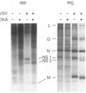
32p
VSV - - + +
[image:5.612.147.234.73.285.2]OKA - + - + + +
FIG. 7. Urea-polyacrylamide gelelectrophoretic separationof pro-teins from VSV-infected and uninfected cells made in the presence and absenceofOKA. CHOcells,uninfectedorinfected withVSVat
anMOIof125at37°C,wereradiolabeled between 3and4hp.i.with either32p(A)or [35S]methionine(B). Halfofeachsamplewasalso treated with 5 ,uM OKA at the same time as the labelingperiod. Cytoplasmicextracts werethenprepared bysuspendingcells in NEB buffercontaining 1% NonidetP-40and1% sodiumdeoxycholate, and nuclei were removed by centrifugation. Aliquotsofcytoplasmic
ex-tracts from eachsamplewereelectrophoresed inanSDS-10% poly-acrylamide gelcontainingurea.
mine whether therewas a relation between the inhibition of viral RNA replication and inhibition of phosphatases, the effect of OKA on protein phosphorylation was examined. VSV-infectedcellswerelabeled witheither
32p
or[35S]methi-onine in the presence and absence of OKA. Total proteins
were analyzed in SDS-polyacrylamidegelscontaining urea as describedbyClinton etal. (7).Inthisgelsystem,NSproteins
of VSVare resolved into NS1 and the more phosphorylated form,NS2. As shown inFig.7, when infected cellsweretreated with OKA, NS2 became the dominantspeciesofNS,
suggest-ing that much of NS1 became hyperphosphorylated when
phosphataseactivitywasinhibited. The other VSV
phosphor-ylated protein, M,was not as dramatically affected, showing onlyaslightmobility shift indicating possibleincreased phos-phorylation. This is consistent with the mobilityshift seen in
Fig.5. However,since thereisnoevidence tosuggestthatM
playsanyroleduring full-lengthRNAreplication,therewasno reason to implicate M protein in
any
role during RNAreplication. Also, in lanes containing
3%P,
OKA effected an overall increase in incorporation, whether cellswere infected or not, as evidencedby thebackground radioactivity seen inFig.7.This increase in
phosphate
labelingwas notmatchedbyasimilarincrease in [3
Slmethionine
labeling, which suggests thatpreviously synthesized proteinsbecamehyperphosphory-lated in the presence of OKA.
Conversion of NS1 to NS2 in OKA-treated infected cells.
Clintonetal.
(7)
haveshown that thetwoforms of NSproteins
areinterconvertible in vitro.Todetermine whether the hyper-phosphorylated NS2 in OKA-treated cells was derived from theless phosphorylated NS1, apulse-chase experiment using
[35S]methionine
and32p
wasperformed. As shown inFig.
8,conversion of NS1 toNS2 didnot occurin theabsence of the
drugevenwithachaseperiodof 60min; in contrast,no matter which radioisotope was used, NS1 converted to NS2 in the infected cells after 15 min oftreatmentwithOKA, butsome residual NS1 alwaysremained.Inother experiments(datanot
shown), conversion of NS1 to NS2 occurred within 5 min,
G
--NS
1---NS.2
on November 9, 2019 by guest
http://jvi.asm.org/
[image:5.612.365.511.74.232.2] [image:5.612.127.245.556.645.2]VSV RNA SYNTHESIS 4985 A. (-) OKA
.Pp ,S
'!r C I5 IC 6 C
|I'
I-N
---NS 1
-NS2
B. (±) OKA
.p
r,in C C I55 3 C 6 -n 5, 3 C)C
|
k
-~G
___-_
NS
-NS2
-M
[image:6.612.148.472.75.270.2]-M
FIG. 8. Proteins from VSV-infectedcells from a pulse-chase experiment. VSV-infected cells were labeled with32Por[35S]methionine at2.5 h p.i.At3.5 hp.i.,4,000-foldunlabeledphosphate or 1,000-fold L-methionine was added to the samples, and one half of each sample was treated with OKA at 5 ,uM.Aliquots were removed at 0, 15, 30, and 60 min of further incubation. Cytoplasmic extracts were prepared from each sample
asdescribed forFig.7.Aliquots of cytoplasmicextracts wereanalyzed in anSDS-10%polyacrylamide gel containing urea.
indicating that hyperphosphorylation was quite rapid. This result showed that NS1 wasaprecursorof NS2, indicating that
hyperphosphorylationof NSproteindoesnotrequirede novo
protein synthesis. Since the inhibition of total viral RNA
synthesiscouldnotbe reversedinattemptstowashoutOKA, NS specieswere observed under similar conditions to deter-mine whether hyperphosphorylation was similarl, not re-versed. Although there was a30% loss of NS2, by [
5S]methi-oninequantification, the loss occurred very slowlyover a4-h chaseperiod (datanotshown).
Association of NS protein with nucleocapsids in OKA-treatedinfectedcells. NS1 isreported tobindtonucleocapsid
templatesfromvirions and from infected cells(6).To reexam-ine thisbindingnowthatmoredetectable NS2wasproduced in OKA-treatedcultures,infected cellsweredoublylabeledwith
32Pand
[14C]uridine
in the presenceorabsence ofOKA. Cellextracts werepreparedat4.5 hp.i.,and thenucleocapsidswere
purified by centrifugation through a 15 to
30%
sucrosegradi-ent. Thefraction containing nucleocapsidswas collected and
analyzed by urea-SDS-PAGE (Fig. 9). In infected cells not treated with OKA, in which NS1 is usually found in greater abundance than NS2, both NS1 and NS2 were found to be associated with nucleocapsids, but by far the predominant species was NS1. With OKA, both NS1 and NS2 were also
bound, with NS2 predominating. Phosphorylated M protein
was not detectable in the nucleocapsid fractions, supporting
theassumptionthathyperphosphorylatedMdidnotplayarole inRNAreplication.
DISCUSSION
OKA had a pleiotropic effect on VSV-infected cells. It inhibited anearly stage of infection during eitherpenetration
or uncoating. Total VSV RNA synthesis was also inhibited
maximallyby 80%in adose-dependentmanner.Theresidual RNAsynthesis represented productsofprimary transcription.
The inhibition ofRNAreplication byOKAwas notdueto a block in protein synthesis. Analysis of viral RNA species showed that RNA inhibition was the result of reduced
full-lengthRNAsynthesis,withasubsequentindirect inhibition of
secondarytranscription and progeny formation. Both plus- and minus-strand RNA syntheses were inhibited. Detailed exami-nation of VSV proteins synthesized in the presence of OKA showed a shift in the migration of three of the five VSV
proteins.Theglycoprotein migratedfaster asaresult ofablock in carbohydrate processing. M proteinchanged its migration rate, which was most noticeable in urea-containing
SDS-polyacrylamide gels. Themost dramatic result of OKA treat-mentwasonNSprotein:mostofNS1became
hyperphospho-OKA- +
NS1
*-NS
2FIG. 9. Nucleocapsid-associated proteins from OKA-treated in-fected cells.VSV-infectedcellswerelabeled with["4C]uridineat0.5 h p.i. and half of eachsamplewastreatedwith OKA(2
pLM)
at1.5 hp.i. p was added at 2.5 h p.i. At 4.5 h p.i., cytoplasmic extract wasfractionated in a sucrose gradient as described in Materials and Methods. The locationofnucleocapsidswasdeterminedonthebasis of the pattern of optical density at 260 nm and the profile of
['4C]uridine
incorporation. The fractions containing nucleocapsidswerecollected and32P-labeledproteinswereanalyzedinanSDS-10% polyacrylamide gel containingurea.
VOL.68, 1994
-?. 11
9 S,. .f
it
-'v -'- L X.on November 9, 2019 by guest
http://jvi.asm.org/
[image:6.612.402.476.446.626.2]rylated to NS2 even in the absence of de novo protein synthesis. Both phosphorylated NS species were found to be associated with nucleocapsid templatesirrespective of whether OKAwas present. Taken together, these results confirm that VSV RNA synthesis is regulated by NS proteins and suggest that the binding of NS2 leads to an inhibition of RNA
replication but does not affect transcription.
The findingthatNS2 bound to templates contrasts with the report of Clinton et al. (6), who found that only the less
phosphorylated form-NS1 associated with VSV templates. It maybe that their use of a higher radioactive background and the verysmall amount of NS2 found in cells led to difficulties indetecting NS2, much less its association with nucleocapsids. In alaterpublication, Williams and Emerson (32) showedthat both NS1 and NS2,purified by DEAE-cellulose
chromatogra-phy, rebind to ribonucleocapsids in vitro. In contrast to the
intracellularpreponderance of NS1 over NS2, Clintonet al. (6) found much more NS2 than NS1 in virions. These findings
coupledwith the correlation of inhibition of RNA replication with theaccumulation of NS2 suggest that NS2 may playa role in progeny maturation by shutting off further use of full-length RNAas atemplate for replication. This step and the budding ofvirions withexcessNS2 maybe in addition to the inhibitory function ofM protein on RNA transcription (10) and coales-cenceofthe templates into progeny.
Besides the effect of OKA on VSV RNA replication, there werealsoeffectsonviralpenetration and/or uncoating and on the transportof newly synthesized glycoprotein. Theseresults maybeexplained by the many other reported effects ofOKA oncellular functions, particularly membranes. Changes inpH occur,since OKA affects cellular cotransport of potassiumand chloride(22) andalters the exchange of sodium and hydrogen ions (3, 27). Moreover, OKA induces Golgi apparatus frag-mentation(21)andinhibits vesicular fusion of endosomes (33). OurstudiesonVSVglycoprotein transport aresimilar to those
reported for the tsO45 VSV glycoprotein; in that case, trans-portbetween the endoplasmic reticulum and Golgi apparatus is blocked in cells treatedwith OKA (12). Another possibility for the role of OKA in Gproteintrafficking might be its effect on calcium ions,which may cause proteins to fold incorrectly
leadingtoretention ordegradation (reviewed in reference 26). Whatrole,if any,phosphorylationof M protein plays during the VSV life cycle remains unknown. Hyperphosphorylated forms of M are seldom seen under normal conditions of infection in the absence of OKA. Evidence presented here and elsewhere suggests that M proteins do not bind tightly with
ribonucleocapsid inside the cell. Recently, an organizing role for M protein during the winding of ribonucleoprotein into
bullet-shapedcoreshas been suggested (2), and it may be that
phosphorylationhelps Mproteins to recognize each other and coalesce intocores.
Given theabilityof bothspeciestobind to templates, we can
postulateasimple modelforhow NS1 and NS2regulate VSV RNA synthesis. Templates bound to homodimers of NS1 support transcription, whereas RNA replication requires the
bindingof bothNS1 and NS2. Excess NS2 would drive NS1 off,
leading to binding of homodimers and inhibition of RNA
replication. Transcription remains in the presence of excess NS2 ifwepostulate that homodimers of NS1 are irreversible. This model fits with the relative ratios of NS1 and NS2 in
cells,
invirions, and in OKA-treated cells (6, 7) (Fig. 7). Sucha model naturally leads to experiments on coinfections with
wild-type VSV andts mutants containing lesions in theP/NS
gene, in vitro RNA assays with controlled concentrations of
phosphorylatedNS speciestogether with viral ribonucleopro-teintemplates,andcharacterizations of host proteins that bind
to NS1 and NS2 and might alter VSV RNA synthesis. Such
experiments arein progress.
ACKNOWLEDGMENTS
We thank John Lenard for helpful discussions, Stuart Mendelson for technical assistance, and Elizabeth Schmalz for photographic assis-tance.
This work was supported byNIH researchgrants 5R37 AI20896to A.S.H. andA118083 to C.S.R.
REFERENCES
1. Banerjee, A. K., and S.Barik. 1992. Gene expression of vesicular stomatitis virus genome RNA. Virology 188:417-428.
2. Barge, A., Y. Gaudin, P. Coulon, and R. W. H. Ruigork. 1993. Vesicular stomatitis virus M proteins may be inside the ribo-nucleocapsid coil. J. Virol.67:7246-7253.
3. Bianchini, L., M.Woodside, C. Sardet, J. Pouyssegur, A.Takai, and S. Grinstein. 1991. Okadaic acid, a phosphatase inhibitor, induces activation and phosphorylation of the Na+/H+ antiport. J. Biol. Chem. 266:15406-15413.
4. Carroll, A. R., and R. R. Wagner. 1979.Role of the membrane(M) protein in endogenous inhibition of in vitro transcription by vesicular stomatitis virus. J. Virol. 29:134-142.
5. Chomczynski, P., and N. Sacchi. 1987.Single-stepmethod of RNA isolation by acid guanidinum thiocyanate-phenol-chloroform ex-traction.Anal. Biochem. 162:156-159.
6. Clinton, G. M., B. W. Burge, and A. S. Huang. 1978. Effects of phosphorylation and pH on the association of NS protein with vesicular stomatitis virus cores. J. Virol. 27:340-346.
7. Clinton, G. M., B. W. Burge, and A. S. Huang. 1979. Phosphopro-teinsofvesicular stomatitis virus: identity and interconversion of phosphorylated forms. Virology 99:84-94.
8. Clinton, G. M., N. G. Guerina, H.-Y. Guo, and A. S. Huang. 1982. Host-dependent phosphorylation and kinase activity associated withvesicular stomatitis virus. J. Biol. Chem. 257:3313-3319. 9. Clinton, G. M., and A.S. Huang. 1981.Distribution of
phospho-serine, phosphothreonine, and phosphotyrosine in proteins of vesicular stomatitisvirus. Virology 108:510-514.
10. Clinton, G.M., S. P. Little, F. S. Hagen, and A. S. Huang. 1978. The matrix (M) protein of vesicular stomatitis virus regulates transcription.Cell 15:1455-1462.
11. Cohen, P., C. F. B. Holmes, and Y. Tsukitani. 1990. Okadaicacid: a new probe for the study of cellular regulation. Trends Biochem. Sci. 15:98-102.
12. Davidson, H. W., C. H. McGrowan, and W. E. Balch. 1992. Evidence for the regulation of exocytic transport by protein phosphorylation. J. CellBiol.116:1343-1355.
13. Emerson, S.U.1987.Transcriptionofvesicularstomatitis virus, p. 245-269. InR. R.Wagner(ed.),Therhabdoviruses. Plenum Press, NewYork.
14. Emerson, S.U., andY.-H. Yu. 1975. Both NS and L proteins are required for in vitroRNAsynthesisbyvesicularstomatitis virus. J. Virol. 15:1348-1356.
15. Huang, A. S., D. Baltimore,and M. Stampfer. 1970. Ribonucleic acidsynthesis ofvesicularstomatitisvirus. III. Multiple comple-mentarymessenger RNA molecules. Virology 42:946-957. 16. Huang, A.S., andE.K. Manders.1972. Ribonucleic acid synthesis
of vesicular stomatitis virus. IV. Transcription by standard virus in thepresenceofdefectiveinterferingparticles. J. Virol. 9:909-916. 17. Kingsbury,D. W., C.-H.Hsu,andE. M.Morgan.1981. Arole for NS-protein phosphorylation in vesicular stomatitis virus transcrip-tion, p. 821-827. In D. H. L. Bishop and R. W. Compans (ed.), The replication of negative strand virus.Elsevier/NorthHolland, Inc., New York.
18. Kingsford, L., and S. U. Emerson. 1980. Transcriptional activities ofdifferentphosphorylated speciesofNS protein purified from vesicularstomatitis virions andcytoplasmofinfectedcells. J. Virol. 33:1097-1105.
19. Laemmli, U. K.1970. Cleavage ofstructural proteins during the assembly of the head of bacteriophage T4. Nature (London) 227:680-685.
20. Lesnaw, J. A., L. R. Dickson, and R. H. Curry. 1979. Proposed
on November 9, 2019 by guest
http://jvi.asm.org/
replicative role of the NSpolypeptide of vesicular stomatitis virus: structural analysis ofanelectrophoretic variant.J.Virol.31:8-16. 21. Lucocq, J., G. Warren, and J. Pryde. 1991. Okadaic acid induces Golgi apparatus fragmentation andarrest ofintracellular trans-port.J.Cell Sci. 100:753-759.
22. Orringer,E.P., J.S. Brockenbrough, J. A. Whitney,P.S. Glosson, andJ.C.Parker. 1991. Okadaic acid inhibits activation of K-Cl
cotransport in red blood cells containinghemoglobins S and C. Am J. Physiol.261:C591-C593.
23. Perlman, S. M., and A. S. Huang. 1973/1974. Virus-specific RNA specified by group I and IV temperature-sensitive mutant of vesicularstomatitis virus. Intervirology 2:312-325.
24. Repik, P., A. Flamand, and D. H. L. Bishop. 1976. Synthesis of RNA bymutantsofvesicularstomatitis virus (Indiana serotype) and theability of wild-type VSVNewJerseytocomplement the VSVIndianatsGI-114 transcription defect. J. Virol. 20:157-169. 25. Sambrook, J., E. F. Fritsch, and T. Maniatis. 1989. Molecular cloning:alaboratorymanual, 2nd ed. Cold Spring Harbor
Labo-ratory,Cold Spring Harbor, N.Y.
26. Sambrook, J. F. 1990. The involvement of calcium intransportof
secretoryproteins fromtheendoplasmic reticulum. Cell 61:197-199.
27. Sardet, C. P. Fafournoux, and J. Pouyssegur. 1991. Alpha-throm-bin, epidermal growth factor, and okadaic acid activate the
Na+/H+exchanger, by phosphorylatingasetofcommonsites.J.
Biol. Chem.266:19166-19171.
28. Stampfer,M.,D.Baltimore, and A. S. Huang.1969. Ribonucleic acidsynthesis of vesicular stomatitis virus. I. Species of ribonucleic acidfound inChinese hamsterovarycells infected with
plaque-forming and defective particles. J. Virol. 4:154-161.
29. Szilagyi, J. F., and C. Pringle. 1972. Effect of
temperature-sensitive mutationsonthe virions-associatedRNAtranscriptaseof
vesicular stomatitis virus. J. Mol. Biol. 72:281-292.
30. Unger, J. T., and M. E. Reichmann. 1973. RNA synthesis in
temperature-sensitive mutants of vesicular stomatitis virus. J. Virol. 12:570-578.
31. Wertz,G.,N.L.Davis,and J. Patton. 1987. The role of proteinsin
vesicular stomatitis virus RNA replication, p. 271-296. In R. R.
Wagner(ed.), The rhabdoviruses. Plenum Press, New York. 32. Williams,P. M., andS. U. Emerson. 1984.Binding studies of NS1
andNS2 of vesicularstomatitis virus,p.79-85.InD. H. L.Bishop
and R. W.Compans(ed.),Nonsegmented negativestrand viruses:
paramyxoviruses and rhabdoviruses. Academic Press Inc.,
Or-lando,Fla.
33. Woodman,P.G., D.I. Mundy, P. Cohen,andG.Warren. 1992.
Cell-free fusion ofendocytic vesicles isregulated by
phosphoryla-tion.J.CellBiol. 116:331-338.