COMPARATIVE ANALYSIS OF FUNCTIONAL OUTCOME OF DYNAMIC HIP SCREW VERSUS PROXIMAL FEMORAL NAILING
IN INTERTROCHANTERIC FRACTURES
DISSERTATION SUBMITTED FOR M.S DEGREE EXAMINATION
MS ORTHOPAEDICS APRIL 2011
TIRUNELVELI MEDICAL COLLEGE
CERTIFICATE
This is to certify that the work entitled “COMPARATIVE ANALYSIS OF FUNCTIONAL OUTCOME OF DYNAMIC HIP SCREW VERSUS PROXIMAL FEMORAL NAILING IN INTERTROCHANTERIC FRACTURES” which is being submitted for M.S. Orthopaedics, is a bonafide work of Dr. R.Naveen Kumar, Post Graduate Student at Department of Orthopaedics, Tirunelveli Medical College, Tirunelveli.
DEAN
Tirunelveli Medical College
CERTIFICATE
This is to certify that the work entitled “COMPARATIVE ANALYSIS OF FUNCTIONAL OUTCOME OF DYNAMIC HIP SCREW VERSUS PROXIMAL FEMORAL NAILING IN INTERTROCHANTERIC FRACTURES” which is being submitted for M.S. Orthopaedics, is a bonafide work of Dr. R.Naveen Kumar, Post Graduate Student at Department of Orthopaedics, Tirunelveli Medical College, Tirunelveli.
He has completed the necessary period of stay in the Department and has
fulfilled the conditions required for the submission of this thesis according to
the University regulations. The study was undertaken by the candidate himself
and the observations recorded have been periodically checked by us.
Recommended and forwarded
Prof. R. RAMAKRISNAN
Prof. & HOD, Dept. Of Orthopaedics,
ACKNOWLEDGEMENT
The most pleasant part of writing a thesis is acknowledging once
gratitude to all those who have helped in its completion.
I take this opportunity to express my deep sense of gratitude although I
find words inadequate to express the greatness of
Prof. R. RAMAKRISHNAN, Prof. and Head Department of Orthopaedics, Tirunelveli Medical College who has been a pillar of discipline, courage and
immense kindness and who was instrumental in guiding me throughout the
course of this thesis. I consider myself fortunate and privileged to work under
his affectionate guidance, superb supervision and sustained support.
I am immensely thankful to Prof. Elangovan Chellappa and Prof. R. Arivasan, Prof. of Orthopaedics for their guidance and ingenious
suggestions and ever available help. But for their co-operation, this study
would not have been possible.
I am extremely thankful to Dr. N. Manikandan, Asst. Prof. of Orthopaedics, who had been a constant source of inspiration to me and whose
excellent guidance, day to day help and dedication paved the way for successful
completion of this study.
I humbly acknowledge and express my thanks to Dr. A. Sureshkumar, Dr. Ajith Inigo, Dr. Senthil Kumar and Dr. Prabhu for their excellent encouragement and constructive criticism without which it would not have been
I am extremely thankful to all my Assistant Professors for their constant
help, guidance and expert advice towards the successful completion of this
study.
Last, but not the least, I extend my thankfulness to all the patients who
have participated in this study. But for their co-operation this exercise would
CONTENTS
Sl. NO TITLE Page No
1. 2. 3. 4. 5. 6. 7. 8. 9. 10. 11. 12. 13. 14. 15. 16. 17. INTRODUCTION
REVIEW OF LITERATURE
ANATOMY
BIO-MECHANICS
MECHANISM OF INJURY
CLASSIFICATION
INVESTIGATIONS
PRINCIPLES OF MANAGEMENT
METHODS OF TREATMENT
COMPLICATIONS
PREAMPLE
AIM OF STUDY
MATERIALS AND METHODS
OBSERVATIONS
FUNCTIONAL OUTCOME
1
INTRODUCTION
With the tremendous improvements achieved in the field of medicine
over the decades, life span of an individual has also increased. Gediatrics is
anew field in its own. Intertrochanteric fractures are one of the most common
and most devastating injuries in the elderly. The incidence of these fractures
have increased with the advancing age.
These patients are limited to home ambulation and are dependent for their
basic day to day activities either on a family member or a walking aid, hence
become a liability. Mortality rates are very high due to limited ambulation. Due
to improved treatment, early ambulation is possible and better functional
outcome is achieved with reduction in the morbildity rates. Incidence is gender
and race dependent and varies from country to country. In the United States
ratio is 63 per 100,000 in females and 34 per 100,000 in males. In India with the
incidence is increasing due to the increased life span.
Femur is the most important weight bearing bone of the lower limb.
Proximal femur has two ridges the greater trochanter and the lesser trochanter.
A fracture involving the area between the two trochanter is called the
intertrochanteric fracture.
Intertrochanteric fractures are caused by road traffic accidents, even low
velocity fall injury, especially in elderly patients with osteopenic bone.
2
methods. Non-operative method includes skeletal traction and derotation boot.
Operative methods are by dynamic hip screw, intramedullary nailing and
prosthetic replacement.
Two main mode of operative management are dynamic hip screw and
intramedullary nailing mainly proximal femoral nailing. Operative treatment has
better prognosis and reduces mortality due to fracture. Different types of
implants are used according to type of intertrochanteric fracture.
This is a study mainly to analyse the functional outcome of dynamic hip
screw and proximal femoral nailing when used in all types of intertrochantric
3
REVIEW OF LITERATURE HISTORY:-
This history of proximal femur starts as early as:-
• 1564 - AMBROSE PARE described the fracture of proximal femur.
• 1882 - SIR JACOB ASTLEY COOPER - was the first to distinguish
between intra and extra capsular fracture. In those days therapeutic
options were few and patients were treated with bed rest.
• 1960's - operative management consisting of fracture reduction and
stabilization which permits early mobilization, minimising many of the
complications of prolonged bed rest, became the treatment of choice.
• 19th century the concept of traction was introduced with the goal of
minimizing limb shortening and deformity. But prolonged bed rest in
traction, until fracture healing, followed by a lengthy prolonged
ambulation training was associated with high complication rates
especially elderly with decubitus ulcers, UTI, joint contractures,
pneumonia and thromboembolism resulting in high mortality rate. In
addition, fracture healing was generally acompanied by varus deformity
and shortening, in order to counteract the deforming forces.
• 1930 - SMITH PETERSON introduced a nail which allows immediate
4
Unstable fractures still remain a big problem, so in 1960 various
osteotomies were advocated by Dimon Hugston. Sarmiento used rigid fixation
device to create a stable fracture from an unstable configuration. Unfortunately
both of the procedures have been asociated with increased morbidity and
mortality due to increased surgical time and post operative shortening which is
unacceptable to the patients.
CLAWSON and MASSIE introduced sliding devices that allowed
impaction of fracture fragments. This led to superior results in the treatment of
intertrochantric fracture. Intramedullary devices where introduced in 1970's in
the form of Ender's nail a condylocephalic nail for fixation of intertrochantric
fracutres. These devices are traced retrograde from entry site near the knee
using percutaneous techniques under fluoroscopic control. Theorotical
advantage include decreased bending movement on the device as previously
described for the gamma nail, elastic fixation which was proposed to aid
fracture healing. Later series shows a high incidence of varus deformity and
knee pain caused by migration of pins, this lead to high incident of reparation.
Some surgeons believe there is place for Ender's nail in elderly with stable
fracture. Most recent devices are the gamma nail and proximal femoral nail. The
gamma nail (RUTHERFORD New Jersy) was developed to circumvent these
drawbacks by combining the advantages of intramedullary fixation with those
5
ANATOMY
Proximal femur is an important part of the lower limb it forms the major
part of the hip joint. Intertrochanteric region is the important part of the
proximal femur greater trochanter is the prominent projection from the junction
of the upper end of the shaft and upper part of the neck. Anterior surface of the
trochanter has a rough impression for the insertion of gluteus minimus, lateral
surface has an oblique strip for insertion of gluteus medius, upper body receives
the insertion of piriformis, posteromedial to the trochanter is an hollow fossa
called trochantric fossa where the obturator externus gets inserted into it, more
anteriorly the medial surface of the trochanter receives the insertion of common
tendon of obturator internus and two gamelli.
Lesser trochanter is a conical projection at the junction of posteroinferior
part of the neck with the shaft the iliacus and posas major are inserted into it the
intertrochanteric line is continuous below with spiral line the following
structures are attached to it - capsule of the hip joint, upper and lower bands of
iliofemoral ligament to its upper and lower parts respectively upper part gives
origin to the highest part of the vastus lateralis, the lowest part gives origin to
the highest fiber of vastus medialis, intertrochanteric crest connect the two
trochanters posteriorly at the junction of the shaft with the neck a little above its
middle is the prominence called quadrate tubercle which receives the insertion
6
BIOMECHANICS
Extra capsular fractures (intertrochanteric) fractures primarily involved
cortical and cancellous bones because of the complex stress configuration in this
region and its homogenous nature osseous structure and geometry, fractures
ocur along the path of least resistance through the proximal femur. The amount
of energy absorbed by the bone determines the fracture whether it i simple or is
characterized by a more extensive communited pattern.
Bone is stronger in compression than in tension cycle of repetitive
loading of bone. At loads lower than its tensile strength can cause a fatigue
fracture, each load causes microscopic cracks that can coalesce into a single
macro crack which inturn functions as stress riser Failure can, thus occur if
healing of these micro fractures doesn't take place in repetitive loading the
fatigue process is affected by the frequency of loading as well as by magnitude
of the load and number of repetition.
Muscle force place a major role in the biomechanics of the hip joint
during gait or stance, bending movement are applied to the femoral neck by the
weight of the body resulting in the tensile stress and strain on superior cortex,
the contraction of gluteus medius however generates axial compression causing
stress and strain in the femoral neck that acts as a counter balance to the tensile
stress and strain when the gluteus medius is strained and fatigue and opposed
7
substantiated as a result of continuous strenuous physical activity that causes the
muscles to gradually fatigue and loose their ability to conteract and neutralize
8
MECHANISM OF INJURY
Intertrochanteric fractures in younger individuals are usually a result of
high energy injury such as motor vehicle accidents or fall from heights. Ninety
percent of intertrochanteric fractrures in the elderly results from simple fall. The
tendancy to fall increases with patients age and is exacerbated by several factors
like poor vision and decreased muscle power. Labile blood pressure, decreased
reflexes, vascular diseases and coexisting musculoskeletal pathology.
Laboratory research indicates that faith in an elderly individual from an
erect position typicaly generates atleast 16 times. The energy necessary to
fracture the proximal femur.
Although these datas suggests that such falls should cause fracture almost
every time they occur, only 5% to 10% of falls occur in older people. The
factory that the majority of falls do not result in a hip fracture implies that
mechanism of fall are important in determining whether fracture will occur.
According to cummings, four factors contribute to whether a particular
fall results in a fracture of the hip. (1) The fall must be oriented so that person
lands on or near hip. (2) Protective reflexes must be inadequate to reduce the
energy of fall below a certain critical point threshold. (3) Local shock absorbers
must be inadequate. (4) Bone strength at the hip must be insufficient.
Person must land on or near the hip for the energy of the fall to be
9
the greater trochanter is much more likely to cause hip fracture than impacts
anywhere. Such falls are also such common likely when there is little or no
forward movement as the person is standing still or walking slowly. Further
more the reaction time is late and less muscle strength, less protective responses
which is all seen in older people.
Skin, fat and muscle surrounding the hip can absorb large amounts of
energy from an impact. There is decline in the muscle mass around the hip has
accounted for the increased incidence of the fracture with aging. Although the
muscle surrounding the hip gives protection, the contraction of the muscles
during fall may actually lead to increased rates of hip fractures. In a laboratory
study, Hayes found that muscle relaxed during falls has a decreased incidence of
10
CLASSIFICATION
There are 3 classifications given for intertrochanteric fractures. These
helps to study the fracture pattern and also helps to plan the surgery also.
The most important classification is the BOYD AND GRIFFIN
CLASSIFICATION. It is divided into 4 types.
TYPE 1- Fracture line extends from greater trochanter to lesser trochanter.
Reduction usually is simple and is maintained with little difficulty. Results
Generally are satisfactory.
TYPE 2 - Comminuted fractures, the main fragment being alone the
intertrochanteric line but with multiple fractures in the cortex. Reduction of
these fractures is more dificult because the comminution can vary from slight to
extreme. A particularly deceptive form is the fracture in which an
anteroposterior linear intertrochanteric fracture occurs as in type - 1, but with an
additional fracture in the coronal plane, which can be seen in lateral radiograph.
TYPE 3 - Fractures that are basically subtrochanteric with atleast one fracture
passing across the proximal end of the shaft just distal to or at lesser trochanter.
Varying degrees of comminution are associated. These fractures usually are
more difficult to reduce and result in more complications at operation and
during convalescence.
TYPE 4 - fractures of the trochanteric region and proximal shaft with fracture in
11
routine anteroposterior radiographs. If open reduction and internal fixation are
used, two plane fixation is required because of the spiral oblique or butterfly
12
EVANS CLASSIFICATION It is divided into 2 types:
1. Stable fractures
2. Unstable fractures
STABLE FRACTURES:
These are divided into
1. Stable, undisplaced
2. Displaced, reduced
3. Displaced, not reduced
4. Comminuted
13
AO FRACTURE CLASSIFICATION 31-A - Femur, Proximal Trochanteric
31-A1 - Peritrochanteric simple
31-A1-1 - Along - Intertrochanteric Line
31-A1-2 - Through greater trochanter
31-A1-3 - Below lesser trochanter
31-A2 - Peritrochanteric Multifragmentary
31-A2-1 - With one intermediate fragment
31-A2-2 - With several intermediate fragments
31-A2-3 - Extending more than 1cm
Below lesser trochanter
31-A3 - Intertrochanteric
31-A3-1 - Simple Oblique
31-A3-2 - Simple Transverse
14
INVESTIGATIONS
The most important investigation is the X-ray.
1. Standard anteroposterior view of the pelvis.
2. Cross table lateral view of the proximal femur.
Ap pelvis view allows comparison of the affected side with the normal
side and helps to identify the nondisplaced fractures. The lateral x-ray helps to
assess the posterior communition of the proximal femur. A cross table lateral
view is preferred to a for lateral view because the later requries abduction,
flexion and external rotation of the lower extremity and involves risk of fracture
displacement. A traction and internal rotation view helps to study the fracture
pattern also. Internally rotating the involved femur 10-15 degree offsets the
anteversion of the femoral neck and provides a view of the fracture. A second
ap view is also taken for preopertive planning.
When a hip fracture is suspected, but not on standard x-rays, a technetium
bone scan or a magnetic resonance imaging scan would be obtained. CT scan
can be taken for severely comminuted fractures to study the fracture pattern for
15
PRINCIPLES OF MANAGEMENT
Before the introduction of fixation devices, Treatment of intertrochanteric
fractures were of non-operative measures. Prolonged traction with bed rest and
lengthy ambulation. In elderly patient, morbidity rate was incresed many folds,
typical problems included decubitus ulcers, urinary tract infection, joint
contractures resulting pneumonia and thrombo embolic complications resulting
in a high mortality rate. In addition fracture healing was accompanied by varus
deformity and shortening because of inability of fracture to effectively counter
act the deforming mascular forces.
Techniques of operative fixation have changed dramatically since the
1960's and problems associated with early fixation devices have been overcome.
Operative management consists of fracture reduction and stabilization, which
permits early patients mobilization and minimizes many of the complication of
prolonged bed rest, has consequently become the treatment of choice for
intertrochanteric fractures.
Fracture fixation mainly depends on the type of fracture the implant is
selected. Mostly the fraction fixation varies between stable and unstable
trochanteric fractures. Stable fractures can be fixed with dynamic hip screw
plate fixation. Unstable fractures can be fixed with dynamic condylar screw
16
Factors mainly determining the type of treatment are as follows: 1. Type of fractures
2. Stability of the fractures
3. Degree of comminution
4. Extent of soft tissue injury
5. Presence of multiple trauma
6. Degree of osteoporosis
7. Complex associated injuries
Main Objectives:
1. Anatomical reduction and rigid fixation
2. Early ambulation and reduced morbidity
3. Regain full range of function of limb
17
METHODS OF TREATMENT
The treatment of intertrochanteric fracture are mainly classified into two
types. They are:
Non-operative and Operative methods.
Aim of the treatment is to get a stable and rigid fixation of the proximal
femur with return of near normal function with adequate soft tissue healing and
prevention of late degenerative changes.
Conservative Management
Skeletal Traction
Derotation boot immobilization
Operative Treatment
Dynamic hip screw platting fixation
Conventional sliding hip screw fixation
Variable angle sliding hip screw fixation
Talon compression hip screw fixation
Trochanteric stabilizing plate
Medoff plate fixation
Percutaneous compression plate fixation
18
Trochanteric fixation nailing
Proximal femoral nailing
External fixation
Prosthetic replacement
Indications for conservative management
1. Elderly person whose medical condition caries an ecessively high risk of
mortality from anesthesia and surgery.
2. Non ambulatory patient who has minimal discomfort following fracture.
Conservative Management Skeletal Traction
This technique involves the use of Steinmann pin inserted in the upper
tibial shaft. The limb is kept in a splint. Traction is applied for nearly
10-12 weeks with bed rest until fracture healing occurs.
Derotation Boot immobilization
After the fracture the limb is flexed abducted and externally rotated hence
by means of conservative method, Derotation boot is applied by reducing the
fracture by bringing the limb to the neutral position weight is also applied to the
derotation boot. It can be applied to a range of 8-12 weeks till fracture healing
19
Surgical Management Introduction
Techniques of operative fixation have changed dramatically since the
1960's and the problems associated have been overcome. The combination of
properly designed implants, better understanding of thepersonality of fracture
minimal soft tissue handling techniques, pre operative antibiotics have made
surgical fixation safe and practical while treating fractures. The goals of
operative treatment are as follows:
1. Anatomical reduction and stable fixation
2. Early mobilization and reduced morbidity
3. Return normal functional recovery
Operative treatment is indicated in all types now a days and until
any medical contra indication for surgery exists.
Pre Operative Planning
Pre operative planning is mandatory and gives better results.
Proper x-rays and also traction views gives better idea to the surgeon
about the personality of the fracture and operative strategy. Good radiological
evaluation is needed, proper instrumentation planning is mandatory.
1. Anatomical reduction by direct means
2. Stable fixation
20
Surgical Exposure
Exposure of the trochanter is mainly done by means of lateral approach.
It is the best approach for any kind of fixation. Planned surgical approach
should provide adequate trochanter visualization with preservation of all vital
structures and minimal soft tissue handling and osseous devitalisation. Skin
incision for trochanteric fractures are vertical incision on the lateral thigh.
Upper third with greater trochanter as marking point. Incision length varies on
the type of fixation also. The exposure varies depending on fixation type.
Reduction Techniques
Reduction of trochanteric fractures can be attained by direct or indirect
means. Direct reduction can be done by either open or percutaneous means
indirect reduction mainly done with the help of fluoroscopy on the traction
table. Reduction adjusted witht he help of fluoroscopy best and recent method is
mainly indirect reduction by help of fluoroscopy and then fixation. So after
indirect reduction, both dynamic hip screw fixation and proximal femoral
nailing fixation can be done.
Post Operative Protocal
Limb elevation should be given immediately after surgery, hip
mobilization and ambulation training be initiated on post operative day 1. Hip
abduction and knee mobilization exercises are started, weight bearing is allowed
as much as tolerated. Full weight bearing is allowed after stable fixation and
21
COMPLICATION
The complication following operative treatment has been reduced due to
better operative technique and post operative care.
Complication of Fracture
1. Malunion
2. Non union
3. Varus deformity
4. Shortening
5. Post traumatic arthritis
6. Osteo necrosis of femoral neck
Complications of Operative Treatment
1. Infection
2. Improper reduction
3. Hardware Failure
4. Malrotation deformity
5. Periprosthetic fractures
22
Infection
The major drawback of operative fixation is infection. Incidence of
infection is 3-38%. Incidence is more in unstable fractures and long surgical
incision surgeries. Bad post operative care and improper surgical procedure
leads to infection. Even with infection, implant should be retained. As stable
infected fractures can be managed better than unstable fracture. If the infection
is severe then the implant should be removed and other means should be tried.
Improper Reduction
Another important complication is the improper reduction whatever it is
direct or indirect means. Improper reduction can lead to malunion and also non
union. Even if the fracture fixation is stable in improper reduction, the fractrures
united with deformity.
Hardware Failure
Another problem is the failure of the implant like cut through of the
screws from the femoral head (z-effect). Breakage of the screws, plates and
nails can lead to refracture and deformity.
Malunion
Most common-complication after surgery is mal union. Very much
common in unstable and comminuted fractures. More of varus deformity occurs
23
Post Traumatic Arthiritis
One of the preventable complications after surgery. Mostly it occurs due
to improper physiotherapy to the adjoining joints. Better avoided by starting
mobilization of the adjacent joint from the first post operative day itself.
Non union
A rare complication in operative treatment compared with other fractures.
Various causes are implant failure, improper reduction and improper
physiotherapy. Osteoporosis (gross) is also important cause of non union. If the
24
PRE AMBLE
The intertrochanteric fractures are the most common hip fractures in the
elderly in whole world. These factors affect the hip function and stability. A
well aligned and stable fixation is the prime goal of treatment of all operative
fixation methods. It helps to restore and preserve good hip function following
operative fixation. Anatomical restoration of proximal femur, maintainence of
mechanical axis and restoration of hip function can be achieved.
Both stable and unstable fractures are fixed now-a-days by various
fixation methods. Complex anatomical features, associated complications,
patients general condition all have bearing on early surgical management of
these fractures.
The study includes 20 patients all of whom were adults. It includes all
four types of intertrochanteric fractures < boyd and Griffin Classification >
fixed either with dynamic hip screw fixation and proximal femoral nailing
fixation.
Based on our findings, we here by submit
Comparitive study of functional outcome of intertrochanteric fractures of
25
AIM OF STUDY
Intertrochanteric fractures are one of the most important fractures which
increases morbidity in a person manifolds as the person is bedridden. So there is
absolute necessity to fix the fracture and to start early ambulation of the patient.
So early fixation is very much essential in all types of intertrochanteric
fractures to reduce the morbidity and early ambulation and to bring near normal
hip function.
Our aim is to study the functional outcome of fixation of intertrochanteric
fractures with both dynamic hip screw fixation and proximal femoral nailing
fixation. All types of intertrochanteric fractures are included in this study to
26
MATERIALS AND METHODS
This is a prospective study of 20 cases of intertrochanteric fractures
treatedby early surgical fixation with both dynamic hip screw fixation and
proximal femoral nailing.
The period of survey and follow up extends from July 2008 to September
2010.
It includes all types of intertrochanteric fractures.
The time protocol extends from within 24 hours of injury to 14 days of
injury.
The cases were analysed as per the following criteria.
AGE DISTRIBUTION
SEX DISTRIBUTION
SIDE OF INJURY
MODE OF INJURY
CLASSIFICATION OF FRACTURES
IMPLANT USED
TIME INTERVAL BETWEEN INJURY AND SURGERY
ASSOCIATED INJURIES
27
DURATION OF POSTOPERATIVE STAY
DURATION OF UNION - 6 WEEKS, 10 WEEKS, 14 WEEKS
RANGE OF MOVEMENTS
POSTOPERATIVE COMPLICATIONS
28
DYNAMIC HIP SCREW PLATE SYSTEM DHS PLATES
Standard Barrell - 38mm.
Standard plate with barrel angles - 135, 10, 145, 150 degrees. Most common -
135 degrees.
135 degree DHS plates are available in 2, 4, 6, 8, 10, 12, 14, 16 holes.
Lengths from 46mm to 206mm.
Thickness - 5.8mm.
Width - 19mm.
Hole spacing - 16mm.
Barrell outside diameter - 12.6mm.
DHS PLATE
135 degrees, 25mm Barrell.
Short Barrel available with 4, 5, 6 holes.
Length 78mm to 110mm.
DHS Screws
Smooth shaft and partially threaded and cannulated.
Thread tapered at the tip and has reverse cutting flute.
29
Thread diameter - 12.5mm.
Thread length - 22mm.
30
PROXIMAL FEMORAL NAIL
The proximal femoral nail is a cephalomedullary nail in which the larger
diameter lag screw has been replaced with a 6.5mm superior and an 11mm
inferior screw.
Material -steel or titanium
Proximal diameter - 17mm.
Distal diameter - 10mm, 11mm, 12mm, standard > 11mm <long>
Length - 170 to 235mm <standard>, 300 to 460mm <long>
Lag screw insertion angle - 125, 130, 135 degrees.
MI angle - 6degrees.
Lag screw diameter 11mm neck screw and 6.5mm hip pin.
Distal screw diameter 4.9mm.
31
PROCEDURE AND POSTOPERATIVE PROTOCOL General Measures:
All patients received in the emergency ward were resuscitated for
hypovolemia with fluids and blood. Major injuries were treated first. After the
general condition of the patient is improved, x-ray pelvis anteroposterior view
and the affected hip anteroposterior and lateral views are taken. Then the
fracture was immobilized in bohler brawn splint with upper tibial pin traction.
Once the patient is assessed by the anaesthetist for surgery, all 4 types of
intertrochanteric fractures are fixed with both dynamic compression screw
fixation and proximal femoral nailing. Most of the cases are taken up for
elective surgery before 5th day. Its taken after 5 days if there is any associated
injuries or factors affecting the assessment for surgery.
Fixation with Dynamic Hip Screw:
All 4 types of intertrochanteric fractures are fixed with dynamic
compression screw fixation. The preoperative lag screw size and length of plate
also was assessed. The fracture table was used. Patient is positioned in supine
position with traction was given in affected limb with 15 degrees of internal
32
Reduction:
Reduction of the intertrochanteric fracture is done with the help of
fluoroscopy. Fragment position is checked in both anteroposterior and lateral
views. Reduction is done with traction, adduction and internally rotate. Thus
reduction is done and confirmed by fluoroscopy on both the views.
Draping:
Draping is done only after reduction of the fracture.
Exposure:
Proximal femur is approached laterally from the greater trochanter and
extend distally. Length of incision depends on length of implant used.
Elevate the vastus laterails off the intermuscular septum with coagulating
the branches of profounda femoris.
Guide Pin Insertion:
Entry point is mainly 2cm below the vastus lateralis ridge for the
135 degree angle plate. Guide pin is inserted in the femoral head. Confirm the
placement of guide pin in both views.
Reaming of the Head:
After confirming the pin position, the triple reamer is adjusted the size
after measuring with the direct measuring device. Then slowly reaming of the
33
Insertion of Lag Screw:
After tapping, the lag screw is fixed of proper length without piercing the
subchondral bone, short barrel or long barrel is fixed according to the length of
the lag screw.
Plate Attachment:
Length of the plate depends on the extension of the fracture line. Plate is
fixed with cortical screws to the bone. Then traction is released and
compression screw on the lag screw is applied. Wound closed in layers. Suction
34
FIXATION WITH PROXIMAL FEMORAL NAILING
All 4 types of trochanteric fractures are fixed with proximal femoral
nailing. Nail size and the size of the lag screws are measured preoperatively.
Patient Position:
Patient on fracture table in supine position with traction on injured limb.
Other limb is flexed and abducted.
Reduction:
Reduction is done with the help of fluoroscopy. Fracture is reduced by
adjusting to adduction and also rotation. Reduction is confirmed with
fluoroscopy in both the views.
Draping done only after reduction of the fracture.
Incision made 3 to 4cm above greater trochanter adequate enough to
make entry point. Entry point for this nail is the greater trochanter. Bone awl is
used for the entry point. Once confirmed in both views, guide wire is inserted.
After checking the position of the guide wire in both the views, the
adequate length nail is fixed.
Nail has proximally 2 holes for cancellous screws in the head. Incision is
made for the fixation of 2 cancellous screws. First the antirotation screw is fixed
and then larger lag screw is fixed. Length of both the screws is checked on both
views. Proximal screw should be shorter than the distal lag screw.
Always distal locking should be done with help of cortical screws.
35
POSTOPERATIVE PROTOCOL Dynamic Hip Screw:
Postoperative rehabilitation was decided by the stability of the fracture. In
all types of trochanteric fractures with dynamic hip screw fixation, mobilization
exercises started in day one. Touch down weight bearing by 10th day.
Partial weight bearing allowed after radiological evidence of callus by 4-6
weeks. Full weight bearing is allowed only after radiological evidence of union.
Proximal Femoral Nailing:
In type-1, and type-2 fractures, postoperative rehabilitation started by
starting mobilization exercises on post operative day one. Touch down weight
bearing is started by 6th day. Partial weight bearing is started by 2-3 weeks with
crutches. Full weight bearing is allowed only by radiological evidence of union.
In type-3 and 4 fractures, partial weight bearing is allowed by 4-5 weeks. Full
36
PITFALLS AND THEIR MANAGEMENT Infection:
4 cases developed wound infection, 3 of them were superficial stitch
abscess and one was deep infection. The treatment protocol for superficial
infection was continuation of antibiotics and daily dressing. All 3 healed
without complications.
A case of deep infection was treated with thorough irrigation, excision of
slough and debridement of infective material with continuation of antibiotics
sensitive to the organism. Once the wound started granulating secondary suture
is done.
Malunion:
Malunion occurred in 2 cases. Since the patient was more than 60 years
and his functional disability was minimal with existing malunion, his hip
movements are painfree and good, they are left without any intervention.
Delayed Union:
Delayed union occurred in 2 cases. It took 5 months to get complete
union in both these cases. Active physiotherapy is given regularly for delayed
37
Bed Sores:
It developed in one patient who has 70 years old. It was of grade-1 and it
healed with proper dressing and antibiotics.
Limb Length Inequality:
Shortening of 1-2cm occurred in 5 patients, none of them had any
functional deficit.
Lag Screw Pull out:
of orth observ This study hopaedics vations and Sl. No. 1. 2. 3. 4. 0 5 10 15 20 25 30 35 40 4 y compris Tirunelv
d the resul
AGE
Age (in Y
40 - 50 Y
50 - 60 Y 60 - 70 Y
70 – 80 Y
40 ‐50 Yrs 2
10
OBS sed of 20 p
veli Medic lts compil T WISE DI Group Years) Yrs Yrs Yrs Yrs
50 ‐60 Yrs 4 20 38 SERVATI patients w cal Colleg
led at the e
Table No. ISTRIBU
No.
60 ‐70 Yrs 8
40
IONS who were a
ge hospita
end of stud
1: UTION (n of Cases 2 4 8 6
70 – 80 Yrs 6
30
admitted in
al. The fo
dy.
n = 20)
Percen
No Per
n the depa
ollowing a
ntage (%)
10
20 40
30
. of cases rcentage
artment
Sl. No.
1.
2.
8
SEX
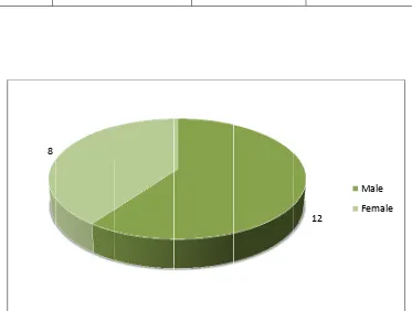
S
Male
Female
T WISE DI
Sex
[image:46.595.111.484.289.571.2]39
Table No. ISTRIBU
No. 2:
UTION (n
of Cases
12
8
= 20)
Percen
12
ntage (%)
60
40
D DISTRIB Sl. No. 1. 2. 3. 0 10 20 30 40 50 60 UTION A Type Fall Inju Road Tr Acciden Fall of H Objects
Fall Injury 12 60 T ACCORD of Injury ury raffic nts Heavy
[image:47.595.114.485.178.324.2]Road Traf Accident 6 3 40 Table No. DING TO No. o ffic ts
Fall of Obj 2 30 3: MODE O of Cases 12 6 2
f Heavy
jects 2 10 OF INJUR Percent 6 3 1
No. of Percen RY (n=20 tage (%) 60 30 10
f Cases ntage (%)
41
Table No. 4:
DISTRIBUTION ACCORDING TO THE SIDE (n = 20)
Sl. No. Side No. of Cases Percentage (%)
1. Left 12 60
2. Right 8 40
12 8
42
TYPE OF FRACTURES
CLOSED 100%
Typ Type Type Type Type 0 1 2 3 4 5 6 7 8 BOYD A pe - 1 - 2 - 3 - 4
Type ‐1 6
CLAS
AND GRI No. of 6 8 4 2Type ‐2 8 43
SSIFICA
IFFIN CL f Cases 6 8 4 2Type ‐3 4
ATION
LASSIFIC
Type ‐4 2 CATION Percenta 30% 40% 20% 10% age
0 2 4 6 8 10
I Dynamic
Proxima
Dynam
Dyn
IMP
mplant U c Hip Scre
al Femoral
ic Hip Screw 10
namic Hip Scre
44
PLANT U
Used ew
l Nailing
Proxim
ew Prox
USED
No. o
mal Femoral N 10
imal Femoral
of Cases 10
10
ailing
TIME
< 2 da 2 ‐5 da 5 ‐7 da
INTERV
Time Int < 2 da
2 - 5 da 5 - 7 da
0 ys ys ys
VAL BETW
terval N ays ays ays 2 4 2 45 WEEN IN
No. of Ca 2 10 8 6 NJURY A ases Pe 8 10 8 AND SUR ercentage 10% 50% 40% 10 No. RGERY
intertro
Pubic Ram
ochanteric
Pubic Ram
Shaft of F
Both Bon Pneumoth Head Inju 1 1 mus Fract c fractures mus Fractu
Femur - 1
nes Leg fra
horax - 1 c
ury - 1 cas
1
ASSOCI tures are t
s.
ures - 3 ca
case
acture - 1 c
case
e
1
46
IATED IN the most c
ases case NJURIES commonly 3 Pu Sh Bo Pn He S y associate
ubic Ramus Fr haft of Femur oth Bones Leg neumothorax
ead Injury
ed fracture
actures
g fracture
Oper Type Type Type Type -rating Tim - 1 - 2 - 3 - 4 0 0.5 1 1.5 2 2.5 T 0 0.5 1 1.5 2 2.5 me Pr 1.4 2.1 2.4 2.5
Type ‐1 Typ 1.45
P
Type ‐1 Ty 1.3 OPER roximal Fe 45 Hrs 0 Hrs 45 Hrs 50 Hrs
pe ‐2 Type ‐
2.1
2.45
roximal
ype ‐2 Typ 1.5
Dynam
47
RATING emoral N
3 Type ‐4
5 2.5
l
Femora
pe ‐3 Type
2.1 2
mic
Hip
TIME Nailing 1 1 2 2
al
Nailin
Pro
e ‐4 2.2
Screw
Dynam .30 Hrs .50 Hrs .10 Hrs .20 Hrsng
oximal Femor
Dynamic H
ic Hip Sc
al Nailing
Hip Screw
Bl Type Type Type Type -lood Loss - 1 - 2 - 3 - 4 0 50 100 150 200 250 300 350 400 0 50 100 150 200 250 T
s D
200
350
400
380
Type ‐1 Typ 200
D
Type ‐1 Typ 90
Prox
BL Dynamic H 0ml 0ml 0ml 0mlpe ‐2 Type
350
40
Dynamic
pe ‐2 Type ‐
180
220
ximal
Fe
48
LOOD LO Hip Screw
‐3 Type ‐4 00
380
c
Hip
Sc
‐3 Type ‐4 0 200
emoral
OSS w Pro 90m 180m 220m 200m 4
crew
(m
D
Nailing
Pro (m oximal Fe ml ml ml ml
l)
Dynamic Hip Sc
(ml)
oximal Femor ml)
emoral Na
crew (ml)
al Nailing
Blood Type Type Type Type -d Transfu - 1 - 2 - 3 - 4 UNI sion D 1 U 1 U 2 U 2 U 2
Dy
1Proxi
IT OF BL Dynamic H Unit Unit Units Units 2
ynamic
0 1
imal
Fem
49
LOOD TR Hip Screw
2
Hip
Scre
0
1
moral
N
RANSFUS w Pro Nil 1 Un 1 Un 1 Un 1
ew
(uni
1
Nailing
(
SED oximal Fe nit nit nit 1
ts)
Unit)
emoral NaType ‐1 Type ‐2 Type ‐3 Type ‐4
Type ‐1 Type ‐2 Type ‐3 Type ‐4
E Type Type Type Type -Exposure - 1 - 2 - 3 - 4 0 5 10 15 20 25 T 0 5 10 15 20 25 30 T F D 10 15 20 22
ype ‐1 Type 10
D
ype ‐1 Type 20
Prox
FLUROSC Dynamic H min min min mine ‐2 Type ‐
15
20
ynamic
e ‐2 Type ‐
25
30
ximal
Fe
50
COPIC EX Hip Screw
3 Type ‐4 22
Hip
Scr
3 Type ‐4 28
moral
N
XPOSUR w Pro 20 m 25 m 30 m 28 m
rew
(mi
Dy
Nailing
(
Pro (m RE oximal Fe min min min min
n)
ynamic Hip Scr
(min)
oximal Femor min)
emoral Na
rew (min)
al Nailing
Dynam
Proxim
Time mic Hip Sc
mal Femor 0 10 20 30 40 50 60 70 80 90 100 e crew ral Nailing
6 Weeks 28
36
TIM
6 W 2
g 3
10 Weeks 68
80
51
ME OF UN
Weeks 28%
36%
14 Weeks 84 92 NION 10 W 68 80 Dy Pro Weeks 8% 0%
ynamic Hip Scr oximal Femor
14 We 84%
92%
rew al Nailing
eeks %
52
FUNCTIONAL OUT COME
HARRIS HIP SCORE Pain (maximum score 44)
None or ignores it (44)
Slight, Occasional, no compromise in activities (40)
Mild pain, no effect on average activities, rarely moderate pain with unusual activity; may take aspirin (30)
Moderate pain, tolerable but makes concession to pain. Some limitation of ordinary activity or work. May require Occassional pain medication stronger than aspirin (20)
Marked pain, serious limitation of activities (10)
Totally disabled, crippled, pain in bed, bedridden (0)
Limp (maximum score 11) None (11)
Slight (8)
Moderate (5)
Severe (0)
Support (maximum score 11) None (11)
Cane for long walks (7)
Cane most of time (5)
One Crutch (3)
Two canes (2)
53
Distance Walked (maximum score 11) Unlimited (11)
Six blocks (8)
Two or Three blocks (5)
Indoors only (2)
Bed and Chair only (0)
Sitting (maximum score 5)
Comfortably in ordinary chair for one hour (5)
On a high chair for 30 minutes (3)
Unable to sit comfortably in any chair (0)
Enter Public Transportation (maximum score 1) Yes (1)
No (0)
Stairs (maximum score 4)
Normally without using a railing (4)
Normally using a railing (2)
In any manner (1)
Unable to do stairs (0)
Put on Shoes and socks (maximum score 4) With ease (4)
With difficulty (2)
54
Absence of Deformity (All yes = 4; Less than 4 = 0) (maximum score 4)
Less than 30o fixed flexion contracture Yes No
Less than 10ofixed abduction Yes No
Less than 10o fixed internal rotation in extension Yes No
Limb length discrepancy less than 3.2cm Yes No
Range of Motion (* indicates normal)
Flexion (*140o) _______________
Abduction (*40o) _______________
Adduction (*40o) _______________
External Rotation (*40o) _______________
Internal Rotation (*40o) _______________
Range of Motion Scale (maximum score 5) 211o - 300o (5) 61o - 100o (2)
161o - 210o (4) 31o - 60o (1) 101o - 160o (3) 0o - 30o (0) Range of Motion Score: _______________
M 3 mon 6 mon Months ths ths 0 5 10 15 20 25 30 35 40 POS Dyn
Dynamic Hi 20
STOPER HARR
PAIN - M
amic Hip 20
30
p Screw 30
55
RATIVE F RIS HIP S
MAX SCO
p Screw
Proximal F Nailin
30
FOLLOW SCORE
ORE - 44
Prox Femoral ng 40 W UP ximal Fem 30 40 3 6 moral Nai 0 0
3 months 6 months
M 3 mon 6 mon Months ths ths 0 1 2 3 4 5 6 7 8 Dyn
Dynamic Hip 0
LIMP - M
amic Hip 0
5
p Screw P 5
56
MAX SCO
p Screw
Proximal Femo 5
ORE – 11
Prox
oral Nailing 8 1 ximal Fem 5 8 3 6 moral Nai 5 8
3 months 6 months
M 3 mon 6 mon Months ths ths 0 1 2 3 4 5 6 7 S Dyn
Dynamic Hip 3
SUPPORT
amic Hip 3
5
p Screw P 5
57
T - MAX
p Screw
Proximal Femo 5
SCORE 1
Prox
oral Nailing 7 11 ximal Fem 5 7 3 6 moral Nai 5 7
3 months 6 months
M 3 mon 6 mon Months ths ths 0 1 2 3 4 5 6 7 8 DISTAN Dyn
Dynamic Hip 2
NCE WA
amic Hip 2
3
p Screw P 3
58
ALKED - M
p Screw
Proximal Femo 5
MAX SC
Prox
oral Nailing 8 ORE 11 ximal Fem 5 8 3 6 moral Nai 5 8
3 months 6 months
M 3 M 6 M M 3 mon 6 mon MONTHS MONTHS MONTHS P Months ths ths 0 0.5 1 1.5 2 2.5 3 3.5 4 4.5 5 DY S S PUBLIC T Dyn
DYNAMIC HI 0 SITTI YNAMIC SCREW 0 3 TRANSP amic Hip 0 1
P SCREW 3 59 ING MAX HIP W PORT USE p Screw
PROXIMAL FE NAILIN
3
X SCORE
PROX
E - MAX
Prox
EMORAL
NG 5
E - 5
M 3 M 6 M 0 0.2 0.4 0.6 0.8 1 1.2 1.4 1.6 1.8 2 MONTHS MONTHS MONTHS
DYNAMIC HI 0
DY
S S
STAIRS
P SCREW 1 YNAMIC SCREW 0 1 60 MAX SC
PROXIMAL FE NAILIN
1
C HIP W
CORE - 4
M 3 mon 6 mon PUT Months ths ths 0 0.5 1 1.5 2 2.5 3 3.5 4
T ON SHO
Dyn
Dynamic H 0
OES AND
amic Hip 0
2
ip Screw 2
61
D CHAPP
p Screw
Proximal F Naili
2
PELS – M
Prox Femoral ng 4 MAX SCO ximal Fem 2 4 3 6 RE 4 moral Nai 2 4
3 months 6 months
M 3 mon 6 mon A Months ths ths 0 0.5 1 1.5 2 2.5 3 ABSENCE Dyn
Dynamic H 2
E OF DEF
amic Hip 2
3
ip Screw 3
62
FORMIT
p Screw
Proximal F Naili
3
TY - MAX
Prox Femoral ng 3 X SCORE ximal Fem 3 3 3 6 4 moral Nai 3 3
3 months 6 months
M 3 mon 6 mon Months ths ths 0 0.5 1 1.5 2 2.5 3 3.5 4 RANG Dyn
Dynamic H 2
GE OF MO
amic Hip 2
3
ip Screw 3
63
OTION
-p Screw
Proximal F Naili 3 MAX SC Prox Femoral ng 4 CORE 5 ximal Fem 3 4 3 6 moral Nai 3 4
3 months 6 months
M 3 mon 6 mon Months ths ths 0 10 20 30 40 50 60 70 80 90 TO Dyn
Dynamic Hi 29
OTAL HA
amic Hip 29
57
p Screw 57
64
ARRIS H
p Screw
Proximal F Nailin 58 HIP SCOR Prox Femoral ng 82 RE ximal Fem 58 82 3 6 moral Nai 8 2
3 months 6 months
accoun
easy t
multip
OVER
The rating
nt pain, m
to follow ple factors RALL RE E G F F 0 2 4 6 8 10 12 ANALYS g system f
movement, and anal for analys ESULTS Gradin Excellent Good Fair Failure Excellent 12
SIS OF F followed w
, function
lyse. Othe
sis.
ng N
Good 3
65
UNCTIO was that o
n, shorteni
ers system
No. of Cas 12 3 3 2 Fair 3 ONAL OU f Harris H
ing and a
ms are m
ses Pe
Failure 2
UTCOME Hip Score w
ngulation more confu ercentage 60% 15% 15% 10% No E which tak
. This sys
fusing and
e
. of Cases
ken into
stem is
66
RESULTS ACCORDING TO SUBTYPE
Types Grading Type - 1 Excellent to Fair
Type - 2 Excellent to Fair
Type - 3 Excellent to Failure
Type - 4 Excellent to Failure
RESULTS ACCORDING TO THE IMPLANT USED
Implant No. of Cases Grading Percentage
Dynamic Hip Screw 6 Excellent 60%
2 Good 20%
1 Fair 10%
1 Failure 10%
Proximal Femoral Nail 7 Excellent 70%
1 Good 10%
1 Fair 10%
67
DISCUSSION
The aim of study is to evaluate the functional outcome of the fixation of
intertrochanteric fractures fixed with both dynamic hip screw and proximal
femoral nailing.
We selected 20 cases of intertrochanteric fractures during the time of July
2008 to September 2010. All 4 types of intertrochanteric fractures are included.
We had 6 cases of type-1, 8 cases of type-2, 4 cases of type-3, and 2 cases of
type-4 fractures. 3 cases of type-1, 4 cases of type-2, 2 cases of type-3, and 1
case of type-4 fractures. Two groups each were fixed with dynamic hip screw
and proximal femoral nailing for each groups consisting of 10 cases.
10 cases were fixed with dynamic hip screw and 10 cases were fixed with
proximal femoral nailing. The youngest patient in our series is 45 years and
oldest patient in our series is 75 years. Average age is 60 years.
We had 12 male cases and 8 female cases. With a ratio of 1.5:1. All the
20 cases were closed fractures. Most common mode of injury is accidental fall
injury. In our study we had 12 cases right sided and 8 cases left sided.
Most common associated injuries are 3 public ramus fractures. Others are
1 shaft of femur, 1 both bones leg fractures, 1 pneumothorax and 1 head injury.
Duration between injury and hospitalization, 10 cases were between 6-12
hrs and 7 cases were more than 12 hours, 2 cases were within 3-6 hrs and 1 case
68
All cases were evaluated with x-ray pelvis with both hips anteroposterior
view and the affected hip were both anteroposterior and lateral views.
Routine blood investigations with ECG and x-ray chest also taken for
assessment for surgery. Traction and internal rotation special view is also taken
for the study of the fracture fragments for fixation plan. All 4 types of fractures
are fixed with both types of fixation. All fractures are fixed by lateral approach.
Preoperative antibiotics are given before surgery. Dynamic hip screw fixation is
by lateral approach with fixation of cancellous screw in the femoral head with
the side plate to the shaft. Proximal femoral nailing incision is more smaller just
for entry point and screw fixation. 2 cancellous screws, one as lag screw and
one as hip pin with distal locking in the shaft. Each step of fixation in both these
methods is checked with help of fluoroscopy in both anteroposterior and lateral
views.
Operating time is longer for proximal femoral nailing than dynamic hip
screw fixation. Type-3 and 4 fractures have longer operative time. Blood loss is
more for type-3 and 4 fractures and also for dynamic hip screw fixation. 2 units
of blood transfusion done for type-3 and 4 fractures. Rest are given only 1 unit
and mainly 2 units are given for dynamic hip screw fixation. Fluroscopic
exposure is more for the proximal femoral nailing than dynamic hip screw.
Duration of postoperative stay is 12 days for dynamic hip screw and 6
days proximal femoral nailing. All postoperative cases were started with
mobilization on first postoperative day itself. Postoperative x-ray is taken and
69
Time of union for dynamic hip screw fixation at 6 weeks is 28%, 10
weeks is 68% and 14 weeks is 84%. Time of union for proximal femoral nailing
at 6 weeks is 36%, 10 weeks 80% and 14 weeks is 92%. Full weight bearing
allowed only after evidence of full radiological union.
Postoperative outcome of both fixation is measured by Harris Hip Score.
Pain is mild in proximal femoral nailing compared to dynamic hip screw
nailing. Limping is less in proximal femoral nailing. Support distance walked,
using public transport, absence of deformity, sitting, using stairs, range of
motion are better in proximal femoral nailing in both 3 and 6 months of follow
up using harris hip score than dynamic hip screw.
Functional out come is excellent in 12 cases, good in 3 cases, fair in 3
cases and failure in 2 cases. Type-1 and 2 fractures have excellent to fair results.
Type-3 and 4 fractures have excellent to failure results. Dynamic hip screw has
60% excellent results, whereas proximal femoral nailing has 70% excellent
results.
Postoperative complications was infection in 4 cases, malunion in 2
cases, delayed union in 2 cases, bed sores in 1 patient, limb length inequality in
5 patients, none of them had any funcitonal deficit, lag screw pull out in one
case.
No vascular and neurological complications were noted in these 20 cases.
In our study, outcome of fixation is studied extensively from operation
70
CONCLUSION
In the present study assessing the functional outcome of intertrochanteric
fractures, we reached the following conclusions.
1. Intertrochanteric fractures commonly occur in men around age of 6th
decade due to accidental fall injury.
2. Conventional radiographs are not essential to study the fracture pattern,
traction and internal rotation view is needed to classify the fractures.
3. Boyd and Griffin classification is essential for classification of
intertrochanteric fractures.
4. Fracture stabilization by rigid internal fixation by both methods results
in early functional recovery and early ambulation. Perfect anatomical
reduction gives excellent results.
5. Blood loss and unit of blood transfusion is less in case of proximal
femoral nailing compared with dynamic hip screw.
6. Operative time is longer in proximal femoral nailing than dynamic hip
screw.
7. Fluroscopic exposure is longer for proximal femoral nailing than
dynamic hip screw.
8. Duration of postoperative stay is longer in dynamic hip screw than
71
9. Results of both fixation are better in type-1 and 2 fractures compared
with type-3 and 4 fractures.
10. Union rates are also better in type-1 and 2 fractures with both fixation
than type-3 and 4 fractures.
11. Postoperative follow up was measured by Harris Hip Score for a follow
up of 3 and 6 months.
12. Pain, limp, support, distance walked, sitting, public transport, walking
stairs, put chapels, absence of deformity. All these factors are better in
proximal femoral nailing for 3 and 6 months follow up than dynamic
72
ADVANTAGES OF PROXIMAL FEMORAL NAILING 1. Less blood loss and blood transfusion.
2. Early weight bearing.
3. Union results better in all 4 types of trochanteric fractures.
4. Postoperative complication is less.
5. Postoperative functional mobility is better.
ADVANTAGES OF DYNAMIC HIP SCREW 1. Less operative time.
2. Shorter fluoroscopic time.
3. Screw pull out is less. <no z-effect>
In our series, proximal femoral nailing has better results than dynamic hip
screws. Proximal femoral nailing has better union rates and functional results
than dynamic hip screw. It has very good results even in type-1 and 2 stable
fractures. Introperative and postoperative complications are less in proximal
femoral nailing. But disadvantages is screw pullout <z-effect> is seen and also
the operative time and fluoroscopic time is longer which is hazardous to the
BIBLIOGRAPHY
1. ALBAREDA.J. LADERIGA A. PAVANEAD et. al. Int ortho op20 47-50, 1996.
2. BAUNGAETNER MI, CROSTOWSKI JH, LEVY RN Inter trochanteric Hip#
BROMER BD eds. Skeletal trainna Vol. 2 PHILADELPHIA.
WB SASUNDERS 1992 - 1833 - 1881.
JBJS 1995 (2 Articles)
3. BELLA BARBA.C NERSLOVICI. Jn D RICCI WM Percutaneous treatment of pei
trochanteric # using a 'r' nail
Clinical ortho p375:34 2000.
4. BOYD, HB GRIFFIN LL, CLASSIFICATION A Treatment of Trochanteric # Arch
surg 1949.
5. BRIDE Sn, PASTEL AD Bircher M. CCalvert PT. Fixation of intertrochanteric # of
Tnefemur. A randomnised prospective companison of the 'r' nail & the dynamic hip
screw. Journal of Bone Joint Surgery 1991.
6. Calvert PT 'r' nail A significant advance or as passing failure? JBTs - 1992.
7. CEENG CL, cnowsp PUNWK et. al., long term results complications of treatment.
8. Augmentation in the treatment of unsteable Frochanteric # injunj.
9. Cunnings SR. WEVITT. MC Non skeletal determinants of # the potential
importance of the mechanice of fall Osteoporosis int 1994 suppl 1.
10. Davis TR SnER JL. Hersman A et.al., intertrochanteric femoral # Mechanical
failure after Int' fixation. JBJs 1990
11. Dimon JN hugnston JC unstable inter Frochanteric # of hip JBJs 1967 clinical
orthop 92:1973.
12. Evan €The Treatment of trochanteric # of femur. JBJS.
13. Flores LA NAMINGTON Menor M the stability of intertrochanteric #s treatment c
sliding screw plate JBJS 1990.
14. Gold Nagers Pr, 'O' Connor Pr, Scnwarzed. Journal of orthopaedic trauma 1994. A
prospective comparative study 'r' nail ADMS.
MASTER CHART
DYNAMIC HIP SCREWS
S. NO. AGE/ SEX BODY/ GRIFFIN CLASSIFICATION TYPE MODE OF
INJURY SIDE
ASSOCIATED INJURIES INTERNAL BETWEEN INJURY & SURGERY TYPE OF FUNCTION OPERATION TIME BLOOD LOSS FURROSIVE EXPOSURE COMPLIC- ATIONS TIME OF UNION (14weeks) HARRIS H/P SCORE MAX SCORE -100 (6MONTHS)
1. 63 / M I FALL
INJURY RIGHT NIL 5 DAYS DHS 1.30 HRS 200ML 12 MIN SHORTENING 78% 61
2. 73 / M III FALL
INJURY LEFT NIL 3 DAYS DHS 2.20 HRS 420 ML 22 MIN INFECTION 71% 64
3. 65 / M II FALL
INJURY RIGHT
START OF
FEVER,FRACTURE 7 DAYS DHS 1.50 HRS 330 ML 17 MIN MALUNION 76% 59
4. 74 / M I RTA LEFT NIL 6 DAYS DHS 1.30 HRS 190 MIL 24 MIN NIL 83% 68
5. 64 / M II FALL
INJURY RIGHT
PULIC
FRACTURE 4 DAYS DHS 1.40 HRS 360 ML 15 MIN INFECTION 74% 72
6. 58 / M III RTA LEFT NIL 1 DAY DHS 2.10 HRS 310 ML 18 MIN DELAYED
UNION 80% 71
7. 47 / M II
HEAVY OBJECT
FALL RIGHT
NIL 3 DAYS DHS 1.50 HRS 370 ML 18 MIN INFECTION 78% 64
8. 73 / M II FALL
INJURY LEFT NIL 4 DAYS DHS 1.40 HRS 310 ML 15 MIN SHORTENING 75% 68
9. 68 / M IV RTA LEFT PUBLIC RAN
FRACTURE 6 DAYS DHS 2.20 HRS 380 ML 22 MIN SHORTENING 76% 65
10. 75 / M I FALL
PROXIMAL FEMORAL NAILING S. NO. AGE/ SEX BODY/ GRIFFIN CLASSIFICATION TYPE MODE OF INJURY SIDE ASSOCIATED INJURIES INTERNAL BETWEEN INJURY & SURGERY TYPE OF FIXATION OPERATION TIME BLOOD LOSS FURROSIVE EXPOSURE COMPLIC- ATIONS TIME OF UNION (14weeks) HARRIS H/P SCORE MAX SCORE -100 (6MONTHS)
11. 56 / M I FALL INJURY LEFT LEG BONES
LEG FRACTURE
6 DAYS PFN 1.45 HRS 90 ML 20 MIN NIL 89% 88
12. 67 / M II FALL INJURY RIGHT NIL 1 DAY PFN 2.10 HRS 190 ML 25 MIN DELAYED
UNION
86% 84
13. 78 / M I RTA LEFT NIL 2 DAYS PFN 1.35 HRS 80 ML 18 MIN SHORTENING 79% 83
14. 44 / M III FALL INJURY LEFT PUBLIC RAWS
FRACTURE
5 DAYS PFN 2.45 HRS 220 ML 35 MIN INFECTION 81% 88
15. 66 / M I FALL INJURY RIGHT NIL PFN 1.55 HRS 86 ML 22 MIN SHORTENING 94% 94
16. 53 / M II HEAVY
OBJECT FALL
LEFT PNE 7 DAYS PFN 2.20 HRS 180 ML 27 MIN MALUNION 83% 89
17. 61 / M II RTA RIGHT 3 DAYS PFN 2.15 HRS 170 ML 29 MIN NIL 93% 83
18. 70 / M II FALL INJURY LEFT HEAD INJURY 6 DAYS PFN 2.00 HRS 160 ML 26 MIN BED SORE 81% 81
19. 63 / M III RTA RIGHT NIL 5 DAYS PFN 2.40 HRS 220 ML 62 MIN NIL 90% 79
20. 59 / M IV FALL INJURY LEFT NIL 4 DAYS PFN 2.50 HRS 200 ML 28 MIN LAG SCREW
PULL OUT
DYNAMICS HIP SCREW AND PLATE
SURGICAL INCISION STEP 1
STEP 3
DYNAMIC HIP SCREW INSTRUMENTS
PROXIMAL FEMORAL NAILING FIXATION CASE 1
PRE OPERATIVE X-RAY
INTRAOPERATIVE PICTURE
10 WEEKS
CASE 2
PRE OPERATIVE X-RAY
INTRAOPERATIVE PICTURE
POST OPERATIVE X-RAY
6 WEEKS 10 WEEKS
CASE 3
PREOPERATIVE X-RAY
C-ARM PICTURE
6 WEEKS
10 WEEKS
CASE 4
PREOPERATIVE X-RAY
C-ARM PICTURE