AND EFFICACY OF 0.5% ROPIVACAINE WITHOUT EPINEPHRINE, 0.5% ROPIVACAINE WITH EPINEPHRINE
AND 0.5% BUPIVACAINE IN PATIENTS RECEIVING SUBCLAVIAN PERIVASCULAR BRACHIAL PLEXUS BLOCK
Submitted to
THE TAMILNADU DR. M.G.R. MEDICAL UNIVERSITY
In partial fulfillment of the requirements for the award of degree of
MD (BRANCH - X) ANAESTHESIOLOGY
GOVERNMENT STANLEY MEDICAL
COLLEGE & HOSPITAL
THE TAMILNADU Dr. M.G.R. MEDICAL UNIVERSITY
CHENNAI, TAMILNADU
This is to certify that this dissertation entitled“COMPARATIVE
STUDY FOR THE PURPOSE OF SAFETY AND EFFICACY OF 0.5% ROPIVACAINE WITHOUT EPINEPHRINE, 0.5% ROPIVACAINE WITH EPINEPHRINE AND 0.5% BUPIVACAINE IN PATIENTS RECEIVING SUBCLAVIAN PERIVASCULAR BRACHIAL PLEXUS BLOCK” is bonafide
record work done by Dr.K.SATHISHAKUMAR under the guidance of
Prof. Dr.Ponnambala Namachivayam, Additional Professor, Department
of Anaesthesiology, submitted to the Tamil Nadu Dr. M.G.R. Medical
University in partial fulfillment of University regulation for MD,
Branch X –Anaesthesiology.
Prof. Dr. Ponnambala Namachivayam, Prof.Dr. R. Subramania Bharathiyar,
M.D.,D.A., M.D., D.A.,
Additional Professor of Anesthesiology, Professor and Head,
Department of Anesthesiology, Department of Anesthesiology,
Stanley Medical College and Hospital, Stanley Medical College and Hospital,
Chennai. Chennai.
Prof.Dr.C.Vamsadhara M.D.,Ph. D
Dean,
I Dr.K.SATHISHAKUMARsolemnly declare that this dissertation
titled “COMPARATIVE STUDY FOR THE PURPOSE OF
SAFETY AND EFFICACY OF 0.5% ROPIVACAINE
WITHOUT EPINEPHRINE, 0.5% ROPIVACAINE WITH
EPINEPHRINE AND 0.5% BUPIVACAINE IN PATIENTS
RECEIVING SUBCLAVIAN PERIVASCULAR BRACHIAL
PLEXUS BLOCK” has been done by me. I also declare that this
bonafide work or a part of this work was not submitted by me or any other
for any award, degree, diploma to any other University board either in
India or abroad.
This is submitted to The Tamilnadu Dr. M. G. R. Medical
University, Chennai in partial fulfillment of the rules and regulation for the
award of Doctor of Medicine degree Branch –X (Anaesthesiology) to be
held in APRIL 2011.
I wish to express my sincere gratitude to Prof.Dr.C.Vamsadhara,
M.D.,Ph.D., Dean, Government Stanley Medical College for having
kindly permitted me to utilize the facilities of the hospital for the conduct
of the study.
I am deeply indebted to Prof.Dr.R.Subramania Bharathiyar.
M.D., D.A, Professor and Head of the Department of Anaesthesiology,
Stanley Medical College, Chennai for his guidance and encouragement in
preparing this dissertation.
My sincere thanks to Prof.Dr. Ponnambala Namachivayam,
M.D., D.A., Additional Professor of Anaesthesiology, for his able
guidance in completing this study and Prof.Dr. R. Lakshmi. M.D., D.A.,
Additional Professor of Anaesthesiology for her constant support in
performing this study.
My heartful thanks to Prof.Dr. P. Chandrashekar. M.D., D.A,
Additional Professor of Anaesthesiology, Stanley Medical College,
My special gratitude to Dr.N.Baskar M.D., for taking personal
interest and guiding me in this study.
My heartfelt thanks to Dr.S.S. Sukumar M.D., D.A., Dr.K.Arul.
M.D., D.A., and Dr. P.Mahendran D.A.,for helping me at every level
while conducting this study.
I also thank all my Assistant Professors and postgraduate
colleagues, Department of Anaesthesiology, for their kind cooperation for
helping me in doing this study.
I thank Mr. Albert joseph who helped me in arriving at the statistical
conclusions of this study.
I gratefully acknowledge my patients who gave their consent and
S. NO. TITLE PAGE NO. 1. 2. 3. 4. 5. 6. 7. 8. 9. 10. 11. 12. INTRODUCTION
AIM OF THE STUDY
HISTORY
ANATOMY OF BRACHIAL PLEXUS
PERIPHERAL NERVE STIMULATOR
TECHNOLOGY
PHARMACOLOGY
REVIEW OF LITERATURE
MATERIALS AND METHODS
OBSERVATION AND RESULTS
DISCUSSION SUMMARY CONCLUSION ANNEXURES BIBLIOGRAPHY PROFORMA MASTER CHART INFORMED CONSENT
ETHICAL COMMITTEE APPROVAL CERTIFICATE
Peripheral nerve blocks are gaining widespread popularity for
perioperative pain management because of their distinct advantages over
general and central neuraxial anesthesia. Pain relief with peripheral nerve
block (PNB) is devoid of side effects such as somnolence, nausea,
vomiting, hemodynamic instability and voiding difficulties inherent to
general and central neuraxial anesthesia.
Brachial plexus blocks (interscalene, supraclavicular and
axillary blocks) 1-10 provide a useful alternative to general anesthesia for
upper limb surgery. Each of these routes has its own particular
advantages, disadvantages and complications. They achieve ideal
operating conditions by producing complete muscular relaxation,
maintaining stable intraoperative hemodynamic, and the associated
sympathetic block. The sympathetic block decreases postoperative pain,
vasospasmand edema. Supraclavicular perivascular brachial plexus block
is a very commonly practised technique in patients undergoing surgery on
forearm and hand. Supraclavicular block as opposed to infraclavicular
brachial plexus block generally offers denser and complete upper limb
blocks (11,12). Its use avoids paraesthesia, decreases the chance of nerve
injury and gives high success rate.
Introduction of long acting local anesthetic with better safety
profile as well as better equipment has further increased the usage of
peripheral nerve blocks. Bupivacaine hydrochloride, a long acting local
anaesthetic agent, has been used extensively for supraclavicular brachial
plexus block 13. Ropivacaine 14-26, the S- enantiomer of
S-1-propyl-2,6-pipecoloxylidide, is an amino-amide local anaesthetic with chemical
structure similar to that of bupivacaine. Ropivacaine is a potent blocker of
A delta and C fibres (pain fibres). It has been reported to be less toxic
than Bupivacaine.
In contrast to Bupivacaine which is a racemic mixture of R and S
enantiomers, Ropivacaine is the first local anesthetic that has developed
as a pure enantiomer. S enantiomer is considered less neurotoxic and
cardiotoxic 42 than the R enantiomer of local anesthetics, perhaps
reflecting different pharmacodynamics, pharmacokinetics and toxicities.
Studies18,23-26 have shown that Ropivacaine and Bupivacaine when given
in equal volumes was similar in terms of onset and duration of sensory
The addition of epinephrine does not prolong the duration of
Ropivacaine in subclavian perivascular brachial plexus27-28 or epidural
block. Low concentrations of Ropivacaine may produce clinically
significant vasoconstriction which is not further increased by the addition
of epinephrine.
This study attempts to compare the safety and efficacy of 0.5%
Ropivacaine without epinephrine and 0.5% Ropivacaine with epinephrine
and 0.5% Bupivacaine in patients receiving subclavian perivascular
AIM OF THE STUDY
To study the efficacy and safety of 0.5% Ropivacaine without
epinephrine and 0.5% Ropivacaine with epinephrine and 0.5%
Bupivacaine in patients receiving subclavian perivascular brachial
HISTORY
The first effort for the achievement of local or regional anesthesia
dates back to the middle ages, where Ambroise Pare described in 1564 in
France “that anesthesia was used to carry out operations at the
extremities”, with the help of the compression mechanism intended to
bring about analgesia, while in 1846 in Italy M.A. Severino brings about
local anesthesia by putting ice or snow on the surgical field29.
Nevertheless, the real regional anesthesia started upon discovery of the
holed metallic wire in 1854 by Wood, as well as the local anesthetic
properties of cocaine29.
Cocaine is contained in the leaves of a plant, the mahogany of
coca, which is grown in the South American countries 29,30.Cocaine was
isolated from the leaves of the of the coca’s mahogany for the first time in
1860, by A. Niemann in Germany. The substance was crystalline,
colourless, odourless and with a sour taste. In 1884 Karl Koller from
Vienna described at the conference of German ophthalmologists in
Haideberg the anesthetic qualities of a 2% cocaine solution after it was
instilled in the eye 31. In 1885 Halstead, on the one hand, gave a
description of the exclusion of the nerve through infiltration with cocaine,
and Corning from the United States, on the other hand, gave a description
to the paraverterbral exclusion. Thus, cocaine, managed to acquire the
prestige it deserved as a local anesthetic after a number of fatal
implications was recorded from the acid action of the solutions with a
high cocaine content that they were using at that time.
The revolution in the history of local anesthetics broke in 1904,
when Einhorn introduced novocaine (procaine), a local anesthetic with
fewer side effects than cocaine. Nevertheless, the duration of its action
has been short-lived, a fact which has limited its use for the most part to
operations with a short duration. This problem was solved by Braun, who
proposed the addition of adrenaline to the local anesthetic, for the purpose
of prolonging both the duration and the validity of the local anesthetic.
In 1943 Nils Loefgren composed lidocaine, which belongs to the
group of aminoalcylamides . Beyond any doubt, the lidocaine has been
the main substance and, at the same time, the base for all the later studies
of the local anesthetics that followed. The later researches led to the
discovery of new local anesthetics such as etidocaine, the prilocaine, and
the bupivacaine.
THE DEVELOPMENT OF THE MODERN LOCAL ANESTHETICS
Albright’s announcement on the quest of new local anesthetics
in 1979 with regard to the heart failures following the administration of
announcement, there followed a constructive discussion with regard to
the toxicity of the local anesthetics 33, 34. There followed experimental
studies, which confirmed the assumption of Albright that deaths were the
result of the high cardiotoxicity of the long-lasting local anesthetics 35-38.
During the search for new local anesthetics, the researchers utilized
the characteristic fabrication of the 3-dimensional molecular structure that
the bupivacaine can demonstrate 39. The meaning of “stereo-isomery” had
been introduced at the end of the 19th century by the Swede chemist von
Berzelius (1779 through 1848). Thus, the Bupivacaine (racemic mixture
with R- and S-form in the same percentage) helped produce ropivacaine
(net S-enantiomer).
Ropivacaine is produced through the halcyliosis of the
S-enantiomer of L-staphylitic-dibenzoic-acid-ester. Its chemical structure
has a great similarity with that of bupivacaine and of mepivacaine, while
its two-dimensional form has a few only differences from that of
bupivacaine. Ropivacaine does not differ as regards the physical and
chemical properties from racemic mixture. The only difference that it
presents as compared with bupivacaine is its lipophilia, which is
comparable with that of mepivacaine.
The significant progress that the introduction of this short-lived
reduced toxicity on the cardiovascular and central nervous system and, on
the other hand, on its dose-dependent differential closeout (the qualities
of which we can utilize in the postoperative analgesia and the
administration of epidural anesthesia in the obstetrics) 40-43. The wide
acceptance of ropivacaine in the clinical practice was followed by the
preparation of a number of other substances by the group of amides, for
the purpose of introducing them into the clinical practice. Meanwhile, an
S-enantiomer of bupivacaine, the levobupivacaine, which, based on the
first clinical experiences, has proven the same profile of action, such as in
the racemic mixture which has been used so far in the various techniques
of the anesthesia area 44, 45. Levobupivacaine is governed by an equally
large lipophilia such as the bupivacaine and that for this reason it differs
to a great extent from the ropivacaine, mainly as to the power, the
kinetics on the receptor level and, quite possible, as to the cardiac
toxicity. Due to the high lipophilia which governs it, the power of the
levobupivacaine is higher that than of ropivacaine, while its kinetics at
the level of the receptor is quite probably comparable to that of
Anatomy of
ANATOMY OF BRACHIAL PLEXUS
46-49Knowledge of the formation of the brachial plexus and its
distribution is essential to the intelligent and effective use of the brachial
plexus blockade for the surgeries of the upper limb. Close familiarity with
the vascular, muscular and fascial relationship of the plexus throughout
the formation and distribution is equally essential to the mastery of
various techniques of Brachial plexus Blockade.
In its course from the intervertebral foramina to the arm, the fibre’s
that constitute the plexus are composed consecutively of roots, trunks,
divisions, cords and terminal branches, which are formed through a
complex process of combining, dividing, recombining and finally
redividing.
The brachial plexus is formed by the union of the anterior primary
rami of the fifth, sixth, seventh, eighth cervical nerves and the first
thoracic nerve, with variable contributions from the fourth cervical
(pre fixed) above and second thoracic nerve (post fixed) below. These
nerves unite to form trunks, which lie in the neck above the clavicle. Its
roots pass between the scalenus anterior and scalenus medius which is
enclosed by fascia accompanied by the subclavian artery and then
invaginates the scalene fascia to form a neurovascular bundle. This fascia
RELATION OF BRACHIAL PLEXUS:
Anterior relations:
The skin, superficial fascia, platysma, supraclavicular branches of
the superficial cervical plexus, deep fascia and external jugular vein. The
clavicle is in front of the lower part and scalenus anterior is in the front of
the upper part.
Posterior relations:
Scalenus medius and the long thoracic nerve of bell.
Inferior relations:
Related to the first rib.
Superior relations:
Lies first above and then lateral to the subclavian artery.
SYMPATHETIC CONTRIBUTION TO THE PLEXUS:
Close to the emergence, the fifth and sixth cervical nerves, each
receive a grey ramus from the middle cervical sympathetic ganglion. The
seventh and eighth cervical nerves each receive a grey ramus from the
ROOTS:
Anterior primary rami of C5, C6, C7, C8 and T1 (occasionally C4
or T2)
TRUNKS:
Upper trunk - Anterior rami C5 and C6
Middle trunk – Anterior ramus of C7
Lower trunk – Anterior rami of C8 and T1
DIVISIONS:
Behind the clavicle each trunk divides into anterior and posterior
divisions.
CORDS:
Lateral cord – Anterior divisions of upper and middle trunks
(C5-C7)
Medial cord – Anterior divisions of lower trunk (C8-T1)
Posterior cord – Posterior divisions of all the three trunks (C5-T1)
BRANCHES:
From roots:
Nerve to Serratus anterior (C5, C6, and C7)
Muscular branches to longus cervices (C5, C6, C7, and C8)
Nerve to Scalene (C5, C6, C7 and C8)
Nerve to Rhomboids(C5)
From trunk:
Nerve to subclavius (C5-C6)
Suprascapular nerve (C5-C6)
From cords:
Lateral cord:
Lateral root of median nerve (C5, C6 and C7)
Lateral pectoral nerve (C5, C6 and C7)
Musculocutaneous nerve (C5, C6 and C7)
Medial cord:
Medial root of median nerve (C8, T1)
Medial cutaneous nerve of arm (C8, T1)
Medial cutaneous nerve of forearm (C8, T1)
Medial pectoral nerve (C8, T1)
Ulnar nerve (C8, T1)
Posterior cord:
Radial nerve (C5- T1)
Axillary nerve (C5-C8)
Upper and lower subscapular nerve (C5, C6)
Nerve to lattismus dorsi (C6, C7, C8)
Familarity with the perineural structures that surround and
accompany the brachial plexus as it leaves the vertebral column on its
and distribution of the neural plexus itself. Palpable muscular and
vascular landmarks allow accurate location of the plexus percutaneously.
An appreciation of the fascial relations is absolutely essential since this is
the basis for all the perivascular techniques.
After leaving the intervertebral foramina, the anterior primary rami
of the nerves destined to become the brachial plexus travel in the gutter
formed by the anterior and posterior tubercles of the corresponding
transverse processes of the cervical vertebra. After leaving the transverse
processes, the roots of the plexus descend in front of the middle scalene
muscle, which arises from the posterior tubercles of the transverse
processes of lower six cervical vertebra. The insertion of this muscle on
the first rib is separated from that of the anterior scalene muscle by the
inferior trunk of the brachial plexus. The anterior scalene muscle arises
from the anterior tubercles of the transverse processes of the third to sixth
cervical vertebrae and inserts on the scalene tubercle of the first rib, thus
separating the subclavian artery from the subclavian vein.
The fascia covering both the scalene muscle is derived from the
prevertebral fascia, which splits to invest these muscles and then fuses
again at their lateral margins to form an enclosed inter scalene space.
Therefore as the roots leave the transverse processes, they emerge
between the two walls of the fascia covering the anterior and middle
scalene muscles. In their descend towards the first rib to form the trunks
of the plexus the roots may be considered to be sandwiched between the
of the plexus. As the trunks approach the first rib they are arranged one
above the other vertically.
As the trunks of the plexus cross the first rib, they are joined by the
subclavian artery, which lies in a plane anterior to the trunks, so that the
inferior trunk lies behind the artery in the subclavian groove with the
middle and superior trunks above the level of the vessel. At this level the
artery and the trunks are moving laterally, across the ribs and invaginate
the scalene fascia to form the subclavian perivascular space, which is
continuous medially and superiorly with the interscalene space and
inferiorly and laterally with the axillary perivascular space.
The important concept is that there is a continuous fascial enclosed
perineural and perivascular space extending from the cervical transverse
processes to several centimeters beyond the axilla; this space has been
divided into a axillary perivascular space and interscalene space. The
existence of such a continuous perineural space renders brachial plexus
block simple. The space described may be entered at any level, and the
volume of the anesthetic injected at this level would determine the extent
of anesthesia. Thus, the technique to be used in any case should be
determined on the basis of the surgical site, the required level of
anesthesia, the physical status and habitus of the patient.
Brachial plexus can be blocked at the level of roots, trunks, cords,
peripheral branches. The block at each level has a distinct distribution of
Peripheral
Nerve
PERIPHERAL NERVE STIMULATOR
TECHNOLOGY
11,12,50-56The ability of a nerve stimulator to evoke a motor response
depends on the intensity, duration, and polarity of the stimulating current
used and the needle (stimulus)-nerve distance. To propagate a nerve
impulse, a threshold current must be applied to the nerve fibre. Peripheral
nerve stimulation is typically performed using a rectangular pulse of
current. When a square pulse of the current is used to stimulate a nerve,
the total charge delivered is the product of the current strength and the
duration of pulse.
RHEOBASE-is the minimal threshold current required to stimulate
a nerve with a long pulse width.
CHRONAXIE- is the duration of the stimulus required to stimulate
at twice the rheobase. Chronaxie is used to express the relative
excitabilities of different tissues. It is possible to stimulate A- (motor)
fibres without stimulating A- and C fibres that transmit pain. Moreover,
mixed nerves can be located by evoking a motor response without
causing patient discomfort. Stimulation intensity will be variable as
determined by coulomb’s law. A very high stimulus current is required to
stimulate the nerve when the needle tip is far away from the nerve. If the
nerve may produce significant pain and systemic effects. An EMR at a
stimulating current of <0.5mA is associated with high rates of success of
PNS assisted PNB.
Characteristics of an ideal PNS:
1. Constant current output-A particular current not the voltage
stimulates the nerve. Therefore, the current delivered by the
device should not vary with changes in the resistance of the
external circuits.
2. Digital display of the delivered current
3. Variable output control
4. Clearly identifiable polarity
5. Option for different pulses
6. A wide range of current output 0.1-5.0mA
7. Battery indicator
Peripheral nerve stimulator settings:
MIXED NERVE(most PNB)
a. Current(dial) - 1mA
b. Current duration - 0.1ms
SENSORY NERVE
(eg- Lateral femoral cutaneous and saphenous nerves)
a. Current (dial) - 2-5mA
b. Current duration - 1ms
c. Frequency - 1Hz
DIABETIC NEUROPATHY(PNB)
a. Current(dial) - 2mA
b. Current duration - 0.3ms
PHARMACOLOGY
20,43,57-59LOCAL ANESTHETICS
Mechanism of action of local anesthetics:
Impulse blockade by local anesthetics may be summarized by
the following chronology
Solutions of local anesthetic are deposited near the nerve. Removal
of free drug molecules away from this locus is a function of tissue
binding, removal by the circulation, and local hydrolysis of
aminoester anesthetics. The net result is penetration of the nerve
sheath by the remaining free drug molecules.
Local anesthetic molecules then permeate the nerve’s axon
membranes and reside there and in the axoplasm. The speed and
extent of these processes depend on a particular drugs pKa and on
the lipophilicity of its base and cation species.
Binding of local anesthetic to sites on voltage gated Na+ channels
prevent opening of the channels by inhibiting the conformational
changes that underlie channel activation. Local anesthetics bind in
During onset of and recovery from local anesthesia, impulse
blockade is incomplete and partially blocked fibers are further
inhibited by repetitive stimulation, which produces an additional,
use- dependent binding to Na+channels
One local anesthetic binding site on the Na+ channel may be
sufficient to account for the drugs resting and use-dependent
actions. Access to this site may potentially involve multiple
pathways, but for clinical local anesthetic, the primary route is the
hydrophobic approach from within the axon membrane.
The clinically observed rates of onset and recovery from blockade
are governed by the relatively slow diffusion of local anesthetic
molecules into and out of the whole nerve, not by their much faster
binding and dissociation from ion channels. A clinically effective
block that may last for hours can be accomplished with local
anesthetic drugs that dissociate from Na + channels in a few
seconds.
BUPIVACAINE HYDROCHLORIDE
It is an amide local anesthetic synthesized by B.O. Af. Evenstam in
1957 of AB Bofor in Sweden. First came into clinical use in 1963 by
Chemistry:
An amino amide local anesthetic having aromatic moiety ( benzene
ring), which offers lipophilicity to one end of the molecule. It is linked by
an amide to a tertiary amine, which is hydrophilic on the other
molecule. IUPAC Name:
carboxamide
It displays stereoisomerism.
optically active enantiomers R and S. S
have slightly longer dura
compared to R-type.
Presentation:
As a clear solution of 0.25/05% bupivacaine hydrochloride. The
hyperbaric solution contains 8
Physico chemical properties
Molecular Weight
pKa(25 °C)
Protein Binding
An amino amide local anesthetic having aromatic moiety ( benzene
ring), which offers lipophilicity to one end of the molecule. It is linked by
an amide to a tertiary amine, which is hydrophilic on the other
IUPAC Name: 1-buty1-N-(2,6-dimethylpheny
It displays stereoisomerism. Marked as a racemic mixture containi
optically active enantiomers R and S. S-enantiomers have been noted to
have slightly longer duration of action and lower systemic toxicity when
type.
As a clear solution of 0.25/05% bupivacaine hydrochloride. The
hyperbaric solution contains 8-mg/ml of glucose.
Physico chemical properties:
Molecular Weight : 288
: 8.1
Protein Binding : 95%
An amino amide local anesthetic having aromatic moiety ( benzene
ring), which offers lipophilicity to one end of the molecule. It is linked by
an amide to a tertiary amine, which is hydrophilic on the other end of the
dimethylphenyl)piperidine-2-racemic mixture containing
have been noted to
tion of action and lower systemic toxicity when
Lipid Solubility : 28
Fraction nonionized at pH 7.4 - 17%
pH7.2 - 11%
Pharmacokinetics:
Local anesthetics are weak bases with pK values somewhat above
physiologic pH. As a result < 50% of the local anesthetic exists in a lipid
soluble non ionized form at physiologic pH. Intrinsic vasodilator activity
of bupivacaine will also influence the potency and duration of action.
The absorption of local anesthetics is related to
The site of injection ( Intercostals > Epidural> Brachial Plexus >
Subcutaneous)
The dose: a linear relationship exists between the total dose and the
peak blood concentration achieved.
The presence of vasoconstrictors which delay absorption.
Pharmacologic characteristics of the drug.
Lipid solubility, tissue blood flow, age, cardiovascular status and
hepatic function will influence the absorption and the resultant plasma
concentration of the drug. Protein binding of the drug will influence the
important protein binding site of Bupivacaine. Volume of distribution is
73 liters.
The possible pathways for metabolism of Bupivacaine include
aromatic hydroxylation, N-dealkylation, amide hydrolysis and
conjugation. Only the N-dealkylated metabolite N-desbutylbupivacaine
has been measured in the blood or urine. Clearance is 0.47 L/min and the
elimination half time is about 210 minutes.
Routes of administration/ Doses:
Bupivacaine is used for infiltration, peripheral nerve block,
epidural and spinal anesthesia.
Concentration Used:
Infiltration - 0.25%
Peripheral nerve Block - 0.25% - 0.5%
Epidural Anesthesia - 0.25%-0.5%
Spinal Anesthesia - 0.5% Heavy
ROPIVACAINE HYDROCHLORIDE
Ropivacaine Hydrochloride is a member of the amino amide class
of local anesthetics and is supplied as the pure
S-(-)-1-propyl-2',6'-pipecoloxylidide hydrochloride monohydrate. The drug substance is
a white crystalline powder, with a molecular formula of
C17H26N2O•HCl•H2O and the following structural formula:
Physico chemical properties:
Molecular weight - 328.89
pKa - 8.1
Protein binding - 94%
Fraction nonionized % at pH 7.4 -17
Pharmacokinectics:
Ropivacaine is 94% protein bound, mainly to Alpha1-acid
glycoprotein. The lipid solubility is intermediate between lidocaine and
bupivacaine. Volume of distribution is 59 liters. Ropivacaine is
extensively metabolized in the liver, predominantly by aromatic
hydroxylation. It is metabolized to 2,6-pipecoloxylidide and
3-hydroxyropivacaine by hepatic cytochrome P-450 enzymes. Only a small
fraction of ropivacaine is excreted unchanged in the urine. Clearance is
of ropivacaine is higher than that determined for bupivacaine and its
elimination half –time is shorter. The higher clearance of ropivacaine
may offer an advantage over bupivacaine in terms of systemic toxicity.
Presentation:
As a clear solution of 0.25% /0.5%/0.75% Ropivacaine (Naropin)
hydrochloride.
Routes of administration / Doses:
Infiltration - 2.5 and 5 mg/ml
Peripheral nerve blockade - 5 and 10 mg/ml
Epidural - 5 and 7.5 mg/ml
SIDE EFFECTS OF LOCAL ANESTHETICS:
The principal side effects related to the use of local anesthetics are
allergic reactions and systemic toxicity due to excessive plasma
concentrations of the local anesthetic.
Allergic reactions:
Allergic reactions to the amide type local anesthetics are extremely
rare when compared with esters of local anesthetics due to the metabolite
methylparaben or similar substances used as preservatives in commercial
preparations of ester and amide local anesthetics.
Systemic toxicity:
Systemic toxicity of local anesthetics is due to an excess plasma
concentration of the drug, accidental intravascular injection. Systemic
toxicity of local anesthetics involves the central nervous system and
cardiovascular system.
Central nervous System:
The principal effect of Bupivacaine is reversible neural blockade.
This leads to a characteristically biphasic effect in the central nervous
system. During accidental over dosage or direct vascular injections the
clinical signs are numbness of tongue and circumoral tissue, light
headedness, visual and auditory disturbances, muscular twitching and
tremors. Skeletal muscle twitching is often first evident in the face and
extremities and signal immediate tonic- clonic seizures. The signs may
progress to generalized convulsion of the tonic clonic nature. When
plasma levels continue to rise, CNS excitation is rapidly superseded by
depression (drowsiness, disorientation and coma). The typical plasma
concentration of Bupivacaine associated with seizure is 4.5 to 5.5 ug/ml.
In studies which subjected volunteers to continuous intravenous infusions
volunteers tolerated a 25% greater total dose of ropivacaine than
bupivacaine, and plasma levels of ropivacaine were greater at onset of
symptoms.90
Cardiovascular System
The cardiovascular system is more resistant to the toxic effects of
high plasma concentrations of local anesthetics than is the CNS.
Bupivacaine is markedly cardiotoxic. It results from blockade of cardiac
sodium channels. At high plasma concentrations sufficient cardiac
sodium channel is blocked so that conduction and automaticity becomes
adversely depressed. Effects of the drug on calcium ion and potassium
ion channels and local anesthetic induced inhibition of cyclic adenosine
monophosphate (cAMP) production may also contribute to cardio
toxicity. Accidental IV injection may result in precipitous hypotension,
cardiac dysrhythmias and atrioventricular heart block. It may cause
ventricular cardiac dysrhythmias through direct brain stem effect.
Cardiotoxic plasma concentration of Bupivacaine is 8-10 ug/ml.
Commercial bupivacaine is a 50:50 racemic mixture of the S- and
R-enantiomers. Because of its greater affinity for and dwell time at
voltage-gated sodium channels, the R configuration confers greater
cardiotoxicity to racemic bupivacaine. Compared to the S-enantiomer,
unbinds 4.4 times as slowly. R-bupivacaine is also more arrhythmogenic,
and slows ventricular conduction 4.6 times as much as S-bupivacaine.
Ropivacaine is manufactured as the pure S-enantiomer in order to take
advantage of the decreased cardiotoxicity of the S-configuration. The
decrease in cardiotoxicity is due to S-ropivacaine's steroselectivity.
USE OF VASOCONSTRICTORS
Epinephrine (1:2,00,000 or 5µg/ml)when added to local anesthetic
solution , produce vasoconstriction which limits systemic absorption and
maintains the drug concentration in the vicinity of the nerve fibres to be
anesthetized. Thus it prolongs the duration of block and also by lowering
the peak blood level, it reduces the incidence of systemic toxicity of the
local anaesthetic. Most local anesthetics with the exception of ropivacaine
possess intrinsic vasodilator properties and it is possible that epinephrine
induced vasoconstriction will slow clearance from injection site and thus
prolonging the time the drug is in contact with the nerve fibres.
The extent to which epinephrine prolongs the duration of
anesthesia depends on the specific local anesthetic used and the site of
injection. In addition to decreasing systemic absorption to prolong
conduction blockade, it may also enhance conduction blockade by
increasing neuronal uptake of local anesthetic. The alpha-adrenergic
that could contribute to the effects of conduction blockade. The addition
of epinephrine to local anesthetic solution has little, if any, effect on the
onset rate of local analgesia.
Systemic absorption of epinephrine may accentuate systemic
hypertension in vulnerable patients.
EPINEPHRINE
It’s a non selective agonistic of all adrenergic receptor including
alpha1, alpha2, beta1, beta2and beta3receptors. Epinephrine is synthesized
in the adrenal gland in an enzymatic pathway that converts amino acid
tyrosine in a series of intermediates and ultimately epinephrine. Tyrosine
is first oxidized to L-dopa, which is subsequently decarboxylated to give
dopamine. Dopamine on oxidation gives norepinephrine, which is
methylated to give epinephrine.
Formula: C9H13NO3
Functions:
Regulation of myocardial contractility, heart rate, vascular and
bronchial smooth muscle tone.
Potentiates glandular secretions and metabolic processes like
Pharmacokinetics:
Oral administration is not effective because epinephrine is rapidly
metabolized in the gastrointestinal mucosa and liver. Absorption after SC
injection is slow because of local epinephrine induced vasoconstriction.
Epinephrine is poorly lipid soluble, preventing its ready entrance into the
CNS and accounting for the lack of cerebral effects. Half life is 2
minutes.
Clinical uses:
Addition to local anesthetic solutions to decrease systemic
absorption and prolong the duration of action of the anesthetic
Treatment of life-threatening allergic reaction
During cardiopulmonary resuscitation as the single most important
therapeutic drug, and
Continuous infusion to increase myocardial contractility.
Side effects:
Palpitation, Tachycardia, Anxiety, Headache, Tremors,
REVIEW OF LITERATURE
1. A comparison of ropivacaine 0.5% and bupivacaine 0.5% for
brachial plexus block. Anesthesiology. 1991 Apr; 74(4):639-42.
Hickey R, Hoffman J, Ramamurthy S.
Forty-eight patients received a subclavian perivascular brachial
plexus block for upper-extremity surgery. One group (n = 24) received
ropivacaine 0.5% (175 mg) and a second group (n = 24) received
bupivacaine 0.5% (175 mg), both without epinephrine. Onset times for
analgesia and anesthesia in each of the C5 through T1 brachial plexus
dermatomes did not differ significantly between groups. Duration of
analgesia and anesthesia was long (mean duration of analgesia, 13-14 h;
mean duration of anesthesia, 9-11 h) and also did not differ significantly
between groups. Motor block was profound, with shoulder paralysis as
well as hand paresis developing in all of the patients in both groups. Two
patients in each group required supplemental blocks before surgery.
Conclusion: Ropivacaine 0.5% and bupivacaine 0.5% appeared equally
effective in providing brachial plexus anesthesia.
2. 0.75% and 0.5% ropivacaine for axillary brachial plexus block: A
clinical comparison with 0.5% bupivacaine. Reg Anesth Pain Med
1999; 24:514-8. Laura Bertini M.D, Vincenzo Tagariello M.D.,
It’s a double blinded , randomized , prospective study . 32 mL of
the local anesthetic solution into the midaxilla, by a nerve-stimulator
technique. The rate of complete sensory and motor block observed with
both ropivacaine groups was higher at 10, 15, and 20 minutes post
injection (P < .001). The mean peak time was shorter with ropivacaine
than with bupivacaine (R50 = 16.37 minutes, R75 = 14.7 minutes, B =
22.3 minutes, P < .05). The quality of the anesthesia was higher with
ropivacaine, as measured by the intraoperative needs for opioids and the
overall patient's satisfaction (P < .05). Conclusions: Ropivacaine showed
advantages over bupivacaine for axillary brachial plexus block. Because
no statistical differences were found between the two ropivacaine groups,
we therefore conclude that 0.75% does not add benefit and that 0.5%
ropivacaine should be used to perform axillary brachial plexus blocks.
3. Brachial plexus block with a new local anaesthetic: 0.5 per cent
ropivacaine. Can J Anaesth. 1990 Oct; 37(7):732-8. Hickey R,
Candido KD, Ramamurthy S, Winnie AP et al
0.5 % ropivacaine was used in 32 patients receiving a subclavian
perivascular block for upper extremity surgery. One group (n = 15)
received 0.5 per cent ropivacaine without epinephrine and a second group
(n = 17) received 0.5 per cent ropivacaine with epinephrine in a
concentration of 1:200,000. Anaesthesia was achieved in 87 per cent of
dermatomes. Motor block was profound with 100 per cent of patients in
both groups developing paresis at both the shoulder and hand and 100 per
cent developing paralysis at the shoulder. There was a rapid initial onset
of sensory block (a mean of less than four minutes for analgesia) with a
prolonged duration (a mean of greater than 13 hr of analgesia). The
addition of epinephrine did not significantly affect the quality or onset of
sensory or motor block. The duration of sensory block was reduced by
epinephrine at T1 for analgesia and at C7, C8, and T1 for anaesthesia.
The duration of sensory block in the remaining brachial plexus
dermatomes as well as the duration of motor block was not affected by
epinephrine. There was no evidence of cardiovascular or central nervous
system toxicity in either group with a mean dose of 2.5-2.6 mg/kg
ropivacaine.
4. Comparison of S (-) Bupivacaine and racemic (RS) - bupivacaine
in supraclavicular brachial plexus block. BJA 1998; 80:594- 598. C.R
Cox, M.R. Checketts, N. Mackenzie, N.B.Scott et al
This study compared S () Bupivacaine with racemic RS
-bupivacaine in supraclavicular brachial plexus block in 75 patients
undergoing elective hand surgery. Patients received 0.4 ml/kg of either
0.25% or 0.5% S (-) bupivacaine or 0.5% RS bupivacaine in a
randomized double blind study. There were no significant differences in
between three groups. Duration of sensory block was prolonged with
wide inter- patient variation. Conclusion: S (-) bupivacaine was suitable
for local anesthetic use in brachial plexus block.
5. A comparative study of 0.25% ropivacaine and 0.25% bupivacaine
for brachial plexus block. Anesth-Analg. 1992 Oct. 75(4) 602-6.
Hickey.R,Rowley.C.L, Ramamurthy.S, Winnie.A.P et al
The randomized double blind study compares the effectiveness of
0.25% ropivacaine and 0.25% bupivacaine in 44 patients receiving a
subclavian perivascular brachial plexus block for upper extremity
surgery.. Onset times for analgesia and anesthesia did not differ
significantly between the two groups. The mean onset time for analgesia
ranged from 11.2 to 20.2 min, and the mean onset time for anesthesia
ranged from 23.3 to 48.2 min. The onset of motor block differed only
with respect to paresis in the hand, with bupivacaine demonstrating a
shorter onset time than ropivacaine. The duration of sensory and motor
block also was not significantly different between the two groups. The
mean duration of analgesia ranged from 9.2 to 13.0 h, and the mean
duration of anesthesia ranged from 5.0 to 10.2 h. Both groups required
supplementation with peripheral nerve blocks or general anesthesia in a
large number of cases. Conclusion: In view of the frequent need for
bupivacaine, we do not recommend using the 0.25% concentrations of
these local anesthetics to provide brachial plexus block.
6. A Comparison of 0.5% Bupivacaine, 0.5% Ropivacaine, and
0.75% Ropivacaine for Interscalene Brachial Plexus Block. Anesth
Analg 1998; 87:1316-9. Stephen M. Klein, Roy A. Greengrass, Susan
M. Steele, Fran J. D’Ercole, Kevin P. Speer, David H. Gleason, et al
Seventy-five adult patients were entered into this double-blind,
randomized study. Patients were assigned (n = 25 per group) to receive an
interscalene block using 30 ml of 0.5% bupivacaine, 0.5% ropivacaine, or
0.75% ropivacaine. All solutions contained fresh epinephrine in a
1:400,000 concentration. The mean onset time of both motor and sensory
blockade was <6 min in all groups. Duration of sensory blockade was
similar in all groups as defined by the three recovery measures.
Conclusion: There is no clinically important difference in times to onset
and recovery of interscalene block for Bupivacaine 0.5%, ropivacaine
0.5%, and ropivacaine 0.75% when injected in equal volumes. In
addition, increasing the concentration of ropivacaine from 0.5% to 0.75%
fails to improve the onset or duration of interscalene brachial plexus
block.
7. A clinical comparison of ropivacaine 0.75%, ropivacaine 1% or
European Journal of Anaesthesiology (1999), 16: 11: 784-789. A
Casati, G Fanelli, G. Cappelleri, P. Beccari, L.Magistris, et al
45 patients, undergoing elective shoulder surgery, were randomly
allocated to receive interscalene brachial plexus anaesthesia with 20 mL
of either ropivacaine 0.75% (n=15), ropivacaine 1% (n=15), or
bupivacaine 0.5% (n=15). Readiness for surgery was achieved later with
bupivacaine 0.5% (28±15 min) than with ropivacaine 1% (10±5 min)
(P=0.005) and ropivacaine 0.75% (15±8 min) (P=0.0005). 7 patients
receiving bupivacaine 0.5% required intra-operative analgesic
supplementation (fentanyl 0.1 mg intravenous) compared with one patient
receiving ropivacaine 0.75%, and two patients treated with ropivacaine
1% (P=0.02). The time from the block placement to first request for pain
medication was similar in the three groups. Conclusion: 0.75% or 1%
ropivacaine allows for a prolonged post-operative pain relief, similar to
that provided by bupivacaine 0.5%, with short onset time of surgical
anaesthesia.
8. A Clinical and Pharmacokinetic Comparison of Ropivacaine and
Bupivacaine in Axillary Plexus Block. Anesth Analg 1995; 81:534-8.
Vilho A. Vainionpaa G et al
The clinical and pharmacokinetic properties of ropivacaine and
in 60 patients in this randomized, double-blind, parallel-group study. The
axillary plexus was identified with a nerve stimulator and 30, 35, or 40
mL of drug, depending on body weight, was injected into the perivascular
sheath. In 20 patients, venous blood samples for the pharmacokinetic
measurement were obtained over 24 h. The median onset time for
anesthesia and complete motor block were in the range of 12-48 min and
5-20 min, respectively. Thirty-eight percent of patients in the ropivacaine
group and 29% in the bupivacaine group needed additional nerve block(s)
or supplementary analgesia and 7% in the bupivacaine group needed
general anesthesia for surgery. Anesthesia was achieved in 52%-86% of
the evaluated six nerves in the ropivacaine group and in 36%-87% in the
bupivacaine group; the lowest figures were seen in the musculocutaneous
nerve. In the pharmacokinetic study the mean peak plasma concentrations
were 1.28+/- 0.21 mg/L in the ropivacaine group and 1.28 +/-0.47 mg/L
in the bupivacaine group and the median times to peak plasma
concentration were 0.86 h and 0.96 h, respectively. Conclusion: No
statistically significant differences were found between ropivacaine and
bupivacaine in either the clinical or the pharmacokinetic comparisons.
9. Plasma concentrations of ropivacaine given with or without
epinephrine for brachial plexus block. Can J Anaesth 1990 / 37:8 /
pp878-82. Rosemary Hickey, Janna Blanchard, Joan Hoffman, Jan
Single injection of 33 ml ropivacaine for subclavian perivascular
block and 5 ml to block the intercostobrachial nerve in the axilla was
given. One group (n = 8) received 0.5 per cent ropivacaine without
epinephrine and the other (n = 9) received 0.5 per cent ropivacaine with
epinephrine 1:200,000. Plasma ropivacaine concentrations were measured
from peripheral venous blood samples taken for 12 hr after drug
administration. The mean peak plasma concentration was 1.6 +/- 0.6 mg/
L and 1.3 +/- 0.4 mg /L after administration of ropivacaine with and
without epinephrine. The median time to peak plasma concentration was
0.75 hr and 0.88 hr. The differences were not statistically significant.
Conclusion: Addition of epinephrine does not alter tire pharmacokinetic
properties of ropivacaine when used for subclavian perivascular brachial
plexus block.
10. Epinephrine Does Not Prolong the Analgesia of 20 mL
Ropivacaine 0.5% or 0.2% in a Femoral Three-In-One Block.
Anesth Analg. 1988 Nov; 67(11):1053-8. Anne Weber, Roxane
Fournier, Elisabeth Van Gessel, Nicolas Riand et al
41 patients undergoing total knee replacement under combined
peripheral block/general anesthesia were randomly allocated to two
groups. After insertion of a femoral catheter, 21 patients in the
Ropivacaine-Epinephrine (ROPI-EPI) group received 20 mL ropivacaine
group (ROPI) received 20 mL plain ropivacaine 0.5%. Thereafter, a
sciatic block with 30 mL bupivacaine 0.5% plus epinephrine 1:200,000
was performed in all patients, followed by general anesthesia. After
surgery, patient-controlled analgesia (PCA) with ropivacaine 0.2% plus
epinephrine 1:200,000 for Group ROPI-EPI and plain ropivacaine 0.2%
for Group ROPI was available via the femoral catheter (200 mL
ropivacaine 0.2% ± epinephrine, bolus 20 mL, lockout 120 min). The
interval between the initial ropivacaine injection and the first PCA
injection determined the duration of 20 mL ropivacaine 0.5% ±
epinephrine, whereas the interval between the first and second PCA
injection determined the duration of 20 mL ropivacaine 0.2% ±
epinephrine. The average duration of ropivacaine 0.5% was 657 ± 345
min for the ROPI-EPI group and 718 ± 423 min for the ROPI group (NS),
whereas for ropivacaine 0.2%, the average duration was 409 ± 245 min
for the ROPI-EPI group and 419 ± 339 min for the ROPI group (not
significant). Conclusion: We conclude that epinephrine does not
influence the duration of analgesia of the ropivacaine concentrations
investigated.
11. High-dose bupivacaine, levobupivacaine and ropivacaine in
axillary brachial plexus block. Acta Anaesthesiologica Scandinavica
In 90 patients scheduled for hand and forearm surgery, a
perivascular axillary brachial plexus block was performed with 45 ml of 5
mg ml of either racemic bupivacaine-HCl, levobupivacaine-HCl, or
ropivacaine-HCl. After similar onsets of sensory block, the sum of
completely anaesthetized innervation areas of the four main nerves at 45
min was greater in the ropivacaine group than in the levobupivacaine
group (P < 0.01). Complete motor block at the elbow was more frequent
in the ropivacaine group (67%) than in the bupivacaine (47%) and
levobupivacaine groups (30%).In the hand, the corresponding results
were 83%, 77%, and 57%, respectively. Mean duration of the blocks was
similar in the bupivacaine, levobupivacaine and ropivacaine groups at
19.3 h, 19.5 h, and 17.3h, respectively. Conclusion: Ropivacaine-HCl 5
mg /ml produced slightly better sensory and motor block intensity than
the same dose of levobupivacaine-HCl.
12. Ropivacaine: An Update of its Use in Regional Anaesthesia.
Drugs: November 2000 - Volume 60 - Issue 5 - pp 1065-1093.
McClellan, Karen J.; Faulds, Diana
In patients about to undergo upper limb surgery, 30 to 40ml
ropivacaine 0.5% produced brachial plexus anaesthesia broadly similar to
that achieved with equivalent volumes of bupivacaine 0.5%, although the
time to onset of sensory block tended to be faster and the duration of
tolerated regional anaesthetic with an efficacy broadly similar to that of
bupivacaine. However, it may be a preferred option because of its
reduced CNS and cardiotoxic potential and its lower propensity for motor
block
13. The place of ropivacaine in anesthesia. Acta Anaesth. Belg., 2003,
54, 141-148. R. STIENSTRA.
Ropivacaine has a lower systemic toxicity than both racemic and
levobupivacaine. Especially its better cardiotoxic profile has been well
documented and is an important advantage when using techniques with a
potential for high plasma concentrations. The lower systemic toxicity of
ropivacaine compared to bupivacaine is not offset by a lower potency, as
ropivacaine in a 50% higher dose is still less cardiotoxic.
14. The subclavian perivascular technique of brachial plexus
anesthesia. Aneathesiology 1964; 25:353-363. Winnie A, Collins V
The prevertebral fascia envelops the brachial plexus from the
cervical vertebrae to the distal axilla, forming a subclavian perivascular
space that is continuous with the axillary perivascular space. By applying
the concept of the axillary perivascular technique to the supraclavicular
approach, subclavian perivascular technique was developed which affords
entered a single injection is necessary and the extent of anesthesia will
depend on the volume of anesthetic and the level at which it is injected.
15. The Supraclavicular Block with a Nerve Stimulator: To Decrease
or Not to Decrease, That Is the Question. Anesth Analg 2004;
98:1167–71. Carlo D. Franco, Vitaliy Domashevich, Gennadiy
Voronov, Amir B. Rafizad, and Tanyu J. Jelev
This study concluded that during the performance of a
supraclavicular block eliciting a clearly visible response of the fingers at
0.9 mA can be immediately followed by the injection of local anesthetic,
because decreasing the output to 0.5 mA does not seem to improve the
overall quality of the block as measured by the onset and duration of
MATERIALS AND METHODS
This is a prospective randomized double blinded study conducted
at Government Stanley Hospital, attached to Stanley Medical College,
Chennai from April 2010 to October 2010 after approval by the
departmental dissertation committee and the hospital’s ethical committee.
Seventy five patients of ASA grade I or II of either sex undergoing plastic
surgery (both elective and emergency) on forearm or hand were randomly
allocated into three groups. Each group comprises of 25 patients.
Inclusion criteria:
Age 18 - 45 yrs
Sex (Male/ Female)
Weight (50-65 kg)
ASA grade I & II scheduled to undergo forearm and hand
surgery under subclavian perivascular brachial plexus block
Exclusion criteria
Coagulopathy
Infection at the puncture site
Hypertension
Diabetes mellitus
Pregnancy
Severe pulmonary pathology
Mental incapacity or language barrier
BMI more than 35
Anatomical variations
Drugs and Equipment:
Oxygen source
BRAUN –Stimuplex DIG – Nerve locator
Disposable Braun – Stimuplex (insulated) needle
A50(22Gx2”)
0.5 % Bupivacaine and 0.5% Ropivacaine
Epinephrine (1:2,00,000)
Midazolam and Fentanyl injection
Appropriate size intravenous cannula and IV fluids
Standard monitors
Airway equipments
All emergency drugs.
Procedure
All patients were preoperatively evaluated for any systemic
explained in detail and written consent was obtained. Procedure was
carried out in theatre where facilities for resuscitation were available.
After obtaining informed consent, patients were randomly assigned
to receive 30 ml of one of three different solutions.
GROUP I – Receives 0.5 % Ropivacaine without epinephrine.
GROUP II-Receives 0.5% Ropivacaine with epinephrine
(1:2,00,000)
GROUP III – Receives 0.5% bupivacaine.
0.5% ropivacaine was prepared by adding 10 ml of distilled water
to 20 cc of 0.75% ropivacaine (ROPIN 0.75% - 7.5 mg/l, Preservative
free 20 ml amp). Epinephrine is available as 1 mg in 1ml amp. 9 ml of
distilled water is added to it thus each ml now contains 100 micro
gram.1.5 ml of this is added along with 8.5 ml of distilled water to
available 0.75% ropivacaine to get 30 ml of 0.5% Ropivacaine with
epinephrine(1:2,00,000).The randomization involved the use of sealed
envelopes. A sealed envelope was randomly selected by the patient and
opened by an assistant, with instructions to draw up the relevant drug.
The syringe was labelled with the patient’s name and it was handed to the
investigator.
All patients were brought to preoperative holding area. After
patients in the contralateral arm with 18G intravenous cannula and IV
fluid was started. Patient was sedated with IV Midazolam 0.01 to 0.02
mg/kg and Fentanyl 1 to 2 microgram / kg. Each patient received
subclavian perivascular brachial plexus block using nerve locator.
SUBCLAVIAN PERIVASCULAR BRACHIAL PLEXUS BLOCK:
Patient was placed on the table and proper illumination was done
at the site of block.
POSITION:
Patient was placed in supine position with head turned 30 to the
opposite side to be injected.
The arms were placed at the patient’s side with hands pointing
towards the knee. The arm on the side to be injected may be pulled
to depress the clavicle and the shoulder.
A rolled towel was placed lengthwise between the shoulders along
the spine to give the best exposure of the area.
STEPS:
The block site was aseptically prepared and draped.
The patient was asked to lift the head slightly to bring the
clavicular head of the sternomastoid muscle into prominence.
The index finger was placed lateral to the muscle and the patient is
asked to relax. The index finger was rolled laterally across the
belly of the muscle until the interscalene groove is palpated.
Finger was then moved inferiorly down the groove until the pulse
of the subclavian artery was palpated between the scalene
muscles.
A skin wheal was raised at this point with 2 ml of 2 % lignocaine
with a 24 G needle about 2-3 cms above the midpoint of the
clavicle.
Pulsation of the subclavian artery against the palpating finger is a
guide to supraclavicular block.
Positive pole of the cable is connected to the patient’s arm on the
side of block. Negative pole of the cable is connected to stimulating block
needle. Injecting syringe with local anesthetic solution is connected with
extension catheter of the block needle. Then, with the nerve stimulator
output set at 0.9 mA at 1 Hz, the needle was inserted just above the
palpating finger and was advanced directly caudad (parallel to the table)
fingers was obtained, at which point the output was reduced to 0.5 mA. If
the response was still visible at this level of stimulation, the needle is
stabilized and 2-3ml of local anesthetic is injected after a negative
aspiration for blood according to the group
Group I patient receives 30 ml of 0.5% of ropivacaine without
epinephrine,
Group II patient receives 30 ml of 0.5% of ropivacaine with
epinephrine (1:2,00,000)
Group III patient receives 30 ml of 0.5% bupivacaine.
If the needle is in the perivascular space, the volume of the local
anesthetic will produce a “pressure paraesthesia”. The rest of the local
aesthetic should then be injected with frequent aspiration to prevent an
intravascular injection.
If the needle penetrates the artery it simply indicates that it is too
far anterior.
The intercostobrachial nerve was blocked separately at the
axilla anterior to the axillary artery by subcutaneous injection of
local anesthetic to ensure complete anesthesia of the upper
The needle should not be advanced beyond 2.5 cm to avoid the
risk of complications (cervical cord injury, pneumothorax,
carotid artery puncture) A cough by the patient is a warning that
the pleura is being irritated by the needle.
After injecting the local anesthetic, the block was tested for both
sensory (using pin prick) and motor (using muscle power). Motor block
was evaluated by thumb abduction (Radial nerve), thumb adduction
(Ulnar nerve), thumb opposition (Median nerve) and flexion of the elbow
in supination and pronation of the forearm (Musculocutaneous nerve).
The following grade was used in the study for assessing sensory and
motor blockade.
Sensory blockade
Grade 0 = No loss of sensation to pin prick
Grade 1 = Analgesia (Patient feels touch, but not pain)
Grade 2 = Anesthesia (Patient does not feel touch)
Motor blockade
Grade 0 = No weakness
Grade 1 = Paresis (loss of wrist or elbow flexion)
Evaluation of sensory and motor function was carried out prior to
block and then every minute till the onset of blockade and thereafter
every 5 min till one hour and then hourly until complete recovery.
PARAMETERS OBSERVED:
Onset of sensory blockade: Onset of sensory blockade is
considered when grade 1is achieved.
Onset of motor blockade: Onset of motor blockade is considered
when grade 1 is achieved.
Duration of sensory blockade: Duration of sensory blockade is
defined as a time interval between the onset of the sensory
blockade till attainment of grade 0.
Duration of motor blockade: Duration of the motor block is defined
as the time interval between the onset of motor block till attainment
of grade 0.
Vital signs: In addition patients is monitored continuously for any
signs of cardiovascular or CNS toxicity. Heart rate, systolic,
diastolic and mean arterial pressure is monitored every ten minutes
for one hour, every 30 min till three hours, every three hours till 12
Complications (Pneumothorax, accidental vascular puncture,
heamatoma) and side effects (nausea, vomiting, convulsions,
shivering, pruritus, perioral numbness) are looked for.
Statistical analysis:
Data’s like age weight and onset and duration of sensory and motor
block are analyzed using analysis of variance (ANOVA)
Sex is analyzed using Chi square test.
P value <0.05 is taken as statistically siginifcant.
Mean values of hemodynamic parameters ( heart rate, systolic BP,
diastolic BP and Mean arterial BP ) are analyzed over a period of six
hours after giving block. These are compared with the base line values.
Any deviation of more than 30 % from the base line values is considered
OBSERVATION AND RESULTS
In this prospective randomized double blind study we compared
the safety and efficacy of 0.5% Ropivacaine without epinephrine
(group I) and 0.5% Ropivacaine with epinephrine (group II) and 0.5%
Bupivacaine (group III) in patients receiving subclavian perivascular
[image:72.595.137.483.359.559.2]brachial plexus block in seventy five patients( twenty five in each group).
TABLE 1: AGEWISE COMPARISON
Age Group I Group II Group III
Mean 26.60 27.52 25.32
Range 18-45 18-42 18-45
SD 8.52 7.33 7.57
F value 0.50
p value 0.61(not significant)
The mean age group of patient is 26.60 ± 8.52 SD, 27.52 ± 7.33SD
and 25.32 ± 7.57 SD in group I, group II and group III respectively. The
TABLE 2: SEX DISTRIBUTION
Sex
Group I Group II Group III Total
No % No % No % No %
Male 19 76 14 56 21 84 54 72
Female 06 24 11 44 04 16 21 28
Total 25 100 25 100 25 100 75 100
There are 19 male and 6 females in group I; 14 Males and 11
females in group II and 21 males and 4 females in group III. p value is
0.08 The sex distribution between groups is not statistically significant
TABLE3: WEIGHT
Weight
(kg)
Group I Group II Group III
Mean 60.20 60.04 61.40
Range 50-65 50-65 51-65
SD 5.06 5.25 3.70
p value 0.54 (not significant)
The mean weight of patient is 60.20± 5.06 SD, 60.04± 5.25SD
and 61.40± 3.70 SD in group I, group II and group III respectively. The
TABLE 4. ONSET OF SENSORY BLOCK
Onset of sensory block
(min)
Group I Group II Group III
Mean 8.32 8.36 8.40
Range 7-15 5-12 7-10
SD 1.57 1.29 0.82
F value 0.03
p value 0.98 (not significant)
The onset of sensory block is 8.32 min, 8.36 min and 8.40 min
respectively for group I, group II and group III respectively. This is not
TABLE 5. ONSET OF MOTOR BLOCK
Onset of motor block
(min)
Group I Group II Group III
Mean 6.84 7.24 6.92
Range 4-11 5-10 4-8
SD 1.34 1.09 1.19
F value 0.76
p value 0.47 (not significant)
The onset of motor block is 6.84 min, 7.24 min and 6.92 min
respectively for group I, group II and group III respectively. This is not
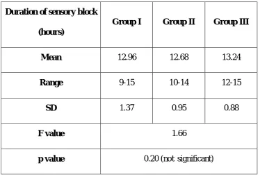
TABLE 6. DURATION OF SENSORY BLOCK
Duration of sensory block
(hours)
Group I Group II Group III
Mean 12.96 12.68 13.24
Range 9-15 10-14 12-15
SD 1.37 0.95 0.88
F value 1.66
p value 0.20 (not significant)
The duration of sensory block is 12.96 hours, 12.68 hours and
13.24 hours respectively for group I, group II and group III respectively.
TABLE 7. DURATION OF MOTOR BLOCK
Duration of motor block
(hours)
Group I Group II Group III
Mean 13.80 13.56 14.12
Range 11-15 9-15 13-16
SD 0.87 1.26 0.88
F value 1.90
p value 0.16 (not significant)
The duration of motor block is 13.80 hours, 13.56 hours and 14.12
hours respectively for group I, group II and group III respectively. This is
TABLE 8. HEMODYNAMIC PARAMETERS
Parameters
GROUP-I GROUP-II GROUP-III
Mean over 6 Hours Base Line Deviation from Base Line Mean over 6 Hours Base Line Deviation from Base Line Mean over 6 Hours Base Line Deviation from Base Line Heart Rate bpm 76.44 ±4.67 79.84 ±5.99 -3.40 ±1.32 73.60 ±5.03 76.08 ±5.76 -2.48 ±0.73 75.96 ±5.82 79.04 ±6.09 -3.08 ±0.27 Systolic BP mm hg 114.8 ±6.56 118.65 ±7.27 -3.85± 0.71 114.88 ±8.02 118.24 ±8.39 -3.36 ±0.37 115.52 ±5.98 120 ±6.14 -4.48 ±0.16 Diastolic BP mm hg 73.52 ±4.63 77.12 ±5.29 -3.85 ±1.18 72 ±5.29 75.36 ±5.16 -3.36 ±0.13 71.92 ±4.42 78.08 ±4.67 -6.16 ±0.25 Mean Arterial Pressure mm hg 101.08 ±5.32 104.88 ±6.57 -3.8 ±1.25 100.48 ±6.28 104.04 ±6.79 -3.56 ±0.51 101.08 ±4.55 106.12 ±5.19 -5.04 ±0.61
The base line and first six hours mean hemodynamic parameters
(heart rate, systolic and diastolic BP and mean arterial pressure) are
observed during the study period for three groups. From these the
baseline value, mean value over the period of six hours and deviation
from baseline (+ indicates increase, - indicates decrease) values are
derived. There is no hemodynamic instability seen in three groups since