0022-538X/95/$04.0010
Copyrightq1995, American Society for Microbiology
Bipartite DNA-Binding Region of the Epstein-Barr Virus BMRF1
Product Essential for DNA Polymerase Accessory Function
ANITA KIEHLANDDAVID I. DORSKY*
Division of Infectious Diseases, University of Connecticut Health Center, Farmington, Connecticut 06030
Received 21 October 1994/Accepted 7 December 1994
The Epstein-Barr virus (EBV) BMRF1 gene product is necessary for DNA polymerase catalytic subunit (BALF5) activity in 100 mM ammonium sulfate. To map regions of BMRF1 necessary for polymerase accessory function, linker insertion and deletion mutant BMRF1 polypeptides were expressed by in vitro transcription-translation and assayed for DNA polymerase elongation activity and binding to double-stranded DNA (dsDNA)-cellulose. Amino-terminal deletions up to residue 303 were defective for stimulation of elongation. Deletions between residues 44 and 194 and residues 238 and 303 abolished binding to dsDNA-cellulose. The region from residues 194 to 238, therefore, is necessary for stimulation of BALF5 elongation but dispensable for dsDNA-cellulose binding. Deletion analysis also localized reactive epitopes of two neutralizing monoclonal antibodies to BMRF1 to a carboxy-terminal region which is dispensable for activity. These data suggest that a bipartite DNA-binding region is an essential component of the DNA polymerase accessory function and that the two noncontiguous regions are separated by a region (residues 194 to 217) which is essential for stimu-lation; therefore, it may interact with the BALF5 catalytic subunit of EBV DNA polymerase. Both EBV BMRF1 and herpes simplex virus UL42 gene products are DNA polymerase accessory proteins which bind dsDNA and increase the processivity of their corresponding catalytic components. Outstanding similarities between their primary amino acid sequences are not evident. However, it appears that their structural organizations are similar.
The in vitro activity of the Epstein-Barr virus (EBV) DNA polymerase (EBVpol) is dependent on the functional interac-tion between the catalytic subunit (BALF5 product) and the accessory subunit (BMRF1 product) (16, 19, 36, 37). The genes for both components are required for in vivo replication of the viral lytic origin of replication, oriLyt, but not for oriP, which is responsible for the maintenance of latency (9). Therefore, it is likely that the in vitro cooperation between the BALF5 and BMRF1 products reflects their functions in vivo.
The 110-kDa BALF5 catalytic component of EBVpol carries out nonprocessive elongation at low salt concentrations in the absence of the 50-kDa BMRF1 accessory product (37). In 100 mM ammonium sulfate, the BALF5 activity is negligible unless the BMRF1 product is present (16, 36, 37), and then the activity is highly processive (37). The BMRF1 product binds strongly to double-stranded DNA (dsDNA) (35); therefore, the manner by which it increases BALF5 processivity is most likely by acting as a ‘‘sliding clamp’’ to tether the catalytic subunit to the template. The Escherichia coli polymerase IIIb protein, which acts as a sliding clamp for the core catalytic machinery, does so by forming a closed circular structure that can encircle duplex DNA, but thebprotein monomer does not have any intrinsic affinity for DNA (17, 31). Other DNA poly-merase accessory proteins such as phage T4 gp45 and eukary-otic proliferating cell nuclear antigen are likely to act similarly to the E. coli b protein (18). The other class of DNA poly-merase accessory proteins, represented by the E. coligd com-plex, the phage T4 gp44-gp62 comcom-plex, and eukaryotic repli-cation factor C, do not directly tether the catalytic subunit to DNA but are ATP-dependent DNA-binding complexes which act to position their respective toroidal sliding clamps onto the
template (1, 15, 23, 34). EBV BMRF1 purified from insect cells lacks ATPase activity (35). The herpes simplex virus (HSV) UL42 product is similar to EBV BMRF1 in that it is a DNA polymerase accessory protein which binds strongly to dsDNA (10, 11, 13). Although there is no direct experimental evidence that the dsDNA-binding activity of HSV UL42 is directly re-sponsible for the increased processivity of the UL42-UL30 complex, the affinity of the polymerase for a synthetic primer-template junction is increased 10-fold by the presence of UL42, and this supports the notion that UL42 acts as a sliding clamp for the polymerase (13). Therefore, EBV BMRF1 and the other herpesvirus DNA polymerase accessory proteins seem to have modes of action which have no obvious homologs in other DNA replication systems.
The BMRF1 accessory protein is a major component of the EBV EA-D complex and is the major early phosphoprotein induced during EBV infection (8, 24). In Burkitt lymphoma cells induced to undergo lytic EBV replication, BMRF1 and BALF5 colocalize to intranuclear replication compartments where DNA synthesis occurs (16). The specific blockade of viral DNA synthesis by the inhibitors phosphonoacetic acid and acyclovir leads to a diffuse nuclear distribution for both antigens (16). Therefore, continuous viral DNA synthesis is necessary for physical colocalization of BALF5 and BMRF1 in infected cell nuclei. Despite demonstrations of cooperation of BALF5 and BMRF1 in vitro and colocalization in nuclei of infected cells, immunoprecipitation of a complex of the two proteins has not been demonstrated (6, 16). This may be for more than trivial reasons, since at least two monoclonal anti-bodies (MAbs) against BMRF1 neutralize EBVpol activity (19, 33, 35), suggesting that the MAbs interfere with functional interaction between the two components. The interacting BALF5-BMRF1 system may, therefore, be a target for the
* Corresponding author. Phone: (203) 3587. Fax: (203) 679-4701.
1669
on November 9, 2019 by guest
http://jvi.asm.org/
rational design of antiviral agents which interfere specifically with subunit interactions.
This study correlates the structures of insertion and deletion mutants of BMRF1 with the two known functional properties of BMRF1: the stimulation of BALF5 elongation activity and dsDNA-binding activity. Mutant BMRF1 polypeptides were expressed by a modification of a previously described in vitro transcription-translation (IVT) expression system (16), screened for reactivity with two anti-BMRF1 MAbs, assayed for binding to dsDNA-cellulose, and assayed for stimulation of IVT-ex-pressed BALF5 elongation activity. The epitopes recognized by two MAbs were localized by deletion mapping to the car-boxy-terminal third of the polypeptide. This approach permit-ted the identification of two noncontiguous regions in the ami-no-terminal two-thirds of BMRF1 which were essential for dsDNA binding. All regions essential for dsDNA binding were also necessary for elongation stimulation; however, several mutants in the region from residues 194 to 237 (194–237 re-gion) bound to dsDNA but were defective in elongation stim-ulation activity. Thus, the analysis identified a central region of BMRF1 which is essential for functional interaction with BALF5 but is not required for dsDNA binding.
MATERIALS AND METHODS
Plasmids.Plasmids were constructed by previously described methods (29), using restriction enzymes, bacterial alkaline phosphatase, S1 nuclease, T4 polynucleotide kinase, and T4 DNA ligase purchased from New England Biolabs and Bethesda Research Laboratories. Plasmids were propagated in E. coli HB101, except that the Dam2strain GM1634 was used to prepare plasmids for cutting with methylation-sensitive restriction enzymes. The expression plasmid pYES2 was purchased from Invitrogen.
(i) pYES2-M1.The BMRF1 open reading frame (ORF) was mobilized on a
BclI-EcoRI fragment from p19-BMRF1 (16) and inserted into the pYES2
polylinker downstream from the T7 promoter.
(ii) pYES2-X2.The BALF5 ORF was mobilized on an EcoRI-HindIII frag-ment from p18-BX5 and inserted into the pYES2 polylinker downstream from the T7 promoter to yield pYES2-X1. A rare MseI site occurs just 7 bp upstream from the BALF5 translational initiation site. An MseI-Bsu36I fragment filled in with Klenow enzyme at MseI was obtained from p18-BX5 and inserted into a complementary fragment of pYES2-X1 cut with EcoRI (filled in with Klenow enzyme) and Bsu36I, to yield pYES2-X2.
(iii) pEX-EBVpol3.The BALF5 ORF was mobilized from pYES2-X2 on a
HindIII-EcoRI fragment, blunt ended with Klenow enzyme, and inserted into
pGEMEX-1 (Promega Biotech) at the unique NheI site downstream from the T7 promoter. The BALF5 ORF was fused to the first seven T7 gene 10 codons (MARASLT-BALF5). The expression vector contains the phage T7 transcrip-tion terminatranscrip-tion signal distal to the cloning site, so in vitro transcriptranscrip-tion with T7 RNA polymerase could be carried out efficiently without linearizing the tem-plate. The translational initiation site of phage T7 gene 10 (59CATATGG) has been shown to function efficiently on uncapped in vitro transcripts translated in rabbit reticulocyte lysate (7).
Linker insertion mutagenesis.The BMRF1 expression plasmid pYES2-M1 was pseudorandomly linearized with the frequently cutting enzyme HaeIII (27 sites in the BMRF1 ORF) in the presence of 1mg of ethidium bromide per ml (12). The unit-length plasmid was gel purified, treated with bacterial alkaline phosphatase, and ligated with an excess of the EcoRI linker, 59CCGGAATTC CGG, which had been 59phosphorylated with T4 polynucleotide kinase. After digestion with EcoRI, the DNA was gel purified, ligated with T4 DNA ligase at a DNA concentration of 0.01 mg/ml, and transformed into E. coli HB101. Ampicillin-resistant colonies were selected, and DNA minipreps were screened for EcoRI sensitivity (pYES2-M1 has no EcoRI sites). HindIII and BglII sites define the proximal and distal ends of the BMRF1 ORF, so plasmids containing the linker were subjected to triple digestion with EcoRI, HindIII, and BglII in order to map the insertions within the BMRF1 ORF. Insertions within BMRF1 were then mapped exactly by end labeling small restriction fragments either containing the insertion site or having one EcoRI end and determining the length on a 6% sequencing gel, using a known sequence ladder for exact size determi-nation. Insertion sites were then confirmed by direct DNA sequencing (30). Linker insertion mutants were named for the BMRF1 codon which they altered.
Deletion mutagenesis.The amino-terminal deletionDN129 was constructed by digesting pYES2-M1 (I-122) with HindIII and EcoRI, filling in with Klenow enzyme, and religating. The result was that the BMRF1 ORF began with me-thionine 130 (59CTAGG), which appeared to function efficiently in vitro as a translational initiation site, as judged from the size of the in vitro translation product. Carboxy-terminal deletions were constructed by digestion at unique
restriction sites and a unique XbaI site distal to the BMRF1 ORF, filling in with Klenow enzyme, and religating (NotI [DC303], AvrII [DC342], and EcoRI [DC371] of I-371).DC279 was obtained by digesting with AatII and NotI, treating with S1 nuclease, religating, and selecting the mutant from sequenced plasmids. Internal deletions were created by digestion at linker insertion sites and naturally occurring unique restriction sites, creating blunt ends with either Klenow enzyme or S1 nuclease when necessary, and religating. Constructions were carried out as follows:D45-98, EcoRI of I-44/Klenow and EcoN1/Klenow;D80-204, EcoRI of I-79/sticky and EcoRI of I-205/sticky;D124-217, EcoRI of I-122/sticky and EcoRI of I-217/sticky;D194-217, BspEI/Klenow and EcoRI of I-217/Klenow;D206-236,
EcoRI of I-205/sticky and EcoRI of I-237/sticky;D218-233, EcoRI of I-217/ Klenow and EcoRI of I-234/Klenow;D238-276, EcoRI of I-237/sticky and EcoRI of I-277/sticky;D278-306, AatII/S1 and NotI/S1;D302-306, NotI/S1 only; and D317-378, SmaI and NruI (both blunt). Restoration of reading frame by internal deletions was confirmed by dideoxy DNA sequencing and by demonstrating that the polypeptide specified reacted with MAb 90E2, which recognizes an epitope at the carboxy terminus of BMRF1.
IVT.In vitro transcription templates were prepared from CsCl gradient-puri-fied plasmids by linearizing downstream from their ORFs at unique XbaI sites. Closed circular pEX-EBVpol3 was used, since it contains a phage T7 transcrip-tional termination signal downstream from the BALF5 ORF. DNA templates were transcribed with T7 RNA polymerase and translated in modified rabbit reticulocyte (Promega) as previously described (16). [35S]methionine labeling was carried out as previously described, using ICN Translabel or NEN/Dupont Express Protein Labeling Mix (1,200 mCi/mmol) (16). After 90 min of in vitro translation at 308C, the radioactivity labeled polypeptides were precipitated in 4 volumes of cold methanol, dried in vacuo, boiled in sodium dodecyl sulfate-polyacrylamide gel electrophoresis (SDS-PAGE) sample buffer, and analyzed by SDS-PAGE (7.5 or 10% gel) and fluorography, using prestained high-range protein molecular weight standards (Gibco-BRL).
DNA-cellulose chromatography.[35
S]methionine-labeled wild-type and mu-tant BMRF1 polypeptides were expressed by IVT (18-ml samples). A small (3-ml) aliquot was retained for analysis, and the rest was diluted 1:10 in TE buffer (50 mM Tris-HCl [pH 7.5], 1 mM EDTA) containing 100mg of RNase A and applied to a 200-ml bed volume of dsDNA-cellulose (Sigma) in an Isolab minicolumn equilibrated in the same buffer. The column was washed with 1 bed volume of TE and then eluted with a 2-bed-volume step gradient of 100, 200, 300, 400, and 500 mM NaCl in TE. The eluates were then precipitated with 4 volumes of methanol in the presence of 10mg of bovine serum albumin (BSA) and analyzed by SDS-PAGE and fluorography.
Antibodies and immunoblotting.The IVT-expressed radiolabeled BMRF1 polypeptides were analyzed by SDS-PAGE (10% gel) and electrophoretically transferred to nitrocellulose membranes for 1 h at 200 mA in 25 mM Tris-HCl (pH 8.0)–200 mM glycine–20% (vol/vol) methanol. The membranes were blocked by a 1-h incubation in Tris-buffered saline (pH 7.6) containing 0.5% Tween 20 (TBST) and 1% BSA. MAb R3 at 100mg/ml (24) was purchased from NEN/Dupont, and MAb 90E2 hybridoma fluid was a gift from Ronald Glaser (Ohio State University) (33). Blocked nitrocellulose filters were incubated for 1 h at room temperature in blocking buffer with MAb R3 at 1:1,000 or MAb 90E2 at 1:300, subjected to three 10-min TBST washes, and then incubated for 30 min with alkaline phosphatase-conjugated anti-mouse immunoglobulin G (Promega) at 1:3,000 in blocking solution. Detection was carried out by incubation in 100 mM Tris-HCl (pH 9.5)–100 mM NaCl–20 mM MgCl2containing 0.33 mg of nitroblue tetrazolium (Promega) per ml and 0.16 mg of 5-bromo-4-chloro-3-indolyl phosphate (BCIP; Promega) per ml. After drying, filters containing ra-diolabeled proteins were exposed for autoradiography.
DNA polymerase assays.BALF5 and BMRF1 in vitro transcripts (4mg per assay) were translated in 25-ml aliquots of reticulocyte lysate in the presence of unlabeled amino acids. DNA polymerase activity assays were carried out in 100-ml volumes containing 10mg of activated calf thymus DNA, 100 mM am-monium sulfate, 50 mM Tris-HCl (pH 8.0), 7.5 mM MgCl2, 5mM each dATP, dTTP, and dGTP, and 2.5mCi of [a-32P]dCTP (3,000 Ci/mmol; NEN/DuPont) for 20 min at 378C. The reaction was stopped by the addition of 0.8 ml of 10% trichloroacetic acid, cooled in ice for 10 min, spotted onto Whatman GFA glass filters, and washed with three 2.0-ml washes of 10% trichloroacetic acid and once with 2.0 ml of ethanol, using a Hoefer vacuum filter manifold. Filters were dried, and acid-precipitable radioactivity was measured by scintillation counting. DNA polymerase activity intrinsic to the unprogrammed reticulocyte lysate was sub-tracted as background, and enzyme activity was measured in units, defined as femtomoles of acid-precipitable [32
P]dCMP per hour per milliliter of reticulocyte lysate assayed at 378C (16). Results are expressed as averages of three separate assays.
Computerized protein sequence analysis.EBV, HSV-1, and GH5 (globular domain of histone H5) DNA sequences were obtained from GenBank and manipulated on a VAX computer, using the University of Wisconsin Genetics Computer Group software. Protein sequences were compared by using the BESTFIT program, and secondary structure predictions were carried out with the PEPTIDESTRUCTURE program (38).
on November 9, 2019 by guest
http://jvi.asm.org/
RESULTS
IVT expression of BALF5 and BMRF1.In our earlier study, IVT expression of BALF5 and BMRF1 from plasmids p18-BX5 and p19-BMRF1, respectively, was relatively inefficient, probably because of long 59untranslated regions (16). To im-prove the IVT expression efficiency, the BALF5 and BMRF1 ORFs were inserted into the pYES2 vector. Both 59 untrans-lated regions were coincidentally shortened by 128 bp. The BALF5 ORF was also inserted into the T7 expression vector pGEMEX-1 (Promega) to yield pEX-EBVpol3, which allowed IVT expression of BALF5 from a nonlinearized template. The polypeptides expressed by IVT from plasmids pYES2-M1 (BMRF1) and pEX-EBVpol3 (BALF5) are shown in Fig. 1. The intensity of the band of the 113-kDa BALF5 polypeptide, with 15 methionine residues, is comparable to that of the HSV DNA polymerase (HSV-pol) 130-kDa polypeptide specified by plasmid pEX-HSVpol2 (7, 16), with 21 methionine residues, and the 48-kDa BMRF1 polypeptide specified by plasmid pYES2-M1. The efficiency of IVT expression and in vitro en-zymatic activity of the T7 gene 10-BALF5 fusion specified by pEX-EBVpol3 were identical to those of the authentic BALF5 polypeptide specified by pYES2-X2 (not shown). Table 1 shows the results of assaying the polypeptides shown in Fig. 1 for DNA polymerase activity in 100 mM ammonium sulfate. The activity of the BALF5 polypeptide expressed from pYES2-X2 had over 10-fold more activity than the polypeptide expressed from p18-BX5, and the BALF5 polypeptide ex-pressed alone had less than 1% of the activity demonstrated in the presence of BMRF1. Under the same experimental condi-tions, the HSV DNA polymerase expressed from pEX-HSVpol2 showed comparable activity in the absence of a viral DNA polymerase accessory protein. Comparison of SDS-PAGE gel
fluorogram band intensities obtained under similar expression conditions indicated that IVT expression of BMRF1 from pYES2-M1 and BALF5 from pYES2-X2 was about eightfold more efficient than from p19-BMRF1 and p18-BX5, respec-tively (data not shown).
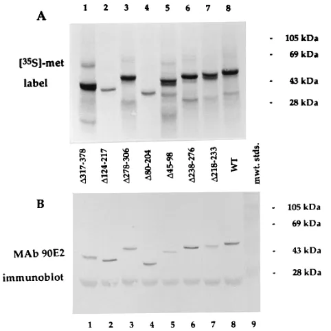
Mutagenesis of BMRF1. Ten distinct EcoRI linker inser-tions within the intact BMRF1 ORF in pYES2-M1 were re-covered from the mutagenesis scheme, which yielded over 50 independently obtained insertions in BMRF1. Also recovered were two additional insertions (at codons 116 and 352) which were accompanied by rearrangements in BMRF1. The BMRF1 linker insertions and their predicted amino acid changes are shown in Table 2. Deletion mutants of BMRF1, constructed with some of the EcoRI linker insertions and other naturally occurring restriction sites in BMRF1, are also shown in Table 2. The mutant polypeptides were expressed by IVT and analyzed by SDS-PAGE, as shown in Fig. 2 to 4. IVT-expressed [35S]methionine-labeled BMRF1 polypeptides re-solved by SDS-PAGE were electroblotted to nitrocellulose filters in order to study the same samples by autoradiography and immunoreactivity. All of the BMRF1 mutants in this study appeared to be stable to 90-min incubations in rabbit reticu-locyte lysate, and all of the linker insertion mutant polypep-tides had electrophoretic mobilities indistinguishable from that of wild-type BMRF1 by SDS-PAGE (7 and 10% gels) (not shown). In Fig. 2, the presence of the carboxy-terminal MAb 90E2 epitope was demonstrated for most of the internal dele-tion mutants, verifying restoradele-tion of the BMRF1 ORF. Mu-FIG. 1. IVT expression of EBV BALF5 and BMRF1. DNA templates
[image:3.612.113.246.68.158.2]pEX-HSVpol2, pYES2-M1, and pEX-EBVpol3, respectively, were used to express HSVpol (lane 1), EBV BMRF1 (lane 2) and EBV BALF5 (lane 3). Radiolabeled polypeptides were analyzed by SDS-PAGE (7.5% gel) and fluorography, using prestained molecular weight standards.
TABLE 1. Activities of in vitro-translated DNA polymerasesa
Plasmid(s) used as
template for RNA Gene product(s)
Activity (U)
p18-BX5 EBV BALF5 0
p19-BMRF1 EBV BMRF1 0
p18-BX51p19-BMRF1 EBV BALF5 and BMRF1 84
pEX-HSVpol2 HSV UL30 (pol) 1,935
pYES2-M1 EBV BMRF1 0
pYES2-X2 EBV BALF5 0
pYES2-X21pYES2-M1 EBV BALF5 and BMRF1 2,427
a
In vitro transcripts prepared from templates with T7 RNA polymerase were used to program rabbit reticulocyte lysates which were assayed for DNA poly-merase activity on activated calf thymus DNA. Activity intrinsic to the lysate was subtracted as background, and activity was measured in units of femtomoles of acid-precipitable [32
[image:3.612.316.554.90.358.2]P]dCMP per hour per milliliter of reticulocyte lysate assayed at 378C.
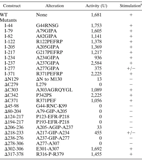
TABLE 2. Stimulation of BALF5 elongation activity by BMRF1 mutantsa
Construct Alteration Activity (U) Stimulationb
WT None 1,681 1
Mutants
I-44 G44RNSG 1,753 1
I-79 A79GIPA 1,605 1
I-82 A82GIPA 1,141 1
I-122 R122PEFRP 1,378 1
I-205 A205GIPA 1,369 1
I-217 G217PEFRP 1,217 1
I-234 A234GIPA 936 1
I-237 A237GIPA 2,584 1
I-277 A277GIPA 375 1/2
I-371 R371PEFRP 2,225 1
DN129 DN to M130 13 2
DC279 L279 0 2
DC303 A303AGRQYGL 1,089 1
DC342 P342PS 2,225 1
DC371 R371PEF 1,056 1
D45-98 G44-RNC-K99 0 2
D80-204 A79-GIP-A205 0 2
D124-217 P123-EFR-P218 0 2
D194-217 P193-EFR-P218 0 2
D206-236 A205-AGIP-A237 33 2
D218-233 A217-GIP-A234 455 1/2
D238-276 A237-GIP-A277 0 2
D278-306 A277-A307 0 2
D302-306 E301-A307 1,692 1
D317-378 R316-P-R379 1,455 1
aReticulocyte lysates programmed with BALF5, BMRF1, and BMRF1
mu-tant in vitro transcripts were assayed for DNA polymerase activity on activated calf thymus DNA as previously described (16). Data are the means of three assays. Mutants are designated by the codon numbers disrupted by the muta-tions, with the wild-type (WT) residue preceding the codon number and the substitution following it.
b1, wild-type activity;2, less than 5% of wild-type activity;1/2, 20 to 30%
of wild-type activity.
on November 9, 2019 by guest
http://jvi.asm.org/
[image:3.612.59.298.585.677.2]tantsDN129,D194-217, andD206-236 and the insertions are not shown in Fig. 2 but were found to contain the MAb 90E2 epitope by immunoblotting.
Mapping of the MAb R3 epitope. The mutant BMRF1 polypeptides were expressed by IVT and analyzed by autora-diography (Fig. 3A) and for immunoreactivity by immunoblot-ting (Fig. 3B and C). Figure 3B shows that reactivity with MAb R3 is absent for mutantsDC303,D317-378,DC342, and I-371. In Fig. 3B, lane 6 shows faint MAb R3 reactivity of DC371 which is seen only with loading of twice the sample volume, compared with lane 7, where no reactivity is demonstrated. The absence of reactivity of I-371 (even with double sample volumes; not shown), the faint reactivity of DC371, and the absence of reactivity ofD317-378 are all consistent with the linear MAb R3 epitope lying immediately upstream of, and probably including, BMRF1 amino acid residue 371. The faint reaction of MAb R3 with a 47-kDa protein in rabbit reticulo-cyte lysate seen in this figure has been previously described (24).
Mapping of the MAb 90E2 epitope.Figure 3C shows that MAb 90E2 reactivity is absent fromDC371, present in D 317-378, and preserved in every BMRF1 mutant which contains carboxy-terminal residues distal to residue 378. Therefore, the MAb 90E2 epitope lies within BMRF1 residues 379 through 404. The identification of a carboxy-terminal linear epitope provided a convenient method for proving restoration of the BMRF1 ORF in internal deletion mutants.
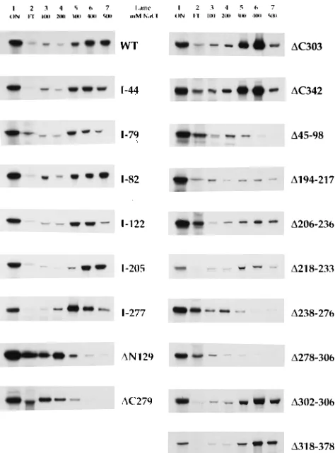
dsDNA binding by BMRF1 mutants.BMRF1 expressed by baculovirus has intrinsic dsDNA-binding activity (35). To map the regions involved in dsDNA binding, the BMRF1 mutant polypeptides were screened for binding to dsDNA-cellulose by determining the elution profile from a dsDNA-cellulose mi-crocolumn with an NaCl step gradient. As shown in Fig. 4, the IVT-expressed wild-type BMRF1 polypeptide bound effi-ciently to dsDNA-cellulose, and most of the material eluted at
400 to 500 mM NaCl. All of the linker insertion mutants showed wild-type dsDNA-binding activity (I-217, I-234, I-237, and I-371; not shown), with the possible exception of I-79, I-122, and I-277, which eluted maximally at 300 mM NaCl, in contrast to the wild-type maximal elution at 400 mM. The deletions DC303,DC342, DC371, D194-217, D206-236, D 218-233,D302-306, andD318-378 also showed wild-type dsDNA-binding activity (data forDC371 not shown). Two of the mu-tant polypeptides (D194-217 and D206-236) which bound to dsDNA-cellulose and eluted at 300 to 500 mM NaCl were also partially eluted in the flowthrough or 100 mM NaCl fractions. In the case of D194-217, little material eluted in the flowthrough, but about one-third of the material eluted in the 100 mM NaCl fraction. In the case of D206-236, about one-third of the material eluted in the flowthrough, but most of the material eluted in the 300 to 500 mM NaCl fractions. These findings were confirmed on three separate experimental runs. The amino-terminal deletionDN129 and carboxy-terminal de-letionDC279 mutants eluted from dsDNA at 100 to 200 mM NaCl, as did the internal deletions D45-98, D238-276, and
[image:4.612.60.301.70.311.2]D278-306. MutantD124-217 (not shown) also eluted at 100 to 200 mM NaCl. The dsDNA-binding activities of the mutant FIG. 2. Internal deletion mutants of BMRF1. Radiolabeled wild-type (WT)
[image:4.612.324.549.71.424.2]and mutant BMRF1 polypeptides were expressed by IVT, resolved by SDS-PAGE (10% gel), using prestained molecular weight standards (mwt. stds.), and electroblotted to a nitrocellulose filter which was stained for reactivity with MAb 90E2 (B) and autoradiographed (A).
FIG. 3. Immunoreactivity of BMRF1 deletion mutants. Radiolabeled wild-type (WT) and mutant BMRF1 polypeptides were expressed by IVT, resolved by SDS-PAGE (10% gel), using prestained molecular weight standards, and elec-troblotted to nitrocellulose filters which were autoradiographed (A), stained for reactivity with MAb R3 (B), or stained for reactivity with MAb 90E2 (C). Lanes 6 and 7 contain, respectively, double and unit sample volumes ofDC371.
on November 9, 2019 by guest
http://jvi.asm.org/
BMRF1 polypeptides were grouped into two categories of salt sensitivity: high affinity (400 to 500 mM) and low-affinity (100 to 200 mM). In the cases of mutantsD194-217 andD206-236, the presence of significant amounts of high-affinity binding material distinguished them from mutants such asDN129 and
DC279. The dsDNA-binding activity mapping is summarized in Fig. 5. Carboxy-terminal residues distal to residue 302 were dispensable for dsDNA binding. No dispensable amino-termi-nal region could be identified by deletion aamino-termi-nalysis.
Effects of BMRF1 mutations on in vitro EBVpol activity.
The BMRF1 mutants were assayed for their capacity to stim-ulate the elongation activity of BALF5 in vitro as described in Materials and Methods. The results of these assays are shown in Table 2. The activities of the BMRF1 mutants fell into three categories: wild type, less than 5% of wild-type activity, and 20 to 30% of wild-type activity. Mutants were designated as stim-ulating if their activity was at least fivefold above background activity. All mutants which failed to stimulate had activities below (tabulated as zero) or less than 1.2-fold background activity. Mutants designated as partially stimulating wereD 218-233 (2.7-fold above background, 22% of wild-type activity) and I-277 (3.7-fold, 27% of wild-type activity). All of the linker insertions, with the exception of I-277, showed wild-type activ-ity in the DNA polymerase assay. Deletion mutants DC303,
DC342,DC371,D302-306, andD317-378 had wild-type activity, while all other deletion mutants exceptD218-233 showed less than 5% of wild-type activity. The deletion mutant analysis, comparing the dsDNA-binding and BALF5 stimulation activ-ities of the BMRF1 mutants, is summarized in Fig. 5. As for dsDNA-binding activity, the carboxy-terminal residues distal to residue 302 were dispensable for in vitro EBVpol activity. Three internal deletion mutants, D194-217, D206-236, and
D218-233, showed high-affinity dsDNA binding but absent (or severely diminished, in the case ofD218-233) EBVpol activity. The behavior of these three overlapping internal deletions in the central portion of BMRF1 demonstrated a discordance between the structural requirements for dsDNA binding and EBVpol activity. Binding to dsDNA appeared to be necessary but not sufficient for EBVpol activity.
DISCUSSION
[image:5.612.60.297.70.390.2]The EBV BMRF1 product is a DNA polymerase accessory protein which binds strongly to dsDNA (35) and is required for processive elongation activity of the EBV DNA polymerase catalytic component (36). Like several other well-characterized DNA polymerase accessory proteins, BMRF1 seems to func-tion as a sliding clamp in that it binds to DNA and stimulates processive DNA synthesis by the catalytic component of EBVpol, but it differs from the E. colibprotein, the bacterio-phage T4 gp45, and eukaryotic proliferating cell nuclear anti-gen in that BMRF1 binds tightly to DNA while the others do not (15, 18, 31, 35). In support of the sliding-clamp explanation is that the dsDNA-binding activity of BMRF1 appears to be essential for elongation activity; however, it has not yet been demonstrated that BMRF1 actually tethers the catalytic com-ponent to the primer-template junction. The HSV UL42 gene product is also a dsDNA-binding protein which increases the FIG. 4. dsDNA binding by BMRF1 mutants. Radiolabeled wild-type (WT)
[image:5.612.317.556.73.274.2]and mutant BMRF1 polypeptides expressed by IVT were applied to a dsDNA-cellulose minicolumn and eluted with an NaCl step gradient. Eluted samples were precipitated with methanol and analyzed by SDS-PAGE (7.5% gel) and fluorography. Lane 1, material applied onto column (ON); lane 2, column flowthrough (FT); lane 3, 100 mM NaCl elution; lane 4, 200 mM NaCl elution; lane 5, 300 mM NaCl elution; lane 6, 400 mM NaCl elution; lane 7, 500 mM NaCl elution.
FIG. 5. Summary of properties of BMRF1 deletion mutants. A schematic representation of the BMRF1 ORF is shown on the left. Shaded regions repre-sent the expressed portions of the ORF, and open regions reprerepre-sent the dele-tions. Numbering refers to the amino acid residues at the boundaries of the expressed portions. BMRF1 deletion mutants are more fully described in Table 2. Black shading indicates wild-type (WT; high-affinity) dsDNA binding and elongation-stimulatory activity (1). Gray shading indicates absence of both ac-tivities (2). Cross-hatching indicates wild-type dsDNA binding but deficient elongation-stimulatory activity. MutantD218-233 has finer cross-hatching to in-dicate that it has detectable (22% of wild-type) elongation stimulatory activity (1/2). Mutant in vitro properties are shown at the right.
on November 9, 2019 by guest
http://jvi.asm.org/
affinity of the catalytic subunit for the primer-template junction by 10-fold, consistent with the explanation that UL42 stimu-lates processive DNA synthesis by the UL30 catalytic compo-nent by decreasing the rate of dissociation of the polymerase from the primer-template junction after each elongation cycle (13). To begin to understand how BMRF1 causes processive DNA synthesis by BALF5, and to compare it with HSV UL42, we have carried out a structure-function relationship study with mutants of BMRF1, assaying for stimulation of elongation activity and binding to dsDNA. The heterologous expression system used in this study was IVT in rabbit reticulocyte lysate, which has been previously described for the study of HSVpol and its accessory protein and has been used to show the re-quirement of BMRF1 for BALF5 elongation activity in 100 mM ammonium sulfate (16). The IVT expression system for BALF5 and BMRF1 was vastly improved over the system used in the previous study by removal of most of the 59untranslated regions of both ORFs, such that the expression efficiency was comparable to that described for HSVpol (7, 16). A particu-larly useful feature of the IVT expression assay system was that the elongation activity of BALF5 in the absence of BMRF1 was undetectable at 100 mM ammonium sulfate. The proper-ties of the IVT-expressed polypeptides were consistent with the properties of purified BALF5 and BMRF1 expressed in insect cells from recombinant baculoviruses: at low salt con-centrations, the BALF5 elongation activity is weak and non-processive, but at high salt concentrations, it is inactive unless BMRF1 is present, and the elongation is highly processive (6, 36, 37). The DNA polymerase assay used in this study did not distinguish between processive and distributive elongation ac-tivity.
The analysis of the properties of BMRF1 mutants identified two noncontiguous regions of the polypeptide which are es-sential for dsDNA binding and stimulation of BALF5 elonga-tion activity. We identified a region in the central porelonga-tion of BMRF1 which is dispensable for dsDNA binding but required for stimulation of BALF5 elongation activity. Also, the dele-tion analysis localized the linear epitopes for two anti-BMRF1 MAbs (which neutralize EBVpol activity) to the carboxy ter-minus of BMRF1, which is dispensable for both dsDNA bind-ing and BALF5 elongation activity at high salt concentration.
Two noncontiguous regions required for dsDNA binding.
BMRF1 expressed in insect cells from recombinant baculovi-ruses eluted from dsDNA-cellulose at 0.24 M NaCl (35), and the pp58 and pp50 BMRF1 phosphoproteins from NC 37 cells eluted at 0.3 M NaCl (28). BMRF1 expressed by IVT bound efficiently to denatured DNA-cellulose and was eluted with a step gradient at 200 to 300 mM NaCl (16). In the present study, the peak of BMRF1 dsDNA-cellulose binding activity eluted at 400 mM NaCl. The broadness of the elution profile probably reflected the use of a step gradient, which facilitated the com-parison of chromatographic behaviors of the mutant polypep-tides. Although the dsDNA-cellulose elution profiles of the BMRF1 polypeptides were not sharply monophasic, they were sufficiently informative to allow identification of dsDNA-bind-ing and nonbinddsDNA-bind-ing behavior which was reproducible and in-ternally consistent. The behavior of the mutant polypeptides which eluted from dsDNA-cellulose at low salt,DN129,DC279,
D45-98,D238-276, andD278-306, could easily be distinguished from the more salt-resistant profiles of wild-type BMRF1, the linker insertion mutants, and the deletions DC303, DC342,
D194-217,D206-236,D218-233,D302-306, andD318-378. Some of the insertion mutants (I-79, I-122, and I-277) displayed subtle differences from the wild-type elution profile in that the maximum elution was at 300 mM NaCl. By this deletion anal-ysis, two noncontiguous regions of BMRF1 required for
dsDNA binding were identified, as shown in Fig. 5. The distal boundary of the amino-terminal region (residues 1 to 193) was defined by mutantD194-217. No proximal amino-terminal de-letions preserved dsDNA binding; therefore, the requirement for an intact amino terminus for dsDNA binding could not be ruled out. The three overlapping central internal deletions which bound dsDNA,D194-217,D206-236, andD218-233, de-fined residues 194 to 236 as dispensable for dsDNA binding. The requirement for residues 238 to 302 for dsDNA binding was consistently defined by the behaviors of the dsDNA-bind-ing mutantsDC303 andD302-306 and the nonbinding mutants
DC279,D238-276, andD278-306.
The contention that residues 194 to 236 are not essential for dsDNA binding rests on the interpretation of the chromato-graphic behaviors of theD194-217 andD206-236 polypeptides, since their elution profiles differed from that of wild-type BMRF1 in subtle ways, whileD218-233 appeared to be iden-tical to the wild type for dsDNA binding. Significant amounts of the mutant polypeptidesD194-217 andD206-236 eluted in the flowthrough and low-salt fractions, but for both mutants, about half of the protein eluted in the 300 to 500 mM NaCl fractions. The preservation of apparent molecular weight on SDS-PAGE and the absence of enhancement of minor bands upon overexposure of the autofluorograms (not shown) sug-gest that the material eluting in the low-salt fractions was not degraded. The reproducible elution of these two mutant polypeptides in both low-salt and high-salt fractions could be due to impaired dsDNA binding caused by the mutations, but that would not adequately explain why some of the material still eluted in the high-salt range. Another interpretation, which we favor, is that theD194-217 andD206-236 polypep-tides misfold partially in such a way as to make the DNA-binding domain less stable than the wild-type polypeptide, so that only a certain fraction of the molecules maintains a prop-erly folded conformation which allows salt-resistant binding to dsDNA. One way in which this might happen is if the central 194–236 region linking the 1–193 (proximal) and 237–303 (dis-tal) subdomains can interact with either or both subdomains. Loss of this interaction might partially destabilize, but not destroy, the conformation of the DNA-binding region. The proximal and distal essential regions may both contact DNA, or one may stabilize the DNA binding by the other. The data do not allow a distinction between the two models, but the analysis of dsDNA binding by glutathione S-transferase– BMRF1 domain fusions may identify smaller dsDNA-binding subdomains. The elucidation of the correct structural explana-tion of dsDNA binding by BMRF1, though, will most likely require X-ray crystallography.
Regions of BMRF1 required for stimulation of BALF5 elon-gation activity. The analysis of BMRF1 mutants assayed for stimulation of BALF5 elongation activity, shown in Table 2 and summarized in Fig. 5, indicated that the mutations which prevented binding to dsDNA also interfered with stimulation of BALF5 elongation activity. However, the properties of three central internal deletion mutants indicated that the structural requirements for dsDNA binding and stimulation of BALF5 elongation activity are different. MutantsD194-217 andD 206-236 bound to dsDNA but lacked elongation-stimulatory activ-ity, and mutantD218-233 bound dsDNA but had 22% of wild-type elongation-stimulatory activity. About half of the in vitro-translatedD194-217 andD206-236 polypeptides failed to bind to dsDNA in a salt-resistant manner, suggestive of partial mis-folding, but neither mutant had any elongation-stimulatory activity, unlikeD218-233, which had partial activity. If deletions
D194-217 andD206-236 caused loss of elongation stimulation entirely as a result of impaired dsDNA binding, then partial
on November 9, 2019 by guest
http://jvi.asm.org/
elongation stimulation activity might have been observed, but it was not. We cannot rule out that the misfolded non-DNA-bindingD194-217 andD206-236 polypeptides somehow inhib-ited the remaining polypeptide molecules capable of binding dsDNA from interacting with BALF5 to produce elongation activity. However, the interpretation that we favor is that the 194–236 region supplies a function other than involvement with DNA binding, possibly providing a site of interaction with BALF5. The deletion analysis indicated that the 194–217 re-gion was necessary for elongation stimulatory activity and that the 218–233 region immediately distal to it influenced the activity. Despite the effects of these small central deletions on elongation-stimulatory activity, the linker insertions in this re-gion, I-205, I-217, and I-234, were found to have nearly wild-type activity, or in the case of I-234, 56% of wild-wild-type activity. Mutant I-277, located near the distal boundary of the regions identified by deletion analysis as essential for dsDNA binding and BALF5 stimulation, showed nearly wild-type dsDNA-binding activity and 22% of wild-type BALF5 stimulation ac-tivity. As several of the other insertion mutants showed subtle differences from the wild type in their dsDNA column elution profiles, it is difficult to determine if the diminished elongation stimulation activity of I-277 is due to altered DNA-binding properties or some other effect.
Localization of MAb epitopes.Both the anti-BMRF1 MAbs R3 and 90E2 reacted with BMRF1 by immunoblotting, indi-cating that they recognized linear epitopes (Fig. 2 and 3). The MAb R3 epitope was localized by mutational analysis of BMRF1 to the region immediately proximal to, and probably including, residue 371. This conclusion is based on the absence of reactivity withD317-378, the absence of reactivity with I-371, and the faint reactivity withDC371. The MAb 90E2 epitope was mapped unambiguously to the region distal to residue 378, by virtue of its reactivity withD318-378 and absence of reac-tivity with DC371. The localization of both of these MAb epitopes to the carboxy-terminal third of BMRF1 is significant, since both MAbs have been shown to neutralize EBVpol ac-tivity in vitro (19, 33, 35). However, the structure-function analysis of BMRF1 showed that the carboxy-terminal 304–404 region was dispensable for both dsDNA-binding activity and stimulation of BALF5 elongation activity. The presence of neutralizing epitopes on a region of BMRF1 dispensable for in vitro function suggests that the carboxy-terminal 102 residues are situated in the native structure such that they stabilize an essential region of BMRF1 or such that the binding of the antibody interferes with BMRF1 function through steric hin-drance. MAb 6898 against HSV UL42, in contrast, recognized a carboxy-terminal epitope in UL42 residues 363 to 369 (which are not required for UL30 stimulation or binding) (21) and precipitated a UL42-UL30 complex efficiently (20). Because of the requirement of both BALF5 and BMRF1 components for processive EBVpol activity, it seemed likely that a physical complex between them could be detected by immunoprecipi-tation, as has been described for the HSVpol UL30-UL42 complex (3–5, 20). However, attempts to detect coimmunopre-cipitation of BALF5 by anti-BMRF1 antibodies failed to iden-tify such a complex with MAbs R3 and 90E2 (6). It seems likely that although both MAb epitopes lie within a portion of BMRF1 not required for in vitro function, binding of either of the MAbs is sufficient to prevent complex formation through steric hindrance. Another possibility is that the MAbs prevent binding of BMRF1 to DNA, which may be essential for for-mation of a BALF5-BMRF1 complex. The effects of these and other MAbs on BMRF1 function seem to be significant and can be studied further by using the purified components.
Comparison with HSV UL42.EBV BMRF1 appears to have
in vitro properties and a gross structural organization which are analogous to those of the 65-kDa HSV UL42 product. For both proteins, the amino-terminal two-thirds of the molecule contains a dsDNA-binding region and a region required for functional interaction with the corresponding catalytic subunit. Deletions scanning the amino-terminal two-thirds of both mol-ecules interfere with both dsDNA binding and elongation stim-ulation activity. For both, in vitro activity does not require the presence of the carboxy-terminal third of the molecule. The analysis has been taken further in the case of HSV UL42, since the carboxy-terminal third of UL42 was found to be dispens-able for complementation in vivo with a UL42 null mutant in transfected Vero cells (4). Despite substantial protein se-quence similarities between the HSV-1 UL30 catalytic subunit of HSVpol and EBV BALF5, no outstanding sequence simi-larities between HSV UL42 and EBV BMRF1 have been de-tected by using the BESTFIT analysis. In contrast, stronger sequence similarities have been detected between the HSV-1 UL42 and varicella-zoster virus ORF16 products (4, 32). It seems clear that the herpesvirus DNA polymerase accessory proteins have similar functions and grossly similar structural organizations and arose from a common ancestral precursor. However, there are significant functional differences between the HSVpol and EBVpol catalytic subunits. The HSVpol UL30 product shows significant in vitro elongation activity in 100 mM ammonium sulfate which is stimulated and made more processive by UL42, while EBV BALF5 has negligible elongation activity in the absence of BMRF1 in 100 mM am-monium sulfate, and the reconstituted activity is highly proces-sive. The HSV UL30 and UL42 products associate in vitro to form a 181-kDa holoenzyme (14) which is precipitable by an-tibodies against either component (3), while anan-tibodies against BALF5 and BMRF1 neutralize the holoenzyme activity and fail to precipitate a BALF5-BMRF1 complex (6, 16).
Deletion analyses of HSV UL42 and EBV BMRF1 identi-fied large carboxy-terminal regions for both proteins which were dispensable for activity. The analyses differed in that significant central deletions which preserved dsDNA binding were identified for BMRF1 but not for UL42. Mutational analysis of UL42 indicated that the carboxy-terminal 172 res-idues are dispensable for dsDNA binding and UL30-stimula-tory activity (4, 32), while the largest internal deletions de-scribed for HSV UL42 which preserved dsDNA binding were
D9-20 and D242-250 (4). Two regions of UL42 required for functional interaction with the catalytic subunit were identified by deletion mutations D129-163 and D202-337, and the up-stream mutant retained its ability to coprecipitate the catalytic subunit, while the downstream did not, suggesting that residues 202 to 337 were important for direct interaction with UL30 (20). Another mutational analysis of UL42 mapped dsDNA binding to within the carboxy-terminal 340 residues but showed that DC282 still partially bound UL30, whereas D250-308 lacked both dsDNA- and UL30-binding activities (4); loss of functional activity (stimulation of UL30 processivity) corre-lated with loss of either dsDNA binding or UL30 binding. Two interesting linker insertion mutants of UL42 failed to stimulate UL30 processivity. I-160 bound dsDNA, failed to bind UL30, and lacked UL30-stimulatory activity (4). I-140, in contrast, failed to stimulate UL30 but could coprecipitate UL30 (20). Because UL42D129-163 bound to UL30 (20), it appears un-likely that the I-160 mutant identified a UL30-binding region of UL42.
Possible structures of functionally essential regions. The structural organization of BMRF1 suggested by this analysis is that of a bipartite DNA-binding domain consisting of residues 1 to 303, which is interrupted by the 194–236 region, which is
on November 9, 2019 by guest
http://jvi.asm.org/
not essential for dsDNA binding. Because it is not required for dsDNA binding, the 194–236 region may form a loop protrud-ing from a globular bipartite DNA-bindprotrud-ing domain consistprotrud-ing of residues 1 to 193 and 237 to 303. The carboxy-terminal residues 304 to 404 are not essential for in vitro activity but probably play a role in maintaining holoenzyme structure, since neutralizing MAb epitopes are located within this region. The proximal boundary of the 194–236 region was defined by only one internal deletion (D194-217); therefore, the region may actually extend further toward the amino terminus. The justification for calling this a bipartite dsDNA-binding region is that an overlapping series of deletion mutants between resi-dues 194 and 236 possessed dsDNA-binding activity, creating a gap of 42 nonessential residues in a region of 303 residues identified as essential by carboxy-terminal deletion analysis. The data do not allow determination of whether one or both of the regions essential for dsDNA binding actually make contact with DNA. One region may be necessary for stabilization of the other, with only one interacting directly with DNA. Be-cause the region is required for full BALF5 elongation stimu-lation activity, BMRF1 residues 194 to 236 may interact di-rectly with BALF5 or stabilize the interaction of BALF5 with another region of BMRF1. Another interpretation is that the BMRF1 194–236 region may function only as a linker between the two regions essential for DNA binding, such that the in-teraction with BALF5 occurs at one or both of these regions, allowing the formation of a complex which partially or fully encircles the duplex DNA template. In either case, this map-ping information should be useful for studying the physical interactions between BALF5 and BMRF1 and to design pep-tidic inhibitors of EBVpol subunit-subunit interaction.
There are no known structural models for the dsDNA-bind-ing domain of BMRF1. Another dsDNA-binddsDNA-bind-ing protein, for which the crystal structure is known, is GH5 (25, 26), which binds as a monomer and is non-sequence specific. It has been proposed that GH5 binds to dsDNA through two separate interaction regions which correspond to the bipartite DNA-binding regions found in the eukaryotic helix-turn-helix variant proteins such as HNF-3g(2). By examining the DNA-binding properties of BMRF1 subdomains, it may be possible to de-termine if there are more than superficial similarities between BMRF1 and this class of DNA-binding proteins.
Although the carboxy-terminal 102 residues of BMRF1 are dispensable for in vitro function, this region may have a role in regulating the activity of EBVpol in vivo or may bind to cel-lular proteins involved in DNA replication. It appears unlikely that the carboxy terminus, with 23 of 102 basic residues, is needed for nuclear localization, since preliminary studies indi-cate that residues 98 to 124 contain a unique nuclear localiza-tion signal (16a). The BMRF1 carboxy terminus may be phos-phorylated or play a role in kinase regulation, since one proline-rich region (330-HTVSPSPSPPPPPRTPTWESPAR PETPSAIPSHSSNT) appears to contain several Ser/Thr-Pro or Pro-X-Ser/Thr-Pro substrate recognition sequences for cell cycle-regulated kinases, such as mitogen-activated protein ki-nase and p34cdc2(22, 27). BMRF1 is highly phosphorylated in vivo, although neither the function nor regulation of this phos-phorylation is understood (8, 24, 28). It will be necessary to examine the in vivo properties of the BMRF1 carboxy terminus by analyzing BMRF1 mutants for their ability to participate in replication of oriLyt in a multiplasmid transfection system (9).
ACKNOWLEDGMENTS
Catherine Plourde, Micky Zickefoose, and Trung Le provided tech-nical assistance. We thank Ron Glaser and Deborah Parris for pro-viding MAb 90E2.
This work was supported by Public Health Service grant R29-AI29009 from the National Institutes of Health and a faculty research grant from the University of Connecticut Health Center to D.I.D. and by National Research Service Award F32AI08704 from the National Institutes of Health to A.K.
REFERENCES
1. Capson, T. L., S. J. Benkovic, and N. G. Nossal. 1991. Protein-DNA cross-linking demonstrates stepwise ATP-dependent assembly of T4 DNA poly-merase and its accessory proteins on the primer-template. Cell 65:249–258. 2. Clark, K. L., E. D. Halay, E. Lai, and S. K. Burley. 1993. Co-crystal structure of the HNF-3/fork head DNA-recognition motif resembles histone H5. Na-ture (London) 364:412–420.
3. Digard, P., W. R. Bebrin, K. Weisshart, and D. M. Coen. 1993. The extreme C terminus of herpes simplex virus DNA polymerase is crucial for functional interaction with processivity factor UL42 and for viral replication. J. Virol.
67:398–406.
4. Digard, P., C. S. Chow, L. Pirrit, and D. M. Coen. 1993. Functional analysis of the herpes simplex virus UL42 protein. J. Virol. 67:1159–1168. 5. Digard, P., and D. M. Coen. 1990. A novel functional domain of ana-like
DNA polymerase. J. Biol. Chem. 265:17393–17396. 6. Dorsky, D. I. Unpublished observations.
7. Dorsky, D. I., and C. Plourde. 1993. Resistance to antiviral inhibitors caused by the mutation S889A in the highly-conserved 885-GDTDS motif of the HSV-1 DNA polymerase. Virology 195:831–835.
8. Epstein, A. L. 1984. Immunochemical characterization with monoclonal an-tibodies of EpsteBarr virus-associated early antigens in chemically in-duced cells. J. Virol. 50:372–379.
9. Fixman, E. D., G. S. Hayward, and D. Hayward. 1992. trans-acting require-ments for replication of Epstein-Barr virus ori-Lyt. J. Virol. 66:5030–5039. 10. Gallo, M. L., D. I. Dorsky, C. S. Crumpacker, and D. S. Parris. 1989. The essential 65-kilodalton DNA-binding protein of herpes simplex virus stimu-lates the viral encoded DNA polymerase. J. Virol. 63:5023–5029. 11. Gallo, M. L., D. H. Jackwood, M. Murphy, H. S. Marsden, and D. H. Parris.
1988. Purification of the herpes simplex virus type 1 65-kilodalton DNA binding protein: properties of the protein and evidence of its association with the virus-encoded DNA polymerase. J. Virol. 62:2874–2883.
12. Goff, S. P., and V. R. Prasad. 1991. Linker insertion mutagenesis as probe of structure-function relationships. Methods Enzymol. 208:586–603. 13. Gottlieb, J., and M. D. Challberg. 1994. Interaction of herpes simplex virus
DNA polymerase and the UL42 accessory protein with a model primer template. J. Virol. 68:4937–4945.
14. Gottlieb, J., A. I. Marcy, D. M. Coen, and M. D. Challberg. 1990. The herpes simplex virus type 1 UL42 gene product: a subunit of DNA polymerase that functions to increase processivity. J. Virol. 64:5976–5987.
15. Jarvis, T. C., J. W. Newport, and P. H. von Hippel. 1991. Stimulation of the processivity of the DNA polymerase of bacteriophage T4 by the polymerase accessory proteins. J. Biol. Chem. 266:1830–1840.
16. Kiehl, A., and D. I. Dorsky. 1991. Cooperation of EBV DNA polymerase and EA-D(BMRF1) in vitro and colocalization in nuclei of infected cells. Virol-ogy 184:330–340.
16a.Kiehl, A., and D. I. Dorsky. Unpublished data.
17. Kong, X. P., R. Onrust, M. O’Donnell, and J. Kuriyan. 1992. Three-dimen-sional structure of thebsubunit of the E. coli DNA polymerase III holoen-zyme: a sliding DNA clamp. Cell 69:425–437.
18. Kurian, J., and M. O’Donnell. 1993. Sliding clamps of DNA polymerases. J. Mol. Biol. 234:915–925.
19. Li, J. S., B. S. Zhou, G. E. Dutschman, S. P. Grill, R. S. Tan, and Y. C.
Cheng.1987. Association of Epstein-Barr virus early antigen diffuse compo-nent and virus-specified DNA polymerase activity. J. Virol. 61:2947–2949. 20. Monahan, S. J., T. F. Barlam, C. S. Crumpacker, and D. S. Parris. 1993. Two
regions of the herpes simplex virus UL42 protein are required for functional interaction with the viral DNA polymerase. J. Virol. 67:5922–5931. 21. Murphy, M., P. Schenk, H. M. Lankinen, A. M. Cross, P. Taylor, A.
Ow-sianka, R. G. Hope, H. Ludwig, and H. S. Marsden.1989. Mapping of epitopes on the 65K DNA-binding protein of herpes simplex virus type 1. J. Gen. Virol. 70:2357–2364.
22. Norbury, D., and P. Nurse. 1992. Animal cell cycles and their control. Annu. Rev. Biochem. 61:441–470.
23. O’Donnell, M. E. 1987. Accessory proteins bind a primed template and mediate rapid cycling of DNA polymerase III holoenzyme from Escherichia
coli. J. Biol. Chem. 262:16558–16565.
24. Pearson, G. R., B. Vroman, B. Chase, T. Sculley, M. Hummel, and E. Kieff. 1983. Identification of polypeptide components of the Epstein-Barr virus early antigen complex with monoclonal antibodies. J. Virol. 47:193–201. 25. Ramakrishnan, V. 1994. Histone structure. Curr. Opin. Biol. 4:44–50. 26. Ramakrishnan, V., J. T. Finch, V. Graziano, P. L. Lee, and R. M. Sweet.
1993. Crystal structure of globular domain of histone H5 and its implications for nucleosome binding. Nature (London) 362:219–223.
27. Reed, S. I. 1992. The role of p34 kinases in the G1 to S-phase transition. Annu. Rev. Cell Biol. 8:529–561.
on November 9, 2019 by guest
http://jvi.asm.org/
28. Roeckel, D., and N. Mueller-Lantzsch. 1985. Biochemical characterization of two Epstein-Barr virus early antigen-associated phosphopolypeptides. Virol-ogy 147:253–263.
29. Sambrook, J., E. F. Fritsch, and T. Maniatis. 1989. Molecular cloning: a laboratory manual, 2nd ed. Cold Spring Harbor Press, Cold Spring Harbor, N.Y.
30. Sanger, F., S. Nicklen, and A. R. Coulson. 1977. DNA sequencing with chain-terminating inhibitors. Proc. Natl. Acad. Sci. USA 74:5463–5467. 31. Stukenberg, P. T., P. S. Studwell-Vaughn, and M. O’Donnell. 1991.
Mech-anism of the slidingb-clamp of DNA polymerase III holoenzyme. J. Biol. Chem. 266:11328–11334.
32. Tenney, D. J., P. A. Micheletti, J. T. Stevens, R. K. Hamatake, J. T.
Mat-thews, A. R. Sanchez, W. W. Hurlburt, M. Bifano, and M. G. Cordingley.
1993. Mutations in the C terminus of herpes simplex virus type 1 DNA polymerase can affect binding and stimulation by its accessory protein UL42 without affecting basal polymerase activity. J. Virol. 67:543–547. 33. Tsai, C. H. A., and R. Glaser. 1991. A comparison of Epstein-Barr virus
specific proteins expressed by three Epstein-Barr virus isolates using specific
monoclonal antibodies. Intervirology 32:376–382.
34. Tsurimoto, T., and B. Stillman. 1990. Functions of replication factor C and proliferating-cell nuclear antigen: functional similarity of DNA polymerase accessory proteins from human cells and bacteriophage T4. Proc. Natl. Acad. Sci. USA 87:1023–1027.
35. Tsurumi, T. 1993. Purification and characterization of the DNA-binding activity of the Epstein-Barr virus DNA polymerase accessory protein BMRF1 gene products, as expressed in insect cells by using the baculovirus system. J. Virol. 67:1681–1687.
36. Tsurumi, T., T. Daikoku, R. Kurachi, and Y. Nishiyama. 1993. Functional interaction between Epstein-Barr virus DNA polymerase catalytic subunit and its accessory subunit in vitro. J. Virol. 67:7648–7653.
37. Tsurumi, T., A. Kobayashi, K. Tamai, T. Daikoku, R. Kurachi, and Y.
Nishiyama.1993. Functional expression and characterization of the Epstein-Barr virus DNA polymerase catalytic subunit. J. Virol. 67:4651–4658. 38. Wolf, H., S. Modrow, M. Motz, B. Jameson, G. Hermann, and B. Fortsch.
1987. An integrated family of amino acid sequence analysis programs. Comp. Appl. Biosci. 4:187–191.