A SHORT TERM STUDY ON
PELVIURETERIC JUNCTION
OBSTRUCTION IN CHILDREN
Revised and Resubmission of Dissertation
For the final examination of
MCh Branch V (Paediatric Surgery)
August 2006
CERTIFICATE
This is to certify that this is the bonafide dissertation work done by Dr.A.RAVIKUMAR, MS., submitted for the MCh Paediatric surgery examination held on August 2006 under the title of “A SHORT TERM STUDY ON PELVIURETERIC JUNCTION OBSTRUCTION IN CHILDREN” under my guidance and supervision.
Prof. Dr.RAMESH RATHINAM, MS., MCh Professor and Head,
Department of Peadiatric Surgery, Madurai Medical College,
ACKNOWLEDGEMENT
I take this opportunity to express my sincere gratitude to my unit chief Prof. Dr.T. GNANASEELAN MS., MCh. Professor and Head of the department of Paediatric surgery, Madurai Medical College, Madurai for his constant guidance and encouragement throughout the period of this study and without whose help, this study would not have been possible.
I am very much thankful to Prof. Dr. Ramesh Rathinam MS, MCh for extending his help to conduct this study.
I am very much thankful to THE DEAN, Madurai Medical College for permitting me to do the study and extending all help to conduct this study.
I thank our Assistant professors Dr.A.Athigaman M.S, MCh., Dr.A.KachiMohideen M.S,MCh., Dr.S.Ramamoorthy M.S, MCh., Dr.B.Hemanth kumar M.S, MCh for their valuable guidance given during my study.
CONTENTS
1. INTRODUCTION 1
2. MATERIALS AND METHODS 2
3. AIM OF THE STUDY 3
4. REVIEW OF LITERATURE 4
EMBRYOLOGY OF PELVIURETERIC JUNCTION
PHYSIOLOGY
PATHOPHYSIOLOGY ETIOLOGY
5. DIAGNOSTIC WORKUP 17
6. TREATMENT 38
7. RESULTS OF STUDY AND DISCUSSION 89
8. CONCLUSION 96
9. PROFORMA 10. MASTER CHART 11. BIBLIOGRAPHY
INTRODUCTION
Hydronephrosis is defined as dilatation of the renal collecting system as a result of either inadequate drainage or retrograde flow of urine. Pelviureteric junction obstruction (PUJ stenosis) represents 44% of all postnatal causes of hydronephrosis.
Congenital hydronephrosis, caused by pelviureteric junction obstruction, has challenged the ingenuity of paediatric surgeons and remains one of the most enigmatic clinical problems today. Historically, pain, infection, stone and hematuria have been the classic and compelling indications for intervention.
Currently with the increased widespread use of high-resolution real time fetal ultrasonography, antenatal hydronephrosis with minor dilatation of the upper urinary tract is being detected with increased frequency.
The challenge for today’s Paediatric surgeons in the management of hydronephrosis is to decide which patients can be observed, which can be medically managed and which require surgery.
MATERIALS AND METHODS
60 cases of hydronephrosis were studied in the department of Paediatric surgery in Madurai medical college, Madurai during the period from July 2003 to September 2005. Out of 60, 41 had Pelviureteric junction obstruction.
All cases were evaluated by recording antenatal history, age, sex, presenting symptoms, signs and if there is any previous treatment.
All the cases were investigated with urine culture, blood urea, creatinine estimation, ultrasonogram abdomen and intravenous urogram. Some were subjected to CT scan abdomen with contrast and Diuretic renogram.
The conventional open Anderson Hynes pyeloplasty was done in 39 cases. All the patients were followed regularly with ultrasonogram and few of them with intravenous pyelogram and diuretic renogram.
AIM OF THE STUDY
1. To analyse the incidence of pelviureteric junction obstruction 2. To analyse the clinical presentation
3. To analyse the various modalities of treatment
4. To analyse the pre and postoperative function of the affected kidney.
REVIEW OF LITERATURE
EMBRYOLOGY
At around the 5th week of intrauterine life, the ureteral bud develops as a diverticulum from the caudal segment of the mesonephric duct near its entry into cloaca. The ureteral bud grows cephalad and penetrates the metanephric blastema resulting tissue induction of the undifferentiating mesenchyme and transforming it into the functioning nephrons of the metanephric kidney.
The ureteric bud undergoes a series of approximately 15 generations of divisions and by 20 weeks of gestation form the entire collecting system that is, the ureter, renal pelvis, calices, papillary ducts and collecting tubules.
At one time it was thought that the ureter forms a solid cord of tissue by the 6th week of intrauterine life and undergoes canalisation that begins in the midureteric segment and that extends bidirectionally.(1) The pelviureteric junction and the ureterovesical junction (UVJ) are the last segments to canalise. More recently, however, it has been shown that bidirectional recanalisation does not occur. Recanalisation occurs only at the middle portion of the ureter.(2)
physiological hydronephrosis or hydroureteronephrosis, respectively. A delay in the resolution of these membranes can lead to upper urinary tract dilatation.(1,3)
The ability of the kidneys to make urine and to maintain adequate amniotic fluid volume has a profound influence on the growth and development of the fetus, particularly after the 18th week of gestation. Under the inductive influence of ureteric bud, nephron differentiation begins during the seventh week.
By 20 weeks, when the collecting system is completely developed, approximately one third of neprons are present, nephrogenesis is complete by 36 weeks.
HISTOLOGY
The renal pelvis is composed of three layers; mucosa, muscularis and adventitia. The inner layer is mucosal and lined by transitional epithelium supported by a lamina. The epithelium is two to three cells thickness in the renal pelvis and four to five cells thickness in the ureter. The epithelium sits on a thin basal lamina which rests in the lamina propria composed of dense fibro connective tissue with prominent elastic fibres.(95)
The adventitia is external to the muscular layer and is composed of fibroelastic tissue, which is continuous with the capsule of the kidney. The ureter has rich blood supply with extensive vascular and lymphatic plexus within the muscularis and lamina propria. Ganglion cells are present within the ureter and supply motor function to the muscularis. Sensory fibers within the muscularis penetrate between the cells of epithelium.
PHYSIOLOGY
Urine produced in the glomerulus flows into the pelvis via the calyces. At physiologic rates of urine production, the calyces and renal pelvic musculature contracts at a frequency greater than that of the upper ureter.
PATHOPHYSIOLOGY
Anatomic Response to Urine Flow Impairment
Parenchymal Response
Dilatation of the pelvis and calyces is the first anatomic response to urine flow impairment. It may lead to significant and long-standing histological damage of the renal parenchyma and changes in the renal function. This damage is related to the degree and duration of the urine flow impairment.
The ultimate response to urine flow impairment is renal atrophy due to programmed cell death called apoptosis.(5) The SGP – 2-gene expression located in the adventitial layer of the hilar arteries and intrarenal arterioles is responsible for apoptosis.(6)
Histological Changes
found increased levels in the band of interstitium. Type IV collagen is found in the tubular basal membrane of the obstructed kidneys.(8)
Histological Changes in the Contralateral Kidney
1. Immunoglobulin G deposits.
2. Increased in size of the glomerular corpuscle.(9)
Timing of Urinary Flow Impairment
The parenchyma response to urine flow impairment is closely related to the gestational age or the severity of impairment. If it occurs in the early in pregnancy dysplasia will be formed.(10,11) When urine flow impairment occurs in the later gestation, it dilates excretory system without affecting the renal parenchyma.(12,13)
Response of the Ipsilateral Excretory System to Urinary Flow Impairment
Ureteral obstruction leads to dilatation and hypertrophy of the pelvis.
Functional Respose to Urinary Flow Impairment
A reduction of the ipsilateral glomerular filtration rate and an increase of the contralateral glomerular filtration rate is the ultimate response to, significant and durable unilateral urine flow impairment.
The nitric oxide is the prime response for the initial increase in renal blood flow after acute unilateral obstruction.(14) After the acute phase, the Angiotensin II, Platelet activating factor and Thromboxane A are responsible for decrease blood flow and glomerular filtration rate.(15,16) This vascular changes lead to renal hypoxia, which reflected by an increased level of renal Lactate dehydrogenase in the obstructed kidney.(17)
Ipsilateral Glomerular and Tubular Response to Urinary Flow Impairment
The affected kidneys produce a larger volume, lose more sodium and have lower creatinine clearance. The concentrating capacity is also decreased.(18)
Ipsilateral Pelvic Contractility and Pressure Response to Urinary Flow Impairment
Normal human kidney pelves have uniform peristaltic pressure waves of 1 to 4 mm Hg amplitude and 5 to 10 seconds duration. In patients with mild to moderate hydronephrosis but well-preserved function, the pressure wave amplitude and frequency vary only minimally.(19) When function is poor, however, the amplitude and frequency may drastically change. The baseline pressure of the normal pelvis also depends on the urine flow rate; the normal range is 5 to 25 cm of H2O.
dilate and maintain physiologically normal pressures may help to preserve the renal parenchyma. This pelvic compliance is effective until a critical volume or capacity is reached. At this point, smooth muscles, elastin, and connective tissue were become overstretched. At and above this volume, pressure may increase sharply.
Koff and Thrall established pelvi -metric curves that correlate pelvic volume and pelvic pressure and show two phases in cases of urine flow impairment ; the accommodation phase (pelvic compliance) and the over distention phase.(21)
Koff (22) describes a pressure dependent flow pattern, seen when intrinsic pelviureteric junction obstruction is present and characterised by increased flow with increased intrapelvic pressure. This intrinsic obstruction is caused by a narrowing or amuscular segment and is associated with the pressure-dependent restriction to urinary outflow. There is a linear relationship between pressure and flow such that as pressure in the pelvis increases, so does flow across the pelviureteric junction.
caused by increasing pelviureteric junction angulation with intrapelvic volume expansion. As the pelvic volume expands, the pressure also increases, resulting in increased resistance and even more severe obstruction. Flow remains constant or declines.
Contralateral Response to Unilateral Urinary Flow Impairment
Due to some biological events after unilateral obstruction, compensatory hypertrophy occurs in the opposite kidney. (23)
The total number of filtering nephrons however, is decreased in the post-obstructed kidney indicating a significant increase in the single nephron GFR (SNGFR)
of the remaining nephron units. This is because due to the local production of prostacyclin and PGE2 and their total effect on the afferent and efferent glomerular arteriolar vascular resistance. It is obvious that these changes, in turn determine the net effect on renal clearance or GFR.
ETIOLOGY
The etiologies for the pelviureteric junction obstruction are: 1. Intrinsic abnormalities.
This is the most common causes of obstruction. They are usually congenital. The valve like process and polyps may also associate with obstruction at pelviureteric junction. These prevent urine physically from entering the upper ureter from renal pelvis. In this abnormality the pelviureteric junction neither was nor anatomically narrowed and there is no demonstrable extrinsic obstruction.
Histology
Murnaghan noted muscular bundles with abnormal configurations at the site of obstruction.(24) Decreased musculature was noted. All of these are consistent with an aperistaltic segment often associated with replacement of the normal circular musculature with muscle having a primarily longitudinal orientation. In such instances when the renal pelvis is distended, the ureter will elongate and the longitudinal bundles will produce narrowing instead of the normal widening that occurs when circular fibres relax.
Tainio et al have shown the abnormalities of peptidergic innervation with dense innervation of neuropeptide Y and vasoactive intestinal peptide and proposed that these may have a role in intrinsic obstruction (25)
Hanna reported that electron microscopy often reveals disruption of the intercellular relationship between the muscle cells at the pelviureteric junction.(26)
Extrinsic Abnormalities
Aberrant vessels are seen in about one third of cases.
Stephen coined the term ureterovascular tangle, to describe the condition where in the proximal ureter is seemingly angulated and obstructed by the aberrant renal vessels supplying the lower pole of the kidney. (27)A lower hilar segmental vessel may arise from any point along the course of the main renal artery, aorta and iliac artery. In 25% of the cases of obstructive hydronephrosis are associated with this ureterovascular relationship.
During fetal development the maturing kidney ascends to its position in the upper part of the retroperitoneum as the renal pelvis changes its orientation from the anterior to medial. During this stage the kidney acquires its permanent blood supply. The vessels are arranged in a” ladder” pattern from the aorta to the involuting mesonephros, and the maturing metanephros tunnels its way toward an upper retroperitoneal position posterior to these vessels. As the kidney ascends, it sequentially derives its blood supply from the higher vessel and sheds the lower one.(28) It is conceivable that abnormal spatial or temporal progression of renal ascent/ rotation in combination with renal vascular formation may lead to an unfavorable ureterovascular configuration leading to partial obstruction.
Kinks, bands and adhesions are often intraoperative findings even in the absence of inflammation or prior infection. These anomalies cause volume dependent obstruction. As the pelvis distends, the angulation becomes more severe and the ureter may be folded in such a way that most dependent portion of the pelvis does not drain. This anomaly then often becomes an apparent high insertion of the ureter into the pelvis.
CLINICAL PRESENTATION
Incidence
The incidence of pelviureteric junction obstruction is 1 in 1500 of live births. The boys are more commonly affected than girls (65%: 35%). (29) In 60% of cases, the left side is predominate. The bilateral involvement is about 10%. Today, pelviureteric junction obstruction is manifested in less than 19% of neonates with mass abdomen.
Before the advent of prenatal ultrasonography most infants presented with 1. Abdominal mass
2. Hematuria
3. Urinary tract infection
4. Gastrointestinal discomfort (30)
ASSOCIATED ANOMALIES 1. Multicystic dysplastic kidney 2. Duplication of collecting system 3. Imperforate anus
4. Congenital heart disease 5. VATER association 6. Oesophageal atresia
INVESTIGATIONS
1. ULTRASONOGRAPHY
This is the first radiological investigation of choice in a child suspected pelviureteric junction obstruction. No special preparation is required prior to the study. In ultrasonogram the following information should be obtained. (Fig no.4&5)
1. Degree or severity of hydronephrosis
Anteroposterior diameter of pelvis<12mm-mild, 12-20mm-moderate, >20mm-severe
2. Size of renal pelvis 3. Calyceal dilatation
4. Renal length, ipsilateral and contralateral kidney 5. Cortical thickness
7. Bladder wall thickness and bladder volume at the time of study
The USG is simple to perform, noninvasive, easily reproducible. It is important that the child is well hydrated prior to the study. The classical picture seen in pelviureteric junction obstruction is the Mickey Mouse appearance.
Dhillon et al have measured the anteroposterior diameter of renal pelvis in the transverse plane in all children with an antenatal diagnosis of hydroneprosis. This has been unique parameter in the management of antenatally diagnosed problems.(33)
He also maintain that children with renal pelvis of anteroposterior diameter <20mm did not come for surgery and progressive increase in the degree of hydronephrosis may precede functional deterioration on renography after several years.
Ransley et al showed that none of the children being followed for asymptomatic hydronephrosis with an AP renal pelvis of <12mm, required a pyeloplaty.(34)
On USG the longitudinal scan of normal kidney appears as an oval organ with the echo poor (black) parenchyma and white central echogenic area. The parenchyma includes both cortex and medulla. Central echogenic area contains fibro fatty, vessels, lymphatic and most important of all the pelvicalyceal system.
Normally the pelvicalyceal system is collapsed and is not separated. When there is hydronephrosis, fluid distended pelvicalyceal system is seen on longitudinal scan as separation of central echogenic area by lucency of fluid. This separation will increase with increasing hydronephrosis.
On seeing this, next step will be a coronal scan of kidney where the fluid filled calyces will be seen to join the pelvis. After this, trace the pelvis to continue as the ureter if obstruction is beyond PUJ. Visualizing the proximal ureter on one side of the image the ureter is traced down by tilting the probe to find out the level and cause of obstruction.
Hydronephrosis is the most common anomaly diagnosed in utero. With advent of maternal ultrasonography, hydronephrosis is frequently detected is utero and the diagnostic work up is completed shortly after birth. Before the use of maternal sonography, infants would present with urosepsis, abdominal masses, failure to thrive and hematuria. Pelviureteric junction obstruction was the most common cause of hydronephrosis (22%).
Pelviureteric junction obstruction is suspected when there is dilatation of the renal pelvis and no visualisation of the ureter. On ultrasonography the dilated pelvis should also communicate with calices. Lack of communication suggests a multicystic dysplastic kidney.
It is generally agreed that dilation of the urinary tract as seen in ultrasonography, IVP or Retrograde pyelography alone is insufficient to diagnose obstruction of the upper urinary tract. Attempt to use Doppler ultrasound for renal blood flow studies have been made. For example, the renal blood flow decreases with obstruction, a parameter called the resistive Index have been studied.
less than 14cm H2O . To expand the utility of this study further, furosemide can be
administered in the course of this study. An increase of 15% in the postfurosemide resistive index over the prefurosemide resistive index does correlate with the pressure of obstruction with 88% specificity and 76% sensitivity. Thus far, the use of resistive index to define the presence of obstruction has not gained widespread acceptance in paediatric age group.
2. VOIDING CYSTOURETHROGRAPHY
In 9%-14% of patients with pelviureteric junction obstruction have vesicoureteric reflux. Conversely, 1% of patients found to have PUJ obstruction. These associations, albeit low, can affect renal function and change management. Consequently, voiding cystourethrography is the standard of practice for the clinical evaluation of all infants with prenatal hydronephrosis, regardless of age or gender.(37,38)
Recently, several investigators have challenged this standard. They concluded that VCUG should be limited to children with pelviureteric junction obstruction who also have a dilated ureter on ultrasound.(39)
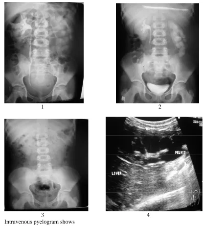
3. INTRAVENOUS PYELOGRAHY
non-visualization of the ipsilateral ureter. Delayed films are important because with better pelvic filling they tend to confirm the diagnosis. In established cases, thickness of the renal parenchyma is reduced and in severe hydronephrosis, a sliver of opacification known as “Shell Nephrogram” or “Rim sign” is seen.(Fig No.11) In severe hydronephrosis, a crescentic collection of contrast medium “Crescent Sign” is observed in the collecting ducts overlying nonopacified calyces. It indicates that some renal function is present.(40)
The drawbacks of IVP include the necessity of dehydration even in infants, which makes it a relatively risky procedure. Of course, a risk of radiation exposure exists, which can be minimised by limiting number of films taken. Problems associated with contrast media exist, such as nephrotoxicity and anaphylactic reactions. The newer nonionic contrast agents that are currently available can reduce these problems.
4.
RETROGRADE PYELOGRAPHY5
. DIURETIC RADIONUCLIDE RENOGRAMDiuretic renogram is a provocative method of evaluating patients found to have dilation of the upper urinary tract in which an obstructive process is suspected. (Fig No.18)
The theoretical basis of this test is two fold: if an obstructive lesion is present, then (a) renal function, more specifically glomerular function may be impaired and (b) a dilated upper urinary tract will retain a larger amount of radionuclide that will not wash out if increased urine flow is generated by the administration of a diuretic.
Three radio pharmaceuticals are primarily used in diuretic renography and their characteristics are linked to their biological activity. Technetium 99m Diethylenetriaminepentaacetic acid (99mTc-DTPA) and technetium 99m- Mercaptoacetyltriglycine (MAG3) are preferentially concentrated by the kidney and filtered by the glomerulus.(41)
tubular cell and is there fore, useful for the detection of differential renal function and clinically significant cortical lesions such as renal scars.
In general, neonates and young infants are placed supine for the study. Ideally, the child should be well hydrated because relative dehydration prolongs parenchymal transit and delays urinary excretion.
MAG- 3 or DTPA is injected intravenously as a bolus. The paediatric dose of MAG-3 is 50mcu/ kg. (43) Subsequently, 4- second posterior images are recorded with a high-resolution collimater. One minute after injection, images of the kidneys are obtained each minute.
Normally, the renal parenchyma is well visualised during the first minute; by 2 (or) 3 minutes, activity seen in collecting system and by 6 to 9 minutes the bladder is visualised.
The activity within each kidney is expressed as a percentage of the total renal counts, and this differential activity is used to compute differential renal function.
To determine whether significant obstruction is present, furosemide is administered intravenously. There are 3 variations in the furosemide administrations.
1. F +20 - furosemide is injected 20 minutes after the injection of tracer. 2. F – 15 - furosemide is injected 15 minutes prior to the tracer.
3. F – 0 - furosimide is injected at the beginning of the study
The most commonly used protocol for diuretic renogram is that in which the furosemide is administered 20 minutes after tracer injection the so-called F+20 protocol. The dose of furosemide in paediatric is 1mg/kg in infants, 0.5mg/kg in children aged 1-16 years.
At completion of the study, a static upright post void image is obtained, especially if the study has been performed supine and persistent tracer pooling seen within the renal pelvis.
Image acquisition must be carried out for at least 20 minutes following furosemide injection to avoid missing flow- dependent intermittent obstruction the so called “ beer drinker’s kidney”.
The renogram curve in this setting may appear to fall normally. In response to furosemide early on, but, as urine flow rates reach maximum, the curve again deflects upward in an obstructive pattern. This produces the “delayed double peak” pattern – Homsy’s sign of intermittent obstruction.(42) Premature termination of the test may result in missing this second upward curve deflection. In these patients, a follow up F – 15 renogram is helpful in confirming the true obstruction. (43). F - 15 renography can be used in patients in whom the diagnosis of significant obstruction is equivocal by F + 20 protocol.
The additional information, which can be obtained, includes clearance half time of the tracer from the pelvicalyceal system, which is used by some workers as a parameter to diagnose obstruction.
The renogram curve
Diuresis renography was pioneered by PH ‘O’ Reilly (1978) and associates as a means of distinguishing between obstructive and non obstructive dilatation.(44)
The characteristics of the uptake and drainage curves fall into four patterns; Type 1: Normal uptake with prompt washout.
Type 2: Rising uptake curve, no response to diuretic. Type 3a: An initially rising curve, which falls rapidly in response to injection of lasix.
Type 3b: An initially rising curve, which neither falls promptly Following the injection of lasix nor continues to rise. This renogram pattern was defined by O’Reilly and Colleagues as equivocal and much of their subsequent work has been aimed at eliminating the type 3b curve
The renogram curve
A. Normal curve shows the three renogram phase.
B. Abnormal curves. These curves are typical for obstructive or nonobstructive dilatation with accumulation of tracer in the collecting system. Impaired renal function with cortical retention of tracer can produce a similar appearance, although the paek parenchymal activity will be lower than normal.(bottom curve) C. In non obstructed collecting system, there is prompt washout of pelvicaliceal
activity after intravenous furosemide.
The renogram curve is a time activity curve describing the transit of tracer through the kidney. The curve is obtained by placing a computer assisted region of interest over the whole kidney or cortex obtaining the counts in the ROI for each period of data acquisition and plotting these counts as a function of time.
The renogram curve is often divided into the period of tracer appearance; tracer extraction and tracer elemination denoted as phase 1, 2 and 3 respectively. Tracer appearance describes the period of blood flow beneath the detector; tracer extraction is proportional to renal plasma flow or glomerular filtration rate according to used tracer. The curve peaks when tracer exits from the kidney at the same rate it is entering the kidney. Normally the intrarenal transit of tracer is less than 5 minutes.
The excreting part of the curve is measure of drainage from the pelvis. Various quantitative parameters are available but the simple one is measuring the half time (T1/2) of excretion. It is generally recognized that a T1/2 < 10 minutes is considered normal, a T1/2 between 10-20 minutes is considered indeterminate and a T1/2 of >20 minutes is likely caused by an obstruction.
technique was also compared with other modalities like perfusion studies described by Whitaker where 85% correlation was obtained. When compared with morphological studies (Goslings and Dixon 1978) the correlation rate was 88%. Factors affecting diuretic renography in the neonate are
Renal maturity Volume of urine in the bladder Renal functioning Outlined regions of interest Hydration status Patient position
Type and dose of tracer Patient movement Dose of diuretic Capacity of upper tract Timing of diuretic administration Severity of obstruction Vesicoureteric reflux Site of obstruction Method of data interpretation
It has become the procedure of choice in the evaluation of suspected obstruction because it is relatively noninvasive, providing information about differential function and reliably diagnosing functional obstruction.
To reduce inter institutional difference the society for fetal urology and society of nuclear medicine council proposed a uniform methodology for performing the diuretic renogram called well - tempered renogram.(45)
To reduce the likelihood of immature renal function interfering with interpretation, infants should be older than 2 weeks. Oral hydration is begun 2 hours before study. Intravenous hydration of 15ml /kg over 30minutes is started as a bolus 15 minutes before injection of the isotope followed by maintenance fluid at a rate of 200ml /kg/24hours.The bladder is catheterised throughout the study.
Diuretic renogram is well tolerated, easily repeatable and appropriate for paediatric patients. It should be done after 4-6 weeks of life.(46)
6.
CT SCAN7. MAGNETIC RESONANCE IMAGING
New developments in MRI technology have made it possible to image kidneys while assessing intracellular metabolic parameters independent of blood flow and tubular function. Relative high cost and the noise during the procedure limit the routine use of MRI for evaluating urinary obstruction in children.(Fig No.15)
8.
ENDOURETERAL SONOGRAPHYBagley and co workers have used endoureteral sonography in the evaluation of the obstructed pelviureteric junction.(47) Catheter based ultrasound probes with single crystal transducers (12.5 Mhz/ 20Mhz) are available in size ranging from 3.5 F to 6.2 F. The transducer is densely radio opaque and can be seen fluoroscopically. The device can be used in conjunction with standard endoscopic equipment. It can provide accurate information of pelviureteric junction anatomy. It can visualise vessels adjacent to the PUJ/ ureter; define the presence of high ureteral insertion including the direction, length and thickness of the septum.
The obstructed PUJ has three distinct patterns seen sonographically. The area of the PUJ can be quite narrow, with a stenotic segment ranging from 1 or 2 to several millimeters.
This can be the only finding or may be associated with the other two patterns of crossing vessels or high insertion, as the presence of one pattern does not exclude the others.
9. PRESSURE PERFUSION STUDY
Whitaker’s test was considered the gold standard for the evaluation of upper urinary tract dilatation.(48) It provides urodynamic evidence of a mechanical obstruction of the upper urinary tract at a given flow rate. With the advent of the diuretic renogram and some of the newer radiopharmaceutical agents, the Whitaker test is not often utilised clinically.
procedure, and its relationship to changes in renal pressure can be significant. Contrast material is given along with the saline solution, making fluoroscopic monitoring of the anatomic site of the obstruction possible .Whitaker’s test
Results are separated into three categories 1. Pressure less than 15 cm H 2 O = non obstructed
2. Pressures of 15 to 22 cm H 2 O = equivocal
3. Pressures greater than 22 cm H 2 O = obstructed
10.
URINARY MARKERS PELVIURETERIC JUNCTION OBSTRUCTION1. Urinary Beta 2 Microglobulin
Disruption of proximal tubular integrity leads to increased urinary concentrations of beta-2-microglobulin (B2M), which normally reabsorbed from
the tubular lumen via phagocytosis and lysosomal digestion.(49)
An increase in urinary concentrations of B2M may indicate tubular
dysfunction as a result of the obstructive insult. Functionally significant obstruction and recovery from obstruction may be determined by following the urinary concentration of B2M.
The potential for B2M to be a marker for significant obstruction is quite
appealing; however, the determination of its levels in obstructed kidneys is not routine, and many different insults other than pelviureteric junction obstruction can lead to increased levels of B2M in the urine. In addition, the immaturity of the nephron and the
high fractional excretion of water in neonates contribute to elevated B2M levels in the
2. N-Acetyl-β- Glucosaminidase
N-acetyl-β-glucosaminidase (NAG) is a tubular lysosomal enzyme present in the urine of children who have various renal diseases. This is currently experimental.(50)
In rats with experimental partial ureteral obstruction, the urinary concentration of NAG increases in the first 2 weeks of obstruction and decreases with the relief of obstruction.
Urinary biochemical markers of renal damage sometimes may aid the diagnosis of clinically significant urinary obstruction.
The assessment of urine for growth factors (eg, epidermal growth factor [EGF], platelet-derived growth factor [PDGF], TGFb, cytokines, p53, p21) and vasoactive substances may be an important adjunct in evaluating obstructive uropathy in the future.(51)
TREATMENT
Goals
symptoms when they exist. There are four therapeutic approaches are available for the treatment of PUJ obstruction
1. Conservative management
2. Temporary diversion of urine (percutaneous nephrostomy) 3. Surgery or endoscopic treatment
4. Fetal surgery or urinary diversion
Conservative Management
Conservative management of pelviureteric junction obstruction is justified in most of the cases during the first year of life. Three conditions are required to treat a unilateral pelviureteric junction obstruction conservatively.
1. The asymptomatic child
2. The pelvic dilatation should be stable or decrease on repeated ultrasonogram
3. The relative function on repeated isotopic studies should be stable or increase.
Temporary Diversion
of bilateral dilated pelvis, unilateral nephrostomy usually improves drainage from both kidneys.
Surgical or Endoscopic Treatment
Indications
1. A symptomatic pelviureteric junction obstruction 2. Declining function of the dilated kidney
3. Increasing pelvic dilatation
4. Bilateral, moderate to severe dilatation of the pelvis
Surgical Treatment
PYELOPLASTY
Historical review
The first PUJ repair was performed by Trendelenburg in 1886. The first successful pyeloplasty is credited to Kuster in 1891. In 1894 Fenzer applied the Heineke- Mickulicz principle to reconstruct the pelviureteric junction. The dismembered procedure of Kuster was modified by Nesbit in 1949 and further modified by Anderson and Hynes by spatulating the ureter and excising the redundant pelvis.
applicable to cases of high insertion and unsuitable for pelviureteric junctions, which were dependent.
Four criteria for success in the repair of a pelviureteric junction obstruction were defined by Foley in 1937 as follows (52)
1. Formation of a funnel 2. Dependent drainage 3. Water tight anastomosis 4. Tension free anastomosis
The following procedures are available to treat pelviureteric junction obstruction 1. Dismembered Procedures
- Open Anderson Hynes pyeloplasty - Laparoscopic pyeloplasty 2. Flap Procedures
- Foley Y plasty - Spiral flap
- Scardino Prince Vertical flap pyeloureteroplasty 3. Ureterocalycostomy
Dismembered Procedures
Dismembered procedures involve excision of pelviureteric junction with an anastomosis of the upper ureter to the dependent renal pelvis. Success rate of this procedure ranges between 90-95%, with failure often due to fibrosis secondary to urinary extravasation or to poor tissue handling.
The Anderson-Hynes pyeloplasty has become the most commonly employed "open" surgical procedure for the repair of pelviureteric obstruction.(53)The principal reasons for the universal acceptance of the dismembered pyeloplasty are
1. Broad applicability, including preservation of anomalous vessels
2. Excision of the pathologic pelviureteric junction and appropriate repositioning
3. Successful reduction pyeloplasty.
This operation is generally easy to perform and can be accomplished by a number of surgical approaches including
1. Anterolateral extraperitoneal approach 2. Posterior lumbotomy approach
Anterolateral extraperitoneal approach
Anderson-Hynes dismembered pyeloplasty, as performed through an anterolateralaproach, is as follows (Fig No.20)
1. The anterolateral incision is a muscle-splitting incision that is made with the patient supine and a roll placed transversely beneath the patient to elevate the flank.
2. Each muscle layer encountered is split in the direction of the muscle fibers until Gerota's fascia is identified by sweeping the peritoneum medially. The fascia is then incised posteriorly over the lateral aspect of the kidney.
3. The renal pelvis is identified by medial retraction of the peritoneum and lateral traction of the kidney.
5. The area of pelviureteric junction is dissected free to allow for a clear area in which to perform the anastomosis. Traction sutures may be placed in the renal pelvis superiorly, medially, laterally, and inferiorly to the pelviureteric junction. Once adequate ureteral length is confirmed and the pathology of pelviureteric junction identified, the ureter can be transected at this level.
6. The ureter is spatulated on the side opposite to the traction suture using Potts tenotomy scissors. The distance over which the ureter is opened is variable, until healthy ureter is encountered, which springs open when forceps are placed into it.
7. A portion of pelvis is excised. It is better to leave too much renal pelvis than too little, especially when resecting along the medial aspect of the renal pelvis. The ureter and renal pelvis are aligned to ensure that the anastomosis can be accomplished without tension. If a nephrostomy tube is to be used, it is placed at this time. An inferior calyx is chosen, preferably where the overlying parenchyma is not too thick.
tissue. The area of the initial anastomosis is critical to ensuring a watertight closure.
9. Before the repair is completed, the renal pelvis is irrigated to remove any blood clots or debris that could obstruct the pelviureteric junction. A drain is placed adjacent to the repair and brought out through a separate stab wound.
10.The kidney is returned to its native position, and perinephric fat if available, is placed over the anastomosis. Wound closed in layers.
11.The transanastomic stent removed usually after 48hours. The drain removed on 4thday.
Anderson-Hynes pyeloplasty
In the following situations, postoperative temporary nephrostomy is advisable. 1. Inflamed renal pelvis (prior nephrostomy, presence of calculus, infection) 2. Thin or hypoplastic ureter.
4. Poor renal function.
5. Solitary kidney or bilateral disease. 6. Transabdominal pyeloplasty.
7. Long segment stricture or distal ipsilateral pathology (VU reflux and when PU junction edema anticipated)
Posterior lumbotomy approach
Posterior lumbotomy approach is more commonly used in infants, small children and lean older preadolescent with normally located operative kidney.(54) The use of muscle splitting rather than cutting makes it almost a minimal invasive procedure. The location of the incision is posterior and in the crease line has a cosmetic advantage.
Procedure
The lateral edge of the lumbodorsal fascia is elevated, and the sacrospinalis muscle is medially retracted. An incision is made through the middle and anterior lamella of the lumbodorsal fascia, taking care not to injure the ileohypogastric nerve. The quadratus lumborum muscle is retracted, exposing Gerota's fascia beneath the paranephric fat, and then this fascia is opened.
The renal pelvis is identified, and several holding stitches are placed in the pelvis. The ureter is identified, a holding stitch is placed in the ureter, and the surgeon proceeds with the dismembered pyeloplasty as usual manner.
After pyeloplasty, a single muscle fascia layer bringing the lumbodorsal fascia back together again does closure.
Flank Approach
The patient is placed over the kidney rest in a flank position; the kidney rest is elevated and the operating room table is flexed. The skin incision is made off the tip of the 12th rib, or, if necessary, a supracostal 12th rib incision is made.
The external oblique and latissimus dorsi muscles are divided. Next the internal oblique and serratus posterior inferior muscle are divided. The transversalis muscle is often thin and can be divided with digital dissection. The peritoneum is identified and retracted medially.
Gerota's fascia is then encountered and opened longitudinally to gain exposure to the perinephric space. After identification of the renal pelvis and the ureter, a dismembered pyeloplasty can be performed as described earlier.
Flap Procedures
The spiral flap is outlined with its base situated obliquely on the dependent aspect of the pelvis. The base of the flap is positioned anatomically lateral to the pelviureretic junction and should lie between the ureteral insertion and the renal parenchyma. The flap is then spiraled posteriorly to anteriorly or vice versa. The medial line of incision through the flap is carried down completely through the obstructed proximal ureteral segment into normal caliber ureter.
The length of the flap is determined by the site of the apex, which in turn should be a function of the length of proximal ureter to be traversed. In order to insure vascular integrity of the flap, however, the ratio of flap length to width should not exceed3-1. The flap is developed with fine scissors and the apex then rotated down to the inferior most aspect of the ureterotomy. The anastamosis is completed over an internal stent, using fine absorbable sutures.
Scardino and Prince described a vertical flap that can be used in the situation of a dependent pelviureteric junction with a large, square-shaped extrarenal pelvis. (56)
In contrast to a spiral flap, however, the base of the vertical flap is situated horizontally rather than obliquely between the pelviureteric junction and renal parenchyma. The flap itself is formed by a convergence of two straight lines from the base vertically to the apex on either the anterior or posterior aspects of the renal pelvis.
As for the spiral flap, the height of the apex determines the length of flap obtained which again is a function of the length of proximal ureter to be bridged. Medially, the ureterotomy is carried through the proximal ureter completely through the strictured area, continuing a few millimeters into normal caliber ureter. The apex of the flap is rotated down and joined to the inferior most aspect of the ureterotomy. The flap is closed over an internal stent by approximating the edges with fine absorbable suture.
Foley -Y- Plasty
In this procedure, the limbs of the Y are widely separated one on the anterior and one on the posterior aspect of the renal pelvis. The laterally placed portion of the pelvis then drops inferiorly. The lateral border of the ureter is opened longitudinally through the pelviureteric junction to at least 1cm below the obstruction where the ureteral caliber is normal. The midportion of the pelvic flap is sutured to the inferior margin of the ureterotomy converting the incision to a V.
Ureterocalicostomy
This procedure generally reserved for situations in which dependent renal drainage cannot be established by conventional techniques. It is primarily used for horseshoe kidney, fusion anomalies and in the repair of secondary pelviureteric junction obstruction. The inferior calyx is more dependent than pelvis in the Horseshoe kidney and it may be a more physiologic procedure than pyeloplasty.
the ureter from entrapment by contracting fibrosis of the renal cortex. (57) There should be no tension on the anastomosis and there should be no renal parenchyma below the level of the repair.
Nephrectomy
Because the recovery potential of the kidney is greater in children, extreme conservation is justified. Salvage pyeloplasty should be considered as renal function shown in renal scintigraphy can recover.(58) During surgery, the renal cortex should be assessed. The severe cystic dysplasia is an indication for nephrectomy, otherwise every effort be made to salvage the kidney.
ENDOSCOPIC MANAGEMENT OF PELVIURETERIC OBSTRUCTION
Historical review
The idea of endopyelotomy was conceived in 1909 by Albarran in France describing the technique of incising the narrowed pelviureteric junction as URETEROTOME EXTERNE in which a stent was left in.(59) The incised ureter heals over time, which is something akin to internal urethrotomy in stricture urethra. Davis 1943 popularised this technique as intubated ureterostomy in which few loose stitches were placed.(60)
Wickham and Miller first described a percutaneous approach to incising the pelviureteric junction termed pyelolysis. Subsequently Arthur smith popularized this technique renaming it endopyelotomy.(61,62)
With the advent of endourological techniques the following minimal invasive procedures are being applied to pelviureteric junction obstruction
1. Antegrade nephroscopic endopyelotomy 2. Retrograde ureteroscopic endopyelotomy 3. Balloon dilatation with cutting device
1. Antegrade Endopyelotomy
Percutaneous techniques were introduced in the early 80’s for the management of nephrolithiasis. Subsequently in 1983 reports began to appear in the literature for endoscopic treatment of pelviureteric obstruction. Success rates vary from 50% to 95% and remain inferior to that of open pyeloplasty.(63) Success appears to be dependent on correct patient selection rather than the type of operation.
Patient Factors
1. Age
Neonates and infants may not be suitable candidates for endourologic management. In addition to technical difficulties, the radiation exposure may be extensive. Children in the adolescent age group are more suitable as the anatomy and caliber of the ureter is similar to the adult urinary tract.
2. Hydronephrosis
Van Cangh (64) in his series of adult patients found the success rate of
endopyelotomy fall from 95% to 77% in the presence of massive hydronephrosis.
3. Crossing Vessels
4.Primary versus Secondary PUJ Obstruction
A review of most recent series including that of Van Cangh and co workers, it appears that secondary UPJ obstruction responds better to antegrade pyelotomy.
In older child or adolescent patient with primary pelviureteric junction obstruction and favorable factors (e.g. good ipsilateral renal function and only mild to moderate hydronephrosis), endopyelotomy is the preferred procedure. Endopyelotomy is the initial therapy for secondary PUJ obstruction in all-pediatric patients
Endopyelotomy
The pelviureteric junction can be incised with various devices. 1. Cold knife.
2. Electrocautery.
3. Laser – Nd YAG / KTP / Holmium
The Incision
This is made on the lateral side and extends down the ureter for a centimeter beyond the area of narrowing. Proximally it extends 1-2 cm up the pelvis and should go through all layers till the retroperitoneal fat is visualised. Either a graduated or a standard stent is passed down the guide wire into the bladder. A nephrostomy tube is placed in the pelvis and remains for 48 hours. A nephrostogram is usually performed prior to removal and the stent is left indwelling for 3-6 weeks.
2. Retrograde Ureteroscopic Endopyelotomy
3.
Retrograde Balloon Cautery Incision of the Pelviureteric
Junction (Acucise Device)
A special balloon catheter with an inbuilt cutting device is used in this technique. Once the waist disappears (after 10 minutes of balloon dilatation) cutting current is activated for 3 seconds to get the desired endopyelotomy. Overall success in this method is around 78% and hospital stay is around 1 day only. In this method the retrograde balloon is used to define the area of stenosis and to carry the cutting wire into the area to be incised. It is designed to accept a maximum of 2.5ml of fluid. The electrically active surface on the cutting wire is 2.8cm in length and 150µm in diameter.
Balloon inflation with contrast indicates the narrowed PUJ segment by presenting as a waist. Then the cutting device which is positioned across the PUJ in such a manner that it points posterolaterally, is activated by cutting current for 3 seconds, so as to cut the stenosed PUJ precisely but surely in one plane only avoiding the vessels.
Patients return 4 weeks following stent removal for postoperative intravenous pyelography to confirm efficacy of the endopyelotomy.
Postoperative bleeding, necrosis of ureter, urinoma, hematoma and urinary tract infections are the frequent complications of this procedure.
Advantages of this technique include 1. Short operating time.
2. Ability to perform it as a day care procedure.
3. No special instrumentation required. The balloon can be passed through a standard cystoscope.
4. The device can also be used through an antegrade percutaneous nephrostomy tract.
4. Balloon Dilatation / Endoburst
the rupture and a 6 to 8 F stent is positioned over the guide wire on removal of the balloon catheter.
Tan reported on the early results of balloon dilatation for primary pelviureteric junction obstruction in 10 children. The age range was 3 months to 9 years and at 22 months follow up had a success rate of 70%.(66)
Pros and cons in endopyelotomy
The concern in high insertion pelviureteric junction endopyelotomy may not yield good results. It is not a suitable method in vessels crossing pelviureteric junction where there can be torrential bleeding. Lingman et al 1993 reported antegrade approach with 100% success when followed for one year. Figenshau’s 4 years follow up shown 100% success. (67)
In children with secondary pelviureteric junction obstruction, the results are more encouraging. It approaches nearly 100% as against available (70 to 95% success) results in primary pelviureteric junction obstruction.
For primary pelviureteric junction obstruction with pelvic size less than 60ml volume, if vessel crossing pelviureteric junction is ruled out by spiral CT scan or endoluminal US scan, definitely endopyelotomy can be attempted.
LAPAROSCOPIC PYELOPLASTY
The latest technique is laparoscopic pyeloplasty where dismembered pyeloplasty is done as in open with the advantage of minimal invasive nature. Laparoscopic pyeloplasty was first reported by Schuesler.(68)
Approach of laparoscopic pyeloplasty
The exposure of pelviureteric junction can be done either by 1. Transperitoneal approach
2. Retroperitoneal approach
By transperitoneal approach pelviureteric junction is exposed by mobilising the colon. But problems in transperitoneal approach are
1. Transgressing the peritoneal cavity carries the potential risk of adhesion.
2. Anastomotic leak can cause intraperitoneal spillage, paralytic ileus and septicemia.
Technique
The principle of the dismembered pyeloplasty is followed in general. The preoperative enema to empty the colon, nasogastric tube to decompress the stomach and Foleys catheter to keep the bladder empty are helpful to improve the available space in the peritoneal cavity in children.
Following induction with general anaesthesia, cystoscopy and retrograde pyelography are performed to confirm the diagnosis and a long indwelling ureteral stent is passed
The patient is placed in a 45-degree lateral decubitus position and secured to the operating table. Insufflation is performed through a veress needle and three laparoscopic ports are passed into the peritoneal cavity.
between the vessels and collecting system should be divided. The renal pelvis is transected circumferentially above the PUJ and the proximally spatulated through the level of the PUJ laterally. Care should be taken not to cut the ureteral stent.
If the crossing vessels are present, the ureter and renal pelvis are transposed to the opposite side of the vessels prior to completion of the anastomosis. All intracorporeal suturing is performed using the endostich device and 4’0’ poyglycolic acid suture. A corner stitch is placed through the most dependent portion of the renal pelvis and through the corresponding corner of the spatulated ureter. The posterior anastomosis is then performed using multiple interrupted sutures followed by the anterior anastomosis.
As in true open pyeloplasty, the goal of surgical repair is to create a dependent, tension free, water tight anastomosis.
Following completion of anastomosis, a 5mm closed suction drain is placed through a posterior stab incision into the perinephric space adjacent to the PUJ. Hemostasis is confirmed, the CO2 is evacuated and port sites are closed.
RETROPERITONEAL PYELOPLASTY
deepened by a blunt dissection with a hemostat and index finger to reach the peritoneum.
Then, peritoneum is pushed away from abdominal wall either with index finger or Hegar dilator (size 12 or 14). The inflating balloon is inserted through this and kept between the peritoneum and muscles. It is inflated with co2 or saline is put to create a space beneath the peritoneum. Balloon is kept for few minutes and is deflated.
A trocar either a Hassons type or a trocar with self-retained balloon at the tip, is chosen to prevent leak from the trocar site. After verifying the correct entry of retroperitoneal space by telescopic examination, two additional ports are made one near the costal margin and another near ileac crest. By blunt and sharp dissection pelviureteric junction is exposed and pyeloplasty is done in the usual way.(100)
In retroperitoneal approach, the main advantage is avoiding entry of peritoneal cavity and thereby urinary leakage related problems can be avoided.
Complications of laparoscopic pyeloplasty.
The complications are similar to open pyeloplasty like anastomotic leak, septicemia in the immediate postoperative period and anastomotic stenosis later.
With experience and improvements in instrumentation and innovative suturing techniques laparoscopic approach will become the procedure of choice.
ROBOTIC PYELOPLASTY
Robotics is another exciting and evolving area for minimally invasive surgery in children. This new technique provides excellent three dimensional visualisation, unprecedented control of endocorporeal instruments, and an ergonomic surgeon’s position. Robots may advance minimally invasive surgery by allowing paediatric urologist with limited laparoscopic experience, to rapidly master the endocorporeal skills necessary to treat pelviureteric junction obstruction.(70,71)
FETAL INTERVENTION
around 24to28 weeks, fetal renal function is adequate, and there are no associated serious anomalies. The male child with posterior urethral valves with good renal function is the usual candidate. Mild uropathy which worsens on serial scans as shown by increasing dilatation or decrease of renal function is also an indication for antenatal decompression. The percutaneous vesicoamniotic shunting (in bladder outlet obstruction),open vesicostomy and open pyelostomy can be used for antenatal decompression.
PYELOPLASTY FOR SPECIFIC ANATOMIC DERANGEMENT
1. Pelviureteric junction obstruction in the Horseshoe Kidney
Because of concern, regarding angulation and obstruction at the isthmus, division of the isthmus and nephropexy to allow more dependent drainage of the PUJ have been proposed.
Ureterocalicostomy may be considered as an alternative to dismembered pyeloplaty for repair of even primary PUJ obstruction in horseshoe kidneys. It permits dependent drainage and avoids any problems related to the isthmus.(74)
Newer minimally invasive techniques have been successfully utilised for PUJ obstruction in horseshoe kidneys, albeit, in small numbers of patients. Antegrade endopyelotomy has been used successfully in seven of eight reported cases.
Laparoscopic pyeloplasty has been successfully applied to horseshoe kidneys (J.H.Ross,H.Winfield 1997). The horseshoe kidney is particularly amenable to laparoscopic approach; because the anteriorly placed PUJ is easily exposed and manipulated with this technique.
2.
Pelviureteric junction obstruction in the lower segment of duplexVesicoureteric reflux is often seen in the lower segment of a completely duplicated system and is some times associated with pelviureteric junction obstruction. The following procedures can be done
1. When the duplication is complete dismembered pyeloplasty may be performed.
2. When the lower pole ureter of a partially duplicated system is short, the entire lower pole ureter is excised and a lower to upper pole pyeloureterostomy is performed.
3. When there is marked hydronephrosis of the lower-pole moiety, particularly if the pelviureteric junction obstruction is difficult to expose, then an ureterocalicostomy may be performed.
4. If the lower pole is poorly functioning, a lower-pole heminephrectomy is appropriate.
5. If a dysplastic upper pole segment is present, owing to an ectopic ureter or ureterocele, if may be excised at the time of the lower pole repair. Alternatively, an upper to lower pole ureteropyelostomy may be performed at the time of the lower pole pyeloplasty.
3. Pelviureteric junction obstruction in the pelvic kidney
include pelvic (55%)(Fig No.14), crossed (32%), lumbar (12%), and thoracic (1%). Approximately half of such kidneys are hydronephrotic. This results from a variety of causes, including PUJ problems (37%), VUR and lower tract problems (26%) and hypoplastic adynamic segments at the UVJ (15%). All cases require lower tract imaging and accurate anatomical upper tract imaging. Surgical correction requires special considerations. Position, incision, and approach must be individualized; in a majority a modified Gibson incision allows an extraperitoneal approach. Open pyeloplasty is usually successful. Experience with less invasive techniques is limited.
ANTENATALLY DETECTED HYDRONEPHROSIS
The clinical presentation of the pelviureteric obstruction has dramatically changed since the advent of maternal ultrasonographic screening. Before the routine fetal ultrasound the commonest presentation was abdominal flank mass.(76) 50% of abdominal masses in newborns are of renal origin with 40% being secondary to pelviureteric obstruction. The newborns are also present with abdominal pain, urinary tract infections, irritability, vomiting, and failure to thrive.
Associated Anomalies
10% of the PUJ is associated with vesicoureteric reflux. The association of multicystic dysplastic kidney and contralateral pelviureteric junction obstruction is well known. The dysplastic kidney reflects the extreme end of the clinical spectrum of pelviureteric junction obstruction. Bilateral pelviureteric junction obstruction is 10-36% of cases.(77)
Differential Diagnosis
Dilatation of urinary tract can be secondary to obstructive or nonobstructive causes. The various obstructive causes include pelviureteric junction obstruction (44%), VUJ obstruction (21%), multicystic dysplastic kidney, ureterocele/ectopic ureter, duplicated collecting system (12%), posterior urethral valves (9%) and hydrometrocolpos.
The non obstructive causes include physiological dilatation, vesicoureteric reflux (14%), prune belly syndrome, renal cyst and megacalicosis.
Indications for maternal fetal ultrasonography
A systematic approach to the prenatal diagnosis of urinary tract abnormalities improves the yield of the ultrasonographic examination. (Schlussel RN, MandellJ et al) (78)
This includes the following steps 1. Assessment of fetal size and maturity.
2. Assessment of amniotic fluid volume (AFV), which is a semi quantitative technique. Currently, there are two methods to estimate AFV; amniotic fluid index (AFI) or measurement of the single deepest vertical pocket of amniotic fluid. Current ultrasonographic criteria for oligohydramnios (actual AFV < 500 ml) include an AFI of 5.0 cm or less and a two - diameter amniotic fluid pocket of less than 15 cm. Both methods are poor predictors of oligohygramnios.
3. Identification of the gender of the fetus.
4. Localisation and characterisation of urinary tract abnormalities (bladder volume, kidney size, anteroposterior diameter of the renal pelvis, echotexture of the kidney, and the presence of cortical cysts).
5. Identification of associated abnormalities.
6. Monitoring the detected lesions and their impact on the overall health of the fetus.
of renal pelvis to AP diameter of kidney > 0.5 has been considered significant. Grigon et al (79) graded the fetal hydronephrosis into 5 grades as follows
Grade I – detectable renal pelvic dilatation. Grade II – dilatation greater than 1cm.
Grade III- IV – further degree of pyelectasis with dilatation greater than1.5cms.
Grade V – association with atropic cortex.
Dilatation of the collecting system can occur in the absence of obstruction and is termed as physiological hydronephrosis.
Typically, the ureter is of normal caliber and is not seen.(80) But if it dilated the size of ureter is also assessed ultrasonographically and graded 1-3 according to ureteral size width < 7mm, 7-10mm, >10mm respectively.(81)
Harrison et al suggested that proportion of more than 1:2 of the pyelon width to the kidney width is pathological and may be diagnosed as fetal hydronephrosis.(82)
The Society for fetal urology grading system is as follows Grade 0 – Normal kidney with no hydronephrosis.
Grade 1 – Slightly dilated renal pelvis without caliectasis. Grade 2 – Moderately dilated pelvis with mild caliectasis.
Grade 3 – Large renal pelvis, dilated calyces, and normal renal parenchyma. Grade 4 – Very large renal pelvis, large dilated calyces, with
thinning of the renal parenchyma.
P. A. Dewan et al describe a new sign in ultrasound examination that may help to identify those fetuses who have high intrarenal pressure and therefore justify more aggressive management, while obviating the need for intervention for those in whom it is not present. The egg-shell sign consists of a thin crescent of increased echogenicity over a distended calyx and, in this case, was documented to be associated with other features of raised intrarenal pressure.(101)
AMNIOCENTISIS AND ASSESSMENT OF FETAL URINE FUNCTION
absence of corticomedullary junction differentiation, which suggest bilateral hydronephrosis and dysplasia of the kidneys.
The goal is to identify fetuses that are risk while in utero, of total renal destruction and pulmonary hypoplasia when there is a reasonable hope of beneficial treatment. This test currently carries of two types.
1. Evaluation of amniotic fluid fetal urine to assess fetal tubular function. 2. Evaluation of proteins in fetal serum or urine to assess
fetal glomerular filtration rate.
Between 16 and 21 weeks of gestation, the fetal urine normally becomes progressively more hypotonic because of selective tubular reabsorption of sodium and chloride in excess of free water. The most quoted values for fetal urinary electrolytes and osmolatity abnormalities indicative of an impaired renal function in the fetus with detectable upper tract dilatation are as follows
1. Osmolality of less than 210mosm/l
5. Urinary calcium levels greater than 8mg/dl (normal <8mg/dl) are the most sensitive indication of renal dyssplasia(100%) but have a demonstrated specificity of only 60%.
6. Beta 2 microglobulin elevation in fetal urine of greater than 4mg/l is pathological.
Management
Antenatal Management
Fetuses with unilateral dilatation of upper tracts and a normal contralateral kidney are simply observed with serial ulrasound examinations at 1-2 weeks intervals till term.
Fetuses with bilateral hydronephrosis who are at higher risk for obstructive uropathy and in whom the Amniotic fluid index should be carefully monitored. Once the oligohydromnios develops, fetal urine sampling is the next step to be evaluated.
Once a fetus with salvageable function is selected, early delivery is considered if it is more than 32 weeks of gestation. If it is between 20-32 weeks various fetal interventions are considered. It is less than 20 weeks with
Scheme of management for antenatal diagnosed bilateral hydronephrosis Bilateral hydronephrosis
Normal amniotic fluid volume oligohydramnios ↓
Detailed sonography and Fetal urine analysis
↓
Ranal dysplasia Poor renal function
Reasonable renal function
Serial monitoring and termination of ↓ ↓ Postnatal management pregnancy
Mature lung immature lungs ↓ ↓
Early delivery in utero decopression
Postnatal evaluation and Management
dilatation is specifically looked for. The bladder is evaluated for size, wall thickness, presence of diverticulum or ureterocele and dilatation of the posterior urethra.
Next VCUG should be performed to rule out posterior urethral valve, a bladder diverticulum, or vesicoureteric reflux. Even if the ultrasound is normal a VCUG should be performed, because reflux may be the cause of fetal hydronephrosis.
If the ultrasound and VCUG are normal, then only a follow up ultrasound in 6-8 weeks is necessary.
If the postnatal sonogram shows grade 1 or 2 hydronephrosis and the VCUG is normal, then the pelvicalyceal dilatation is due to physiological. These children should follow with ultrasonogram in 3-6 months period.
If the sonogram shows grade 3 or 4 hydronephrosis and there is no reflux, the upper tract must be evaluated further with T99 MAG 3 or DTPA diuretic renogram at 4-6 weeks of age.