COLORECTAL MALIGNANCIES
A COMPREHENSIVE STUDY
Dissertation Submitted
for the Degree of
MASTER OF SURGERY
Branch I
(GENERAL SURGERY)
THE TAMIL NADU
Dr.M.G.R. MEDICAL UNIVERSITY
CHENNAI
COIMBATORE MEDICAL COLLEGE
COIMBATORE
CERTIFICATE
Certified that this is the bonafide dissertation done by Dr.L.SANKAR
and submitted in partial fulfillment of the requirement for the
Degree of MASTER OF SURGERY Branch I (GENERAL SURGERY)
of The Tamil Nadu Dr.M.G.R. Medical University, Chennai.
DATE : UNIT CHIEF
DATE : PROFESSOR AND HEAD DEPARTMENT OF SURGERY COIMBATORE MEDICAL COLLEGE
DATE : DEAN
DECLARATION
I solemnly declare that this Dissertation on “COLORECTAL
MALIGNANCIES A COMPREHENSIVE STUDY” was done
by me at Coimbatore Medical College Hospital, Coimbatore under the
guidance and supervision of DR.A.RAMA MOORTHY, M.S.
Place:
Date:
ACKNOWLEDGEMENT
I wish to thank our Dean DR.KALANITHI, M.D., for having
allowed me to conduct the study in this hospital.
I am grateful to Professor and Head of the Department of
Surgery Prof.DR.K.P.ARUN KUMAR, M.S., for his excellent,
expert advice and help in preparing this dissertation.
I am greatly indebted to my unit chief Prof.DR.A.RAMA
MOORTHY, M.S., for his excellent guidance and generous help in
the preparation of this dissertation. Without his guidance and
encouragement this work would not have been completed.
I thank all the surgical unit chiefs Prof.DR.PERUMAL
RAJAN, M.S., Prof.DR.B.EASWARAN, M.S., Prof.DR.PREM
THAMARAI SELVI, M.S., Prof.DR.G.S.RAMACHANDRAN,
M.S., for permitting me to carry out this study in their respective units.
I extend my sincere thanks to all Assistant Professors, Surgical
Department, with special thanks to my Unit Assistant Professors.
Last but not the least I express my gratitude to all the patients
CONTENTS
S.No TITLE PAGE NO
1 2 3 4 5 6
INTRODUCTION AND HISTORICAL DATA
AIM OF STUDY
REVIEW OF LITERATURE
A) SURGICAL ANATOMY
B) SURGICAL PHYSIOLOGY
C) INCIDENCE / EPIDEMIOLOGY
D) PATHOLOGY AND SPREAD
E) STAGING AND CLASSIFICATION
F) CLINICAL FEATURES
G) INVESTIGATIONS
H) DIAGNOSIS AND DIFFERENTIAL DIAGNOSIS
I) TREATMENT
MATERIALS AND METHODS
SUMMARY AND RESULTS
CONCLUSION
BIBLIOGRAPHY
PROFORMA
MASTER CHART
INTRODUCTION AND HISTORICAL DATA
Colorectal cancers though more common in the west are on the
increase in our country for the past decade. The early detection of this
disease is of paramount importance in its outcome. Few topics in
cancer research have engendered more excitement than the recent
discovery of identifiable genetic defect in patients with inherited as
well as sporadic form of colorectal carcinoma.
There are evidences that neoplastic disease has affected humans
since prehistoric times. Mummies from Pre-Columbian, Peru of 2400
years ago as well as Egyptian mummies from 3000 B.C. have
metastatic skeletal deposits. It was Hippocratic (460 – 370 B.C.) who
first propose a theoretical framework to explain cancer invasion.
The Cellular etiology of cancer was first described by Johannes
Peter Mueller in 1828. The following year Joseph calrude Reaemer
proposed that invasion and distant spread were the result of
translocation of cells and he coined the term metastasis. The first
successful resection of colonic growth was performed by Reynoard
After Billroth, Czerny and Mikulicz, the pioneers in abdominal
surgery familiarized, the technique of intestinal resection and
anastamosis, increasing number of colonic resection were attempted.
The combined operation involving abdominal and perineal phases for
excision of the rectum was first performed by Czerny (1883). But it
was undoubtedly the work of Ernest Miles (1908) who established the
abdomino perineal operation.
Cuthberk Dukes (1935) classified carcinomas of rectum into 3
stages and explaining macroscopic variations. These are widely used
by pathologists with minimal changes even now for colorectal cancer
staging Paul of Liverpool (1895) and Mikulicz of Brestan (1903)
devised extra peritoneal resection of carcinoma colon and popularized
the technique in America.
Halstead (1895), Shoemaker (1921), Rankin (1928) and
Wangensteen (1940) developed various method of anastamosis by
which it was hoped to carry out resection and anastamosis in an
entirely sterile manner without opening the bowel lumen till union was
completed. But is was later pointed out by Moynihan that the factor
responsible for sepsis is not contaminated during the operation itself
Whipple (1931) and Turner (1937) favoured intraperitoneal
resection with temporary caecostomy in order to relieve the tension on
the suture lines. Devine (1931) developed preliminary defunctioning
colostomy which helped mechanical cleansing of the distal bowel.
After advent of strong intestinal antiseptics reliance was paced
on them entirely and a primary colostomy was entirely omitted. Lloyd
Davis Morgan and Yollinger (1953) carried out resection with
immediate anastamosis without any form of proximal decompression.
In their series of 109 cases, there were only 3 postoperative death and
none of them due to sepsis.
In recent years, the trend is towards preparation with
mechanical cleansing using balanced salt solutions containing osmotic
purgatives in them with antibiotics, orally or IV. This requires only
single day preoperative preparation.
Surgical resection remains the mainstay of treatment for
colorectal cancer. Radiotherapy and chemotherapy are used as
adjuvant therapeutic options. Turnball at the Cleveland clinic
recommended a no touch technique in which vascular and mesenteric
division was first undertaken, thereby isolating the tumour.
The role of gene and their abnormality are being studied
accumulation of various genetic defects in the form of deletion,
translocation etc. These may help us to find appropriate diagnostic
tool to look for such aberration and early prevention of cancer
progression.
Colorectal surgery had advanced a lot with introduction of endo
AIM OF STUDY
The study was undertaken to find out the pattern of
Incidence - age, sex and site wise
Risk factors
Modes of presentation
Treatment modalities
Adjuvant therapy settings
Follow up of colorectal carcinoma in Coimbatore Medical
SURGICAL ANATOMY OF LARGE
INTESTINE COLON and RECTUM
The large intestine extends from end of ileum to anus and
comprises of the caecum (with appendix) colon, rectum, anal canal,
measuring between 110-170 cm in length1 (on an average 135 cm
long). The caliber is greatest at its commencement at the caecum and
gradually diminished as it is traced distally, but again becomes more
dilated in the lowermost part of rectum just above the collapsed anal
canal.
EMBRYOLOGY
The large intestine develops form both mid gut and hind gut.
Midgut portion extends form caecum to the proximal 2/3 of transverse
colon supplied by superior mesenteric artery.
Hindgut – from distal 1/3 rd of transverse colon to proximal
anus supplied by inferior mesenteric artery. The distal anal canal is
ectodermal in origin and supplied by internal pudendal vessels.
Large guts starts developing by fifth week of gestation and is
completed by eight week of gestation when the anal membrane
over 4 weeks assuming final anatomic position by 10th week of
gestation.
ANATOMY
The Caecum
The caecum lies in the right iliac fossa, app 6 cm in length and
7.5 cm in breadth2. Proximally becomes ascending colon at its
junction with terminal ileum guided by a valve which prevents reflux
(contains muscle).
It lies on iliac and psoas muscle and on genitofemoral, lateral
cutaneous nerve of thigh. Its exact position is variable, may extend
into true pelvis. It is almost completely enveloped by peritoneum but
devoid of mesentery3 and often it is attached to iliac fossa medially
and laterally.
THE ASCENDING COLON
It varies from 10-20 cm (avg. 15 cm app). It lies on iliacus -
muscle, iliac crest, quadratus lumborum and crossing lateral cutaneous
nerve of thigh, ilioinguinal and iliohypogastric nerve. It is usually
covered with peritoneum on all 3 sides except posteriorly where it is
fixed to post abdominal wall. Sometimes it may be fixed by a short
mesentery. It ends at hepatic flexure where it turns left on the lower
THE HEPATIC FLEXURE
At this point the ascending colon turns sharply medially and
slightly forwards and downwards just below the right lobe of liver and
overlapped by it and posteriorly lies on lower aspect of right kidney.
THE TRANSVERSE COLON
It is the longest of all, varying from 40 cm – 70 cm in length
extending from hepatic flexure to splenic flexure forming a dependent
loop between the points. It is suspended by transverse mesocolon
which is attached to descending part of duodenum, lower aspect of
body of pancreas and anterior surface of left kidney.
It contains middle colic vessels and branches of left colic artery,
right colic artery and lymphatics. Its posterior relations from right to
left are anterior surface of descending duodenum, small intestine, part
of left kidney. Just below the spleen it turns down to form splenic
flexure.
THE SPLENIC FLEXURE
This flexure lies at the junction of the transverse colon and the
descending colon. Here the colon bends downwards and backwards.
This flexure lies behind the stomach, and below the anterior end of the
spleen on the lower part of the left kidney and diaphragm. The
THE DESCENDING COLON
It extends from splenic flexure to rim of true pelvis close to
inguinal ligament from where it continues as sigmoid colon measuring
25 cm. Usually it is retroperitoneal. It rests on the same muscle and
related to the same nerve as ascending colon.
At anterior superior iliac spine it turns medially, superior to
inguinal ligament and lies on femoral nerve, psos muscle, genital
vessels, becomes sigmoid colon anterior to external iliac vessels.
THE SIGMOID COLON
It is the most variable part in length (40 - 80 cm) and mobility.
It extends upto rim of true pelvis where it becomes the rectum and is
suspended by sigmoid mesocolon a long mesentery with short base.
THE RECTUM
It lies in the true pelvis measuring about 12-15 cm with a
diameter of 4 cm when empty. It is dilated in the lower part to form
ampulla of rectum.
If follows curve of sacrum and coccyx runs anteriorly, inferiorly
to central perineal tendon lies on levator ani muscles, anococcygeal
ligament. It ends posterior to central perineal tendon and to the apex
If follows the curve of sacrum, coccyx in saggital plane. In
coronal plane it is ‘S’ shaped giving rise to prominent folds in the
lumen known as Houston’s valves. The relationship of pelvic
peritoneum to rectum is of considerable surgical importance. The
upper third has a complete peritoneal investment except for a thin strip
posteriorly where peritoneum is reflected as the two leaves of thick
mesorectum. As rectum descends into pelvis, the uncovered portion
becomes wider until only anterior aspect has a peritoneal coat in
middle 1/3 of rectum. This peritoneum gets reflected forward in the
bottom of rectovesical pouch or rectouterine pouch leaving lower third
of rectum extra peritoneal. Posteriorly the pelvic fascia is thickened to
form fascia of Waldeyer separating rectum from sacrum, coccyx,
blood vessels, and nerves. Anteriorly separated by the fascial layer
known as Denon villier’s fascia.
The upper 2/3 of rectum is separated from pelvic fascia by
posterior cushion of areola tissue which becomes circumferential
below rectovesical / rectourterine pouch carrying blood vessels and its
lymphatics known as mesorectum.
THE ANAL CANAL
It is 3-4 cm long extends from the anorectal junction to the
BLOOD VESSELS OF THE LARGE INTESTINE
(A) ARTERIES
sphincters which keep the lumen closed in the form of an antero
posterior slit, posteriorly anococcygeal ligament separate it from
coccyx, while anteriorly perineal body separates it from membranous
urethra, penile bulb or lower vagina laterally it is releated to
ischiorectal fossa. Its whole length surrounded by sphincters which
keeps it closed.
The mucosa of canal consists of an upper mucosal and lower
cutaneous part, the junction being marked by line of anal valves about
2 cm from anal orifice known as Dentate line or Pectinate line.
BLOOD SUPPLY AND LYMPHATIC DRAINAGE
These two are important subjects in relation to malignancy and
its treatment.
BLOOD SUPPLY
The main arteries supplying the colon, rectum are superior
mesenteric artery, inferior mesenteric artery, middle, inferior rectal
arteries. The caecum, ascending colon, hepatic flexure and proximal
two thirds of transverse colon derives blood supply from superior
mesenteric artery originating from aorta at L2 level via ileo colic, (R)
colic, middle colic vessels. The distal third of transverse colon,
splenic flexure, descending colon, sigmoid and upper third of rectum
colic, sigmoid branches and superior rectal artery. The distal two
thirds of rectum, anal canal get blood supply via middle, inferior rectal
artery of internal iliac artery.
The main colic arteries proceed to colon and bifurcate to form
branches which unite to form arcades an inch or so from mesenteric
border, so that a continuous chain of communicating vessel is formed.
This is the marginal artery from which the ultimate branches to the
colon, the vasarecti are distributed.
These branches ramify between and supply muscular layers,
divide into small submucosal rami and enter the mucosa. The
marginal artery is responsible for bringing the area of supply of the
superior mesenteric artery into communication with that of inferior
mesenteric by connecting the descending branch of the middle colic
with the ascending branch of the left colic by means of long
anastomosis of colon.
The venous drainage follows its arterial blood supply and
empties into portal venous system. The inferior mesenteric vein
diverges from artery and passes behind pancreas to drain into splenic
LYMPHATIC DRAINAGE
The entire colon and rectum are drained by a large number of
lymph nodes numbering 70-100 which are present as a series draining
into a principal nodal group.
A. INTRAMURAL LYMPHATICS
Throughout colon and rectum, continuous lymphatic plexus in
the submucous and subserous layers of the bowel wall are inter
connected and drain into extramural lymphatics.
B. EXTRAMURAL LYMPHATICS OF COLON
These consists of lymphatics channel and glands which are
divided into 4 groups.
Epicolic – Minute nodes on colonic wall, sometimes in appendices
epiploicae
paracolic – Along the medial borders of ascending colon, descending
colon and mesenteric borders of others.
Intermediate colic – Nodes like along right, middle, left ileocolic
arteries
Preterminal colic – Nodes adjoining the main trunks of superior
inferior mesenteric arteries near their corresponding pre-aortic nodes.
groups and hence via efferent channels to thoracic duct into internal
jugular vein.
C. EXTRAMURAL LYMPHATCS OF RECTUM
Likewise lymphatics drain in to pararectal group in the wall of
rectum, then to intermediate group around main arteries and then to
nodes near origin of main vessel.
1. Lymphatics from more than upper half of rectum drain along
the superior haemorrhoidal and inferior mesenteric vessel into aortic
gland after passing through the para rectal and sigmoid nodes.
2. Laterally along middle haemorhoidal vessels on either side to
ischiorectal fossa and thence to internal iliac glands via inferior rectal
and internal pudental vessels (above mucocutaneous junction).
3. Lymphatics of anal canal, below Dentate line descend to
SURGICAL PHYSIOLOGY OF
COLON, RECTUM
In man, the large intestine receives the ileal contents, absorbs
water and electrolytes and acts as a reservoir for the faecal matter until
it is suitable to be discharged through the anus. It was calculated by
Smidday et al (1960) that about 800-1000 ml of fluid enters the large
intestine each day and 150 ml of this is passed in the feces. Complete
loss of colonic and rectal function occurs during ileostomy and total
proctocolectomy procedures. The discharge initially high, slowly
diminishes as the terminal ileum adapts taking over absorptive
function of colon. The importance of terminal 30 cm of ileum was
emphasized by Lillehei and Wangensteen (1956) urging conservation
if possible4.
When ileocaecal valve is removed in right hemicolectomy
bowel function is altered to give an increased stool frequency upto 4
times a day. This is due to colonic reflux with bacterial colonization
of small bowel and loss of regulating valve. After left hemicolectomy
INCIDENCE / EPIDEMOLOGY
It is a dynamically changing disease entity due to multifactorial
reasons. It is predominantly a tumour of old age > 50 yrs and can
occur also in young individuals (genetic inheritance).
90% of carcinoma occurs in people more than 50 yrs old. There
is a definitive male preponderance (more in rectal than colon
carcinoma) averaging 1.3 - 1.8 : 6 sex ratio.
The incidence of colo rectal cancer is much higher in western
countries suggesting environmental and genetic factors. The
incidence is increasing in our country over the last few years possibly
related to changing dietary, social habits. The age standardized rates
of colorectal cancer in India is 4.2 and 3.2 per lakh for males and
females5.
It is observed from earlier statistics Smiddy, Goligher (1967)
that recto sigmoid accounts for more than half cases of colorectal
cancer. Now there is a progressive trend towards disease of right
colon6.
Carcinoma of rectum accounts for nearly one third of all
cancers, followed by carcinoma of sigmoid colon, cancer caecum and
frequency in others are transverse colon, ascending colon, descending
colon, splenic flexure and hepatic flexure (Bailey & Love)7.
In rectum, great controversy exists so as to the distribution of
cancer. According to statistics 36% growth upper third 29.8 % middle
third, 38% lower third occurs.
ETIOPATHOGENESIS
The exact cause of colorectal cancer is not known precisely,
with recent work providing that a complex interaction between genetic
makeup and the environment in which he resides determining the
incidence. Majority of neoplasms are adeno carcinomas, with
malignant melanoma, squamous cells carcinoma being rare variants in
anal canal.
ETIOLOGY
Genetic Predisposition
Approximately 20% of colorectal cancer is familial8. These
include familial adenomatous polyposis, hereditary non polyposis
colon cancer, Peutz Jeghers syndrome, Juvenile polyposis.
Hereditary non polyposis colon cancer
Two distinct clinical presentation were made out.
Lynch Syndrome I - Site specific proximal colon cancer
Lynch Syndrome II - Characterized by the development of
colorectal endometrial, gastric, upper urinary tract, ovarian and other
malignancies.
Both groups are defined using Amsterdam criteria. These
families found to have microsatellite instability in genes due to
mutation in mismatch repair genes9.
Familial adenomatous polyposis
It is an autosomal dominant syndrome, diagnosed when a
patient had more than 100 adenomatous polyps in colon or with a
member of an FAP family ahs any number of colonic adenomas
detected. The basic defect is due to mutation in APC gene located in
chromosome 5 q21 locus10. All patients with this defect will develop
colonic cancer if left untreated, hence recommendation state periodic
colonoscopic examination and prophylactic polypectomy with HPE to
rule out carcinoma or prophylactic proctocolectomy. One marker is
congenital hypertrophy of retinal pigmented epithlium seen in 70-80%
by ophthalmoscopy.
Variants of FAP are
1. Attenuated adnomatous polyposis coli. The patient have less
2. Hereditary flat adenoma syndrome develop small adenoma, less
than 100, frequently dysplatic prone for malignant change.
Others in spectrum of hereditary polyposis syndrome are
A. Gardner’s syndrome – Colonic polyposis, epidermal inclusion cyst,
osteomas of bone, upper GI tumours.
B. Turcot’s syndrome – Colonic polyps with Brain Tumours (medulla
blastoma)
Environmental Factors
Diet
Dietary fat is considered to be an important risk factor for
colorectal cancer. Saturated fats are more carcinogenic than
unsaturated fat11.
Other compounds suggested to be carcinogenic are fecapentanes
produced by gutflora, 3 ketosteriods metabolic product of cholesterol,
pyrrolysis product formed by smoking or deep frying meat products,
rice, etc. Even increased bile acids are thought to be carcinogenic.
Diet containing high fibre, vegetables and fruits are protective
against colorectal cancer12.
This dietary role is proved by people who immigrate from low
risk regions to high risk region acquiring high risk in a generation
Carcinogens
No clear relationship has been established between specific
carcinogens and colorectal cancer. Potential agents under study
include Bile acids, food additives, alcohol, cigarette smoking, ionizing
radiation, oxygen free radicals may serve as promoter or stimulants to
alter gene development.
Pre malignant Conditions
Ulcerative Colitis
The absolute risk of cancer in ulcerative colitis is 5 to 10% after 20
years of disease. Dysplasia is a precursor of cancer13.
Crohn’s Disease
Overall incidence of cancer is 7% over 20 yrs. Risk of cancer is
high in bypassed segments, in fibrotic narrowing and sites of
stricturoplasty.
Previous malignant Disease
Patient who underwent treatment for cancer large bowel have
three fold increased risk of developing colorectal malignancy.
Polyps
Adenomatous polyps more than 2 cm in size, patient with
multiple polyps, villous adenomas when compared to tubular ones
Influence of hormones and growth factors
The lining of colon is exposed to a variety of endogenous
substances exerting tropic effects on mucosa, gastrin appear to be
most directly related to colonic carcinogenesis. Elevated levels
demonstrated in patients with colorectal cancer. Other growth factors
associated are transforming growth factor bombesin, IGF.
Others
Pelvic Radiation
Supportive, but inconclusive evidence found between radiation
and colorectal cancer. Over all risk is very small.
Previous Non Cancer Surgery
Cholecystectomy, uretero sigmoidostomy patients have
PATHOLOGY AND SPREAD
In initial stages, cancer of large intestine takes the form of
localized area of thickening of the normal mucosa or a hard nodule in
a preexisting adenoma or villous papilloma.
There are 4 distinct macroscopic types.
A. Polypoid or cauliflower growth (fungating / Exophytic)
This produces a large fungating mass which projects into the
lumen of the bowel and is not usually associated with much
infiltration of the intestinal wall more commonly seen in proximal
colon, ascending colon, caecum, etc.,
B. Annular or constricting or circumferential growth
These lesion extends around bowel wall and the bowel looks as
if deeply constricted by a string around it more commonly seen in
carcinoma of descending and sigmoid colon.
C. Ulcerating Growth
Presents as a typical malignant ulcer and this infiltrates the
bowel wall producing deformity and narrowing of the lumen.
D. Diffusely infiltrating growth
This corresponds to linitis plastica of stomach and produces
ADENOCARCINOMA – CAECUM
and for more part covered with intact mucosa. More common in the
left side growths.
While all consider the above types as the 4 macroscopic
appearances, Duke classified adenocarcinomas which produces
abundant mucin as colloid carcinoma.
E. Colloid Carcinoma
This forms a bulky growth with gelatinous appearance and may
or may not be associated with ulceration and infiltration.
HISTOLOGIC TYPES
The most common type is adenocarcinoma, the other types are:
Mucinous adenocarcinoma
Signetring cell adenocarcinoma
Squamous cell carcinoma
Adenosquamous carcinoma
Undifferentiated carcinoma
Other types are Carcinoids tumours
Non epithelial tumours
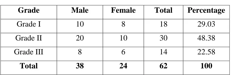
Degree of Differentiation
In general papilliferous growth tend to be better differentiated
BRODERS GRADING14
Broders designated adenocarcinoma into 4 grades based on
percentage of differentiated tumour cells.
Grade I : Well differentials tumours, closely resembling an adenoma
Grade II : Tumour cells more crowded together but still arranged in
fairly regular pattern
Grade III : Less differentiated and arranged in irregularly folded
rings
Grade IV : Anaplastic cells which did not form glandular structures at
all.
Mucoid tumours vary considerably and were graded separately by
Duke
DUKE’S GRADING
Duke considered arrangement of cell and evolved into new
three grade system.
Grade I : Well differented – well formed tubules least nuclear
pleomorphism and mitosis.
Grade II : Moderately differentiated.
Grade III : Least differentiated – occasional glandular structure more
SPREAD OF COLORECTAL CANCER
Most of our knowledge of spread was due to studies by Dukes
(1930). Gordon – Watson.
A. LOCAL INVASION
First, invasion after initial mucosal growth is to protrude into
the lumen. Lateral invasion was more in transverse direction leading
to circumferential growth15. Mural penetration leads to peritoneal
seedling. Additional spread is via perineural spaces with invasion
reaching as far as 10 cm from the primary tumour.
B. LYMPHATIC EXTENSION
Lymph nodal metastasis occurred only after tumours has
penetrated into perirectal / colonic tissues. Lymphatics spread occurs
in an orderly fashion through upwards, laterally and downwards
direction16.
Retrograde spread occurs on blockage of central nodes. The
risk of lymphatics spread increases with increasing tumour grade. As
spread follows the course of blood vessels supplying the
carcinomatous region, appropriate fields of excision is worked out by
C. HAEMATOGENOUS SPREAD
The liver is the primary site of haematogenous spread followed
by lungs in colonic cancer. As venous drainage of rectum is via dual
systems. The liver contains secondary deposits in about one-third to
one half of fatal cases liver17 and lung are involved primarily
depending on site of tumour origin in rectum.
Batson’s vertebral venous plexuses represent another way of
blood spread of metastasis to bone and CNS.
D. IMPLANTATION
It refers to release of tumour cells from primary site and their
deposition on another surface. It can occur transluminally,
transperitoneally after serosal invasion and during surgical
manipulation diminishing curative resection and increasing local
DUKE'S STAGING
STAGE A
STAGE B
STAGING AND CLASSIFICATION
Duke presented the first original staging for rectal carcinoma
which is widely used now to stage both colorectal tumours.
DUKE STAGING (1930)
A – Growth limited to colonic / rectal wall but not through it
B – Growth penetrating through Bowel Wall
C – Involvement of lymph node regardless of extent of Bowel wall
penetration
1935 Modification
A, B as same.
C1 - Locally positive Nodes
C2 - Positive nodes up to the point of ligature
KIRKLIN MODIFICATION (1941)
A - Tumour limited to mucosa, submucosa only
Duke A
B1 - Tumour infiltration into, but not through Muscularis
Propria
B2 - Tumour infiltration through – muscularis propria
D - Defined as disease beyond limit of surgical Resection (Turn
bull addition)
ASTLER and COLLER MODIFICATION18
A - Limited to Mucosa
B1 - Extending into, but not through in propria involved nodes.
B2 - Extending into and through muscularis propria, with
uninvolved nodes.
C1 - Extending into, but not through muscularis propria involved
nodes.
C2 - Extending through M. Propria with involved nodes
This staging allowed separation of wall penetration and nodal
status.
TNM CLASSIFICATION
The AJCC has recommended TNM classification which covers
all possible presentations.
PRIMARY TUMOUR
Tx : Tumour cannot be assessed
To : No evidence of tumour in resected specimen
Tis : Carcinoma in situ
T1 : Tumour invade submucosa
T3-4 : Depends on presence / absence of serosa
Serosa Present :
T3 : Invades through muscularis propria into the serosa
(but not through)
Pericolic fats within the leaves of mesentery
T4 : Invades through serosa into free peritoneal cavity or into
a contingous organ
Serosa Absent :
T3 : Invades through muscularis propria
T4 : Invades other organs
REGIONAL LYMPH NODES
Nx : Nodes cannot be assessed
No : No regional node metastasis
N1 : 1-3 positive nodes (Pericolic / perirectal)
N2 : 4 or more positive nodes
N3 : Central nodes positive
DISTANT METASTASES
Mx : Presence of distant metastases cannot be assessed
Mo : No Distant metastases
STAGING19 :
Stage T N M
0 I II A II B III A III B III C IV T1s T1-2 T3 T4 T1-2 T3-4 Any- T
Any – T
N0 N0 N0 N0 N1 N1 N2
Any - N
CLINICAL FEATURES
There are 3 main ways in which carcinoma of large intestine
may present.
A. As non emergency cases with insidiously developing chronic
symptoms chiefly affecting bowel function and general health.
B. As emergencies with perforation / obstruction of colon with or
without peritonitis.
C. Non specific symptoms
The common symptoms comprise the following
Altered bowel habits in the form of alternating constipation and
Diarrhea, tenesmus etc.
Sense of incomplete evacuation
Bleeding per rectum
Mucus per rectum
Mass per abdomen
History of piles / haemorrhoids
Abdominal pain / dyspepsia
Loss of weight, asthenia, impairment of general health
Loss of appetite
Acute on chronic bowel obstruction
Bowel perforation / peritonitis
Fairly definite correlation between site, type of growth and
symptomatology occurs. Carcinoma of left colon and rectosigmoid
present early due to stenosus / circular growth whereas right colon
growths present late. Local spread may present with related
symptoms like rectovaginal fistula, recto vesical fistula, uterine
obstruction, hemorrhoids etc.,
Carcinoma of caecum and ascending colon
Mostly present with anemia, severe and unyielding to treatment.
A palpable tumour may be present. Sometimes discovered
unexpectedly at operation for acute appendicitis for an appendicular
abscess failing to resolve. Sometimes carcinoma of caecum can be the
apex of an intussusception presenting with intermittent obstruction.
Carcinoma of transverse colon
It may be mistaken for carcinoma of stomach due to position of
tumour together with anemia and lassitude.
Carcinoma of left side of colon
Majority of tumours occur in this location are usually of
stenosing, annular, ulcerative type. The main symptoms are those of
be the only symptom. Alteration in bowel habits, palpable lump,
abdominal distension are the other symptoms.
Carcinoma of rectosigmoid
It presents usually with bleeding per rectum, sense of
incomplete defecation, tenesmus and alteration in bowel habits.
Subacute intestinal obstruction, may induce colicky pain in abdomen.
Pain in the back sciatica indicate local sacral plexus involvement.
History of piles may be the presenting complaint. Symptoms
pertaining to local or distant spreading may be the initial presentation
in some. May present as fistula in ano, single or multiple discharging
pus.
Carcinoma of Anal Canal
Commonly presenting early with bleeding P/R, pain per anal
region, mass per anal region. Patient may present with constipation,
fistula in ano, obstruction, abdominal distension.
Colorectal carcinoma presenting with acute obstruction
About 1/5 of patient complete obstruction occurs either an
aggravation of chronic affairs as acute on chronic obstruction or may
present one day suddenly as pain, distension of abdomen due to the
narrowed lumen plugged by a fecolith, hard stools, undigestable
Chronic obstruction is more commonly encountered with left
colonic carcinoma rather than right sided lesions. Constricting
stenosing types seen more on left side and the faecal nature produces
greater occurrence of acute obstruction.
Symptoms
Complete obstruction of large bowel is often entirely insidious
in onset. If acute on chronic variety with development of acute
obstruction, the patient finds he is constipated without passing motion
for 2-3 days despite use of apperients. He is not able to get rid of
flatus and with progressively increasing abdominal distension. This
state may be prolonged with slow increase in abdominal discomfort
and distension over a period of 6-7 days before finally presenting to
hospital.
Carcinoma colon with Bowel Perforation
Colonic perforation occurs in 3-8 % of patients with colo rectal
cancer. It’s often the result of obstruction followed by perforation.
The common belief was that perforation takes place in a stercoral
ulcer usually in the caecum and less commonly closer to growth. But
in a series of 115 cases of carcinoma of large intestine presenting with
proximal to an obstructing cancer producing localized abscess or
generalized peritonitis.
SYMPTOMS
Obstructed cases with perforation
These patients are usually desperately ill with abdominal
distention, diffuse tenderness, vomiting, gross dehydration and
electrolyte imbalance. The underlying carcinoma may / may not be
palpated or suspected from clinical obstruction. In majority of cases,
unless, supportive measures are done rapidly to stabilize the patient,
patient condition rapidly deteriorates to end fatally.
Unobstructed cases with perforation
Most of these patients are also gravely ill and shows signs of
general peritonitis, but owing to the absence of previous obstruction,
contamination is less. In some, perforation results in localized
peritonitis, abscess formation, producing diagnostic confusion.
If a caecal growth perforate and subsequent development of
subcutaneous abscess, the presentation may mimic an appendicitis or
A) LEFT LATERAL POSITION
B) KNEE - ELBOW POSITION
INVESTIGATIONS
Routine laboratory investigation to be done includes complete
hemogram, B. Urea, B.Sugar, Sr.Creatinine, Urine R/E, E.C.G., Chest
X ray, X-ray abdomen, including liver function test to ascertain
general condition of the patient and treatment of these abnormalities to
allow treatment of cancer.
Definitive test for detection / diagnosis of colorectal malignancy
Test for occult blood
Guaiac test – detects 20 mg hemoglobin per gram or 20 ml
blood / day. High false positive results occur due to presence of other
oxidizing agents.
Immuno fluorescent test – detects conversion of hemoglobin to
fluorescent porphyrins, detects 5-10 mg hemoglobin / gm of stool.
False positive prohibitively high.
Rectal examination
Reaches approximately 8 cm above dentate line upto 75% of all
A B
C D
COLONOSCOPIC VIEW - POLYPOID CARCINOMA
SIGMOID COLON (A), TRANSVERSE COLON (B) BIOPSY TAKEN (C) HAEMORRHAGIC POLYPOID CARCINOMA (D)
Proctosigmoidoscopy
Rigid one is 2 cm in diameter 25 cm long, reaching 20-25 cm
from dentate line, detect 20-25% of tumours. It is more
uncomfortable and the examination is limited to the rectum.
Flexible sigmoidoscopy
It measures 60 cm in length reaching upto splenic flexure and
identify 50% of tumours can be done after 2-3 prep enemas, used only
as a diagnostic tool with biopsy taking with no therapeutic maneuvers
recommended for people over 50 to be years every 3 years in average
risk ones.
Colonoscopy
Allows visualization of mucosa of entire colon, rectum and
usually terminal ileum. Measure about 160 cm in length, it allows
biopsy, polypectomy, heamorrhage control, stricture dilatation. Major
complication rate < 0.2% with (bleeding, anesthetic complications,
perforations) experienced colonoscopists,
Recommended annually or once in 2 yrs in patients with high
risk of colorectal malignancy with detection of upto 100% of all
CT SCAN SHOWING MULTIPLE HEPATIC METASTASES MRI SCAN OF PELVIS SHOWING EXTENSIVE
T3 RECTAL CARCINOMA INVADING LEFT SIDE OF MESORECTUM.
BARIUM ENEMA APPEARANCES OF A COLONIC CANCER WITH TYPICAL ' APPLE CORE' SHOULDERING ULTRASOUND APPEARANCE OF
Plain radiology
Plain abdominal x-ray films are particularly useful in large
bowel obstruction showing typical gas pattern.
Contrast studies
The mainstay of radiological investigation is the barium enema.
Apart from plain Barium enema. Nowadays double contrast barium
enema is used with more accuracy. Water soluble contrast can be
substituted for barium in patients suspected of perforation. The
cardinal finding is filling defect in the barium shadow.
Imaging techniques
These are important in the evaluation, staging and follow up of
colorectal cancer patients.
CT Scan
It allows preoperative evaluation of abdominal cavity to identify
metastasis. Integrity of urinary tract and staging the primary tumour
lesion esp rectal cancer. Angio CT is 95% sensitive to detect
metastasis esp liver. Also 60-80% rectal wall invasion and 75% 1 cm
lymph adenopathy can made out by CT Scan.
MRI Scan
New technique for evaluating colorectal cancer phased array
for differentiation of cancer from normal tissue and fibrotic scar. It is
also more specific to make out liver metastases esp. resectable ones.
Positron – Emission Tomography
It’s still investigational, and may be most helpful in evaluating
recurrent tumour in pelvis when dense scar tissue is present, other
major advantage is to identify extrahepatic / intraperitoneal disease
before surgery.
Abdominal Ultrasound
It is widely used for the detection of hepatic metastases both
preoperatively and in follow up and also used for elucidation of
abdominal mass lesion.
Trans rectal ultrasound
Also known as endosonography of rectum used to stage rectal
cancer USG image allows clear deliniation of rectal wall layers, depth
of invasion, adjacent origin involvement.
Positive predictive value T < 1cm - < 50%
> 1 cm – 70%
Endoluminal USG
Colonoscopic USG is available, allowing determination of
depth of invasion, attachment to adjacent structures, mesenteric lymph
Tumour Markers
CEA Assay
In 1965 Gold and Freedman, isolated a specific antigen in
adenocarcinoma of the endodermally derived epithelium of the GIT20.
They were also able to demonstrate the same antigen in embryonic
and foetal digestive tract tissue upto the end of the first 6 months of
pregnancy circulating antibodies to its were found in the sera of a high
proportion of women throughout pregnancy.
Thomson et al (1960) developed RIA capable minute quantities
of CEA in serum21. Moore et al (1971) further investigated CEA and
noted that apart from colorectal carcinoma it was found in carcinoma
pancreas and other GIT cancers, bronchogenic carcinoma, alcoholic
liver disease and uremia.
The main use of CEA assay will be to monitor patients who had
radical surgery for detect recurrence of disease, persistance of the
lesion in metastatic form22. Other tumour markers include CA 19-9
and CA-50.
Immuno scintigraphy
A recent development in diagnostic tools utilizes the
radiolabelled antibody to target tumours for imaging or detection. It is
TAG 72 has been found to be useful target antigen in colorectal
cancer, the expression of which has been found to be high as 94% in
colonic adenocarcinoma.
A study using indium III labeled TAG 72 for imaging colon
cancer showed sensitivity of 70%.
Specificity – 90% and accuracy 72%.
BIOPSY
HPE of biopsy obtained preoperatively either by open or via
colonoscope or proctosimoidoscope is done to study the type, grade of
DIAGNOSIS AND DIFFERENTIAL
DIAGNOSIS
Most of the cases of carcinoma rectum can be detected by a
proper rectal examination and this should be mandatory for any patient
presenting with suggestive symptoms or a history of piles. Colonic
cancers need further investigations described earlier. Faecal occult
blood should be done for every patient at high risk. Patients at high
risk should be screened by luminal contrast studies.
The investigations available in this hospital apart from routine
basic investigations are luminal contrast studies, colonoscopy, USG
abdomen and CT scan. In patient who presented as acute
emergencies, diagnosis confirmed by exploratory laparotomy.
DIFFERENTIAL DIAGNOSIS
Conditions simulating colonic and rectal carcinoma include
ileocaecal tuberculosis, appendicular masses, diverticulitis, polyps etc.
Rectal carcinoma should be differentiated from squamous cell
TREATMENT
The various modalities of treatment for colorectal carcinomas are :
1. Surgery
2. Radiotherapy
3. Chemotherapy
SURGERY
Surgical resection remains the mainstay of treatment of
colorectal malignancies. Due to biological unfavourable response to
chemotherapeutic drugs and radiotherapy due to various reasons,
surgery is still the best palliative treatment modality.
The objective of surgery in treatment of colonic carcinoma is to
remove cancerous segment of bowel, the mesentery containing its
lymphatics and any organ directly invaded by the tumour. The way
surgical resection is done consists of major steps to give therapeutive
curative resection with as little morbidity and morality to the patient
and starts preoperatively itself.
Preoperative bowel preparations
As large intestine contain the maximum number of pathogenic
clean contaminated surgical procedures. Steps are taken to reduce
bacterial population as much as possible via 2 components.
- Mechanical cleansing
- Antibiotic administration
Mechanical Cleansing
The most commonly used method is dietary restriction to fluids
only for 48 hrs before surgery. On the day before surgery, preparation
of bowel is by using balanced salt solutions containing purgatives,
most commonly mono, dibasic sodium phosphates, polyethylene
glycol and fleet’s phospho soda with liquids are used.
Antibiotic administration
Mechanical cleansing reduce absolute number of colonic
bacteria but wound infection rate is still high (30-60%). Hence
administration of antimicrobials by oral, IV routes to reduce colonic
bacterial concentrations. Most ideally used ones is IV antibiotics
immediately before surgery which reduces postoperative wound
infection to about 10%23.
Surgical Technique
Techniques to minimize tumour spillage, remove adequate
bowel length and bowel continuity restorations are as important as
The abdomen is opened and tumour is assessed for resectability by
- Palpating liver for secondaries by visual, palpatory and
introperative USG if available. (Isolated / 3 metastases with in 1
resectable lobe is not a contraindication for curative surgery).
- Palpate peritoneum draining lymph nodes and mobility and
fixity of tumour to adjacent organs or abdominal wall
structures.
Then intestine to be resected is tied both proximally and distally
to prevent intraluminal spread. The major segmental artery supplying
the cancerous bowel is ligated, and divided. Any adhesion to adjacent
structures are resected in toto if feasible to avoid loss of curative
resection (most adhesion, lymphadenopathy may be inflammatory and
can only be proved histologically).
The extend of resection depends upon primary location of the
tumour lymphatic metastases, adjacent organ invasion. To get a
tumour free margin, usually 2.5 cm margin clearance is ideal, and
anastmosis may be end or end to side which should be tension free.
Treatment options depending on Tumour Location
Carcinoma Caecum or Ascending colon :
AREA TO BE RESECTED WHEN THE GROWTH IS SITUATED IN THE CAECUM
AREA TO BE RESECTED WHEN THE GROWTH IS SITUATED AT THE HEPATIC FLEXURE
Carcinoma Hepatic Flexure
Treated usually by right hemicolectomy or extended right
hemicolectomy.
Carcinoma Transverse colon
Depending on position of tumour on transverse colon.
Segmental excision of transverse colon. Extended right
hemicolectomy may be performed.
Carcinoma splenic flexure
Treated by resection of colon including transverse, descending
if needed sigmoid colon with associated lymphatics and mesentery.
Carcinoma descending colon or sigmoid colon
Treated by left hemicolectomy with anastomosis.
Carcinoma Rectum
The surgical treatment of rectal cancer should accomplish
complete removal of the cancer and involved regional tissue. Basic
principles of cancer surgery is accomplished first and then
consideration is given to restoration of intestinal continuity. In
surgical treatment procedures, rectal cancers are considered in 3
distinct regions in relation to anal verge (sections of roughly 5 cm
is the development of local recurrences, the rates vary from 5% to
45%24.
Upper third lesion
The treatment of distal sigmoid and intra abdominal rectal
(rectosigmoid) cancers. Anterior resection, lower anterior, resection is
done with restoration of intestinal continuity with sutures / staples25.
Cure rate and pattern of recurrent cancer similar to proximal colon
(Sigmoid colon, descending colon).
Middle third lesion
Can be treated by both abdominal perineal resection or
sphincter saving procedures. It depends on tumour location,
infiltration, craniocaudal spread with restoration of intestinal
continuity in latter procedures.
Lower third lesions
Most of the tumours abdomino perineal resection, rarely some
lesion allowing abdomino sacral resection or anterior resection.
Selected patients can be treated by restorative protectomy and colonal
anastamosis.
Summarizing the various optional modalities for rectal
carcinoma are as follows
Low anterior resection
o End to end / side to end
o Sutures / staples
Abdomino sacral resection
Coloanal resection
Localised procedures26
o Local excision
o Fulguration
o Endocavitary irradiation or brachy therapy
Tumour free margins are usually achievable for colon cancer
and usually a marging of 5 cm is deemed adequate, since < 2.5% of
tumour have intramucosal spread 0 more than 2 cm. During low
anterior resection, distal margin of 2 cm is deemed adequate for well
differentiated, small non bulky tumour with favorable prognostic
features thereby aiding restoration of intestinal continuity.
When synchronous colonic cancers are found at different sites
in the colon, the procedure is subtotal colectomy. Resection of
contiguous organs for locally invasive tumous is approximately 10%
of case. Most commonly involved organs are bladder, ovary, ureter,
abdominal wall, less frequently small intestine, spleen, pancreas,
ROLE OF LAPAROSCOPY27
The oncological safety of laparoscopy is of less concern than
the case some years ago. The specter of port site metastasis, once so
alarming, has faded. From all of the large scale studies the
accumulating evidence seems to point to equivalence in terms of
disease specific recurrence and survival between patients treated using
conventional and laparascopic techniques.
Usually laparoscopic assisted method are used and fully
laparoscopic method are also attempted. They are claimed to facilitate
patients convalescence, and reduces stress.
SURGICAL INTERVENTION IN EMERGENCY
PRESENTATION
The major emergency presentation is obstruction, and
perforation.
Obstruction
The incidence of acute obstruction with carcinoma of the right
and transverse colon was 27.4% and with carcinoma of the left colon
40.4% and in the rectum it was only 4.2%. Immediate surgical
treatment is necessary. Obstructing right / transverse colon cancer
treated by resection and primary anastamosis, while left sided cancer
procedure depends on location, presentation findings, surgeons
experience and judgement. It can be treated in stages with
defunctioning colostomy and later curative surgical resection with
anastamosis.
Various modalities available
Primary resection with exterioration of both ends
Hartmann’s procedure
Primary resection with anastomosis
Primary resection with anastamosis protected by colostomy or
ileostomy
Subtotal colectomy with ileosigmoidostomy
Perforation
According to statistics, 7% of all cases of carcinoma of the large
intestine present with perforation of a bowel and consequent
peritonitis. It is usually a life threatening emergency through
exploration of peritoneal cavity is mandatory.
Goal of Surgery
Remove diseased, perforated segment as dehiscence rate is very
high in a contaminated field. Do anastamosis, protected by proximal
colostomy / ileostomy28. The diverting stoma closure is done 10
The survival rate reaches upto 30% of curative resection at 5 yrs in
these patients.
ADJUVANT TREATMENT FOR COLORECTAL CANCER
RADIOTHERAPY
The role of RT is well defined in rectal carcinoma than colonic
carcinoma. Because colonic carcinoma is intra abdominal, makes
radiotherapy less feasible. In rectal cancer local failure with
recurrence is the most common presentation making adjuvant
radiotherapy worth while.
In Rectal carcinoma, local recurrence in pelvis is due to
inability to do wide resection due to narrow confines of pelvis. So
radiotherapy can be used in various regimens to prevent local failure.
can be used preoperatively
sandwich technique
post operative irradiation
Usually 4500 – 5000 cGy is delivered over 5-7 week period and
little complications results during preoperative use and complications
are more after post operative irradiation29.
Unresectable tumours, patients refusing surgery and barely
operable growth may be treated with palliative radiotherapy30. Also
Radiotherapy can be applied using Brachy therapy and Teletherapy.
Most commonly used teletherapy are Cobalt therapy and Neutron
beam irradiation. Radioactive isotopes are used in brachytherapy with
short half lifes.
CHEMOTHERAPY
Chemotherapy is appealing and most effective when tumour
burden is minimal / smallest and when the fraction of malignant cells
in growth phase is highest. It is most commonly given following the
surgical procedures.
Hence maximum response is obtained for postoperative regimen
usually for stage III disease with no significant improvement in patient
with stage II disease. Most experimented drugs are 5 flurouracil alone
or 5 fluroacil and levamisole or 5 Fu and leucovorin combination31.
New combination regimens are underway and results awaited esp.
Methyl – CCNU / vincristine etc.32
Currently systematic CT is used to improve long term survival
by reducing incidence of distant metastases, to improve local disease
control, by combination with external beam radiation, found to have
modest survival benefits in stage II, III patients. The most promosing
drug is 5 Fu which acts as radio sensitizer in the chemoradiation
IMMUNOTHERAPY
Various agents have been tried to modulate or stimulate innate
immune response on enhancement of immune response to destroy,
prevent tumour progression, implantation or metastasis. No
encouraging results are reported after randomized trials. Commonly
used agents are levamisole, BCG vaccination33.
Another strategy is passive – specific immunotherapy using
monoclonal antibodies especially against CEA antigen and various
other tumour antigens which is said to result in improved disease free
and overall survival in patient34. Of recent interest is MCA 17-1A
which resulted in some improvement in Duke stage C patients. Also
MCA – attached with tumoricidal drugs used to target potential
metastases, local recurrences etc. All are still in experimental stages35.
TREATMENT OF HEPATIC METASTASES
It is important to biopsy liver metastases for histological
diagnosis. Patients with upto 2 or 3 liver mets confined to one lobe of
the liver may be offered liver resection. They are not usually resected
at the time of colonic surgery. Multiple painful hepatic metastases can
FOLLOW UP OF COLORECTAL CARCINOMA
The objective of follow up is for early detection of recurrent or
a metachronous carcinoma that can be treated. The follow up program
in general consist of periodic history and physical examination, fecal
occult blood (FOB), liver function tests, tumour markers, colonoscopy
and radiographic studies. Colonoscopy is an important component of
the program. It is a safeguard for detecting anastamotic recurrence,
missed synchronous lesions and metachronous tumours at an early
MATERIALS AND METHODS
Cases of colorectal carcinoma were collected from August 2003
to March 2006 for this study. A total of 62 cases were studied of
which 14 cases presented as emergencies.
Detailed history was elicited from each patient with special
reference to family history, habitations and early symptomatology.
Thorough physical examination of the patient was performed for
evaluation of general condition, detection of signs and per-rectal
examination.
Thorough laboratory investigations were done in all patients,
and every patient except those who presented as emergency underwent
USG abdomen. Luminal contrast radiographic studies and
Colonoscopic evaluation, CT scan abdomen were done in selected
cases. Chest skiagram taken for all patients for preoperative
evaluation as well as detection of secondaries. Liver function test was
done as routine investigation in all patients.
For all possible cases, preoperative biopsy taken via
proctoscopic, colonoscopic guidance and histologic type made out
before planning treatment. Detailed histopathological reports were
available for staging of tumour and assessing the grade of
SUMMARY AND RESULTS
In this series of 62 cases of colorectal carcinoma the following
observations were made.
Risk factors
Diet
Only one of the 62 patients was pure vegetarian. The majority
of patients were illiterate and they could not specify the exact dietary
constituents. But most patient gave history of consumption of fat and
spicy food. Fibre intake was moderately adequate in most patients.
Tobacco
52 patients out of 62 were using tobacco in some form or other.
All the male patients except 4 were regular smokers of beedi or
cigarettes. 18 out of 24 female patients were using tobacco in the
INCIDENCE
Age
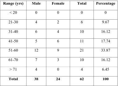
Most of the cases were from older age group. Maximum
incidence was in the sixth decade, but even below the age of 30, 6
cases were reported. The tendency to develop colorectal carcinoma is
[image:72.612.127.508.325.605.2]considered to increase progressively with advancing age.
Table No. 1
AGE INCIDENCE
Range (yrs) Male Female Total Percentage
< 20 0 0 0 0
21-30 4 2 6 9.67
31-40 6 4 10 16.12
41-50 5 6 11 17.74
51-60 12 9 21 33.87
61-70 7 3 10 16.12
> 71 4 0 4 6.45
0 3 6 9 12 15
No. of Patients
< 20 21-30 31-40 41-50 51-60 61-70 > 71
Age Group
AGE INCIDENCE
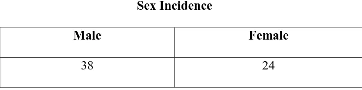
Sex
Incidence was relatively more in males. M : F ratio in this
series was 1:59:1. The incidence of right colon cancer was nearly
equal in both sex but there is definite male preponderance in rectal
[image:74.612.136.506.269.361.2]carcinoma.
Table No. 2
Sex Incidence
Male Female
38 24
M : F = 38 : 24
SEX INCIDENCE
61% 39%
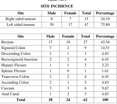
Site
Carcinoma rectum accounted for majority of cases. In 62
patients, 27 were carcinoma of rectum 43.54%. The distribution of
tumour in the other sites in the decreasing order of frequency were as
follows. Sigmoid colon 9, caecum 6, rectosigmoid junction and
transverse colon 4 each, descending colon – 3, ascending colon – 3,
and canal – 3, Hepatic flexure – 2, splenic flexure – 1. 15 out of 62
[image:76.612.123.516.337.687.2]were right sided lesions with 47 being left sided lesion.
Table No. 3
SITE INCIDENCE
Site Male Female Total Percentage
Right sided tumour 8 7 15 24.19
Left sided tumour 30 17 47 75.80
Site Male Female Total Percentage
Rectum 17 10 27 43.54
Sigmoid Colon 7 2 9 14.51
Descending Colon 2 1 3 4.83
Rectosigmoid Junction 2 2 4 6.45
Hepatic Flexure 1 1 2 3.22
Splenic Flexure 1 0 1 1.61
Transverse Colon 2 2 4 6.45
Ascending Colon 2 1 3 4.83
Caecum 3 3 6 9.67
Anal Canal 1 2 3 4.83
SITE INCIDENCE
0 4 8 12 16 20
Rectum Sigmoid colon Descending colon Rectosigmoid junction Hepatic flexure Splenic flexure Transverse colon Ascending colon Caecum Anal Canal
Site
No. of Patients
Male Female
SITE INCIDENCE
24%
76%
MODE OF PRESENTATION
Majority of the patients though had symptoms for sometime,
tend to ignore them and presented at late stages.
Fourteen of the cases in this series are presented as acute
emergencies 22.58%. Twelve of them presented with obstructive
features. Out of which 2 were acute on chronic bowel obstruction. In
all except 1 were left sided lesion. Two patients presented with
features of bowel perforation with peritonitis.
Majority of the cases of right sided colonic tumours had
symptoms of altered bowel habits, increasing constipation, bleeding
per rectum, being the major complaint. Some patient presented with
spurious diarrhea. Majority of bleeding per rectum were seen in
rectum and sigmoid lesions. Pain was relatively late symptom.
Abdominal lump was present in 2 cases of rectosigmoid growth, 1
case of hepatic flexure growth. Rectal growth was palpated in most
cases of carcinoma rectum on per rectal examination.
Transverse colon growth presented with pain abdomen with
typical history of diarrhea alternating with constipation. Anemia,
anorexia and progressive loss of weight was present in majority of
Eight patients presented with symptoms of metastasis /
disseminated disease. Two presented with skeletal metastases mainly
in lumbo dorsal spine out of which one presented with paraplegia.
Five cases presented with hepatomegaly, one patient presented with
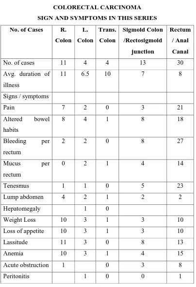
Table No. 4
COLORECTAL CARCINOMA
SIGN AND SYMPTOMS IN THIS SERIES
No. of Cases R.
Colon L. Colon Trans. Colon Sigmoid Colon /Rectosigmoid junction Rectum / Anal Canal
No. of cases 11 4 4 13 30
Avg. duration of
illness
11 6.5 10 7 8
Signs / symptoms
Pain 7 2 0 3 21
Altered bowel
habits
8 4 1 8 18
Bleeding per
rectum
2 2 0 8 27
Mucus per
rectum
0 2 1 4 14
Tenesmus 1 1 0 5 23
Lump abdomen 4 2 1 2 2
Hepatomegaly 1 0
Weight Loss 10 3 1 3 10
Loss of appetite 10 3 1 3 10
Lassitude 11 3 0 8 13
Anemia 10 3 1 4 15
Acute obstruction 1 0 3 8
DIAGNOSIS
Diagnosis in this series was not a problem due to late
presentation in most of cases. Right sided growth tend to present as
mass and left sided growth with features of mass or obstruction and
most of rectal growth were palpated except few.
Thorough laboratory investigations were done in all patients,
and every patient except those who presented as emergency underwent
USG abdomen. Luminal contrast radiographic studies and
Colonoscopic evaluation, CT scan abdomen were done in selected
cases. Chest skiagram taken for all patients for preoperative
evaluation as well as detection of secondaries. Liver function test was
done as routine investigation in all patients.
For all possible cases, preoperative biopsy taken via
proctoscopic, colonoscopic guidance and histologic type made out
before planning treatment. Detailed histopathological reports were
available for staging of tumour and assessing the grade of
differentiation.
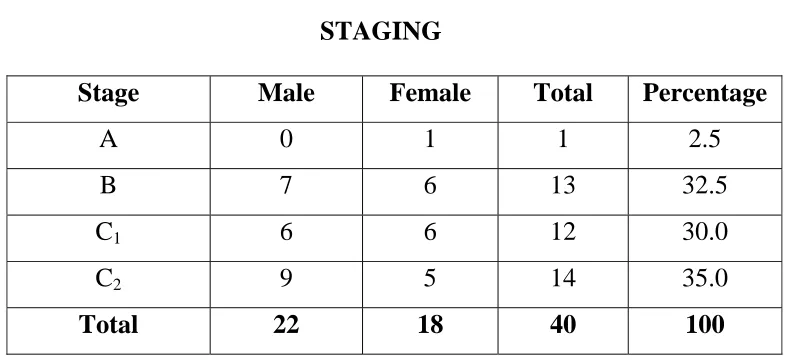
PATHOLOGY AND STAGE
Most of the rectosigmoid and left sided growth were either
annular and stenosing or ulcerative with infiltration type. Almost all