JOURNAL OFVIROLOGY,
0022-538X/97/$04.0010 Apr. 1997, p. 3357–3362 Vol. 71, No. 4
Copyrightq1997, American Society for Microbiology
Differences in Sialic Acid-Galactose Linkages in the Chicken
Egg Amnion and Allantois Influence Human Influenza Virus
Receptor Specificity and Variant Selection
TOSHIHIRO ITO,1,2,3YASUO SUZUKI,4AYATO TAKADA,1,2AYUMI KAWAMOTO,5KOICHI OTSUKI,3
HIROYUKI MASUDA,4MIKA YAMADA,4TAKASHI SUZUKI,4HIROSHI KIDA,1
ANDYOSHIHIRO KAWAOKA2,6*
Laboratory of Microbiology, Department of Disease Control, Graduate School of Veterinary Medicine, Hokkaido University, Sapporo 060,1Department of Veterinary Public Health, Faculty of Agriculture, Tottori University,3
and Tottori Prefecture Institute of Health,5Tottori 680, and Department of Biochemistry,
School of Pharmaceutical Science, University of Shizuoka, Shizuoka 422,4Japan;
Department of Virology and Molecular Biology, St. Jude Children’s Research Hospital, Memphis, Tennessee 38101-03182; and Department of Pathology,
University of Tennessee, Memphis, Memphis, Tennessee 381636
Received 13 November 1996/Accepted 18 December 1996
Human influenza viruses are more efficiently isolated by inoculating patient samples into the amniotic rather than the allantoic cavity of embryonated chicken eggs. This type of cultivation selects virus variants with mutations around the hemagglutinin (HA) receptor binding site. To understand the molecular basis of these
phenomena, we investigated the abundances of sialic acid (SA) linked to galactose (Gal) by thea-2,3 linkage
(SAa2,3Gal) and SAa2,6Gal in egg amniotic and allantoic cells and in Madin-Darby canine kidney (MDCK)
cells. Using SA-Gal linkage-specific lectins (Maackia amurensisagglutinin specific for SAa2,6Gal and
Sambu-cus nigra agglutinin specific for SAa2,3Gal), we found SAa2,3Gal in both allantoic and amniotic cells and
SAa2,6Gal in only the amniotic cells. MDCK cells contained both linkages. To investigate how this difference
in abundances of SAa2,3Gal and SAa2,6Gal in allantoic and amniotic cells affects the appearance of host cell
variants in eggs, we determined the receptor specificities and HA amino acid sequences of two different patient viruses which were isolated and passaged in the amnion or in the allantois and which were compared with
MDCK cell-grown viruses. We found that the viruses maintained high SAa2,6Gal specificities when grown in
MDCK cells or following up to two amniotic passages; however, further passages in either the amnion or
allantois resulted in the acquisition of, or a complete shift to, SAa2,3Gal specificity, depending on the virus
strain. This change in receptor specificity was accompanied by the appearance of variants in the population
with Leu-to-Gln mutations at position 226 in their HA. These findings suggest that lack of SAa2,6Gal linkages
in the allantois of chicken eggs is a selective pressure for the appearance of host cell variants with altered receptor specificities and amino acid changes at position 226.
Clinical samples are inoculated into the amniotic but not the allantoic cavities of embryonated chicken eggs in order to isolate human influenza viruses (12), although recently Madin Darby canine kidney (MDCK) cells have been more commonly used for this purpose. The allantoic cavity is not used because these viruses generally replicate inefficiently there unless they are adapted (3). Once adapted, both human influenza A and B viruses grow well in the allantoic cavity and produce large amounts of virus for laboratory studies and vaccine production (12). Consequently, isolation of human influenza viruses in eggs selects variants with amino acid substitutions that cluster around the receptor-binding site of the hemagglutinin (HA) molecule (16, 18, 21, 23). However, viruses isolated in tissue culture (e.g., from MDCK cells) are homogeneous and iden-tical to those replicating in humans, at least in their HA (17, 22). Thus, host cell factors that could exert a selection pressure on HA have been postulated to exist in ovo, resulting in dif-ferences in HA activity (5). Robertson et al. (25) reported that
clinical influenza B viruses can replicate unrestrainedly within the amnion, whereas allantois-adapted viruses show heteroge-neity among their HA sequences. However, differences in the receptor specificities of influenza viruses passaged in amniotic, allantoic, and MDCK cells remain unknown.
Human influenza viruses preferentially bind cell surface oligosaccharides that contain the 5-N-acetylneuraminic
acid-a-2,6-galactose (Neu5Aca2,6Gal) linkage, while avian and equine influenza viruses bind Neu5Aca2,3Gal (28). Using sialic acid (SA)-galactose (Gal) linkage-specific lectins, Baum and Paulson (1) reported that SA linked to Gal by the a-2,6 linkage (SAa2,6Gal) but not SAa2,3Gal sialyloligosaccharides are present on the surfaces of epithelial cells of the human trachea. By contrast, we found that epithelial cells in duck intestine contain SAa2,3Gal but not SAa2,6Gal (unpublished data). These findings suggest that cell surface sialyloligosac-charides play an important role in the selection and mainte-nance of the receptor specificities of influenza viruses. How-ever, the relative abundances of SAa2,3Gal and SAa2,6Gal in egg amniotic and allantoic cells and in MDCK cells are not known. Recently, Hardy et al. (9) showed that in embryonated eggs two separate mechanisms may exist to select variant in-fluenza viruses: one at the cellular level and the other at the extracellular (fluid) level.
* Corresponding author. Mailing address: Department of Virology and Molecular Biology, St. Jude Children’s Research Hospital, 332 N. Lauderdale, P.O. Box 318, Memphis, TN 38101-0318. Phone: (901) 495-3421. Fax: (901) 523-2622. E-mail address: yoshi.kawaoka@stjude .org.
3357
on November 9, 2019 by guest
http://jvi.asm.org/
In this study, we asked three questions. Do sialyloligosac-charides differ between egg amniotic and allantoic cells, and, if so, does this explain why the isolation of human influenza viruses is more efficient in the amnion than in the allantois? Do differences in sialyloligosaccharides between allantoic and MDCK cells lead to selection of egg variants? And do the receptor specificities of viruses differ according to whether they are grown in the amnion, the allantois, or MDCK cells? To answer these questions, we examined the relative abundances of SAa2,3Gal and SAa2,6Gal on amniotic, allantoic, and MDCK cell surfaces by using linkage-specific lectins. We relate our findings to the receptor specificities and differences in the HA amino acid sequences of viruses grown in these cells.
Analysis of the types of glycosidic linkages between SA and galactose in glycoconjugate on amniotic, allantoic, and
MDCK cells.To compare the abundances of SAa2,3Gal and
SAa2,6Gal on amniotic, allantoic, and MDCK cell surfaces, we incubated frozen sections of these cells with digoxigenin (DIG)-conjugatedMaackia amurensisagglutinin (MAA) (spe-cific for the a-2,3Gal linkage) or Sambucus nigra agglutinin (SNA) (specific for SAa2,6Gal–N-acetylgalactosamine), fol-lowed by incubation with fluorescein-conjugated anti-DIG an-tibody (Fig. 1). Amniotic (Fig. 1D and E) and MDCK (Fig. 1G and H) cells were intensely decorated with both lectins,
whereas allantoic cells were extensively labeled only with MAA (Fig. 1A) and not with SNA (Fig. 1B). These results show that allantoic cells contain mainly SAa2,3Gal, whereas amniotic and MDCK cells contain both SAa2,3Gal and SAa2,6Gal.
Differences in the receptor specificities of viruses grown in
different cells.The differences in the relative abundances of
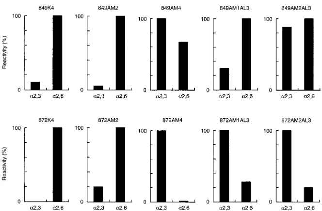
[image:2.612.63.557.68.369.2]SAa2,3Gal and SAa2,6Gal linkages among allantoic, amniotic, and MDCK cells, together with the previous finding that hu-man influenza A and B viruses isolated in eggs and MDCK cells contain different amino acids at the HA receptor binding sites (16–18, 21–24, 34), prompted us to examine the receptor specificities of influenza A viruses grown in these cells. Viruses were isolated from the throat washings of two different patients (patients 849 and 872) infected with H3N2 viruses by inocu-lating samples into the amniotic cavities of embryonated chicken eggs (849AM1 and 872AM1; these and all subsequent similar virus designations are as shown on Fig. 2) and MDCK cells (849K1 and 872K1). After isolation, each virus was pas-saged three more times in its respective host (849AM4, 872AM4, 849K4 and 872K4). Viruses isolated by amniotic in-oculation were also passaged three times in the allantoic cavity with (849AM2AL3 and 872AM2AL3) and without (849AM1AL3 and 872AM1AL3) an additional passage in the amnion prior to the allantoic passages. When 849AM1 virus was passaged in
FIG. 1. Comparison of MAA and SNA staining of allantoic membrane (A, B, C), amniotic membrane (D, E, F), and MDCK (G, H, I) cells. Allantoic and amniotic membranes were obtained from 10-day-old embryonated chicken eggs. Each membrane was washed with phosphate-buffered saline (PBS; pH 7.2) before being cut into 5-mm2squares. The pieces were then embedded in O.C.T. compound (Miles Inc., Elkhart, Ind.) and frozen in liquid nitrogen. The pellet of MDCK cells was also embedded and frozen. Six-micrometer sections of each sample were cut with a microtome cryostat. The sections were air dried and fixed for 15 min with cold acetone before lectin binding. The presence of thea-2,3 ora-2,6 linkage between SA and Gal in each sample was determined by using linkage-specific lectins (Glycan differentiation kit; Boehringer Mannheim Biochemicals, Indianapolis, Ind.). Each section was incubated with 50ml of DIG-labeled SNA (B, E, H) (1mg/ml; specific for SAa2,6Gal–N-acetylgalactosamine) or MAA (A, D, G) (5mg/ml; specific for SAa2,3Gal) for 1 h at room temperature. After three washes with cold PBS, the sections that were incubated with MAA or SNA lectin were mixed with fluorescein-conjugated anti-DIG antibody (Boehringer Mannheim Biochemicals) for 1 h at room temperature. After three more washes with cold PBS, the samples were mounted in buffered glycerol (pH 9.0) for observation. Control slides (C, F, I) were incubated with PBS instead of lectin. The sections were examined under a fluorescence microscope (BH-RFL; Olympus Optics, Tokyo, Japan) with a dark-field condenser and ultraviolet excitation. Magnification,3291.
on November 9, 2019 by guest
http://jvi.asm.org/
the allantoic cavity, virus growth was not detected by hemag-glutination tests. We therefore passaged the allantoic fluid blindly without dilution, which resulted in the detection of virus (HA titer564 U/ml).
Thin-layer chromatography virus-binding assays were done with two different types of gangliosides, 5-N-acetylneuraminic acid-a-2,6-lactotetraosyl ceramide and 5-N-acetylneuraminic acid-a-2,3-lactotetraosyl ceramide. Because the receptor spec-ificity assay requires at least 28 U of HA/ml, the second
pas-sages of the viruses were the earliest paspas-sages we could use to test the specificity of the viruses isolated in the amnion. The viruses isolated and passaged once in the amnion (849AM2 and 872AM2) and those isolated and passaged three more times in MDCK cells (849K4 and 872K4) preferentially bound to Neu5Aca2,6Gal (Fig. 3). Passaging the amniotic viruses two more times in the amnion, however, resulted in a substantial increase in the recognition of Neu5Aca2,3Gal for virus from patient 849 (849AM4) and a complete shift to Neu5Aca2,3Gal recognition for virus from patient 872 (872AM4). Similarly, passaging the virus from patient 872 isolated in the amnion (872AM1) or the virus passaged one more time in the amnion (872AM2) or three more times in the allantois (872AM1AL3 and 872AM2AL3) altered the virus specificity towards Neu5Aca2,3Gal. Interestingly, the allantoic passages of 849AM1 (849AM1AL3) resulted in only a slight increase in Neu5Aca2,3Gal recognition; 849AM1AL3 still preferentially recognized Neu5Aca2,6Gal. The same passages with 849AM2 (849AM2AL3) shifted the specificity more toward Neu5Aca2, 3Gal. These results indicate that egg passage causes a shift in receptor specificities of human influenza A viruses and that this shift may be caused by differences in sialyloligosaccharide among amniotic, allantoic, and MDCK cells.
Differences in the amino acid sequences of HA among
vi-ruses grown in different cells. To assess whether amino acid
substitutions were responsible for the shifts in receptor speci-ficities we had observed after passaging the viruses in different egg compartments, we examined the sequences of the HA genes of these viruses (Table 1). The amino acid residue at position 133 was Asp in the viruses from patient 849, whereas the viruses from patient 872 had Asn at this position
irrespec-tive of their passage histories, providing a signature for the virus strains. HAs of viruses isolated and passaged once in the amnion (849AM2) had Leu at position 226, as did those viruses isolated and passaged three more times in MDCK cells (849K4 and 872K4). However, four of the eight cDNA clones of 872AM2 had Gln at this position and the other four had Leu, indicating that the virus population is mixed with regard to the amino acid residues at this position. Viruses isolated and pas-saged two more times in the amnion (849AM4 and 872AM4) had an amino acid substitution of Gln for Leu at position 226 (Leu3Gln). This change was also found in all three cDNA clones of the virus from patient 872 isolated in the amnion and passaged three times in the allantois (872AM1AL3) but in only two of the seven cDNA clones of virus from patient 849 (849AM1AL3). Since 849AM1AL3, 849AM2, 849K4, 872AM2, and 872K4 viruses preferentially bound to Neu5Aca2,6Gal and 849AM4, 849AM2AL3, 872AM1AL3, 872AM2AL3 and 872AM4 viruses preferentially bound to Neu5Aca2,3Gal (Fig. 3), these results confirm that the amino acid residue at position 226 of HA is responsible for the preferential recognition of Neu5Aca2,6Gal (Leu-226) and Neu5Aca2,3Gal (Gln-226) (29). The HAs of 849AM1AL3, 849AM2, and 849K4 viruses had Ser instead of Asn at position 246. Whether this change is involved in the alteration of receptor specificity is not yet known. Other differences found among these viruses (Tyr3Cys at position 137 in 849AM1AL3 and 872AM2 and Ser3Pro at position 157 in 872K4) did not appear to be critical for recep-tor specificity, but they may be responsible for differences in antigenicity (see below).
Antigenic differences among viruses.The viruses passaged in
[image:3.612.127.494.69.262.2]different compartments of eggs and in MDCK cells had sero-logically distinguishable HAs (Table 2), as reported previously (16–18, 21–24, 34). Monoclonal antibodies (MAbs) raised to the HA of MDCK cell-grown A/Memphis/2/85 (M2-7/2 and M12-2) and egg-grown A/Memphis/2/85 (E12-1) reacted with both HAs of MDCK cell-passaged isolates (849K4 and 872K4) but not with egg-passaged isolates (AM2, AM4, AM1AL3, and AM2AL3) of both viruses. Since 849K4 and 849AM2 have identical HA1 amino acid sequences, the M2-7/2, M12-2, and E12-1 MAbs may recognize oligosaccharide chains in addition
FIG. 2. Diagram of the passage history of the influenza viruses used in this study. Samples of throat washing were collected from two different patients (patients 849 and 872) infected with H3N2 viruses in the winter of 1994 at Tottori Prefecture Institute of Health, Tottori, Japan. The viruses were isolated into the amniotic cavity of 10-day-old embryonated chicken eggs or MDCK cells. After isolation, each virus was further passaged as shown at a 1:100 dilution. Numbers in parentheses indicate the number of passages in MDCK cells (K), in the amnion (AM), and in the allantois (AL). Viruses used in this study are shown in brackets.
VOL. 71, 1997 NOTES 3359
on November 9, 2019 by guest
http://jvi.asm.org/
to the HA backbone. Glycosylation is different for MDCK cell-and egg-grown viruses (14). Two MAbs, 94-49 cell-and 94-79, re-acted with high titers to all of the viruses except the MDCK cell-passaged virus from patient 872 (872K4). A sequence com-parison among the HAs suggested that the Pro at position 157 is responsible for the lack of interaction between the 94-49 and 94-79 MAbs and the 872K4 HA. The 94-63 MAb’s reactivity was associated with Gln at position 226 in these viruses and thus also with the Neu5Aca2,3Gal reactivity. Viruses that reacted with the 94-63 MAb all preferentially recognized Neu5Aca2,3Gal, while those that did not react with the 94-63 MAb preferentially recognized Neu5Aca2,6Gal. These data indicate that the reactivity of this MAb is associated with re-ceptor specificity.
In this study we have demonstrated that the allantoic cells of chicken embryonated eggs contain Neu5Aca2,3Gal but lack Neu5Aca2,6Gal, while amniotic cells contain both linkages. This finding explains why human influenza viruses (which preferentially recognize Neu5Aca2,6Gal) are isolated more efficiently in the amnion than in the allantois (4, 8, 11) and why avian influenza viruses (which preferentially recognize Neu5Aca2,3Gal) are isolated in the allantois efficiently. Our results also provide molecular evidence for the selection of egg variants after the passaging of human influenza viruses in the allantois (16, 18, 21, 23).
Our analysis of the receptor specificities of viruses passaged in the amnion and allantois suggests that the complete conver-sion of receptor specificity from Neu5Aca2,6Gal to Neu5Aca2, 3Gal is not essential for replication of the virus in the allantois (e.g., 849AM1AL3; Fig. 3); the increase in SAa2,3Gal recogni-tion is associated with the presence of variants with Gln at position 226 of their HA (Table 1). In fact, the 849AM1AL3
[image:4.612.141.473.68.286.2]FIG. 3. Binding reactivities to gangliosides of influenza viruses passaged in different egg compartments and in MDCK cells. To determine the receptor specificities of the viruses, a thin-layer chromatography virus binding assay was performed with lacto-series gangliosides containing the type I sugar chain 5-N-acetylneuraminic acid-a-2,6-lactotetraosyl ceramide (a2,6) and 5-N-acetylneuraminic acid-a-2,3-lactotetraosyl ceramide as described previously (32). Each ganglioside (1 nmol) was applied on a silica gel Polygram Sil G plate (Nagel, Germany), developed in chloroform–methanol–12 mM MgCl2(5:4:1, vol/vol/vol), and dried. After being blocked by phosphate-buffered saline (PBS) supplemented with 1% egg albumin and 1% polyvinylpyrrolidone (solution A) at room temperature for 2 h, the plate was incubated with purified virus (28of HA) suspended in PBS for 12 h at 48C. After being washed with PBS, the plate was blocked with solution A and incubated with a pool of 11 MAbs at 48C for 2 h. After being washed with PBS and blocked again with solution A, the plate was incubated at 48C for 2 h with horseradish peroxidase-conjugated protein A and then was incubated with the substrate solution (0.1 M citrate buffer [pH 6.0]–3% 4-chloro-1-naphthol in methanol–3% H2O2aq, 5:1:0.01, vol/vol/vol) at room temperature for 20 min. The binding activity of the virus was determined by scanning the stained chromatogram at 629 nm using a thin-layer chromatography scanner (CSR-9000; Shimazu, Kyoto, Japan). The relative binding reactivities of viruses to gangliosides is shown, taking the highest relative binding reactivity of a virus to a ganglioside as 100%.
TABLE 1. Sequence comparison of the HAs of influenza viruses
Virusa Recognition of
b: Amino acid residue at positionc: Neu5Aca2,3Gal Neu5Aca2,6Gal 133 137 157 226 246
849K4 1 111 D Y S L S
849AM2 1 111 D Y S L S
849AM4 111 11 D Y S Q N
849AM1AL3 11 111 D C S L(Q)d S
849AM2AL3 111 111 D Y S Q N
872K4 2 111 N Y P L N
872AM2 1 111 N C S L(Q)e N
872AM4 111 2 N Y S Q N
872AM1AL3 111 1 N Y S Q N
872AM2AL3 111 1 N Y S Q N
aH3N2 viruses from two different patients (patients 849 and 872) were ana-lyzed in this study. The virus designations are as shown in Fig. 2.
bOriginal data are shown in Fig. 3.2,1,11, and111represent 0, 1 to 30, 31 to 80, and 81 to 100% of maximum binding, respectively.
cViral RNA was isolated from virus-containing amniotic, allantoic, and cul-ture fluid of MDCK cells as previously described (2). cDNA was synthesized with reverse transcriptase and a random hexamer, as described by Katz et al. (17). The HA genes were amplified by PCR with the cDNA, H3 HA-specific oligonucle-otide primers, andPfupolymerase (Stratagene). PCR products were cloned into a plasmid and sequenced with an Autosequencer (Applied Biosystems Inc., Foster City, Calif.) according to the protocol recommended by the company. The sequences of the oligonucleotide primers used will be supplied upon request. At least three independent cDNA clones were sequenced for each virus. When one of the three cDNA clones contained a different nucleotide at a given position, it was considered that an error had been introduced by the polymerase during PCR. Only amino acid residues in the HA1 portion, which differs among the viruses, are shown. Three cDNA clones were sequenced for each virus. Because amino acid residues at position 226 affect receptor specificity (29), additional cDNA clones were sequenced for this position when variations were found among the three clones.
dFive cDNA clones had Leu and two had Gln at this position. eFour cDNA clones had Leu and four had Gln at this position.
on November 9, 2019 by guest
http://jvi.asm.org/
virus still preferentially recognizes Neu5Aca2,6Gal after three passages in the allantois, which is consistent with previous findings that the majority of human isolates preferentially rec-ognize Neu5Aca2,6Gal even when passaged in the allantoic cavity (6, 28). By contrast, when another virus was passaged in the allantois (e.g., 872AM1AL3), its receptor specificity shifted drastically from Neu5Aca2,6Gal to Neu5Aca2,3Gal (Fig. 3). Why the same number of allantoic passages results in only a slight (but significant) increase toward Neu5Aca2,3Gal in one virus (849AM1AL3) yet causes a dramatic change to Neu5Aca2,3Gal in another (872AM1AL3) remains unknown. It may simply reflect the timing of the appearance of Neu5Aca2,3Gal-recognizing variants in the population, or possibly amino acid residues other than that at position 226 in HA are important for Neu5Aca2,3Gal and Neu5Aca2,6Gal preferences. Asp at position 133 may be one such residue, since only at this position is there a difference between the HA1s of the viruses from patients 849 and 872 that contain Gln at position 226. One additional passage of the virus from pa-tient 849 in the amnion (849AM2AL3) resulted in a significant increase in its Neu5Aca2,3Gal recognition. The virus still sig-nificantly recognized Neu5Aca2,6Gal, and there was no evi-dence of a mixed population with respect to the amino acid residue at position 226, which supports our theory that other residues may also be important for linkage preference.
Passaging the AM2 viruses (passaged twice in the amnion) two more times in the amnion resulted in a substantial increase in Neu5Aca2,3Gal recognition for the virus from patient 849 (849AM4) and in a complete shift to Neu5Aca2,3Gal recog-nition for the virus from patient 872 (872AM4). Because the lectin assay is not quantitative, we do not know which type of linkage, Neu5Aca2,3Gal or Neu5Aca2,6Gal, predominates in amniotic cells. Changes in receptor specificity during amniotic passages may be due to the predominance of Neu5Aca2,3Gal in amniotic cells. Alternatively, such variations could arise from exposure of amniotic virus, for example, through a hole between the amniotic and allantoic cavities caused by inocu-lating needles, to SAa2,6Gal-specific inhibitors that may exist in allantoic fluid, as suggested by Robertson et al. (25).
Williams and Robertson (35) found no differences between the binding characteristics of egg- and MDCK cell-grown H1N1 human influenza viruses to allantoic membranes. MDCK cell-derived viruses bound to the membranes, but their internalization was inefficient. They suggested that MDCK cell-derived viruses bound to nonfunctional receptors, which resulted in the failure to trigger endocytosis. Our MDCK-grown viruses preferentially recognized the Neu5Aca2,6Gal linkage, indicating that the nonfunctional receptors in the al-lantois suggested by Williams and Robertson (35) could con-tain Neu5Aca2,6Gal. However, the Neu5Aca2,6Gal linkage was not detected in the allantois, which makes the nonfunc-tional receptor hypothesis less plausible. This discrepancy could be due to differences in the binding assays used by Williams and Robertson (35) (host cell binding) and by us (ganglioside binding). Alternatively, although it is unlikely, the alteration of receptor specificity by allantoic passage may be H3 human virus specific.
This type of host cell-mediated variation occurs with Sendai virus HA-neuraminidase protein (15). Originally mouse-pas-saged and MDCK-pasmouse-pas-saged Sendai viruses did not hemagglu-tinate chicken erythrocytes at 378C, while egg-passaged virus did. Further, the egg-passaged virus had an increased ability to bind Neu5Aca2,3Gal linkages. Thus, host cell-mediated alter-ation of receptor specificity may be a common feature of SA-binding viruses during replication in embryonated eggs.
Katz and Webster (19) reported that there is sequence iden-tity between the HAs of viruses grown in primary chick kidney and other mammalian cells. Although there is no information available on the structure of the receptors on these cell sur-faces, they must contain glycoconjugate with as many Neu5Aca2,6Gal linkages as those on the surfaces of MDCK cells.
MDCK cell- and egg-derived equine influenza viruses differ antigenically (13). In contrast to human isolates, MDCK-de-rived equine viruses display heterogeneity, but egg-deMDCK-de-rived viruses do not, suggesting that eggs are the preferred hosts. Equine influenza viruses recognize Neu5Aca2,3Gal (6, 28). Therefore, allantoic cells, which contain predominantly Neu5Aca2,3Gal, may be favorable hosts. However, the mech-anism of heterogeneity of equine viruses grown in MDCK cells, which contain both Neu5Aca2,3Gal and Neu5Aca2, 6Gal, is unknown. Perhaps, the amount of Neu5Aca2,3Gal is insufficient, and this insufficiency gives variants that recognize Neu5Aca2,6Gal a growth advantage in these cells. A quanti-tative comparison of the amounts of Neu5Aca2,6Gal and Neu5Aca2,3Gal in these cells would address this issue.
The Neu5Aca2,6Gal-specific avian virus variants were stable during passages in MDCK cells, but they exhibited a rapid reversion to the Neu5Aca2,3Gal-specific phenotype when pas-saged only once in the allantoic sacs of eggs (26). This finding is consistent with our results that allantoic cells contain mainly Neu5Aca2,3Gal. Why Neu5Aca2,6Gal-specific human virus isolates are relatively stable to passage in the allantoic cavity remains a mystery. The slight recognition of Neu5Aca2,3Gal seems sufficient for human viruses to replicate in the allantoic cavity. The present study suggests that this recognition is prob-ably conferred by the appearance and eventual dominance of variants with Gln at position 226 in their HA. However, other mutations can also confer the ability to grow in the allantois (thus, presumably, Neu5Aca2,3Gal recognition) (7, 13, 16, 23, 24, 31). It seems that viruses that recognize the Neu5Aca2, 3Gal linkage have a growth advantage in the allantois and predominate after multiple passages in this cavity.
[image:5.612.58.299.89.226.2]After egg adaptation, influenza A and B viruses grow in the allantoic sac and large yields of virus can be produced, but TABLE 2. Antigenic analysis of the HAs of influenza viruses
passaged in different compartments of eggs and MDCK cellsa
Virus HI titer
bwith anti-HA MAbc:
M2-7/2 M12-2 E12-1 94-49 94-63 94-79
849K4 400 400 400 .12,800 ,d .12,800
849AM2 , , , .12,800 , .12,800
849AM4 , , , .12,800 800 .12,800
849AM1AL3 , , , 6,400 , .12,800
849AM2AL3 , , , .12,800 800 .12,800
872K4 3,200 3,200 3,200 200 , ,
872AM2 , , , .12,800 , .12,800
872AM4 , , , .12,800 800 6,400
872AM1AL3 , , , .12,800 6,400 .12,800 872AM2AL3 , , , .12,800 3,200 .12,800
aHemagglutination titration and hemagglutination inhibition (HI) tests were performed in microtiter plates with 0.5% chicken erythrocytes (30).
bReciprocal of the serum dilution that inhibits the activity of four hemagglu-tinating units of virus antigen.
cMouse ascitic fluids were used as the source of MAbs. MAbs were raised against the HAs of MDCK cell-grown A/Memphis/2/85 (H3N2) (M2-7/2) and A/Memphis/12/85 (H3N2) (M12-2) and against the HAs of egg-grown A/Mem-phis/12/85 (H3N2) (E12-1) and A/Memphis/1/94 (H3N2) (94-49, 94-63, and 94-79). MAbs were prepared as described previously by Kida et al. (20).
d,, less than 100.
VOL. 71, 1997 NOTES 3361
on November 9, 2019 by guest
http://jvi.asm.org/
influenza C virus only grows in the amniotic cavity. Influenza C virus, which contains a single envelope hemagglutinin-esterase glycoprotein, recognizes 9-O-acetyl neuraminic acid (10, 27, 33). Although there is no information available on the pres-ence of 9-O-acetyl neuraminic acid in amniotic and allantoic cells, the growth of influenza C virus in allantoic cells may be restricted because of lack of the receptor in these cells. To explain the preferential growth of these viruses in the amniotic cavity, we have initiated a detailed analysis of sialyloligosac-charides in egg membranes.
We thank Krisna Wells for excellent technical assistance, Robert G. Webster for MAbs, and Susan Vallance for editing the manuscript.
This study was supported by a grant-in-aid for scientific research from the Ministry of Education, Science and Culture, Japan, and by a Public Health Service research grant (AI33898) from the National Institute of Allergy and Infectious Diseases, by a Cancer Center Sup-port (CORE) grant, and by the American Lebanese Syrian Associated Charities (ALSAC).
REFERENCES
1.Baum, L. G., and J. C. Paulson.1990. Sialyloligosaccharides of the respira-tory epithelium in the selection of human influenza virus receptor specificity. Acta Histochem.40(89 Suppl.):35–38.
2.Bean, W. J., G. Sriram, and R. G. Webster.1980. Electrophoretic analysis of iodine-labeled influenza virus RNA segments. Anal. Biochem.102:228–232. 3.Burnet, F. M.1940. Influenza virus infections of the chick embryo lung. Br. J.
Exp. Pathol.21:147–153.
4.Burnet, F. M., W. I. B. Beveridge, D. R. Bull, and E. Clarke.1942. Investi-gation of an influenza epidemic in military camps in Victoria, May 1942. Med. J. Aust.2:371–376.
5.Choppin, P. W., and I. Tamm.1960. Studies of two kinds of virus particles which compromise influenza A2 virus strains. I. Characterization of stable homogenous substrains in reactions with specific antibody, mucoprotein in-hibitors, and erythrocytes. J. Exp. Med.112:895–920.
6.Connor, R. J., Y. Kawaoka, R. G. Webster, and J. C. Paulson.1994. Receptor specificity in human, avian, and equine H2 and H3 influenza virus isolates. Virology205:17–23.
7.Daniels, R. S., S. Jeffries, P. Yates, G. C. Schild, G. N. Rogers, J. C. Paulson, S. A. Wharton, A. R. Douglas, J. J. Skehel, and D. C. Wiley.1987. The receptor-binding and membrane-fusion properties of influenza virus variants selected using anti-haemagglutinin monoclonal antibodies. EMBO J.
6:1459–1465.
8.Eatton, M. D., M. Corey, W. Van Herick, and G. Meiklejohn.1945. A comparison of various methods of demonstrating influenza virus in throat washings. Proc. Soc. Exp. Biol.58:6–9.
9.Hardy, T. C., S. A. Young, R. G. Webster, C. W. Naeve, and R. J. Owens.
1995. Egg fluids and cells of the chorioallantoic membrane of embryonated chicken eggs can select different variants of influenza A (H3N2) viruses. Virology211:302–306.
10. Herrler, G., and H.-D. Klenk.1987. The surface receptor is a major deter-minant of the cell tropism of influenza C virus. Virology159:102–108. 11. Hirst, G. K.1942. Direct isolation of influenza virus in chick embryos. Proc.
Soc. Exp. Biol.58:155–157.
12. Hoyle, L.1968. The influenza viruses, p. 1–375.InS. Gard, C. Hallauer, and K. F. Meyer (ed.), Virology monographs, vol. 4. Springer-Verlag, New York, N.Y.
13. Ilobi, C. P., R. Henfrey, J. S. Robertson, J. A. Mumford, B. J. Erasmus, and J. M. Wood.1994. Antigenic and molecular characterization of host cell-mediated variants of equine H3N8 influenza viruses. J. Gen. Virol.75:669– 673.
14. Inkster, M. D., V. S. Hinshaw, and I. T. Schulze.1993. The hemagglutinins of duck and human H1 influenza viruses differ in sequence conservation and in glycosylation. J. Virol.67:7436–7443.
15. Itoh, M., X. L. Wang, Y. Suzuki, and M. Homma.1992. Mutation of the
HANA protein of Sendai virus by passage in eggs. Virology190:356–364. 16. Katz, J. M., C. W. Naeve, and R. G. Webster.1987. Host cell-mediated
variation in H3N2 influenza viruses. Virology156:386–395.
17. Katz, J. M., M. Wang, and R. G. Webster.1990. Direct sequencing of the HA gene of influenza (H3N2) virus in original clinical samples reveals sequence identity with mammalian cell-grown virus. J. Virol.64:1808–1811. 18. Katz, J. M., and R. G. Webster.1988. Antigenic and structural
character-ization of multiple subpopulations of H3N2 influenza virus from an individ-ual. Virology165:446–456.
19. Katz, J. M., and R. G. Webster.1992. Amino acid sequence identity between the HA1 of influenza A (H3N2) viruses grown in mammalian and primary chick kidney cells. J. Gen. Virol.73:1159–1165.
20. Kida, H., L. E. Brown, and R. G. Webster.1982. Biological activity of monoclonal antibodies to operationally defined antigenic regions on the hemagglutinin molecule of A/Seal/Massachusetts/1/80 (H7N7) influenza vi-rus. Virology122:38–47.
21. Robertson, J. S., J. S. Bootman, R. W. Newman, J. S. Oxford, R. S. Daniels, R. G. Webster, and G. C. Schild.1987. Structural changes in the hemagglu-tinin which accompany egg adaptation of an influenza A (H1N1) virus. Virology160:31–37.
22. Robertson, J. S., J. S. Bootman, C. Nicolson, D. Major, E. W. Robertson, and J. M. Wood.1990. The hemagglutinin of influenza B virus present in clinical material is a single species identical to that of mammalian cell-grown virus. Virology179:35–40.
23. Robertson, J. S., C. W. Naeve, R. G. Webster, J. S. Bootman, R. W. Newman, and G. C. Schild.1985. Alterations in the hemagglutinin associated with adaptation of influenza B virus to growth in eggs. Virology143:166–174. 24. Robertson, J. S., C. Nicolson, J. S. Bootman, D. Major, E. W. Robertson, and
J. M. Wood.1991. Sequence analysis of the haemagglutinin (HA) of influ-enza A (H1N1) viruses present in clinical material and comparison with the HA of laboratory-derived virus. J. Gen. Virol.72:2671–2677.
25. Robertson, J. S., C. Nicolson, D. Major, E. W. Robertson, and J. M. Wood.
1993. The role of amniotic passage in the egg-adaptation of human influenza virus is revealed by haemagglutinin sequence analyses. J. Gen. Virol.74:
2047–2051.
26. Rogers, G. N., R. S. Daniels, J. J. Skehel, D. C. Wiley, X. Wang, H. H. Higa, and J. C. Paulson. 1985. Host-mediated selection of influenza receptor variants. Sialic acida2,6Gal specific clones of A/duck/Ukraine/1/63 revert to sialic acida2,3Gal-specific wildtype in ovo. J. Biol. Chem.260:7362–7367. 27. Rogers, G. N., G. Herrler, J. C. Paulson, and H. D. Klenk.1986. Influenza C
virus uses 9-O-Acetyl-N-acetylneuraminic acid as a high affinity receptor determinant for attachment to cells. J. Biol. Chem.261:5947–5951. 28. Rogers, G. N., and J. C. Paulson.1983. Receptor determinants of human and
animal influenza virus isolates: differences in receptor specificity of the H3 hemagglutinin based on species of origin. Virology127:361–373. 29. Rogers, G. N., J. C. Paulson, R. S. Daniels, J. J. Skehel, I. A. Wilson, and
D. C. Wiley.1983. Single amino acid substitutions in influenza haemagglu-tinin change receptor binding specificity. Nature304:76–78.
30. Sever, J. L.1962. Application of a microtechnique to viral serological inves-tigations. J. Immunol.88:320–329.
31. Suzuki, Y., H. Kato, C. W. Naeve, and R. G. Webster.1989. Single-amino-acid substitution in an antigenic site of influenza virus hemagglutinin can alter the specificity of binding to cell membrane-associated gangliosides. J. Virol.63:4298–4302.
32.Suzuki, Y., T. Nakao, T. Ito, N. Watanabe, Y. Toda, X. Guiyun, T. Suzuki, T. Kobayashi, Y. Kimura, A. Yamada, K. Sugawara, H. Nishimura, F. Kitame, K. Nakamura, E. Deya, M. Kiso, and A. Hasegawa.1992. Structural deter-mination of gangliosides that bind to influenza A, B, and C viruses by an improved binding assay: strain-specific receptor epitopes in sialo-sugar chains. Virology189:121–131.
33.Vlasak, R., M. Krystal, M. Nacht, and P. Palese.1987. The influenza C virus glycoprotein (HE) exhibits binding (hemagglutinin) and receptor-destroying (esterase) activities. Virology160:410–425.
34.Wang, M. L., J. M. Katz, and R. G. Webster.1989. Extensive heterogeneity in the hemagglutinin of egg-grown influenza viruses from different patients. Virology171:275–279.
35.Williams, S. P., and J. S. Robertson.1993. Analysis of the restriction to the growth of nonegg-adapted human influenza virus in eggs. Virology196:660– 665.