Copyrightq1997, American Society for Microbiology
Evolution of Mouse Hepatitis Virus: Detection and
Characterization of Spike Deletion Variants
during Persistent Infection
CYNTHIA L. ROWE,1SUSAN C. BAKER,1MEERA J. NATHAN,2
ANDJOHN O. FLEMING2*
Department of Microbiology and Immunology and Molecular Biology Program, Loyola University of Chicago,
Stritch School of Medicine, Maywood, Illinois 60153,1and Departments of Neurology and Medical Microbiology,
University of Wisconsin and William S. Middleton Veterans Hospital, Madison, Wisconsin 537922
Received 30 September 1996/Accepted 13 December 1996
High-frequency RNA recombination has been proposed as an important mechanism for generating viral deletion variants of murine coronavirus. Indeed, a number of variants with deletions in the spike glycoprotein have been isolated from persistently infected animals. However, the significance of generating and potentially accumulating deletion variants in the persisting viral RNA population is unclear. To study this issue, we evaluated the evolution of spike variants by examining the population of spike RNA sequences detected in the brains and spinal cords of mice inoculated with coronavirus and sacrificed at 4, 42, or 100 days postinoculation. We focused on the S1 hypervariable region since previous investigators had shown that this region is subject to recombination and deletion. RNA isolated from the brains or spinal cords of infected mice was rescued by reverse transcription-PCR, and the amplified products were cloned and used in differential colony hybridiza-tions to identify individual isolates with delehybridiza-tions. We found that 11 of 20 persistently infected mice harbored spike deletion variants (SDVs), indicating that deletions are common but not required for persistent infection. To determine if a specific type of SDV accumulated during persistence, we sequenced 106 of the deletion isolates. We identified 23 distinct patterns of SDVs, including 5 double-deletion variants. Furthermore, we found that each mouse harbored distinct variants in its central nervous system (CNS), suggesting that SDVs are generated during viral replication in the CNS. Interestingly, mice with the most severe and persisting neurological disease harbored the most prevalent and diverse quasispecies of SDVs. Overall, these findings illustrate the complexity of the population of persisting viral RNAs which may contribute to chronic disease.
The murine coronavirus mouse hepatitis virus JHM (MHV-JHM) causes acute and chronic neurological disease upon infection of the central nervous system (CNS) of susceptible rodents and has been intensively studied as an experimental model of human demyelinating diseases such as multiple scle-rosis (10, 16, 34). The type of disease induced by MHV-JHM depends on the strain of virus, the age and type of animal, the amount of virus and route of inoculation, the host immune status, and other experimental variables. Infection of mice with attenuated strains of MHV-JHM often causes robust subacute and chronic demyelination (11, 17, 18, 31, 40, 49, 54, 55). For example, recent studies with MHV-JHM 2.2-V-1 have shown that mice have prolonged paralysis and that persisting viral RNA can be rescued from the CNS of these animals by reverse transcription (RT)-PCR for up to 787 days postinoculation (p.i.) (21) whereas infectious virus is typically cleared by 15 to 20 days p.i. Because of possible parallels of this experimental model with chronic CNS diseases of humans and animals, we sought to study the nature of the viral RNA that persists during chronic MHV-JHM 2.2-V-1 infection and to examine its role in pathogenesis.
MHV-JHM is an enveloped virus with a 32-kb single-stranded RNA genome of positive polarity (24, 48). The MHV virion is composed of four structural proteins: spike (S), mem-brane (M), small memmem-brane (sM), and nucleocapsid (N). In addition, some strains of MHV express the hemagglutinin-esterase (HE) glycoprotein. The spike glycoprotein (180 kDa)
is proteolytically processed to two 90-kDa subunits, termed S1 and S2 (50). The spike glycoprotein is critical for receptor binding and fusion with susceptible cells and is the target of neutralizing antibodies (8). Studies with monoclonal antibod-ies (MAbs) directed against the spike found that viruses which escape neutralization were neuroattenuated, suggesting that the spike plays a major role in MHV pathogenesis (11, 17, 55). Furthermore, these MAb escape variants were shown to have deletions in the “hypervariable” region of S1 (nucleotides [nt] 1200 to 1800), implicating this region as a major determinant for MHV neurovirulence (2, 43, 52, 53).
Many investigators have shown that during persistence of MHV in vivo, variants with deletions in the S gene are pro-duced (see Fig. 1). Often, such variants have shown interesting pathogenic properties such as reduced neurovirulence or in-creased tropism for white matter. Also, genetic studies have shown that the deletions in the S gene tend to cluster in a hypervariable region near the same region deleted in MAb escape mutants. The nature and significance of deletions in the S gene remain unclear. One limitation of previous studies is that they have focused exclusively on variants which produce infectious virus. It is possible that these variants represent a selected subpopulation of persisting genomes. Furthermore, such variants have generally been isolated subacutely, at the time that infectious virus is waning. Therefore, we sought to characterize the population of viral RNA which persists at later time points, when disease is chronic and infectious virus typically cannot be recovered.
To address these issues, we developed a system to analyze the population of persisting MHV RNA species and to identify
* Corresponding author. Phone: (608) 5421. Fax: (608) 263-0412. E-mail: [email protected].
2959
on November 9, 2019 by guest
http://jvi.asm.org/
and characterize the MHV spike deletion variants (SDVs) in the population. We rescued the S1 region of the spike RNA, known to be hypervariable, from the brains and spinal cords of experimental mice by RT-PCR. This technique allowed us to examine the population of persisting coronavirus RNA species without bias or requirement that they generate infectious viri-ons. We systematically studied the prevalence and nature of deletions in the S1 region by cloning, differential hybridization, and sequencing individual isolates. We show that SDVs are common but are not required for persistent infection. When SDVs were present during persistent infection, each mouse harbored distinct variants in their CNS, suggesting that SDVs are generated de novo and not from amplification of preexist-ing variants in the input pool. With regard to biological signif-icance, we found that SDVs were most prevalent in the spinal cord RNA of mice with the most severe and prolonged
neu-rological impairment, suggesting an important role for SDVs as a consequence or cause of disease during MHV persistence in the CNS.
MATERIALS AND METHODS
Virus.MHV-JHM 2.2-V-1, a neuroattenuated variant of JHM-DL, was prop-agated in DBT cells and assayed as previously described (17). MHV-JHM 2.2-V-1 causes predominantly subacute and chronic demyelination in mice (17). The entire spike gene of 2.2-V-1 has been sequenced and differs from the parental strain JHM-DL spike gene sequence at one nucleotide (nt 3340) in the S2 region of the spike gene, which results in an amino acid change from leucine to phe-nylalanine (53).
Mice.Six-week-old male C57BL/6J mice (Jackson Laboratories, Bar Harbor, Maine) were used in these experiments. Upon arrival, selected mice were tested and found to be seronegative for murine coronavirus by enzyme-linked immu-nosorbent assay (19). The mice were inoculated intracerebrally with 103PFU of
MHV-JHM 2.2-V-1 (17) and periodically monitored for signs of paralytic disease on a scale of 0 (normal) to 4 (paraplegia) (20). The mice were sacrificed by CO2
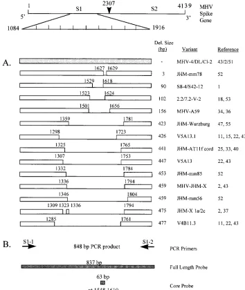
FIG. 1. (A) Schematic representation of the 4,139-nt MHV-JHM spike gene sequence and previously identified variants containing deletions in the 39region of S1. Nucleotide designations represent the first and last positions of the deleted region (i.e., 1627 to 1629 indicates that nt 1627, 1628 and 1629 are deleted). For ambiguous cases, the deletion was defined as the 59-most deletion possible. (B) Location of the PCR primer pair used to amplify S1 cDNA from the brains and spinal cords of mice inoculated with MHV-JHM and of the full-length and core probes (837 and 63 bp, respectively) used in differential colony hybridization to identify individual transformants containing S1 deletions.
on November 9, 2019 by guest
http://jvi.asm.org/
[image:2.612.126.480.71.491.2]inhalation on days 4, 42 and 100 p.i. The brains and spinal cords were removed aseptically, immediately frozen on dry ice, and stored at2708C.
RNA extraction.Brains and spinal cords were thawed, and each half brain and spinal cord was manually disrupted in a Tenbrock homogenizer containing 4 ml (brain) or 2 ml (cord) of RNA STAT (Tel-test Inc., Friendswood, Tex.) and incubated for 5 min at room temperature. RNA was subsequently extracted by addition of 0.2 ml of chloroform/ml of RNA STAT. The phases were separated by centrifugation at 13,8003gfor 15 min at 48C. The aqueous phase was removed, and the RNA was precipitated in the presence of 0.5 ml of isopropanol at room temperature for 10 min. The RNA was pelleted by centrifugation for 10 min at 13,8003gand 48C, and the pellet was washed with 1 ml of 75% ethanol and then air dried for 5 to 6 min. Brain RNA pellets were resuspended in 25ml of diethylpyrocarbonate-treated water, and spinal cord RNA pellets were resus-pended in 20ml of diethylpyrocarbonate-treated water. The RNA quantity was determined by optical density measurements at 260 nm. The integrity of the total RNA was assessed by RT-PCR forb2-microglobulin mRNA. Input virus RNA
was isolated by mixing 53106PFU of MHV-JHM 2.2-V-1 with half of an
uninfected mouse brain and immediately subjecting the mixture to RNA extrac-tion as described above.
RT-PCR of brain and spinal cord RNAs.Brain or spinal cord RNA (2mg per sample) was denatured with 20 mM methyl mercury hydroxide for 5 min at room temperature, andb-mercaptoethanol was added at a final concentration of 0.14 M. For cDNA synthesis, the RNA was incubated at 428C for 60 min in a reaction mixture containing 50 mM Tris-HCl (pH 8.3), 50 mM KCl, 10 mM MgCl2, 10
mM dithiothreitol, 0.5 mM spermidine, 1.25 mM of each deoxynucleoside triphosphate, 40 U of RNasin, 1mg of random hexamer oligonucleotide primers, and 12.5 U of avian myeloblastosis virus reverse transcriptase (Promega, Mad-ison, Wis.). (The RT reaction mixture was preincubated for 30 min on ice prior to cDNA synthesis.) The cDNA synthesis reaction was overlaid with a wax plug (PCR Gems; Perkin-Elmer Cetus, Branchburg, N.J.) and then heat inactivated at 958C for 3 min and quickly cooled on ice.
The synthesized cDNA was amplified by PCR, using a method adapted from that of Saiki et al. (44) and Chang et al. (7) to minimize errors introduced byTaq
polymerase. Briefly, 20ml of RT mixture was mixed with 80ml of a master mix buffer containing 7.5 mM Tris-HCl (pH 9.0), 0.075% Triton X-100, 0.25mM synthetic oligonucleotide primers S1-1 and S1-2 (1), and 2.5 U ofTaqpolymerase (Promega). The PCR mixture was laid over the wax plug and subjected to 40 cycles of amplification, with each cycle consisting of 948C for 1 min, 608C for 1 min, and 728C for 1 min. At the end of this period, an additional extension step at 728C for 7 min was performed. RT-PCR products were purified by the Wizard PCR Preps DNA purification system (Promega), and 1/10 of the reaction prod-ucts were analyzed by electrophoresis on a 1.5% agarose gel for 1 h at 100 V in 13TBE (Tris-borate-EDTA) and visualized by ethidium bromide staining.
Cloning RT-PCR products.Purified RT-PCR products were ligated into the cloning vector pGEM-T (Promega). RT-PCR products which were not initially detected by analysis of 1/10 of the reaction products by ethidium bromide staining of a 1.5% agarose gel were concentrated by precipitation with 1ml of glycogen–1/10 volume of 5 M ammonium acetate–2.5 volumes of 100% ethanol prior to ligation. The ligation reaction mixture included 30 to 50 ng of the RT-PCR products, 1 U of T4 DNA ligase (Gibco-BRL), 50 ng of pGEM-T, 50 mM Tris-HCl (pH 7.6), 10 mM MgCl2, 1 mM ATP, and 1 mM dithiothreitol, and
the reaction was carried out at 148C overnight. The ligation reactions were used to transform high-efficiency competentEscherichia coliDH5acells (Bethesda Research Laboratories). Blue/white selection was used to identify insert-contain-ing colonies, which were picked and stabbed in duplicate in a grid format for colony lifts. Differential colony hybridization was used to identify individual transformants containing deleted forms of the spike gene sequence.
Generation and labeling of probes.An 832-bp wild-type S1 region was PCR amplified with primers S1-1 and S1-2 (1) and subsequently cloned into pGEM-T. An 837-bp full-length probe was generated byEcoRI andBamHI restriction digestion of the S1 wild-type-containing miniprep DNA. The resulting 837-bp fragment was purified by electrophoresis on a 1% agarose gel and eluted from the excised gel slice by centrifugation through glass wool. The gel-purified DNA was concentrated and quantitated on a 1% agarose gel, and 10 ng of it was then labeled.
The 63-bp core probe was generated by PCR amplification of wild-type S1 plasmid DNA with primers S1-5 (59-CCATTTCGCCTCGGCAC-39) (nt 1548 to 1546) and S1-6 (59-TTGGGTTTACAAGTGCACTC-39) (nt 1591 to 1610) under the PCR conditions described above. The 63-bp product was isolated by elec-trophoresis on a 5% nondenaturing polyacrylamide gel. The DNA was eluted from the excised gel slice in 300ml of gel extraction buffer (13TE, 0.4 M NaCl, 0.1% sodium dodecyl sulfate [SDS], 5 mM MgCl2) by gentle mixing on a rotator
for 1 h. The gel-purified DNA was concentrated by ethanol precipitation and quantitated on a 5% polyacrylamide gel. Approximately 10 ng of DNA was ligated in the presence of 1 U of T4 DNA ligase overnight at 148C to join the 63-bp DNA into concatamers and subsequently labeled.
A 10-ng portion of either 837- or 63-bp concatameric DNA was boiled in a total volume of 38ml for 5 min and quickly cooled on ice to generate single-stranded DNA. Denatured DNA fragments were random-primer labeled in a total volume of 50ml by the addition of 10ml of 53labeling buffer, which consists of 20ml of solution A (1.2 M Tris-HCl [pH 8.0], 0.12 M MgCl2, 0.25 Mb
-mer-captoethanol, 0.5 mM dATP, 0.5 mM dGTP, 0.5 mM dTTP), 50ml of solution B
(2 M HEPES), and 30ml of solution C [20 optical density units of random hexamers per ml in 3 mM Tris-HCl (pH 8.0) and 0.2 mM EDTA]; 1mg of bovine serum albumin; 10 to 50mCi of [a-32P]dCTP (3,000 Ci/mmol [Amersham,
Ar-lington Heights, Ill.]); and 5 U of Klenow DNA polymerase (Promega Corp., Madison, Wis.) for 1 to 2 h at room temperature. The probes were purified away from unincorporated nucleotides by elution from a G-50 Sepharose tuberculin syringe column in 200ml of TE (Tris-EDTA [pH 8.0]). The probes were dena-tured in the presence of 1 mg of salmon sperm DNA by boiling for 4 min and quickly cooling on ice, and the hybridization solution was added. The probes were used at a final concentration of greater than 106cpm/ml.
Differential colony hybridization.Approximately 100 colonies per transforma-tion were picked onto a grid format with control colonies of vector alone and vector plus full-length S1 insert. Colony lifts were performed as described by Sambrook et al. (45) with 0.2-mm nitrocellulose filters (Schleicher & Schuell, Keene, N.H.). The filters were dried for 30 min at room temperature and then baked for 2 h in a vacuum oven at 808C. They were soaked in 23SSC (13SSC is 0.15 M NaCl plus 0.015 M sodium citrate) for 2 min prior to prehybridization for at least 2 h at 658C in buffer consisting of 1% SDS, 0.5 M NaCl, 10% dextran sulfate, and 100mg of denatured salmon sperm DNA per ml. The filters were then incubated overnight at 658C with either the full-length 837-bp probe or the core 63-bp probe (10 ng, 106cpm/ml) (see Fig. 1). After hybridization, the blots
were washed for 5 min at room temperature in buffer containing 23SSC and subsequently washed in 23SSC–0.1% SDS at 658C for 20 min. The blots were exposed to X-ray film at2808C for 2 to 12 h. Colonies which hybridized to the full-length 837-bp probe but not the core probe were selected for sequence analysis.
Sequencing.Plasmid DNA was isolated from selected transformants by using the Wizard Minipreps DNA purification system (Promega) and sequenced by the Sanger dideoxy chain termination method (46) with Sequenase Enzyme 2.0 (Sequenase DNA sequencing kit, version 2.0; U.S. Biochemicals) and S1 region primers (S1-2 and S1-3) (1), which flank the previously identified deletions. Thus, the majority of the clones were sequenced from nt 1450 to 1795. In cases where S1 deletions were large and extended beyond these regions, reverse and240 primers as well as primer S1-1 (1) were used to sequence across the deletion. The reaction products were analyzed on 5% Hydrolink (J. T. Baker) gels containing 7 M urea in TBE.
RESULTS
Detection of MHV S1 RNA deletion variants.As a first step toward understanding the role of SDVs in the pathogenesis of persistent MHV infection, we established a system to detect and characterize SDVs from a population of persisting viral RNAs. This system involves amplifying an 848-bp fragment encompassing the MHV S1 hypervariable region from total RNA isolated from the brains or spinal cords of infected mice sacrificed at 4, 42, or 100 days p.i.; cloning the amplified prod-ucts; and identifying deletion variants by differential colony hybridization with a probe to the entire S1 domain (837 bp) and a core probe (63 bp) which represents a domain which is consistently deleted in documented MHV variants (Fig. 1). This system allowed us to detect and characterize individual SDVs from a population of persisting viral RNAs.
We first examined the prevalence of SDVs during the acute stage of infection and within the input virus population. To determine if SDVs are detected in the acute stage of infection, we isolated RNA from the brains and spinal cords of mice at 4 days p.i. and subjected it to RT-PCR amplification, cloning, and differential hybridization (as described above). As shown in Fig. 2, the S1 domain is detected by RT-PCR analysis in both the brains and spinal cords of all 10 MHV-infected mice. Using differential colony hybridization to evaluate approxi-mately 70 cloned isolates per mouse CNS site, we determined that 4 of the 10 acutely infected mice harbored low frequencies (1 to 3%) of SDVs (Tables 1 and 2). To determine if SDVs were present in the input virus inoculum, MHV genomic RNA was isolated and subjected to RT-PCR amplification of the S1 domain, as described in Materials and Methods. The S1 PCR products were cloned, and 849 clonal isolates were examined by differential hybridization as described above. We identified one deletion isolate, indicating that an SDV is detectable, albeit at a low frequency (1 of 849), within the input virus population (Tables 1 and 2). These results indicate that the
on November 9, 2019 by guest
http://jvi.asm.org/
majority of the S1 RNAs are in fact full length both in the input virus and during the acute phase of infection. Indeed, our method of amplification and cloning may enhance our ability to detect deletion variants, since smaller PCR products may be amplified and cloned at a slightly higher efficiency than the full-length sequence. Overall, these results indicate that SDVs are detectable at a low level both in the input virus and in some mice in the acute stage of infection, before the development of a specific immune response to which escape mutants may be selected.
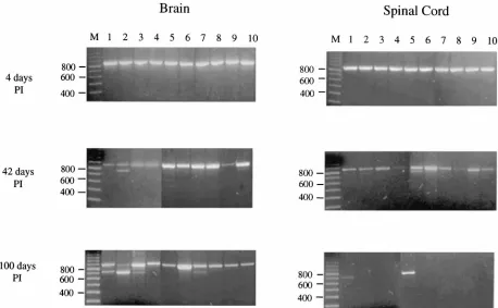
To determine if SDVs are detected during persistent infec-tion, RNA isolated from the brains and spinal cords of mice sacrificed on days 42 and 100 p.i. was subjected to the same analysis. At 42 days p.i., a 848-bp S1 product was detected in both the brains and spinal cords of all 10 mice. However, additional, smaller PCR products were detected in at least five mice (Fig. 2). Cloning of the PCR products followed by differ-ential colony hybridization showed that 7 of the 10 mice har-bored SDVs (Tables 1 and 2). The SDVs represented 4 to 44% of the total S1 products from these mice. Interestingly, 3 of the 10 mice had undetectable levels of SDVs but were clearly persistently infected with MHV. Overall, these results indicate that SDVs are common but are not required for MHV persis-tence.
Similar results were seen in the mice sacrificed at 100 days p.i. RT-PCR amplification of MHV S1 RNA isolated from brains shows that all 10 mice were persistently infected with MHV (Fig. 2). In addition to the wild-type product of 848 bp, we identified putative deletion variant PCR products in 4 of the 10 mice. Differential hybridization of isolated clones again
verified that SDVs were detected in 4 of 10 persistently in-fected mice (Tables 1 and 2). In contrast to the day 100 brain RNA samples, MHV S1 sequences were visualized from only 2 of 10 spinal cord RNA samples, suggesting that MHV RNA was absent or present at only very low levels in the spinal cord in the majority of mice. The integrity of all the spinal cord RNA samples was verified by amplification with primers
spe-cific tob2-microglobulin (data not shown). Interestingly, of the
two mice in which S1 RNA is detected in the spinal cord, one harbored exclusively wild-type S1 (mouse 100-5) and the other harbored exclusively SDVs (mouse 100-1) (Table 1). These results are consistent with our earlier observation that SDVs, while not required for persistent infection, may predominate in some mice during the chronic phase of disease.
[image:4.612.78.536.70.353.2]Correlation of SDVs with neurological impairment.To de-termine if there was an association between clinical signs of demyelination and the presence of SDVs, we evaluated all mice for clinical signs of neurological disease throughout acute and persistent stages of infection. Mice were graded on a scale of 0 to 4 (0 indicating no abnormality, 1 indicating minimal gait abnormality, 2 indicating moderate paraparesis, 3 indicating severe paraparesis, and 4 indicating parapalegia). The majority of infected mice exhibited transient hind-limb paralysis (grade 3 or 4) by day 14 p.i., as expected (Table 1) and subsequently recovered. However, 5 of the 20 persistently infected mice exhibited severe and chronic neurological impairment (clinical score 4 on both days 14 and 24 [Tables 1 and 2]). These mice also harbored the highest frequencies of SDVs in their spinal cords (Fig. 3). In contrast, SDVs were less prevalent in mice with less severe signs of neurological disease (scores 1 to 3 on
FIG. 2. Persistence and evolution of MHV-JHM 2.2-V-1 RNA in the brains and spinal cords of C57/BL6 mice. Brain and spinal cord RNA was isolated from 10 mice each on days 4, 42, and 100 p.i. and subjected to RT-PCR amplification with spike-specific primers (S1-1 and S1-2). Numbers 1 through 10 represent individual mice whose brains and spinal cords were analyzed. Individual samples were later designated by the day of sacrifice and mouse number (example 42-1), and “B” or “C” was added to indicate the origin of the RNA (brain or spinal cord) of that mouse. PCR products were analyzed by electrophoresis on a 1.5% agarose gel and visualized by ethidium bromide staining.
on November 9, 2019 by guest
http://jvi.asm.org/
day 24). Indeed, we found a statistically significant difference
(P50.0025; Mann-Whitney U test) in the percentage of SDVs
in the spinal cords of mice with clinical scores of 4 versus mice with clinical scores of 1 to 3 (Fig. 3). In addition, we found an overall modest but positive correlation between the clinical scores (1 to 4) of mice and the percentage of SDVs in the spinal cord and brain (Fig. 3 [Pearson correlation coefficient;
P50.045 for spinal cord,P50.051 for brain]). These results
suggest that the prevalence of SDVs during persistent infection is associated with more severe clinical manifestations of demy-elination.
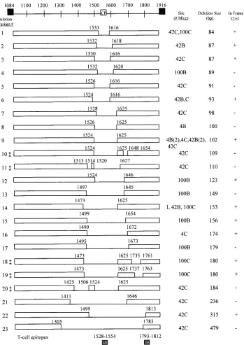
Characterization of MHV SDVs.To characterize the popu-lation of MHV deletion isolates, we sequenced 106 individual SDV clones. Of these, 58 may represent unique species distin-guishable by deletions and flanking point mutations (Table 3). Twenty-three distinct patterns of SDVs were identified and are displayed according to increasing size of the deletion (Fig. 4). We also categorized the SDVs according to the time p.i. during which they were identified and the site from which they were isolated (e.g., 42B indicates brain RNA obtained on day 42 p.i.).
[image:5.612.60.554.82.440.2]Several important points can be derived from this data. First, SDVs appear to arise during viral replication in the CNS. This is supported by the fact that we detected 13 isolates with deletions smaller than the single SDV identified in the input virus. These results indicate that the input SDV we detected was probably not the “founder” virus for all subsequent SDVs in the CNS. In addition, we found that the majority of SDVs were unique to an individual mouse (Fig. 4). These results
TABLE 1. Detection of viral quasispecies during persistent MHV-JHM infection
Mouse and day p.i. sacrificeda
Clinical scorebon p.i. day: Brain Spinal cord
4 14 24 42 100 no. of clones analyzedNo. of clones deleted/ % SDVclones No. of clones deleted/no. of clones analyzed % SDVclones
Day 4
4-1 0 2/71 3 2/71 3
4-2 0 0/71 0 0/73 0
4-3 0 0/76 0 0/73 0
4-4 0 0/80 0 1/78 1
4-6 0 0/82 0 0/82 0
4-8 0 0/82 0 0/70 0
4-9 0 2/76 3 0/79 0
4-10 0 0/78 0 0/78 0
4-7 1 0/85 0 0/72 0
4-5 1 1/76 1 0/83 0
Day 42
42-9 3 2 1 0/73 0 0/114 0
42-8 4 2 1 0/94 0 3/74 4
42-7 3 3 2 0/70 0 3/79 4
42-4 ND 3 2 0/74 0 0/96 0
42-3 ND 3 2 0/77 0 0/75 0
42-6 4 3 2 9/89 10 0/75 0
42-10 4 3 3 6/118 5 0/72 0
42-1c ND 4 2 0/71 0 5/79 11
42-5c 4 4 2 0/92 0 9/77 12
42-2c ND 4 4 23/52 44 17/91 19
Day 100
100-9d 0 1 1 0/83 0 —e —
100-8 4 3 0 0/86 0 — —
100-6 3 2 1 0/166 0 — —
100-7 3 1 2 13/166 8 0/2 0
100-4 3 3 1 0/85 0 — —
100-2 3 3 2 80/82 98 — —
100-10 4 3 2 0/75 0 — —
100-5 3 3 2 0/94 0 0/77 0
100-3c 4 4 3 9/83 11 — —
100-1c 4 4 3 15/66 23 19/19 100
aIndividual samples were designated by day of sacrifice and mouse number (for example, 42-1 indicates mouse number 1 sacrificed on day 42). bClinical scores: 0, normal mouse; 1, minimal gait abnormality; 2, moderate paraparesis; 3, severe paraparesis; 4, parapalegia; ND, not determined. cMice with severe and prolonged neurological disease.
dDetection of viral RNA in the brain indicates that this mouse was infected; however, no significant clinical signs of MHV pathogenesis were observed. e—, viral RNA was not detected by RT-PCR.
TABLE 2. Summary of analysis of SDVs identified from the brains and spinal cords of mice
Time p.i. No. deleted/total no. ofclones analyzed No. of mice withdeleted forms % SDV clonesa
Input virus 1/849 –b 0.1
Day 4 8/1,536 4/10 1–3
Day 42 75/1,642 7/10 4–44
Day 100 136/1,084 4/10 8–100
aRange of percent SDVs in the CNS of mice with at least one deletion. bRNA was isolated from an uninfected mouse brain that was mixed with 53
106PFU of MHV-JHM 2.2-V-1.
on November 9, 2019 by guest
http://jvi.asm.org/
[image:5.612.57.298.635.703.2]suggest that the generation and evolution of SDVs is a random event within each mouse. Furthermore, within some individual mice, we detected SDVs in the brain which were distinct from those in the spinal cord (e.g., mice 42-2 and 100-1 [Table 3]). This de novo generation of MHV SDVs is consistent with the stochastic process of generating viral deletion variants, bottle-necks, and the expansion of quasispecies that has been de-scribed for other viral systems (12, 13, 23, 39).
Second, SDVs probably continue to arise throughout persis-tent infection. We detected a wide range of SDVs at each time p.i. For example, on day 42 p.i., SDVs ranging from 84 to 479 bp were detected. On day 100, we detected nine different SDVs ranging from 84 to 179 bp. We also detected multiple SDVs from a single site (Table 3). About half of the SDVs (52%) were in frame (Fig. 4) and thus have the potential to encode a functional S1 region of the spike glycoprotein. However, 11 of 23 SDVs detected during persistent infection were out of frame, suggesting either that these “defective” SDVs are rep-licated and maintained during persistent infection or that such defective RNAs are generated throughout persistent infection. Ongoing evolution of the MHV SDVs is further supported by the detection of double-deletion variants. Indeed, SDV 9 (de-letion 1524 to 1625) may serve as the parent for the double-deletion variants 10 (double-deletions 1524 to 1625 and 1648 to 1654) and 20 (deletions 1524 to 1625 and 1425 to 1506). Both single and double SDVs were detected in the spinal cord RNA of mouse 42-2 and mouse 100-1 (Table 3). Overall, these results are consistent with continued replication and evolution of SDVs throughout persistent infection.
To determine if a particular sequence is frequently involved in recombination events, we graphed the nucleotide position of the endpoints of the SDVs identified from the 30 mice and plotted the frequency of each in independent deletion events (Fig. 5). Independent deletion events were defined by two criteria: (i) they are identified within an individual mouse or
mouse CNS site, and (ii) they possess independent crossover sites. Because of the possible bias of PCR amplification and introduction of point mutations, the plotted frequency does not reflect the abundance of clones with the same deletion endpoints within an individual mouse, regardless of point mu-tations. We found that the SDVs generated in the mice sacri-ficed on days 4, 42 and 100 all cluster in a hypervariable region of S1 around nt 1500 to 1650. The nonrandom clustering of SDVs may occur as a consequence of the RNA secondary structure in the S1 domain.
We also noted that positions 1524 and 1625 are the most frequent sites involved in recombination events and that, over time, the distribution of the deletion events expands over the hypervariable region. This finding is in contrast to the cluster-ing and eventual loss of recombinants that were generated during coinfection of MHV-A59 and MHV-JHM and passage in DBT cells (2a). This indicates that the selective pressures in the CNS are quite distinct from that of in vitro DBT culture conditions and may favor the “fitness” of different SDVs (12, 14, 23).
Detection of MHV S1 quasispecies in mice with prolonged neurological impairment. As described above, we noted that SDVs were most prevalent in mice which exhibited the most severe and prolonged clinical signs of demyelination through-out persistent infection (mice 42-1, 42-5, 42-2, 1, and 100-3). These severely impaired mice harbored multiple SDVs with flanking point mutations (Table 3). Two of the five mice also harbored double SDVs. These mice were striking because of the prevalence of in-frame SDVs detected in the spinal cord RNA (ranging from 11 to 100% of the S1 isolates). These results are consistent with the earlier observations of Morris et al. (40) suggesting that infectious deletion variants with altered tropism or demyelinating potential may be responsible for the more severe pathogenesis observed in some mice.
DISCUSSION
[image:6.612.58.295.67.283.2]In this study, we examined the population of coronavirus spike deletion variants (SDVs) in the CNS of persistently in-fected mice. The approach we used was to rescue viral RNA from the CNS by RT-PCR, clone the amplified products, and detect SDVs by differential hybridization. Our initial studies were directed to the S1 hypervariable region, since previous investigators have shown that this region is subject to recom-bination and deletion (Fig. 1). We found that the majority of persistently infected mice harbored a mixture or quasispecies of full-length and deleted forms of the MHV S1 region. We detected both in-frame and out-of-frame SDVs and a number of double-deletion variants, suggesting that SDVs may be gen-erated and continue to evolve throughout persistent infection. In the acute phase of infection (day 4), we detected SDVs in 4 of 10 mice. At this stage, SDVs represented no more than 3% of the viral RNA population. Thus, low percentages of SDVs, undetectable by agarose gel electrophoresis, could be detected by differential colony hybridization. These SDVs were proba-bly generated during viral replication in the CNS since they are distinct from the SDV we identified in the input virus. The detection of SDVs on day 4 suggests that deletions are gener-ated at a low frequency even before specific humoral and cellular immune responses can select viral SDVs from the viral population; i.e., SDVs generated acutely may be favored by pressure from the innate immune response or by intrinsic re-quirements of replication in CNS cells. In the chronic phase of infection (days 42 and 100), we detected SDVs in 11 of 20 mice. These results indicate that SDVs are common but are not required for persistent infection. In mice in which SDVs
FIG. 3. Correlation of the frequency of detectable SDVs with neurological impairment. Mice sacrificed on days 42 and 100 were examined for clinical signs of hind-limb paralysis. All mice were examined at the same time point (day 24), after subacute disease commonly resolves, to identify mice that were developing prolonged or unusually severe paralysis. Clinical score: 0, normal mouse; 1, minimal gait abnormality; 2, moderate paraparesis; 3, severe paraparesis; 4, parapalegia. Differential colony hybridization was used to detect deletion vari-ants from the brains (open circles) and spinal cord (solid squares) of mice.
on November 9, 2019 by guest
http://jvi.asm.org/
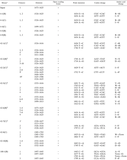
TABLE 3. Characterization of mutations and deletions detected in the S1 region of the spike during persistent MHV-JHM infection
Mousea Clone(s) Deletion Open reading
frame Point mutation Codon change Amino acidchange
Input 1 1473–1625 1
4-1(B) 1, 2 1524–1625 1 1634 G3A CGC3CAC R3H
1654 A3G ATT3GTT I3V
4-1(C) 1, 2 1524–1625 1 1634 G3A CGC3CAC R3H
1654 A3G ATT3GTT I3V
4-4(C) 1 1499–1672 1
4-5(B) 1 1526–1625 2
4-9(B) 1, 2 1524–1625 1 1634 G3A CGC3CAC R3H
1654 A3G ATT3GTT I3V
42-1(C)b 1 1524–1616 1 1636 T3C TGT3CGT C3R
1674 T3C CAT3CAC H3H
1746 T3C AAT3AAC N3N
2, 3 1524–1616 1
4 1530–1616 1
5 1411–1646 2
42-2(B)b 1 1524–1625 1 1744 A3T AAT3TAT N3Y
2 1524–1625 1 1718 A3G GAT3GGT D3G
3–10 1524–1625 1
42-2(C)b 1 1524–1625 1 1829 T3C ATT3ACT I3T
2–8 1524–1625 1
9c 1524–1625 2 1752 T3C CTT3CCT L3P
1648–1654
10c 1425–1506 2
1524–1625
42-5(C)b 1 1533–1616 1 1691 T3A GTT3GAT V3N
1763 G3C TGG3TCG W3S
2 1533–1616 1 1521 T3C CAT3CAC H3H
3 1533–1616 1 1654 A3G ATT3GTT I3V
4 1533–1616 1 1634 G3A CGC3CAC R3H
1501 T3G TGC3GGC C3G
5 1533–1616 1 1485 A3G AAA3AAG K3K
6–8 1533–1616 1
9 1499–1813 1 1891 G3T GTT3TTT V3F
1422 G3C GTG3GTC V3V
42-6(B)b 1–4 1473–1625 1
5–6 1524–1616 1
7–8 1524–1625 1 1654 A3G ATT3GTT I3V
9 1524–1625 1 1654 A3G ATT3GTT I3V
1634 G3A CGC3CAC R3H
42-7(C)b 1c 1520–1627 2
1513–1514
2 1526–1616 2 1456 A3G ATA3GTA I3V
3 1528–1625 2 1797 C3T CCA3TCA P3S
42-8(C) 1 1305–1783 2
2 1305–1783 2 1825 G3A TGG3TAG W3Term
3 1305–1783 2 1864 T3C ATT3ACT I3T
42-10(B) 1–4 1532–1618 1
5 1532–1618 1 1682 G3A GGT3GAT G3D
6 1532–1618 1 1749 T3C GAT3GAC D3D
100-1(B) 1 1497–1645 2 1445 C3T GCA3GTA A3V
2 1497–1645 2 1677 T3G TGA3GGA Term3G
1702 A3G TAA3TGA Term3Term
3 1497–1645 2 1799 A3G CCA3CCG P3P
Continued on following page
on November 9, 2019 by guest
http://jvi.asm.org/
were detected, we found an expansion in the diversity of the SDVs (compared with the acute phase), in terms of both the percentage of the population (4 to 100% of the amplified RNA) and the complexity of the SDVs, as seen in double SDVs. These results are consistent with continued evolution of the viral RNA during persistent infection.
To assess the biological importance of SDVs in the patho-genesis of demyelinating disease, we monitored the mice for clinical signs of neurological disease. Following MHV infec-tion, most mice experienced transient hind-limb paralysis on day 14 p.i. The majority of mice recover, exhibiting less severe clinical signs of neurological impairment. Interestingly, we found the most diverse collection of SDVs in the mice with the most severe clinical disease and most impaired recovery (as defined by clinical score of 4 on both days 14 and 24). However, the reason for this severe disease is currently unclear. The presence of SDVs may reflect an initial high level of viral replication in the CNS and therefore more opportunity to generate SDVs, but the SDVs themselves may not be patho-genic. Alternatively, SDVs may be propagated by selective advantage, such as altered tropism, or reduced fusogenicity and cytopathic effects. A subset of SDVs may be responsible for the chronic demyelination that we and other investigators have reported (40, 54).
Our study suggests that critical determinants of pathogenesis in an individual mouse may be the particular expansion and selection of viral RNA quasispecies which occur within that mouse, especially during the early stages of infection (13, 14, 23). Such stochastic processes may contribute to the diversity of disease outcomes observed when inbred mice are challenged with aliquots of the same viral pool. In fact, several prior studies have suggested that subtle variations in input MHV
may have a crucial influence on disease course. For example, Wege et al. (54) showed that inoculation of uncloned MHV-JHM induced a spectrum of acute and subacute diseases whereas inoculation of plaque-purified MHV-JHM (large or small plaque) predominantly caused a fatal, acute disease. Also, Morris et al. (40) reported that inoculation of the spike deletion isolate AT11f into rats caused a range of pathogenesis as monitored by the pattern of neurological lesions. Taken together, these studies and our present results suggest that minor differences in input virus or virus replicating shortly after inoculation of an individual animal may be progressively amplified and result in major differences in the ultimate dis-ease outcome. Our demonstration that SDVs are infrequent in in vitro viral stocks but often are the predominant viral RNA species in mice with severe and chronic disease is consistent with this hypothesis and suggests that SDVs, once generated in vivo, may be subject to strong positive selection. Future studies will address these issues in more detail by inoculating mice with defined mixtures of viruses with distinct S genotypes and then analyzing subsequent disease course and progeny S ge-notypes.
We note that these SDVs deleted a recently identified T-cell epitope (nt 1528 to 1554) (3, 6, 43a) which overlaps with the core probe (nt 1548 to 1610) used to detect SDVs by differ-ential colony hybridization (Fig. 4). Interestingly, the SDVs exist as a mixed population with wild-type S1 sequences, sug-gesting that loss of the T-cell epitope is not required for per-sistent infection. The persisting mixed population or quasispe-cies may induce “antigenic oscillations” (4, 9, 29, 30, 41) in which subdominant epitopes are recognized by T cells. Anti-genic oscillation has been proposed as a mechanism
contrib-TABLE 3—Continued
Mousea Clone(s) Deletion Open reading
frame Point mutation Codon change Amino acidchange
4 1497–1645 2 1816 T3C ATT3ACT I3T
5 1497–1645 2 1861 C3A ACA3ATA T3I
6–10 1497–1645 2
100-1(C)b 1 1473–1625 1 1677 T3C TGT3TGC C3C
2–5 1473–1625 1
6c 1473–1625 1 1730 G3A GGC3GAC G3N
1737–1763
7–9c 1473–1625 1
1735–1761
10 1533–1616 1 1478 A3G CAG3CGG Q3R
1718 A3G GAT3GGT D3G
100-2(B) 1 1499–1654 1 1494 T3C TCT3TCC S3S
2 1499–1654 1 1695 A3T TTA3TTT L3F
3 1499–1654 1 1720 C3T TCC3TTC S3F
4 1499–1654 2 1763 G3A TGG3TAG W3Term
5–9 1499–1654 1
100-3(B)b 1 1495–1673 2 1690 G3C TGT3TCT C3S
2–4 1495–1673 2
5–10 1532–1620 2
100-7(B) 1, 2 1524–1646 1 1490 T3A ATG3AAG M3K
3 1524–1646 1 1728 G3A AAG3AAA K3K
4–10 1524–1646 1
aIndividual samples were designated by day of sacrifice, mouse number, site of RNA isolation [brain (B), cord (C)], and clone number, for example, 42-1(C)4
indicates mouse 1 sacrificed at day 42 p.i., spinal cord sample, clone 4.
bMice with more than one type of deletion. cClone contains a double deletion.
on November 9, 2019 by guest
http://jvi.asm.org/
[image:8.612.59.555.83.339.2]uting to the inability of the immune system to effectively clear viral infections (42).
The mechanism proposed to be responsible for generating MHV SDVs is copy choice RNA recombination (27, 32, 38). Experimental evidence for copy choice RNA recombination suggests that the recombination of RNA viruses occurs most frequently during negative-strand synthesis (5, 26, 28, 35). Ac-cording to this model, SDVs may be generated when a nascent RNA dissociates from the positive-strand template and rebinds to alternative sites on the template RNA. The rejoining of the
[image:9.612.134.483.67.560.2]nascent RNA may occur at regions of homology at the site of dissociation (homologous recombination), at alternative re-gions of homology (aberrant-homologous recombination), or at distinct, nonhomologous sites (nonhomologous recombina-tion). Aberrant-homologous recombination has been proposed as a mechanism for generating deleted RNA genomes for a number of RNA viruses including MHV (2) and flock house virus (35). Aberrant-homologous recombination appears to re-quire only short stretches of homologous sequence (3 to 7 nt). In our study, we found that seven SDVs (SDV 6, 9, 11, 13, 15,
FIG. 4. Schematic representation of deletions identified in the S1 region of the spike gene (SDVs). The S1 RT-PCR products of 30 mice were analyzed by differential colony hybridization to identify individual clones with deletions. A total of 23 independent SDV patterns were identified by sequencing isolated clones and are designated 1 to 23 by increasing size of deletion. SDVs were designated according to the time p.i. when they were identified. I, input virus; 4B, day 4 brain; 4C, day 4 spinal cord; 42B, day 42 brain; 42C, day 42 spinal cord; 100B, day 100 brain; 100C, day 100 spinal cord. SDVs were identified in only one mouse during the indicated time p.i. unless otherwise indicated. CP, core probe; ‡, double-deletion variants. T-cell epitopes are as identified by Castro and Perlman (6).
on November 9, 2019 by guest
http://jvi.asm.org/
16, and 22) have short (3- to 4-nt) regions of homology be-tween the putative donor and junction sites. Significantly, the majority of the MHV SDVs did not possess regions of homol-ogy at the putative crossover sites and therefore were most probably generated by nonhomologous recombination.
It is currently unclear why the S1 hypervariable region is so frequently involved in generating the SDVs. As previously pro-posed by Banner et al. (2), one possibility is that during RNA synthesis, an RNA secondary structure in the S1 domain causes the RNA polymerase to pause and thereby promotes the dis-sociation of the nascent RNA from the template. The replica-tion complex would then be available to rejoin the template by homologous or nonhomologous RNA recombination. Alterna-tively, copy choice recombination may be occurring randomly throughout the MHV RNA, and only RNA species that are selected during persistent infection are propagated (2a). In-deed, both high-frequency recombination sites and selective pressures may well contribute to the ultimate population of MHV RNAs detected during persistent infection. Future stud-ies will be directed at investigating the role of the RNA sec-ondary structure and selective pressures in the generation of MHV SDVs and quasispecies expansion during persistent in-fection.
ACKNOWLEDGMENTS
We thank Youlian Liao for statistical analysis of clinical data, Joan Pooley for technical assistance, and Tom Gallagher and Jennifer Schiller for helpful discussions.
This work was supported by Public Health Service research grant AI32065 from the National Institutes of Health (to S.C.B.), National Multiple Sclerosis Society research grant RG2283-A-2 (to J.O.F.), and a Department of Veterans Affairs merit review grant (to J.O.F.).
REFERENCES
1.Adami, C., J. Pooley, J. Glomb, E. Stecker, F. Fazal, J. O. Fleming, and S. C. Baker.1995. Evolution of mouse hepatitis virus (MHV) during chronic infection: quasispecies nature of the persisting MHV RNA. Virology209: 337–346.
2.Banner, L. R., J. G. Keck, and M. M. C. Lai.1990. A clustering of RNA recombination sites adjacent to a hypervariable region of the peplomer gene of murine coronavirus. Virology175:548–555.
2a.Banner, L. R., and M. M. C. Lai.1991. Random nature of coronavirus RNA recombination in the absence of selection pressure. Virology185:441–445. 3.Bergmann, C. C., Q. Yao, M. Lin, and S. A. Stohlman.1996. The JHM strain
of mouse hepatitis virus induces a spike protein-specific D-b-restricted cy-totoxic T cell response. J. Gen. Virol.77:315–325.
4.Bertoletti, A., A. Sette, F. V. Chisari, A. Penna, M. Levrero, M. De Carli, F. Fiaccadori, and C. Ferrari.1994. Natural variants of cytotoxic epitopes are T-cell receptor are antagonists for antiviral cytotoxic T cells. Nature369: 407–410.
5.Cascone, P. J., T. F. Haydar, and A. E. Simon.1993. Sequences and struc-FIG. 5. Frequency distribution of crossover sites of SDVs identified from 30 mice sacrificed on day 4 (A), day 42 (B), and day 100 (C) p.i. The nucleotide positions (xaxis) of the endpoints of the left (open bars) and right (solid bars) sequence are indicated. The frequency (yaxis) of endpoints in independent deletion events is shown. Independent deletion events are those arising in an individual mouse in either the brain or spinal cord. Designation of left and right crossover sites is based on copy choice RNA recombination, which may occur during either positive- or negative-strand RNA synthesis.
on November 9, 2019 by guest
http://jvi.asm.org/
tures required for recombination between virus-associated RNAs. Science 260:801–805.
6.Castro, R. F., and S. Perlman.1995. CD81T-cell epitopes within the surface
glycoprotein of a neurotropic coronavirus and correlation with pathogenic-ity. J. Virol.69:8127–8131.
7.Chang, S. Y., A. Shih, and S. Kwok.1993. Detection of variability in natural populations of viruses by polymerase chain reaction. Methods Enzymol. 224:428–432.
8.Collins, A. R., R. L. Knobler, H. Powell, and M. J. Buchmeier.1982. Mono-clonal antibodies to murine hepatitis-4 (strain JHM) define the viral glyco-protein responsible for attachment and cell fusion. Virology119:358–371. 9.Couillin, I., B. Culmann-Penciolelli, E. Gomard, J. Choppin, J.-P. Levy,
J.-G. Guillet, and S. Saragosti.1994. Impaired cytotoxic T lymphocyte rec-ognition due to genetic variations in the main immunogenic region of the human immunodeficiency virus 1 NEF protein. J. Exp. Med.180:1129–1134. 10. Dales, S., and R. Anderson.1995. Pathogenesis and diseases of the central nervous system caused by murine coronaviruses, p. 257–384.InS. G. Siddell (ed.), The Coronaviridae. Plenum Press, New York, N.Y.
11. Dalziel, R. G., P. W. Lampert, P. J. Talbot, and M. J. Buchmeier.1986. Site specific alteration of murine hepatitis virus type 4 peplomer glycoprotein E2 results in reduced neurovirulence. J. Virol.59:463–471.
12. Domingo, E., C. Escarmis, M. A. Martinex, E. Martinez-Salas, and M. G. Mateau.1992. Foot-and-mouth disease virus populations are quasispecies. Curr. Top. Microbiol. Immunol.176:33–47.
13. Duarte, E. A., I. S. Novella, S. C. Weaver, E. Domingo, S. Wain-Hobson, D. K. Clarke, A. Moya, S. F. Elena, J. C. de la Torre, and J. J. Holland.1994. RNA virus quasispecies: significance for viral disease and epidemiology. Infect. Agents Dis.3:201–214.
14. Eigen, M.1993. The origin of genetic information: viruses as models. Gene 135:37–47.
15. Fazakerley, J. K., S. E. Parker, F. Bloom, and M. J. Buchmeier.1992. The V5A13.1 envelope glycoprotein deletion mutant of mouse hepatitis virus type-4 is neuroattenuated by its reduced rate of spread in the central nervous system. Virology187:178–188.
16. Fazakerley, J. K., and M. J. Buchmeier.1993. Pathogenesis of virus-induced demyelination. Adv. Virus Res.42:249–324.
17. Fleming, J. O., M. D. Trousdale, F. A. K. El-Zaatari, and S. A. Stohlman. 1986. Pathogenicity of antigenic variants of murine coronavirus JHM se-lected with monoclonal antibodies. J. Virol.58:869–875.
18. Fleming, J. O., M. D. Trousdale, J. Bradbury, S. A. Stohlman, and L. P. Weiner.1987. Experimental demyelination induced by coronavirus JHM (MHV-4): molecular identification of a viral determinant of paralytic dis-ease. Microb. Pathog.3:9–20.
19. Fleming, J. O., and L. B. Pen.1988. Measurement of the concentration of murine IgG monoclonal antibody in hybridoma supernatants and ascites in absolute units by sensitive and reliable enzyme-linked immunosorbent assay (ELISA). J. Immunol. Methods110:8–11.
20. Fleming, J. O., F. I. Wang, M. D. Trousdale, D. R. Hinton, and S. A. Stohlman.1993. Interaction of immune and central nervous systems: Con-tribution of antiviral Thy-11cells to demelination induced by coronavirus JHM. Reg. Immunol.5:37–43.
21. Fleming, J. O., C. Adami, J. Pooley, J. Glomb, E. Stecker, F. Fazal, and S. C. Baker.1995. Mutations associated with viral sequences isolated from mice persistently infected with MHV-JHM. Adv. Exp. Med. Biol.380:591–595. 22. Gallagher, T. M., S. E. Parker, and M. J. Buchmeier.1990.
Neutralization-resistant variants of a neurotropic coronavirus generated by deletions within the amino-terminal half of the spike glycoprotein. J. Virol.64:731–741. 23. Holland, J. J.1993. Replication error, quasispecies populations and extreme
evolution rates of RNA viruses, p. 203–218.InS. S. Morse (ed.), Emerging viruses. Oxford University Press, Oxford, United Kingdom.
24. Holmes, K. V., and M. M. C. Lai.1996. Coronaviridae: the viruses and their replication, p. 1075–1093.InB. N. Fields, D. M. Knipe, P. M. Howley, et al. (ed.), Fields virology. Lippincott-Raven Publishers, Philadelphia, Pa. 25. Jackson, D. P., D. H. Percy, and V. L. Morris.1984. Characterization of
murine hepatitis virus (JHM) RNA from rats with experimental encephalo-myelitis. Virology137:297–304.
26. Jarvis, T. C., and K. Kirkegaard.1992. Poliovirus RNA recombination: mechanistic studies in absence of selection. EMBO J.11:3135–3145. 27. Keck, J. G., G. K. Matsushima, S. Makino, J. O. Fleming, D. M. Vannier,
S. A. Stohlman, and M. M. C. Lai.1988. In vivo RNA-RNA recombination of coronavirus in mouse brain. J. Virol.62:1810–1813.
28. Kirkegaard, K., and D. Baltimore.1986. The mechanism of RNA recombi-nation in poliovirus. Cell47:433–443.
29. Klenerman, P., S. Rowland-Jones, S. McAdam, J. Edwards, S. Daenke, D. Lalloo, B. Koppe, W. Rosenberg, D. Boyd, A. Edwards, P. Giangrande, R. E. Phillips, and A. J. McMichael.1994. Cytotoxic T-cell activity antagonized by naturally occurring HIV-1 gag variants. Nature369:403–407.
30. Kodama, T., K. Mori, T. Kawahara, D. J. Ringler, and R. C. Desrosiers. 1993. Analysis of simian immunodeficiency virus sequence variation in
tis-sues of rhesus macaques with simian AIDS. J. Virol.67:6522–6534. 31. Kyuwa, S., and S. A. Stohlman.1990. Pathogenesis of a neurotropic murine
coronavirus, strain JHM in the central nervous system of mice. Semin. Virol. 1:273–280.
32. Lai, M. M. C.1992. RNA recombination in animal and plant viruses. Mi-crobiol. Rev.56:61–79.
33. La Monica, N., L. R. Banner, V. L. Morris, and M. M. C. Lai.1991. Localization of extensive deletions in the structural genes of two neurotropic variants of murine coronavirus JHM. Virology182:883–888.
34. Lavi, E., D. H. Gilden, M. K. Highkin, and S. R. Weiss.1986. The organ tropism of mouse hepatitis virus A59 in mice is dependent on dose and route of inoculation. Lab. Anim. Sci.36:130–135.
35. Li, Y., and L. A. Ball.1993. Nonhomologous RNA recombination during negative-strand synthesis of flock house virus RNA. J. Virol.67:3854–3860. 36. Luytjes, W., L. S. Sturman, P. J. Bredenbeek, J. Charite, B. A. M. van der Zeust, M. C. Horzinek, and W. J. M. Spaan.1987. Primary structure of the glycoprotein E2 of coronavirus MHV-A59 and identification of the trypsin cleavage site. Virology161:479–487.
37. Makino, S., F. Taguchi, M. Hayami, and K. Fujiwara.1983. Characterization of small plaque mutants of mouse hepatitis virus, JHM strain. Microbiol. Immunol.27:445–454.
38. Makino, S., J. G. Keck, S. A. Stohlman, and M. M. C. Lai.1986. High-frequency RNA recombination of murine coronaviruses. J. Virol.57:729– 737.
39. Martell, M., J. I. Esteban, J. Quer, J. Genesca, A. Weiner, R. Esteban, J. Guardia, and J. Gomez.1992. Hepatitis C virus (HCV) circulates as a population of different but closely related genomes: quasispecies nature of HCV genome distribution. J. Virol.66:3225–3229.
40. Morris, V. L., C. Tieszer, J. Mackinnon, and D. Percy.1989. Characteriza-tion of coronavirus JHM variants isolated from Wistar Furth rats with a viral-induced demyelinating disease. Virology169:127–136.
41. Moskophidis, D., and R. M. Zinkernagel.1995. Immunobiology of cytotoxic T-cell escape mutants of lymphocytic choriomeningitis virus. J. Virol.69: 2187–2193.
42. Nowak, M. A., R. M. May, R. E. Phillips, S. Rowland-Jones, D. G. Lalloo, S. McAdam, P. Klenerman, B. Koppe, K. Sigmund, C. R. V. Bangham, and A. J. McMichael.1995. Antigenic oscillations and shifting immunodominance in HIV-1 infections. Nature375:606–611.
43. Parker, S., T. M. Gallagher, and M. J. Buchmeier.1989. Sequence analysis reveals extensive polymorphism and evidence of deletions within the E2 glycoprotein gene of several strains of murine hepatitis virus. Virology173: 664–673.
43a.Pewe, L., G. F. Wu, E. M. Barnett, R. F. Castro, and S. Perlman.1996. Cytotoxic T cell-resistant variants are selected in a virus-induced demyeli-nating disease. Immunity5:253–262.
44. Saiki, R. K., D. H. Gelfand, S. Stoffel, S. J. Scharf, R. Higuchi, G. T. Horn, K. B. Mullis, and H. A. Erlich.1988. Primer-directed enzymatic amplifica-tion of DNA with a thermostable DNA polymerase. Science239:487–491. 45. Sambrook, J., E. F. Fritsch, and T. Maniatis.1989. Molecular cloning: a
laboratory manual, 2nd ed. Cold Spring Harbor Laboratory Press, Cold Spring Harbor, N.Y.
46. Sanger, F., S. Nicklen, and A. R. Coulson.1977. DNA sequencing with chain-terminating inhibitors. Proc. Natl. Acad. Sci. USA74:5463–5467. 47. Schmidt, I., M. Skinner, and S. Siddell.1987. Nucleotide sequence of the
gene encoding the surface projection glycoprotein of coronavirus MHV-JHM. J. Gen. Virol.68:47–56.
48. Siddell, S. G.1995. The coronaviridae: an introduction, p. 1–9.InS. G. Siddell (ed.), The Coronaviridae. Plenum Press, New York, N.Y. 49.Stohlman, S. A., and L. P. Weiner.1981. Chronic central nervous system
demyelination in mice after JHM virus infection. Neurology31:38–44. 50.Sturman, L. S., C. S. Ricard, and K. V. Holmes.1985. Proteolytic cleavage
of the E2 glycoprotein of murine coronavirus: activation of cell-fusing activ-ity of virions by trypsin and separation of two different 90K cleavage frag-ments. J. Virol.56:904–911.
51.Taguchi, F., T. Ikeda, and H. Shida.1992. Molecular cloning and expression of a spike protein of neurovirulent murine coronavirus JHMV variant cl-2. J. Gen. Virol.73:1065–1072.
52.Taguchi, F., H. Kubo, H. Takahashi, and H. Suzuki.1995. Localization of neurovirulence determinant for rats on the S1 subunit of murine coronavirus JHMV. Virology208:67–74.
53.Wang, F.-I., J. O. Fleming, and M. M. C. Lai.1992. Sequence analysis of the spike protein gene of murine coronavirus variants: study of genetic sites affecting neuropathogenicity. Virology186:742–749.
54.Wege, H., M. Koga, R. Watanabe, K. Nagashima, and V. ter Meulen.1983. Neurovirulence of murine coronavirus JHM temperature-sensitive mutants in rats. Infect. Immun.39:1316–1324.
55.Wege, H., J. Winter, and R. Meyermann.1988. The peplomer protein E2 of coronavirus JHM as a determinant of neurovirulence: definition of critical epitopes by variant analysis. J. Gen. Virol.69:87–98.