ISCHEMIC FROM NONISCHEMIC DILATED
CARDIOMYOPATHY”
Dissertation submitted for
D.M. DEGREE EXAMINATION
BRANCH II– CARDIOLOGY
MADRAS MEDICAL COLLEGE
AND
GOVERNMENT GENERAL HOSPITAL
CHENNAI – 600 003
THE TAMIL NADU DR.M.G.R MEDICAL UNIVERSITY
CHENNAI – 600 032
CERTIFICATE
This is to certify that the dissertation entitled “UTILITY VALUE OF
TISSUE DOPPLER IMAGING DURING DOBUTAMINE STRESS IN
DIFFERENTIATING ISCHEMIC FROM NONISCHEMIC DILATED
CARDIOMYOPATHY” is the bonafide original work of Dr.C.ARUMUGAM, in
partial fulfillment of the requirements for D.M. Branch-II (CARDIOLOGY)
examination of THE TAMILNADU DR.M.G.R. MEDICAL UNIVERSITY to be
held in August 2007.The period of post-graduate study and training was from August
2004 to July 2007.
PROFESSOR V.JAGANATHAN M.D.,D.M.
PROFESSOR AND HEAD OF THE DEPARTMENT OF CARDIOLOGY
MADRAS MEDICAL COLLEGE AND
GOVERNMENT GENERAL HOSPITAL CHENNAI - 600 003.
THEDEAN
MADRAS MEDICAL COLLEGE
AND
GOVERNMENT GENERAL HOSPITAL
DECLARATION
I Dr.C.ARUMUGAM, solemnly declare that this dissertation entitled,
UTILITY VALUE OF TISSUE DOPPLER IMAGING DURING
DOBUTAMINE STRESS IN DIFFERENTIATING ISCHEMIC FROM
NONISCHEMIC DILATED CARDIOMYOPATHY” is a bonafide work done by
me at the department of Cardiology,Madras Medical College and Government
General Hospital during the period 2004 – 2007 under the guidance and supervision
of the Professor and Head of the department of Cardiology of Madras Medical
College and Government General Hospital,Professor V.Jaganathan M.D.D.M.This
dissertation is submitted to The Tamil Nadu Dr.M.G.R Medical university,towards
partial fulfillment of requirement for the award of D.M. Degree (Branch-II) in
Cardiology.
Place : Chennai
Date :
ACKNOWLEDGEMENT
I would like to thank The Dean, Madras Medical College and Government
General Hospital for permitting me to carry out this study in this institution.
I would like to express my gratitude to Professor Dr.V.Jaganathan
M.D.D.M., Professor and Head of the Department of Cardiology, Madras Medical
College and Government General Hospital for having given me the opportunity to
carry out this study, and for his constant support and encouragement throughout
my course.
I am extremely thankful to Professor Dr.R.Alagesan M.D.D.M., for
giving me valuable guidance and constant support throughout the study.
I am immensely grateful to my Professors and Teachers, Professor Dr. Geetha
Subramanian, M.D.D.M., Professor Dr.A.Balaguru, M.D.D.M., Professor
Dr.B.Ramamurthy M.D.D.M., and Professor Dr.P.Arunachalam M.D.D.M.,
Department of Cardiology, Madras Medical College and Government General
Hospital for their valuable suggestions during the period of study.
I extend my profuse thanks and gratitude to Dr.S.Venkatesan M.D.D.M.,
Dr.G. Gnanavelu M.D.D.M., Dr.M.A. Rajaseakaran, M.D.D.M., Dr. G. Palanisamy
M.D.D.M., Dr.G.Karthikeyan, M.D.D.M., Dr. K.Meenakshi M.D.D.M.,
Dr.C.Elangovan, M.D.D.M., and Dr. Ravishankaran M.D.D.M., Assistant Professors
of Cardiology, Madras Medical College and Government General Hospital for their
valuable suggestions and critical appraisal of this study.
My sincere thanks are to all the patients for their genuine co-operation during
this study. Last but not the least, I would like to thank my family and friends for their
CONTENTS
Sl.No. Title Page No.
1 INTRODUCTION 1-7
2 REVIEW OF LITERATURE 8-38
3 AIM & OBJECTIVES 39
4 MATERIALS & METHODS 40-48
5 OBSERVATIONS & RESULTS 49-59
6 DISCUSSION 60-64
7 CONCLUSIONS 65
BIBLIOGRAPHY
PROFORMA
ABBREVIATIONS
TDI Tissue Doppler Imaging
CAD Coronaty Artery Disease
DCM Dilated Cardiomyopathy
2DE Two Dimensional Echocardiography
LV Left Ventricle
RWMA Regional Wall Motion Abnormalities
DSE Dobutamine Stress Echocardiography
PET Positron Emission Tomography
EBCT Electron Beam Computed Tomography
CMR Cardiovascular Magnetic Resonance
MRI Magnetic Resonance Imaging
PWTD Pulsed-Wave Tissue Doppler
TA Total Amplitude
PES Postejection shortening
SA Systolic amplitude
LVEF Left Ventricular Ejection Fraction
RVEF Right Ventricular Ejection Fraction
WHO World Health Organization
MI Myocardial Infarction
ABBREVIATIONS…
LVIDd Left Ventricular Internal Dimension at end-diastole
LVIDs Left Ventricular Internal Dimension at end-systole
SPECT Single-Photon Emission Computed Tomography
CC Coronary Calcification
MAC Mitral Anular Calcification
AVC Aortic Valve Calcification
TAC Thoracic Aortic Calcification
CAS Coronary Artery Stenosis
S’ Systolic velocity
Ea or E’ Early diastolic velocity
Aa or A’ Late diastolic velocity
LBBB Left Bundle Branch Block
WMSI Wall Motion Score Index
NYHA New York Heart Association
“learn to heal”
INTRODUCTION
Differentiating ischemic from nonischemic cardiomyopathy poses a particular problem, but this is important prognostically and therapeutically. Patients with ischemic cardiomyopathy have worse prognosis than the patients with idiopathic dilated cardiomyopathy. Joseph et al, in 1983 have demonstrated that in patients with severe chronic left ventricular failure, mortality rate in patients with coronary artery was 46% and 69% at 1 and 2 years, respectively compared with 23% and 48% at 1 and 2 years in those with idiopathic dilated cardiomyopathy 1. In 1997, Bradley and
colleagues also have shown ischemic etiology is a significant independent predictor of mortality in patients with cardiomyopahy 2.
Therapeutically also it is important to distinguish between ischemic and nonischemic cardiomyopathy because the diagnosis influences the management. Antiplatelets and lipid lowering therapy are important in management of patients with cardiomyopathy of ischemic etiology. Revascularisation in patients with low ejection fraction and significant coronary artery disease is strongly associated with improved survival 3-5 and should be considered in all patients with ischemic
cardiomyopathy and proven hibernation. Moreover ischemic cardiomyopathy may not respond to medical therapy as favorably as patients with nonischemic cardiomyopathy.
Differentiating ischemic from nonischemic cardiomyopathy clinically is not always easy. Although ischemic cardiomyopthy is generally a late consequence of clinically established coronary artery disease, sometimes the clinical course is really occult and indistinguishable from idiopathic cardiomyopathy. An ischemic cause is probable in patients with history of definite or documented prior myocardial infarction or left ventricular aneurysm in echocardiogram 6,7. However, some patients
evidence of myocardial infarction 8,never complaint of chest pain 9 and shows
diffuse rather than regional hypocontractility 6,10. Conversely many patients with
idiopathic dilated cardiomyopathy report frequent episodes of chestpain 11 and
have electrocardiographic evidence of myocardial infarction 12.
Appropriate differentiation of these two cardiomyopathy is achieved by coronary angiography since it remains the gold standard test to identify coronary artery disease, but coronary angiogram is an invasive method with its attendant risks so a noninvasive method would be preferable which may decrease the unnecessary risk, cost and inconvenience associated with cardiac catheterization. Unfortunately most of the currently used noninvasive methods to distinguish the etiology of left ventricular dysfunction have been of limited value 13 and many non-invasive techniques have been tested with variable
accuracy 14,15,16,17,18,19. There are some limitations in interpretation and availability
of these tests while inconsistent findings have been reported. The relatively low specificity and predictive value of these tests which are further reduced with more pronounced left ventricular dysfunction often preclude definitive clinical decisions in individual cases.
Ischemic cardiomyopthy may be suspected on the basis of clinical and
electrocardiographic data. However, the sensitivity and specificity of these data are often inadequate for individual patient management decisions 20,21,22,23,24 and
further confounded by the frequent presence of a nondiagnostic electrocardiography with conduction abnormalities 24 and high prevalence of
asymptomatic myocardial infarction 8.Exercise stress testing is the standard
dynamic stress. Additionally frequent presence of electrocardiographic conduction abnormalities in these group of patients at rest impairs interpretation of stress electrocardiography.
The detection of regional wall motion abnormalities at rest by echocardiography or ventriculography (contrast or radionuclide) is often thought to be a reliable indicator of presence of significant coronary artery disease obstruction and prior myocardial infarction 25-27, but Robert et al 6 demonstrated
that resting regional wall motion abnormalities detect significant coronary artery disease with high specificity in patients with normal sized ventricle but has low specificity in patients with dilated ventricles. Moreover regional wall motion abnormalities at rest have been demonstrated in as many as two thirds of patients with nonischemic cardiomyopathy whereas patients with ischemic cardiomyopathy might have uniform hypokinesia 6. So resting regional wall
motion abnormalities do not reliably distinguish ischemic from nonischemic cardiomyopathy.
Dobutamine stress echocardiography is an accurate noninvasive technique for evaluating coronary artery disease in nondilated heart with sensitivity and specificities in the range of 80-85% 26, but only limited data
available about the role of dobutamine stress echocardiography in detection of coronary artery disease in dilated cardiomyopathy which had been done in small number of patients. Moreover adequate training is required to obtain these impressive results 29.Also the lack of uniform diagnostic criteria is a significant
common in dilated cardiomyopathy patients further impair the wall motion analysis during stress echocardiography.
Several other noninvasive techniques such as right ventricular function, coronary echocardiography, resting thallium-201 myocardial perfusion scanning, positron emission tomography(PET), coronary calcium score using electron beam computed tomography(EBCT), carotid scanning for measuring carotid atherosclerosis have been used to distinguish these two forms of cardiomyopathy, but distinction has been limited by controversial findings.
Right ventricular function — The right ventricular ejection fraction (RVEF) has been proposed as a means of distinguishing between ischemic and nonischemic
cardiomyopathies. Ischemia and infarction usually spare the right ventricle; as a
result, patients with an ischemic cardiomyopathy should have a relatively normal
RVEF. In contrast, a nonischemic cardiomyopathy is a global process that should
involve the right ventricle and should be associated with a reduced RVEF.
Once again, this theoretical distinction has limited clinical utility 43,44. In one series of
23 patients with idiopathic cardiomyopathy and 36 patients with ischemic
cardiomyopathy, the mean RVEF was, as expected, lower in patients with a
nonischemic cardiomyopathy (31% versus 45% for those with an ischemic
cardiomyopathy)43. However, there was considerable overlap between the two groups:
22 percent of patients with a nonischemic cardiomyopathy had a normal RVEF, while
34 percent of patients with an ischemic cardiomyopathy had a reduced RVEF43. In
another study of 76 patients with proven nonischemic cardiomyopathy, 54 percent had
normal RVEF in the presence of reduced LVEF 44.
visualization of coronary arteries, but it is not possible to visualize coronary arteries in all patients by transthoracic echocardiogram, and also only proximal coronaries can be visualized. Thallium scintigraphy has been used in differentiation of ischemic from nonischemic cardiomyopathy, but it gives false-positive anteroseptal and septal perfusion defects in patients with left bundle branch block 30.Positron emission tomography(PET) has also been shown to
been useful to identify ischemic etiology in patients with dilated cardiomyopathy, but is limited by its availability and expense.
A number of authors have proposed that the development of a user-friendly quantitative approach should overcome the limitations of the subjective evaluation of dobutamine stress echocardiography images 31,32 and other currently
used techniques. Several investigators have shown Tissue Doppler
Imaging(TDI), a novel echocardiographic method that measures the long axis function of the ventricles quantitatively, to be a sensitive alternative to the present echocardiographic and scintigraphic imaging techniques to evaluate stress tests 33,34.Quantification of myocardial function by Pulsed Doppler
myocardial imaging during dobutamine stress have also been demonstrated to be a feasible, accurate, and reproducible technique 34.
Left ventricular fibres are predominantly arranged longitudinally or obliquely in the subendocardium and subepicardium and circumferentially in the intermediate layers. During early systole, the longitudinal and oblique fibres begin to contract first causing the long-axis to shorten and the left ventricular cavity to become more spherical. After a mean delay of 25 ms the circumferential fibres also begin to contract, and throughout the rest of systole both circumferential and longitudinal axes contract synchronously 33.This normal
Therefore abnormalities of long axis function may be of practical use as indicators of ischemia during stress testing 35. Furthermore stress induced
changes in long axis function can be measured objectively 36,37.
By studying left ventricular long axis function by simple M-mode echocardiography, Mishra et al reported that the normal increase in systolic amplitude of mitral annulus occurring with dobutamine administration is attenuated in patients with coronary artery disease and normal left ventricular dimension 38. Simonson and Schiller 39 showed that a reduction in long axis
function, called descent of the base in their study, correlated well with impaired left ventricular ejection fraction in a large group of patients including those with coronary artery disease. Gibson’s group 22 confirmed that, in patients with
ischaemic disease, long-axis shortening was delayed to 85 ms after the Q wave from a normal mean of 55 ms, along with a reduction in the amplitude of long axis shortening. Similar results were found by Henein et al.40 who further showed that these abnormalities resolved after successful angioplasty. This suggested that longaxis function might be useful for the detection of ischaemia. Alam and associates 35 assessed long axis shortening before and after bicycle
exercise and found that there was a blunted increase of long axis shortening in patients with coronary artery disease. They used an upper limit of normal of 3 mm based on inter-observer variability. This gave a sensitivity of 76% for the
cardiomyopathy with greater sensitivity and specificity than standard wall motion score index, particularly in the presence of left bundle branch block. But his study group consist of unselected group of patients with dilated cardiomyopathy which included patients with prior history of myocardial infarction where the etiology of cardiomyopathy is clinically obvious and no further investigations are required for diagnosis itself.
REVIEW OF LITERATURE
DEFINITION OF CARDIOMYOPATHY
WHO defined Cardiomyopathies as diseases of the myocardium associated with cardiac(systolic and/or diastolic) dysfunction 41. Cardiomyopathies constitute a
group of disorders in which the dominant feature is direct involvement of the heart muscle itself. By strict definition, they are a primary myocardial disorder and not related to the effects of conditions such as pre-existing valve disease, hypertension, and coronary artery disease. From a practical standpoint, severe dysfunction due to diffuse coronary artery disease and the effects of coronary ischemia is often considered a form of cardiomyopathy (Ischemic cardiomyopathy).
CLASSIFICATION
The World Health Organisation /International Society and Federation of Cardiology Task Force recommended that cardiomyopathies be classified by the dominant pathophysiological mechanism or etiologic/pathogenic factor 41
By mechanism By Disorder
Dilated cardiomyopthy Ischemic cardiomyopthy
Hypertrophic cardiomyopthy Valvular cardiomyopathy Restrictive cardiomyopthy Hypertensive cardiomyopathy
Arrhythmogenic right ventricular Inflammatory cardiomyopthy
cardiomyopthy Metabolic cardiomyopthy
Unclassified cardiomyopthies General system disease
Muscular dystrophy
Neuromuscular disorders
Sensitive and toxic reaction
DILATED CARDIOMYOPATHY
Dilated cardiomyopathy is characterized by dilatation and impaired contraction of the left or both ventricles. Both end-diastolic and end-systolic dimensions and volumes are increased, and variables of systolic function(ejection fraction, fractional shortening, systolic longitudinal motion of mitral annulus, stroke volume, and cardiac output) are uniformly decreased. With gradual dilatation more along the short axis of the left ventricle, the LV cavity becomes more spherical, with a sphericity index(long-axis dimension/short axis dimension) nearing the value of 1 (normal >1.5). Wall thickness varies but typically is within normal limits; however, the LV mass is uniformly increased. Secondary features include dilated mitral annulus and incomplete coaptation of the mitral valve leaflets responsible for the associated functional mitral regurgitation, evidence of low cardiac output, enlarged atrial cavities, right ventricular enlargement, and apical mural thrombus. Intraventricular conduction delay (left or right bundle branch block) is common and contributes further to cardiac dysfunction by causing intraventricular mechanical dyssynchrony.
It maybe idiopathic, familial/genetic, viral and/or immune, alcoholic/toxic, or associated with recognized cardiovasculardisease in which the degree of myocardial dysfunction is not explained by the abnormal loading conditions or the extent of ischemic damage. Histology is nonspecific. Presentationis usually with heart failure, which is often progressive. Arrhythmias, thromboembolism, and sudden death are common and may occur atany stage.
ISCHEMIC CARDIOMYOPATHY
indistinguishable from those of primary dilated cardiomyopathy. Symptoms of heart failure caused by ischemic myocardial dysfunction and hibernation, diffuse fibrosis, or multiple infarctions, alone or in combination may dominate the picture of coronary artery disease. In some patients with chronic coronary artery disease, angina may be the principal clinical manifestation at one time, but later this symptom diminishes or even disappears as heart failure become more prominent. Other patients with ischemic cardiomyopathy have no history of angina or myocardial infarctions(type I silent ischemia), and it is in this subgroup that ischemic cardiomyopathy is most often confused with dilated cardiomyopathy 42.
Ischemic cardiomyopathy is the term used by some to describe the condition in which coronary artery disease causes multiple infarctions, diffuse fibrosis, or severe ischemia that leads to left ventricular dilatation with congestive heart failure and it may or may not be associated with angina pectoris. They consider ischemic dilated cardiomyopathy is usually a late result of clinically overt coronary artery disease.43
WHO designation of ischemic cardiomyopathy as a specific cardiomyopathy “with impaired contractile performance not explained by extent of coronary artery disease or ischemic damage” has led to confusion. If ischemia is thought not be the cause for impairment, perhaps the adjective should be replaced by one describing the perceived cause, or by idiopathic. Most clinicians use ischemic cardiomyopathy to describe dilatation and dysfunction, which they believe is caused by an ischemic insult.
diagnosis of ischemic cardiomyopathy (57 percent of the total population); CAD was also present in 9 of 38(24%) patients with a pretransplant diagnosis of idiopathic dilated cardiomyopathy and in three of four with presumptive alcoholic cardiomyopathy.
Enlarged and globally hypokinetic left ventricles may at times challenge physicians concerning the etiology of this finding, since the underlying cause cannot always be determined, even after detailed history and skilled clinical evaluation, including echocardiography. Dilated cardiomyopathy, either idiopathic or secondary to specific disorders but without coronary stenoses 41, and ischaemic cardiomyopathy
due to severe coronary artery disease have been used to describe left or biventricular dilatation in addition to impaired contraction. It may be difficult to distinguish between them clinically. An ischemic cause is probable in patients with a history of myocardial infarction (MI) or left ventricular (LV) aneurysm3,4 .However, some
patients with ischemic DCM have neither history nor electrocardiographic (ECG) evidence of MI5 never complain of chest pain 6,and show diffuse, rather than regional,
LV hypocontractility3, 7. Conversely, many patients with idiopathic DCM report
frequent episodes of chest pain s and have ECG evidence of MI 9. Ischemic
cardiomyopathy is usually a late result of clinically overt coronary artery disease 45.
However, sometimes the clinical course of the disease is really occult and, indistinguishable from dilated cardiomyopathy43,45,46. Differentiating between this
latter ‘silent’ ischemic and non-ischemic cardiomyopathy is imperative for both therapeutic and prognostic reasons 47,1,48. Although ischemic cardiomyopathy bears a
worse prognosis than non-ischemic cardiomyopathy 1,48,49, patients with proven
hibernation may show an improved outcome after myocardial revascularization 50.
with variable accuracy 15-19, 51. However, there are some limitations in interpretation
and availability of these tests while inconsistent findings have been reported.
On reviewing the old literatures the following parameters and noninvasive techniques were used to differentiate ischemic from nonischemic dilated cardiomyopathy
1. Clinical and electrocardiographic data
2. Regional wall motion abnormalities detected at rest
3. Wall motion analysis by Dobutamine stress echocardiography 4. Right ventricular function
5. Coronary echocardiography 6. Radionuclide ventriculography
7. Electron beam computed tomography 8. Thallium-Dipyridamole scintigraphy 9. Magnetic Resonance Imaging (MRI) 10. Carotid ultrasound
11. Valvular and thoracic aortic calcification 12. Tissue doppler
CLINICAL AND ELECTROCARDIOGRAPHIC DATA
Patients with severe LV dysfunction due to coronary artery disease or nonischemic cardiomyopathy may present with identical symptoms 52. Chest
pain is considerd to one of the cardinal symptom of coronary artery disease. However, some patients with ischemic DCM never complaint of chest pain9.
Conversely many patients with idiopathic DCM report frequent episodes of chest pain. Kannel and colleagues ,in 1984, analyzed the incidence and prognosis of unrecognized myocardial infarctions and found that almost half of such patients did not have chest pain and such silent infarctions were higher in women and in older men 53. In 1973, Margolis and associates evaluated the
unrecognized myocardial infarctions diagnosed by ECG changes. He observed only one third of men with unrecognized and 58% of men with recognized infarctions had history of angina pectoris (p ≤ 0.001)54. In 1965, Massumi et al
observed as many as 52% of patients with congestive cardiomyopathy report chest pain despite the presence of normal coronary arteries . Pasternac (1982) explained that the chest pain in idiopathic dilated cardiomyopathy is due to decreased myocardial perfusion ,both at rest and during pacing leads to subendocardial ischemia. In his study he observed three of five patients with idiopathic congestive cardiomyopathy had chest discomfort 8. Vigna et al, in
1996, have shown that angina was present in 53% of patients with ischemic DCM and 36% of patients with nonischemic DCM (p= 0.994). From these observations it is evident that patients with ischemic and nonischemic DCM can not reliably be distinguished on the basis of presence or absence of chest pain.
Ischemic cardiomyopathy may be suspected on the basis of
electrocardiographic findings. However, the sensitivity and specificity of these data are often inadequate for individual patient management decisions and further confounded by the frequent presence of a nondiagnostic ECG with conduction abnormalities. In 1964, Tavel et al in his original article of ‘abnormal Q waves simulating myocardial infarction in diffuse myocardial diseases’ he demonstrated that patients with nonischemic myocardial diseases frequently had abnormal Q waves 21. In his study 54% of patients with
demonstrate any ECG changes of coronary artery disease. The sensitivity and specificity of ECG changes is further confounded by frequent presence of conduction abnormalities, especially LBBB. In Duncan’s study, among 73 patients with DCM 26 patients had LBBB, 16 with ischemic DCM and 10 with nonischemic DCM. In other studies also, LBBB was present in comparable numbers in doth types of DCMs 68.
RESTING RWMA
The detection of left ventricular regional wall motion abnormalities by echocardiography or by radionuclide angiography is often thought to be a reliable indicator of the presence of significant coronary artery obstruction and prior infarction 55. However, these findings have been recognized in patients
with nonischemic DCM as well 43,56. In 1984, Wallis et al 56 analyzed fifty
in detecting significant CAD was 95%, l00%, and 95%, respectively. The finding, however, of diffuse LV hypokinesis does not exclude CAD in these patients, especially when the left ventricle is dilated. Though regional wall motion abnormalities had high specificity in detecting CAD in patients with severe LV dysfunction and normal LV dimensions, it had low specificity(57%) in patients with dilated left ventricle. This study has also other limitations. The assessment of wall motion abnormalities by 2DE was qualitative and subjective and may be affected by inter- or intraobserver variability. The finding of CAD in patients with DCM does not necessarily imply that CAD was the cause of congestive cardiomyopathy. A causal relationship between the two entities is not definitely established in this study.
Diaz and colleagues, in 1991, studied the value of two-dimensional echocardiography in differentiation of congestive and ischemic cardiomyopathy. Of 70 total patients with chronic heart failure 36 patients had nonischemic DCM and 34 had ischemic DCM. RWMAs were detected in 44% of nonischemic and 65% of patients with ischemic DCM. Diffuse wall motion abnormalities were present in 56% of patients with nonischemic DCM and 35% of patients with ischemic DCM . Regional dyssynergy in primary DCM is likely because of microcirculatory involvement and myocardial hypoperfusion 6,7.
DOBUTAMINE STRESS ECHOCARDIOGRAPHY (DSE )
Echocardiography performed at rest is widely used to evaluate patients with dilated cardiomyopathy. Unfortunately, the frequency of regional wall motion abnormalities at rest in nonischemic cardiomyopathy and the presence of global dysfunction in patients with advanced ischemic cardiomyopathy prevent accurate identification of coronary artery disease 6,7. Exercise testing is
the standard noninvasive method used for detection of coronary artery disease in patients without dilated cardiomyopathy. It is not routinely used for diagnostic for diagnostic purpose in patients with dilated cardiomyopathy because of their reduced ability to perform exercise testing. Additionally, the frequency of ECG abnormalities at rest impairs interpretation of the stress ECG. Dobutamine stress echocardiography has been found to accurately identifies patients with coronary artery disease with major advantage that it is not dependant on patient’s ability to exercise.
Previous studies have shown that dobutamine stress echocardiography safely and accurately identifies coronary artery disease in patients without dilated cardiomyopathy. All these studies were done in patients with normal sized ventricle. Sharp et al, 1994 15 were the first one attempted to determine the safety
target heart rate or maximal protocol dose in 49 patients (70%), ischemia in 12 (17%), arrhythmia in 4 (6%) and side effects in 5 (7%). No patient had prolonged ischemia or sustained arrhythmia. Of those with angiographic studies, 40 had significant coronary artery disease (> or = 50% diameter stenosis). Use of the change in global wall motion score index from low to peak dose resulted in a sensitivity of 83% for dobutamine stress echocardiography and a specificity of 71% for detection of coronary artery disease. Sensitivity for detection of triple-, double- and single-vessel disease was 100%, 83% and 69%, respectively. He concluded that dobutamine stress echocardiography safely provides diagnostic information in patients with dilated cardiomyopathy. This technique has high sensitivity for multivessel coronary artery disease but only moderate specificity. In this study patients with history of MI were also included where the etiology of left ventricle dysfunction is obvious Moreover, analysis of wall motion was limited to qualitative techniques.
DCM. His data show that analysis of regional contraction by dobutamine stress echocardiography can distinguish between ischemic and nonischemic DCM 16. His
study has some limitations. He did not perform side-by-side comparisons of wall motion on the digitized quad-screen format, so possibility of missing subtle responses to dobutamine in patients with diffuse or severe wall motion abnormalities. Moreover patients with single vessel disease frequently did not develop significant worsening of segments with peak dose dobutamine, so the sensitivity of differentiating ischemic DCM with single vessel disease and nonischemic DCM is low.
Cohen et al, in 1997 studied whether DSE is useful in differentiation of cardiomyopathy 61. Dobutamine stress echocardiography was performed in 56
consecutive patients, mean age: sixty-two +/- twelve years. Twenty-two patients had an idiopathic dilated cardiomyopathy (group 1) and 34 had angiographically proven ischemic dilated cardiomyopathy (group 2). Wall motion score index and left ventricular ejection fraction were determined at baseline, 5 micrograms/kg/min, peak, and ten minutes after stepwise dobutamine infusion. Worsening or no change in global wall motion score was observed in 9 group 2 patients (26%) and 1 group 1 patient (5%, P = .07). No significant difference was observed with regard to wall motion score index decrease between baseline and peak dose. Left ventricular ejection fraction increase during dobutamine infusion was comparable in both groups. Thus, an ischemic response was observed more often in the coronary artery disease group, yielding a good specificity and positive predictive value although sensitivity was low. However, left ventricular function improvement did not help to discriminate patients with or without significant CAD.
dilated cardiomyopathy in 37 patients with suspected myocardial ischemia, low ventricular ejection fraction (23±5%) and heart failure. Coronary artery disease was defined as >50% coronary stenosis in at least one coronary artery. By dobutamine stress echocardiography, ischaemic dilated cardiomyopathy was considered present when either an ischaemic response (biphasic response or direct deterioration) or a scar (fixed dyssynergy) was documented in atleast two segments. Twenty-three patients had ischaemic dilated cardiomyopathy, while 14 had normal coronary arteries. The presence of myocardial ischaemia and/or scar by dobutamine stress echocardiography identified patients with ischaemic dilated cardiomyopathy with a sensitivity of 100% and a specificity of 86%. Three of the four false positive results occurred in patients with left bundle branch block. He concluded that dobutamine stress echocardiography is a sensitive technique for detecting the ischaemic aetiology of dilated cardiomyopathy. However, the specificity is lower particularly when left bundle branch block is present.
THALLIUM –DIPYRIDAMOLE SCINTIGRAPHY
myocardial infarction. They have also been found to be useful in the evaluation of primary hypertrophic, and secondary cardiomyopathies. The purpose of the present study was to determine whether these noninvasive techniques could be used to distinguish ischemic from primary idiopathic congestive cardiomyopathy.
Bernadine and associates, in 1977 62 evaluated radioisotope images of the
heart in 13 patients with ischemic, and eight patients with idiopathic congestive cardiomyopathy, and 14 patients with normal hearts. Diagnosis was established by cardiac catheterization and/or autopsy in each of the 35 patients. The 14 normals could be readily distinguished from cardiomyopathy, and ischemic could be distinguished from idiopathic dilated cardiomyopathy in 20 of 21 patients. All patients with cardiomyopathy showed hypokinetic and dilated left ventricles, but right ventricular dilatation was evident mainly in those with idiopathic CM. TI images in the ischemic type had defects of greater than 40% of image circumference which corresponded to segmental wall motion abnormalities on gated cardiac blood pool scans(GCBPS), whereas those with the idiopathic congestive form were homogeneous or had defects of less than 20% of image circumference. Autopsy studies in 7 of 35 patients correlated TI defects of greater than 20% of circumference with transmural myocardial fibrosis.
Iskandrian et al, in 1986 examined the usefulness of resting thallium-201 myocardial scintigraphy in differentiating patients with primary cardiomyopathy from those with “ischemic” cardiomyopathy 63. The 60 patients included in this study were
arteriography are unlikely candidates for CAD because of their age, sex, symptoms, and coronary risk factors.
Eichhorn and colleagues (1988) assessed the Usefulness of Dipyridamole-Thallium-201 Perfusion Scanning for Distinguishing Ischemic from Nonischemic Cardiomyopathy 17. To determine noninvasively the etiology of left ventricular (LV)
dysfunction, 22 patients with a diagnosis of cardiomyopathy determined via cardiac Cath- eterization and 5 normal control subjects underwent radionuclide ventriculography and intravenous dipyridamole-thallium-201 perfusion scanning. Both ischemically and nonischemically induced LV dysfunction had comparable global LV ejection fractions (24 +/- 6 vs 23 +/- 8%, respectively) and extent of segmental wall motion abnormalities.Right ventricular ejection fraction was significantly better in the group with an ischemic etiology of LV dysfunction (41 +/- 26 vs 13 +/- 10%, p <0.005) but significant group overlap was present. However, computer-assisted analysis of dipyridamole-thallium-201 myocardial perfusion scanning demonstrated more homogeneous myocardial perfusion in idiopathic cardiomyopathy ( mean perfusion defect 25 +/- 11 vs 6+/- 6%, p <0.00l) and successfully predicted the correct etiology of LV dysfunction in 20 of 22 (91%) patients.
than normal myocardial blood flow per unit mass, is still capable of increasing flow to all areas after a metabolic stress such as atria1 pacing. The lower myocardial perfusion found in dilated cardiomyopathy reflects the decreased myocardial metabolic demands resulting from the depressed functional characteristics of the cardiomyopathic ventricle. Although both ischemic and idiopathic cardiomyopathy patients exhibit decreased total coronary blood flow, the coronary artery disease patients have greater heterogeneity of regional myocardial perfusion. This is accentuated by dipyridamole and therefore results in successful delineation of these 2 entities.
Thallium-201 scintigraphy is an accepted modality in the evaluation of atherosclerotic cardiovascular disease; however, uptake is dependent on myocardial mass and cellular extraction in addition to coronary perfusion.4 Therefore, scintigraphic defects at rest may occur independently of perfusion deficits in a dilated left ventricles Tauberg et al, in 1993 51, examined whether idiopathic
analysis, the area of the thallium-201 curve less than normal was greater in ischemic than idiopathic patients (14.8 +/- 9.5% vs 3.3 +/- 2.8%; p = 0.001). The degree and severity of redistribution were similar in both groups. Multivariate analysis identified the qualitative parameters of increased thallium-201 lung uptake, severe defects and large severe defects as the only independent predictors of the presence of ischemic disease. The presence of large severe defects had a 97% predictive value for ischemic cardiomyopathy. The absence of severe defects had a 94% predictive value for idiopathic dilated. This study has the limitation of retrospective data collection.
POSITRON EMISSION TOMOGRAPHY (PET)
multiple myocardial infarctions underlie the process seen as dilated cardiomyopathy in patients with coronary artery disease. These findings indicate that PET permits differentiation of patients with ischemic from those with nonischemic cardiomyopathy.This study is limited by heterogenous and incompletely characterized patient groups, and discrimination between two entities was based only on the presence of discrete infarctions as made by subjective visual analysis
artery disease. The sensitivity for differentiating the two clinical subgroups using circumferential profile analysis was 100% and the specificity 80%. An index incorporating both number and severity of defects derived from circumferential profile analysis was significantly lower in subjects with dilated cardiomyopathy than in ischemic cardiomyopathy (0.3 +/- 0.8 versus 2.7 +/- 2.4; p less than 0.005). Thus, noninvasive positron emission tomographic imaging with N-13 ammonia and F-18 deoxyglucose is helpful in distinguishing patients with severe left ventricular dysfunction secondary to coronary artery disease from those with nonischemic cardiomyopathy, and a semiquantitative index such as circumferential profile analysis is superior to that of visual analysis alone. The main problems with PET are its nonavailability and high cost .
ELECTRON BEAM COMPUTED TOMOGRAPHY(EBCT)
Multiple studies have demonstrated a strong correlation between the presence of coronary calcium and obstructive CAD .Electron beam computed tomography (EBCT), by acquiring x-ray images of the coronary arteries, detects the presence of coronary calcium, which is invariably an indicator of intimal atherosclerosis . Budof et al, in 1998, evaluated the ability of EBCT to distinguish ischemic from nonischemic causes of cardiomyopathy by evaluating heart failure patients for coronary calcification (CC). One hundred and twenty-five patients with cardiomyopathy (ejection fraction <0.40) and known coronary anatomy underwent EBCT coronary scanning to evaluate for CCs within 3 months of coronary angiography.Of the 72 patients who were found to have ischemiccardiomyopathy, 71 patients had CC by EBCT (sensitivity 99%,p < 0.001), mean score 798 ± 899. In comparison, among the 53 patients without significant coronary artery disease(CAD)
CC score of 0, and 92% for scores <80 (p < 0.001). Overallaccuracy for determining the etiology of cardiomyopathy (differentiatingischemic from nonischemic) was 92% for this technique. In this study many many the ischemic patients had hypertension, a potential cause of nonischemic cardiomyopathy.
CAROTID ATHEROSCLEROSIS
Extracranial carotid artery disease has been strongly associated with increased prevalence of significant coronary atherosclerosis and acute coronary events, and vice versa. Furthermore, both coronary and carotid arterial trees share many risk factors that contribute to the atherosclerotic process. Ultrasonic scanning is a reliable and feasible technique to detect carotid disease. Therefore, Androulakis et al , in 2000,prospectively examined the value of carotid atherosclerosis in the prediction of coronary artery disease as the underlying cause of diffuse left ventricular dilatation and dysfunction in cases of undetermined aetiology 65. In this study Seventy-eight
specificity of carotid imaging in predicting coronary artery disease is very much dependent on age and sex. Intima–media thickness was measured in not as detailed a fashion as in most studies implicating it as a risk factor for vascular disease. Instead, the highest value at the distal common carotid artery was taken into account.
MAGNETIC RESONANCE IMAGING (MRI)
The value of cardiovascular magnetic resonance (CMR) in the treatment of heart failure is becoming established in initial functional assessment and in the determination of secondary causes. Gadolinium-enhanced CMR can also characterize areas of myocardial infarction, and limited results suggest that gadolinium enhancement is absent in nonischemic LV dysfunction. Based on these observations, McCrohon and associates , in 2003 66 evaluated whether gadolinium
incorrect assignment of DCM cause in 13% of patients, possibly because of coronary recanalization after infarction.
VALVULAR AND THORACIC AORTIC CALCIFICATIONS
ventriculography were performed percutaneously by Judkins technique. Luminal diameter narrowing over 50% in 1 or more epicardial coronary arteries was considered as significant coronary artery stenosis. Although there was no significant difference between the groups with and without coronary artery stenosis (CAS), with regard to presence of MAC, patients with CAS tended to have MAC more frequently (12/61, 20% vs 4/37, 11%, p>0.05). AVC and TAC were found to be significantly more frequent in patients with CAS compared to those without CAS (AVC, 35/61, 57% vs 4/37, 11%, p<0.001 and TAC, 28/61, 46% vs 2/37, 5%, p<0.001). While all 3 calcifications had sensitivity under 60%, and specificity and positive predictive value over 75% individually, the presence of any of them had a sensitivity of 80%, specificity of 86%, positive predictive value of 91%, and negative predictive value of 73%. Thus the presence of any of these calcifications distinguished patients with coronary artery disease with a sensitivity of 80% and specificity of 86%. The presence of aortic valvular valve and thoracic aortic calcifications seems to be associated with significant coronary arterial stenosis; however, with relatively low negative predictive values these cannot be used in clinical practice for diagnosis of underlying coronary artery disease in patients with dilatated left ventricles and impaired systolic functions.
LONG AXIS FUNCTION OF VENTRICLES
early systole, the longitudinal and oblique fibers begin to contract first causing the long-axis to shorten, moves the mitral annulus towards the apex and the left ventricular cavity to become more spherical. After a mean delay of 25 (SD 40) ms, the circumferential fibres also begin to contract which causes transverse shortening and throughout the rest of systole both circumferential and longitudinal axes contract synchronously. Both the extent and timing of shortening and thickening of longitudinal fibers is essential to normal systolic function. Detailed observations using the superior repetition rate of M mode echocardiography demonstrate that shortening of the long axis begins during the period of isovolumic contraction. Peak long axis shortening occurs at A2 or aortic valve closure, The longitudinal fibers are distributed mainly subendocardially which makes them more vulnerable than the circumferential fibers to the effects of myocardial ischaemia. This is due to a combination of increased wall stress during systole and a limited capacity for the subendocardial arterioles to vasodilate compared with those in the intermediate and epicardial layers. Shortening and lengthening patterns of the longitudinally directed fibres are frequently abnormal in coronary artery disease. These disturbances were both systolic and diastolic in timing. In systole, there was a reduction in total long axis excursion, delayed onset of shortening with respect to the Q wave of the ECG, and reduced shortening velocity. In diastole, the onset of lengthening with respect to the second heart sound was delayed, early diastolic lengthening velocity was reduced, and relative A wave excursion was increased.
thickening of the posterior wall to an extent much greater than would be expected from simultaneous inward movement of the epicardium . This apparent increase in myocardial mass that must underlie the observed extent of thickening can only be explained by concurrent shortening, and thus transverse thickening, of the longitudinally directed fibres. Without this longitudinal component, normal sarcomere shortening would lead to a shortening fraction of 12% and an ejection fraction of less than 30%. Thus, even normal changes in minor axis with ejection can be explained only on the basis of the combined action of the circumferential and longitudinal fibres. This suggests that the function of the latter might well be worth considering in its own right. The ventricular long axes both run from the apex to the base of the heart. The apex is clearly defined anatomically; the exact point used to locate the base has varied in different studies, but the most convenient has been the atrioventricular rings. It is possible to consider points around them separately—for example, septal and left ventricular or right ventricular free wall or to take a mean value. Long axis measurements can thus be made using any imaging method that demonstrates the ventricular cavity. Furthermore, it was demonstrated more than 60 years ago by Hamilton that the cardiac apex is fixed with respect to the chest wall.8 It follows that changes in long axis are actually measured by changes in the position of the atrioventricular rings or planes. As single structures, their position can be followed directly by M mode echocardiography and their velocity determined directly by Doppler. The long axis passes from the fibrous apex to the fibrous atrioventricular ring. Unlike with the minor axis, the extent of long axis shortening is not modified by changes in the minor axis. Measurements are thus simpler to interpret than those of the minor axis.
measured using digitised M modes or tissue Doppler. Because the majority of the longitudinally arranged fibres are located in the subendocardium, long axis function therefore largely reflects subendocardial function. This is therefore useful in assessment of the consequences of ischaemia, to which the subendocardium is particularly sensitive.
Cross sectionally guided M mode echocardiograms of the left ventricular long axis can be obtained by longitudinal placement of the M mode cursor through the lateral aspect of the mitral annulus and central fibrous body, visualized on the apical four chamber view. Similarly, right ventricular long axis function can be obtained by placing the cursor through the lateral aspect of the tricuspid annulus. Motion of the apex, with respect to the transducer placed at the apex, is insignificant, thus overall lengthening and shortening of the long axis of the left or right ventricles is truly reflected by atrioventricular annular motion.
Pattern of long axis motion
The timing and coordination of events is characteristically disturbed in coronary artery disease with delayed onset of contraction, prolonged shortening beyond aortic closure, and abnormal lengthening during early systole.These effects can be reversed by successful coronary angioplasty.The subendocardial fibres are particularly at risk in patients with dilated cardiomyopathy, both from the effects of any large or small coronary disease which may be present, but also from the indirect effects of raised left ventricular diastolic pressures In severe ventricular disease there are further characteristic abnormalities. Motion of the mitral annulus is greatly reduced or absent,7 and this has been used both in predicting prognosis,14 and as a surrogate for ejection fraction.5Typically, not only is the extent of shortening reduced, but the rates of both shortening and lengthening are as well.
TISSUE DOPPLER IMAGING (TDI)
cm/sec or even lower) to optimize TDI signals. Also, TDI can be displayed in the color mode, just as in color imaging of blood flow. Tissue velocities are color-coded by autocorrelation: red for tissue moving towards the transducer and blue for tissue moving away from transducer. Movement and velocities of cardiac structures are regulated by the underlying systolic function and diastolic function of the heart.
The extent of systolic movement of mitral annulus correlates with LV systolic function and stroke volume. Normally, the systolic velocity (Sa or S’ ) of annulus is more than 6 cm/sec. Early diastolic velocity (Ea or E’ ) of the mitral annulus measured with TDI is a good indicator of LV myocardial relaxation. Longitudinal motion of mitral annulus can be appreciated visually from parasternal long-axis and apical four chamber views, but TDI records and demonstrates the velocity of longitudinal motion in numerical value. In the normal heart with normal myocardial relaxation, E’ increases with exercise and dobutamine. Velocities of longitudinal mitral annulus motion are best obtained from apical views. Although various locations of the mitral annulus can be interrogated with TDI, the two most frequently used locations are septal (or medial) and lateral mitral annulus. Usually, E’ from the lateral annulus is higher (normally >15 cm/sec) than the medial annulus (normally >10 cm/sec). Late diastolic velocity (Aa or A’ ) of the mitral anulus at the time of atrial contraction increases during early diastolic dysfunction, as is the case for the mitral inflow A wave, but decreases as atrial function deteriorates. A’ has been correlated with left atrial function
MITRAL ANNULUS
leaflets.It is not a rigid circumferential fibrous ring, but is pliable 11 and incomplete
anteriorly.The annulus has two major collagenous structures, the right and left fibrous trigones. The right fibrous trigone, or central fibrous body, lies in the midline of the heart and represents the confluence of fibrous tissue from the mitral valve,tricuspid valve, membranous septum and posterior aspect of the root of the aorta The left fibrous trigone is composed of fibrous tissue at the confluence of the left margin of the aortic and mitral valves. Between the trigones ventrally, the anterior leaflet of the mitral valve is in direct fibrous continuity with the aortic root. mitral valve annulus is elliptical or nearly elliptical and changed shape during the cardiac cycle, being more circular in late diastole and flatter in systole.
LEFT VENTRICULAR LONG AXIS RESPONSE TO DOBUTAMINE
by Wiggers, who suggested that normal left ventricular contraction relies on organized activation to develop coordinate tension in both circumferential and longitudinal fibers. Overall, therefore, dobutamine had a combined effect in the normal persons, both on activation and directly on the myocardium, so that the extent and velocity of long axis shortening increased and became more synchronous with ventricular ejection.
Experimental and clinical studies have shown that during acute ischaemia, myocardial peak systolic velocity and strain rate were notably reduced or reversed within 5 seconds after coronary occlusion and were delayed. In addition, there was positive velocity after the end of ejection.6Post-systolic shortening or thickening is a sensitive marker of ischaemia and can be easily recognised by high velocity, strain rate or strain occurring during the isovolumic relaxation period, often extending into the early filling period. Accurate timing of the aortic valve closure is crucial for recognition of post-systolic shortening.However, post-systolic shortening is a normal finding in healthy subjects occurring in approximately a third of myocardial segments and, thus, is not always a marker of disease. Pathologic post-systolic shortening has high magnitude with coexisting reduction in systolic strain and strain rate, and its peak occurred later than in control subjects. Asynchronous left ventricular long axis function is common in patients with chronic stable coronary artery disease. On the left side of the heart, both the start and the end of septal long axis shortening are delayed with an ECG pattern of left bundle branch block (LBBB) (fig 1), so that both isovolumic contraction and isovolumic relaxation become uncoordinated.
LONG-AXIS FUNCTION IN DILATED CARDIOMYOPATHY
the base of the heart, and filling of the left atrium and ventricle were studied by Doppler echocardiography in 12 normal subjects and 28 patients with dilated cardiomyopathy by Keren et al in 1988.During ventricular systole, the descent of the mitral anulus is towards the ventricular apex, the extent of which is 12.8 ± 1.4 mm. The end of the descent of the anulus occurs at the cessation of aortic ejection. About 100 msec later, a rapid recoil of the mitral anulus toward the atrium occurs. This rapid recoil contributes to the displacement of blood from the atria into the ventricles in early diastole. During atrial contraction, the mitral anulus moves slightly (2.4 ± 0.7 mm) toward the atrium and then returns toward its initial position within 120 msec. flow. In patients with dilated cardiomyopathy, mitral anulus motion are markedly altered in comparison with normal subjects. In all patients, motion of the mitral anulus is either reduced or absent.
AIM OF THE STUDY
(1) To differentiate Ischemic from Nonischemic Cardiomyopathy using Tissue Doppler Imaging- long axis function during dobutamine stress, and
MATERIALS AND METHODS
Setting : Department of Cardiology, Government General Hospital,
Chennai-3.
Study Design : Single center, nonrandomized, Observational and Prospective Study.
Period of Study : From January’ 2005 to December’ 2006.
I. Patient selection
Patients with Dilated Cardiomyopathy who were referred to our department for the further evaluation and management were evaluated systematically by history, physical examination, electrocardiography and echocardiography along with thorough analysis of past history and risk factors for coronary artery disease.
Inclusion criteria
Patients were included in this study if they fulfilled the following inclusion criteria :
(1) Dilated cardiomyopathy with LV end-diastolic dimension > 5.6 cm and end-systolic dimension >4.0 cm.
(2) Left Ventricular ejection fraction of ≤ 40% .
(3) Patients must be in NYHA class I – III status, and
Both male and female patients are included. Patients with both regional dyssynergy and diffuse hypokinesia are included. Patients with LBBB also included in our study.
Exclusion criteria
Patients were excluded from the study if they have any of the following.
1. Definite history or ECG evidence of prior MI
2. Patients in heart failure
3. Significant structural valvular heart diseases
4. Atrial fibrillation
5. LV aneurysm or large tissue scarring
6. Patients with congenital heart disease, prosthetic heart valves, history of myocarditis, anthracycline or other antineoplastic drug exposure, chronic renal failure
7. Uncontrolled hypertension (systolic BP>200 mmHg,diastolic BP>110 mmHg )
8. Second or third degree AV block
9. Contraindications to dobutamine infusion
10. Peripartum cardiomyopathy
Those with anterior Q waves on surface electrocardiogram or complaints of angina pectoris were included in the study in the absence of clear history of myocardial infarction.
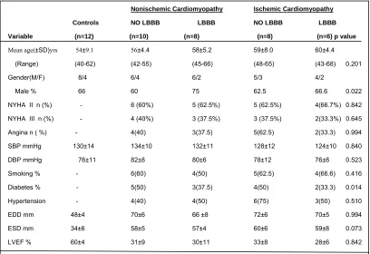
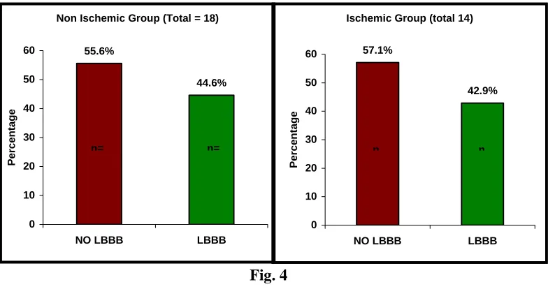
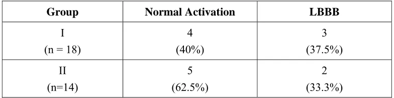
patients and 18(56.2%) had normal activation, i.e no LBBB. Twelve healthy persons without any symptoms and no risk factors for coronary artery disease are considered controls. Informed consent and approval of Institutional Committee were obtained.
II. STUDY DESIGN
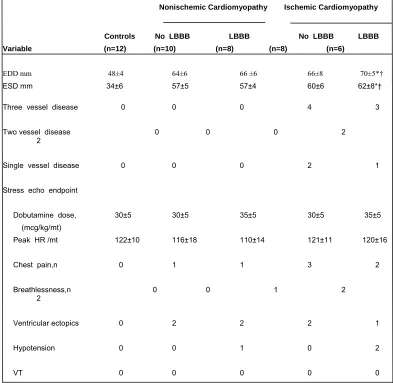
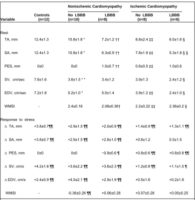
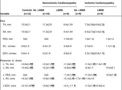
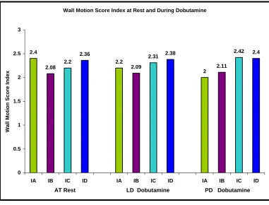
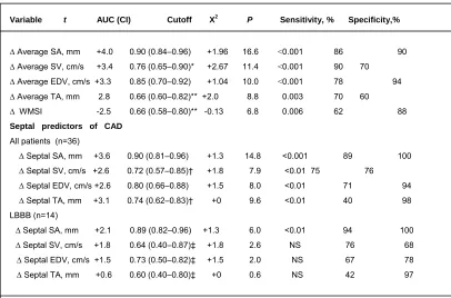
All eligible patients underwent Tissue Doppler Imaging at rest and during peak dose dobutamine stress. LV long-axis M-mode recordings and pulsed-wave, tissue Doppler (PWTD) measurements were obtained with the cursor positioned at the septal, lateral , and posterior angles of the mitral ring. Total amplitude, systolic amplitude and postejection shortening were measured from longaxis M-mode. Both systolic and early diastolic velocities were recorded at rest and at the end of each stage of dobutamine infusion from PWTD. Measurements from the three longaxis sites were combined and average value was taken for the study. Left ventricle was divided into 16 segments, according to recommendations by American Society of Echocardiography 67. For each
segment, wall motion(systolic thickening and endocardial shortening) was qualitatively analyzed and Wall motion score index (WMSI) was calculated at rest and during peak dose dobutamine stress . Ischemia was defined as worsening of wall motion score or failure of a normal segment to develop hyperkinesis during dobutamine stress.
as, having normal activation (no LBBB-subgroup A) or left bundle branch block(subgroup B).
Complete left bunble branch block
LBBB was diagnosed from ECG by the following diagnostic criteria
1. QRS duration ≥120 msec
2. Broad, notched R wave in lateral precordial leads (v 5, v 6) and usually leads I and aVL
3. Small or absent initial ‘r’ wave in right precordial leads (v 1,v 2) followed by deep S wave
4. Absent septal q wave in left sided leads
5. Prolonged intrinsicoid deflection (≥ 60 msec) in v 5 and v 6
Dobutamine stress echocardiography protocol
Dobutamine stress echocardiography was performed using a standard protocol66 in all patients after obtaining informed consent. Cardiovascular drugs were stopped 48 h before the study, apart from short-acting nitrates which patients could
continue until the morning of the investigation. No patient was taking a beta-receptor
antagonist at the time of the study. Before the infusion was begun, a resting
Dobutamine was infused intravenously for 3-minute stages at 5, 10, 20, 30 and
40 µg/kg/min with a maximum of 40 µg/kg/min. In patients not achieving 85% of
their age-predicted maximal heart rate(defined as 220 beats . mt minus age in years for both men and women) or another end-point and without symptoms or signs of myocardial ischaemia, atropine was administered, starting with 0·25 mg intravenously and repeated up to a maximum of 1·0 mg within 4 min with a continuation of the dobutamine infusion. Throughout the dobutamine infusion heart rate and rhythm were monitored from a precordial modified lead I or II or III, and blood pressure was
recorded every 3 min. Reasons for interruption of the test were: severe angina,
symptomatic reduction in systolic blood pressure >40 mmHg from baseline, hypertension (blood pressure >240/120 mmHg), significant tachyarrhythmias such as atrial fibrillation or ventricular tachycardia, akinesis or dyskinesis affecting more than two segments and any serious side effect regarded as being due to dobutamine. Later side-by-side comparisons of wall motion on the digitized quad-screen format was analyzed.
LV dimensions and function
Two-dimensional echocardiography was performed from the parasternal long- and short-axis views and apical 4- and 2-chamber views. LV internal dimension at end diastole was measured at the onset of the QRS complex and systolic internal dimension was measured at the maximal excursion of the ventricular septum from a parasternal, 2-dimensional guided M-mode echocardiogram, with the cursor at the tips of the mitral valve leaflets. Left ventricular ejection fraction was calculated using modified simpson method
M-mode of mitral annulus
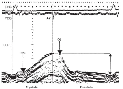
obtained at a paper speed of 100 mm/s. For measuring septal and lateral mitral annular motion ,two-dimensional echo guided M-mode recordings were made from the apical four-chamber view with the cursor placed at the septal and lateral sides of the mitral annulus. (Figure 1A). Posterior mitral annulus movement is recorded from apical long axis view. The amplitude of long axis shortening (mm) was defined as the maximum excursion of the mitral annulus during systole. Total amplitude(TA) was defined as maximum displacement of the ring between the onset of QRS and peak inward movement at or after end of systole. Postejection shortening (PES) was measured as the amplitude of shortening after the systole. Systolic amplitude(SA), representing long-axis displacement during ventricular ejection, was calculated by subtracting PES from total amplitude(Figure 1B).
[image:54.612.133.280.329.526.2]
Figure.1A - M-mode cursor
placement for measurement of mitral annular motion from apical four chamber view. The M-mode cursor is placed first at the septal and then at the lateral side of the mitral annulus
Figure.1B - An example of M mode recording of
[image:54.612.298.500.364.511.2]Parameters are measured at rest and end of each stage of dobutamine stress. Measurements from the three longaxis sites were combined and average value was taken for the study.
Pulsed-wave tissue Doppler of LV long axis
LV long-axis, pulsed-wave, tissue Doppler (PWTD) measurements were obtained with the cursor positioned at the lateral, septal, and posterior angles of the mitral ring.(Figure 2). A Doppler velocity range of 30 cm/s was selected to display systolic and early diastolic velocities. Both systolic and early diastolic velocities were recorded at rest and at the end of each stage of dobutamine infusion .All PWTD recordings were obtained at a paper speed of 100 mm/s. To make data comparable with previous studies of wall-motion score index (WMSI), measurements from the 3 longaxis sites were combined for PWTD Because LBBB is frequently associated with asynchronous septal contraction, septal long-axis values are also presented separately.
[image:55.612.126.515.431.637.2]
A B
Figure. 2 – Tissue Doppler imaging of the septal (A) and lateral mitral annulus in a normal Subject.
In tissue Doppler imaging of the annulus , there are three major velocities: Systolic velocity (S’ or Sa) ,early diastolic velocity (E’ or Ea) and late diastolic velocity (A’or Aa).IVC-Isovolumic contraction. IVR-Isovolumic relaxation
Wall Motion Score Index
Two dimensional echocardiography was performed from the parasternal
long-and short-axis views long-and from apical four- long-and two-chamber views. A full sequence
of images was recorded at the end of each 3 min stage. Baseline and peak images
were analysed using the 16-segment model of the left ventricle as recommended by
the American Society of Echocardiography and WMSI (summation of all segments
scored divided by the number of segments scored) was calculated67. Segmental wall
motion was graded 1 to 4 (where 1=normal wall motion at rest and hyperkinesis with
stress, 2=hypokinesis at rest or segments with normal amplitude at rest but a reduction
with dobutamine, 3=akinesis, and 4=dyskinesis).Ischemia was defined as worsening
of wall motion score or failure of a normal segment to develop hyperkinesis during
dobutamine stress. Multivessel involvement was defined as a new wall motion
abnormality in an arterial site distant from the resting wall motion abnormality.
III. CORONARY ANGIOGRAM
Coronary angiography was carried out via the right femoral approach using
the Judkins technique and standard projections. Significant CAD was defined as
≥50% reduction in the absolute lumen diameter of a major epicardial artery or major
branch vessel(e.g. first diagonal of LAD or obtuse marginal of Cx) estimated by eye.
The results were interpreted by observers blinded to the results of stress