Dissertation work entitled
A COMPARATIVE STUDY ON EFFECTIVENESS OF MOTOR
IMAGINARY TECHNIQUE ON IMPROVING UPPER LIMB
FUNCTION IN MIDDLE CEREBRAL ARTERY STROKE
Submitted by
Reg no: 27102317
Under the guidance of
Prof. M.SHANKAR, M.P.T .,(Neuro)., MIAP,
Dissertation submitted to
THE TAMILNADU DR. M. G. R. MEDICAL UNIVERSITY,
CHENNAI-32.
Dissertation work evaluated on ---
CERTICATE I
This is to certify that the dissertation work entitled“A COMPARATIVE STUDY ON EFFECTIVENESS OF MOTOR IMAGINARY TECHNIQUE ON IMPROVING UPPER LIMB FUNCTION IN MIDDLE CEREBRAL ARTERY STROKE” was carried out by Reg. no.27102317 P.P.G College of physiotherapy, Coimbatore-35, affiliated to The Tamilnadu Dr. M.G.R medical university, Chennai-32, under the guidance of Prof.M.SHANKAR M.P.T(Neuro)., MIAP,
Dr. K. RAJA SENTHIL M.P.T (Cardio-Resp).,MIAP.,PhD
Principal
CERTIFICATE II
This is to certify that the dissertation work “A COMPARATIVE STUDY ON EFFECTIVENESS OF MOTOR IMAGINARY TECHNIQUE ON IMPROVING UPPER LIMB FUNCTION IN MIDDLE CEREBRAL ARTERY STROKE” Was carried out by Reg.No.27102317 P.P.G College of physiotherapy Coimbatore-35, affiliated to The Tamilnadu Dr. M.G.R. Medical University, Chennai-32, under my Guidance and direct supervision.
ACKNOWLEDGEMENT
MY FAMILY MEMBERS were always been a source of all my belonging in all my aspects I am very grateful to them throughout my life.
I express my upgraded thanks to chairman Dr. L.P. THANGAVELU, M.S., F.R.C.S., and correspondent MRS. SHANTHI THANGAVELU, M.A., P.P.G group of institutions, Coimbatore, for their encouragement and providing the source for the successful of the study.
I express my sincere thanks to my principal Dr. K.RAJA SENTHIL M.P.T(Cardio-Resp),.MIAP,M.B.A,PhD, principal of P.P.G.College of physiotherapy who extend his guidance and encouragement throughout this project.
I express my special thanks to my Guide Prof. M.SHANKAR M.P.T(Neuro).,MIAP,who has suggested me to take this valuble topic. Without his interest, precious guidance, creative suggestion, constant encouragement and supported the study would have never taken up shape.
I extend my heartfelt gratitude to my PG coordinator Assistant. Prof K.RAJESH KANNAN M.P.T (ortho)MIAP., fortheirguidance and encouragement for my studies.
My heartful thanks to PHYSIOTHERAPY FACULTY members for their guidance and encouragement for my studies.
I would like to thank my brothers and sisters whose psychological support and motivation and prayers have been my strength out.
I express my thanks to all members whose prayers have been my strength to fulfill this thesis successfully.
Also I privileged to thank my dearest friends, seniors and juniors for their infinitive support and encouragement given to fulfill this thesis successfully.
CONTENTS
S.NO. TITLE PAGE
NO.
I INTRODUCTION
1.1
Introduction
1.2
Need for the study
1.3
Operational definition
1.4
Aims & Objectives
1.5
Hypothesis
01
04
05
06
07
II REVIEW
OF
LITERATURE
08
III MATERIALS
AND
METHODOLOGY
3.1 Materials
3.2 Methodology
3.2.1. Study design
3.2.2. Sampling design
3.2.3. Population
3.2.4. Sample
3.2.5. Selection criteria
•
Inclusion criteria
•
Exclusion criteria
3.2.6. Study setting
3.2.7. study method
3.2.8. Study duration
3.2.9.Parameter
IV DATA
PRESENTATION
19
V
DATA ANALTSIS AND INTERPRETATION
21
VI RESULTS
29
VII DISCUSSION
31
VIII SUMMARY
AND
CONCLUSION
33
IX LIMITATIONS
AND SUGGESTIONS
34
X BIBLIOGRAPHY
35
XI REFERENCE
37
XII APPENDIX
39
APPENDIX
I
39
APPENDIX
II
40
APPENDIX
III
42
LIST OF GRAPHS
Graph
No
Content
Page
No
1
Pre-test values of control group and experimental
group
22
2
Post-test values of control group and experimental
group
24
3
Pre-test and post-test values of control group
26
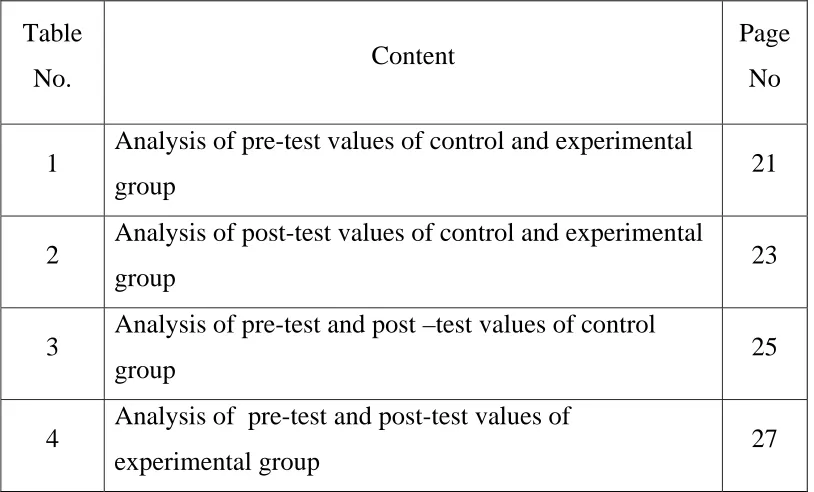
LIST OF TABLES
Table
No.
Content
Page
No
1
Analysis of pre-test values of control and experimental
group
21
2
Analysis of post-test values of control and experimental
group
23
3
Analysis of pre-test and post –test values of control
group
25
4
Analysis of pre-test and post-test values of
[image:8.612.105.512.152.398.2]1
A COMPARATIVE STUDY ON EFFECTIVENESS OF MOTOR
IMAGINARY TECHNIQUE ON IMPROVING UPPER LIMB
FUNCTION IN MIDDLE CEREBRAL ARTERY STROKE
ABSTRACT
STUDY OBJECTIVES: The effectiveness of motor imaginary technique in middle cerebral artery stroke .
DESIGN: Pre-test and post-test two group experimental study design.
PARTICIPANTS: Thirty subjects aged 40-55 years with middle cerebral artery stroke patients were selected under purposive sampling technique and assigned in to two groups with 15 subjects each,one group received conventional physiotherapy and other group received conventional physiotherapy with motor imaginary technique for a period of 4weeks
INTERVENTION: Motor imaginary technique is given to middle cerebral artery stroke patients for 20 minutes per session twice a week .
OUTCOME MEASURES: Fugl-meyer scale is to measure functional outcome before and after treatment
RESULT: Patients in experimental group shows significantly better performance than control group.
2
CHAPTER I
1.1INTRODUCTION
Brain is the major organ of the central nervous system that has control center for all the body’s voluntary and involuntary activities.
The brain has high energy requirement and little metabolic reserves. Interruption of blood flow only few minutes sets in motion a series of pathological events and damage to brain tissue.
Stroke (or) cerebro vascular accident is defined as a rapidly developing clinical sign of focal (or) global disturbance of cerebral function lasting more than 2 hours (or) leading to death with no apparent cause other than the vascular origin (WHO 1991) (Susan B.O Sullivan 2009).
A stroke is a brain attack. It is a third leading cause of death. It occurs when blood clot or ruptured vessel prevent oxygen from reaching the brain resulting in destruction of brain cells.
The disturbance of cerebral function is caused by 3 morphological abnormalities, i.e. stenosis, occlusion or rupture of the arteries. Dysfunction of the brain (neurological deficit) manifests itself by various neurological signs and symptoms that are related to the extent and site of the area involved and to the underlying causes.
Warning signs of stroke can be numbness, weakness or paralysis of face, arm, and leg especially on one side of the body sudden severe head ache, loss of balance and many factor contribute to delay in seeking medical treatment for stroke.
3
The major modifiable risk factor for stroke are transient ischemic attacks especially in presence of 70-99% carotid artery stenosis, hypertension, arterial fibrillation or other source of cardiac emboli, left ventricular hypertrophy, congestive heart failure, cigarette smoking, alcohol consumption, cocaine use, obesity, diabetes mellitus, high serum cholesterol and non modifiable risk factor are age, race, gender and family history of stroke.
Middle cerebral artery occlusion is more common site of occlusion in ischemic stroke. Post stroke hemi paralysis in middle cerebral artery syndrome leads to impairment of upper extremity functions more than the lower extremity. Early activation and forced use of involved upper extremity is effective in counter balancing this effect.
The recovery from stroke takes place in initial 3-6 months after the attack.(UMPHERD, 1998) however , research has show there can be recovery of useful motor function year’s later.
Physiotherapeutic measure on Stroke has been revolutionized in the last decade through a combination of new techniques looking at brain recovery. Advances in basic sciences and clinical research are beginning to merge and show that the human brain is capable of significant recovery after stroke, provided that the appropriate treatments and stimuli are applied in adequate amounts and at the right time. To improve functional activity there is a challenge to implement newer techniques, in that motor imaginary technique shows an important role.
Naturally the challenge in managing middle cerebral artery is to improving functional activity. Physiotherapy with it recent literatures are designing a newer or comprehensive techniques to improve the functional activity of middle cerebral artery stroke.
4
1.2 NEED FOR THE STUDY
Cerebro vascular accident is among the most frequent of all neurological disorder.
The major goal of stroke rehabilitation is functional enhancement by maximizing the independence, life style, and dignity of the patient.
The mortality due to stroke is very severe. Owing to high incidence of middle cerebral artery stroke, upper limb is severely affected than lower limb. About 20% of individual paralyzed by stroke fail to regain the functional use of affected limb, Physiotherapy techniques and approaches improves functional activity following stroke traditionally.
In recent advances shows that motor imaginary technique will play a role on improving upper limb function in stroke.
5
OPERATIONAL DEFINITIONS Stroke
“Rapidly developed clinical sign of focal (or) global disturbance of cerebral function lasting more than 24 hours or leading to death with apparent cause other than vascular origin”
-WHO (1996) Hemiplegia
Motor defects are characterized by paralysis (hemiplegic) or weakness (hemi paresis) typically on one side of the body opposite to side of lesion.
- Susan B.Sullivan(1996)
Fugl-Meyer scale
It is an impairment based test with items organized by sequential recovery stage. A three point ordinal scale is used to measure Impairments of volitional movement with grades from 0 to 2 with sub test for upper extremity function, lower extremity function, balance, sensation, pain and range of motion.
- Brunnstorm(1987) Motor imaginary technique
Motor imaginary refers to the active process by which humans experience sensations with or without external stimuli. It is an active process during which a specific action is reproduced with in working memory without any real movements
6
1.4.AIMS AND OBJECTIVES
AIM OF THE STUDY
The aim of the study is to find out the effectiveness of motor imaginary technique on improving upper limb function in middle cerebral artery stroke.
OBJECTIVE OF THE STUDY
To study the effectiveness of conventional physiotherapy on improving upper limb function in middle cerebral artery.
To study the effectiveness of conventional physiotherapy with motor imaginary technique on improving upper limb function in middle cerebral artery.
7
1.5 HYPOTHESIS
Null Hypothesis
There is no significant difference between conventional physiotherapy and conventional physiotherapy with motor imaginary technique on improving upper limb function in middle cerebral artery stroke.
Alternative Hypothesis
There is significant difference between conventional physiotherapy and conventional physiotherapy with motor imaginary technique on improving upper limb function in middle cerebral artery stroke.
8
CHAPTER-II
REVIEW OF LITERATURE
Walter. G.Bradley et al., 1993
Ischemic stroke account for approximately 85% of all strokes and common cause of death or disability in adult living in industrialized nation. Especially in developed countries like USA and UK stroke is one of the leading disorders. In india stroke is the third leading cause for death. It leads to paralytic and leads to death. It mainly affect the antigravity muscles leads to spasticity.
Bour Bonnais et al., 1995
Movements deficit in hemi paretic in upper extremities may be more a problem of agonist muscles weakness then antagonist muscles spasticity. If it progressed it leads deformity and its difficult to reduce the muscle tone .spasticity gradually increases it doesn’t mean muscle tone increases it gradually weaken the muscle tone.
Deweerdt WJG, 1998
Described the fugl meyer Measure scale is used to measure the patient’s progress and assess rehabilitation outcomes. This scale is useful in clinical settings of rehabilitation of stroke. It used to assess the functional capacity of stroke patient and also to analyse the progression of stroke. this scale is the universal scale to assess functional level of all disability commonly used stroke patients.
Keith RA et al.,1999
9
reliability, validity and sensitivity. this scale is the universal scale to assess functional level of all disability. commonly used stroke patients. The result of this scale provides perfect progression level of patients.
Maria Strokes, 1999
Following stroke there is predominantly spasticity setting on antigravity muscles. Ex: flexor of arm and limb tend to assume a flexed and pronated . antagonist muscles extensor of arm and limb and supinator. lower limb antigravity muscles flexor of hip , extensor of knee and plantar flexor of ankle. Antagonist muscles hip extensor , knee flexor and dorsi flexor of ankle.
Rene Cailiet et al., 2001
A stroke passes through a “Flaccid stage” which lasts for seconds, two minutes or a long period during this stage. All voluntary movements are lost, lost reflex and tone . initially muscle are in flaccid stage later it leads to spasticity. It shows the muscle tone abnormality and regulation of muscle tone will not happen immediately.
Kulyte Grut 2003
Proposed that physiotherapy with task-oriented rehabilitation of stroke patients represented by relearning programme is preferable to physiotherapy with facilitation/inhibition strategies, such as bobath programme in the rehabilitation of stroke. Other methods to rehabilitate stroke patients are movement based therapy, constraint induced method, mental imaginary technique.
Janeth carr 2003
10
Kenneth W . Lindsay et al., 2003
Postulated that site and size of Cerebrovascular lesion and the amount of initial collateral blood flow determines the degree of motor deficits of upper and lower limbs and face.
Steven J. Vehulst 2004
Suggested mental practice combined with neurological approaches are efficacious in treating pure motor hemiparetic strokes in terms of functional outcomes, upper and lower extremity motor skills.
Morris et al., 2006
Concluded that among patients who has stroke within previous 3 to 9 months motor imaginary technique produced statically significant and clinically relevant improvement in upper limb motor function that persisted for at least 3 to 6 months.
SusanB.O. Sullivan 2006
Clinical manifestation of middle cerebral artery includes involvement of upper extremity and face more than lower limb in stroke.
Duncan et al.,2008
Fugl-Meyer motor assessment is widely used in outcome studies and a recommended assessment of motor function in clinical practice for post stroke rehabilitation
Jerrold scott petrofsky 2009
11
Klaus kaae andeson 2009
Suggested that stroke most often occurs within the age range 40 to 55 years and about 85% of strokes are caused by ischemia.
Murray E.Brandstater et al.,2009
Fugl-Meyer scale of Motor assessment post stroke evaluated strength, reflexes, co-ordination and is composite score. It is a reliable, repeat score reflects motor recovery over time in stroke patients.
Andrea zimmermann 2008
Motor imaginary technique can also practiced in all stages of recovery. In an early stage of recovery, motor imaginary technique allows patients to mentally practice a task which they cannot yet carry out physically due to motor impairment .
Sheton to ,mahoey 2009
Suggested that Stroke often strikes after age 40, involved more than 38000 veterans aged 40and older.
jeannerod 2010
Motor imaginary refers to the active process by which humans experience sensations with or without external stimuli. It is an active process during which a specific action is reproduced with in working memory without any real movements
12
CHAPTER III
MATERIALS AND METHODOLOGY
3.1 MATERIALS
• Table
• Pillows
• Ice
• Chair
• Towel
• Couch
• Peg board
• Needle and thread
• Audio tape
3.2 METHODOLOGY 3.2.1. Study Design
Pre-test, post-test two group Experimental study design
3.2.2 Sampling design
Purposive sampling technique.
3.2.3 population
The sample size consist of 30 subjects with middle cerebral artery stroke were selected assigned in to control group and experimental group.
Control group:
consist of 15 middle cerebral artery stroke subjects treated with conventional physiotherapy .
Experimental group:
13
3.2.4Sample
30 subjects who fulfilled inclusion and exclusion criteria were selected for the study.
3.2.5 Criteria for selection of subjects Inclusion criteria
• Ability to walk indoor without a stick indicating no major balance problem
• Hemi paretic patient within the involvement of middle cerebral artery.
• Above one month post-stroke and within one year.(Brunnstorm stage 2)
• Ischemic type of stroke.
• Age groups between 40-55 years
• Both gender
• Both sides of involvement
Exclusion criteria
• Serious sensory or cognitive and aphasic deficit
• Other type of stroke (hemorrhagic, lacunars)
• Comatosed patients
• Bilateral involvement
• Balanced disorder
• Medical instability
• Any recent fracture or surgery
• Recent myocardial infarction
• Auditory impairment
• perceptual defects
• reflex sympathetic dystrophy
14
3.2.6 Study setting
Study was conducted at
• ASHWIN MULTI-SPECIALITY HOSPITAL.
• VIVEKANANDHA INSTITUTE OF MEDICAL
SCIENCES , THIRUCHENCODE.
• OUT PATIENT DEPARTMENT PPG COMMUNITY CENTRE.
3.2.7 Study method
Subjects were divided into control group and Experimental group .
CONTROL GROUP :
15 subjects were treated with conventional physiotherapy. EXPERIMENTAL GROUP :
15 subjects were treated with conventional physiotherapy and motor imaginary technique
3.2.8 Study duration
Study was conducted for a period of 6 months.
3.2.9Parameters
15
3.2.10 Statistical Tools
To compare control Group and Experimental Group : Independent ‘t’ test:
Statistical analysis is done by using Independent ‘t’ test
t =
(
)
2 2 2 1 2 1
n
n
n
n
S
X
X
+
−
2 2 S = (x1 – x1 ) + ( x 2 – x 2 ) n 1 + n 2 - 2
x1 = mean value of control group
x2 = mean value of experimental group n1= number of observations in control group n2= number of observations in experimental group S = standard deviation
Intra group analysis:
Statistical analysis is done by using Paired‘t’ test
s
n
d
t
=
s
=1
)
(
2 2−
−
∑
∑
n
n
d
d
d = difference between the pre-test Vs post test values d = mean difference
16
3.2.11 TREATMENT TECHNIQUE : CONTROL GROUP :
IN SITTING:
Sitting on a firm flat surface, hands rests over bed, feet flat on floor, while therapist place one hand over elbow and other over wrist.
(i) Weight shifting to both sides.
(ii) Clasping both hands forward, turning to sound side. While lifting the affected leg and crossing it over the sound side.
(iii)Clasping both hands forward, turning to affected side. While lifting the sound leg and crossing it over the affected side.
(iv) Sitting with crossed legs. The affected leg over the sound one. While both hand clasps and places over knee.
(v) Flexion and extension of knee. Therapist places one hand over foot other hand over knee.
FROM SITTING TO STANDING:
(i) Clasping both hands forward. Affected foot parallel with sound one. Therapist place one hand over sacrum and other hand over knee.
(ii) Patient stands up weight bearing over affected leg.
Stage 1:Therapists assists in holding patient and help them to raise up. Stage 2: Assist by clasping hands forward and without therapist support. Stage 3: With one hand support.
Stage 4: Without hand support.
IN STANDING:
17 FOR MOVEMENTS OF ARM:
(i) Elevation of arms with clasped hands.
(ii) Moving clasped hands to face, while therapists hand prevents retraction of shoulder.
(iii)Moving clasped hands above head, while therapists hand prevents retraction of shoulder.
(iv) Mobilizing shoulder girdle with extended arm. (v) Bilateral shoulder flexion exercises.
(vi) Sitting push-ups to full elbow extension.
ICE THERAPY
placing the patients hand in a bucket of melting ice for a few seconds brings intense awareness of the part , reduces spasticty and often improves movement.
STRETCHING
all spastic muscles especially biceps brachii ,wrist and finger flexors. LOWER LIMB EXERCISE
mobilising the leg and toes ,bridging exercise, activities on mat, weight bearing exercise, activites on tilt board.
TREATMENT DURATION AND REPETATION 60 minutes and 20 repetition per exercise HOME EXERCISE
needle and thread activity , button activity, peg board activities.
EXPERIMENTAL GROUP:
Conventional physiotherapy given as same as control and motor imaginary technique given.
18
provided during 6 weeks interventions, one during first 2 weeks , second during second 2 weeks , third during third 2 weeks).
Internal, cognitive images were used in which patient received audio tape command imagine himself from third person perspective executing the tasks specified on mental practice audio tape .the intervention was intended to target and improved functional use the patients affected wrist and fingers as well as to secondary improve his ability to move out of synergy with affected arm.
During first 2 weeks , the audio taped functional task was reaching for grasping a cup , during the second 2 weeks , functional tasks practiced was turning pages in large book. during third 2 weeks task practiced was reaching for and grasping a item on a high self and bringing an item to himself, for each of this task the patient was urged to use all of his senses (eg. Feel your fingers grasp around the edge of the cup )
The duration of treatment is 20 minutes per session two session per week.
3.2.12Procedure
The subjects of both control group and experimental group were involved for pre test assessment by fugl- meyer assessment scale (hand component).
The subjects of control group were given conventional physiotherapy and experimental group were given conventional physiotherapy and motor imaginary technique
19
CHAPTER IV
DATA PRESENTATION
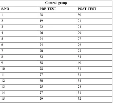
TABLE I
Pre test and Post test values of control group using Fugl meyer scale
Control group
S.NO PRE-TEST POST-TEST
1 28 30
2 19 21
3 22 24
4 26 29
5 24 27
6 24 26
7 20 22
8 32 34
9 38 40
[image:27.612.145.521.245.588.2]20
TABLE II
Pre test and Post test values of Experimental group using Fugl Meyer scale Experimental group
S.NO PRE-TEST POST-TEST
1 19 24
2 26 31`
3 25 30
4 32 36
5 28 37
6 28 35
7 27 34
8 25 32
9 24 29
21
CHAPTER –V
DATA ANALYSIS AND PRESENTATION
TABLE-III
ANALYSIS OF PRETEST DATA OF CONTROL GROUP AND EXPERIMENTAL GROUP
TESTS CONVENTIONALPHYSIOTHERAPY AND CONVENTIONAL PHYSIOTHERAPY WITH
MOTOR IMAGINARY TECHNIQUE
Pre test mean value
Control Group Experimental Group
26.6 26.13 Independent ‘t’ test
0.24
P value and its
significance P value > 0.05 is insignificant
For 28 degrees of freedom at 5% level of significance, the calculated pre test ‘t’ value between control group and Experimental group was 0.24 and the critical value was 1.701, which states that there is no significant difference between two groups.
22
GRAPH - I
PRE –TEST VALUES OF CONTROL GROUP AND
EXPERIMENTAL GROUP
26.6
26.13
0
5
10
15
20
25
30
Pre Test
Pre Test
F
U
G
L-
M
E
Y
E
R
S
C
A
L
E
Pre Test
23
[image:31.612.104.509.255.432.2]
TABLE IV
ANALYSIS OF POST TEST DATA OF CONTROL GROUP AND
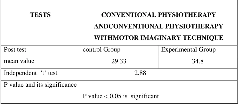
EXPERIMENTAL GROUP
TESTS CONVENTIONAL PHYSIOTHERAPY ANDCONVENTIONAL PHYSIOTHERAPY WITHMOTOR IMAGINARY TECHNIQUE
Post test mean value
control Group Experimental Group
29.33 34.8 Independent ‘t’ test 2.88
P value and its significance
P value < 0.05 is significant
24
GRAPH - II
POST –TEST VALUES OF CONTROL GROUP AND
EXPERIMENTAL GROUP
29.33
34.8
0
10
20
30
40
Post Test
Post Test
Pre Test
Post Test
25
[image:33.612.104.507.236.357.2]
TABLE V
ANALYSIS OF PRETEST AND POSTTEST DATA OF CONTROL
GROUP
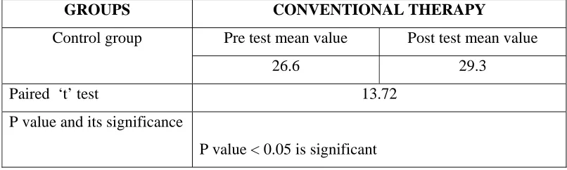
For 14 degrees of freedom at 5% level of significance, the student ‘t’ test value for control group (CONVENTIONAL PHYSIOTHERAPY) was 13.72 and the critical value was 1.761, which states that there exists significant difference between the pre test and post test values of control group
GROUPS CONVENTIONAL THERAPY
Control group Pre test mean value Post test mean value
26.6 29.3
Paired ‘t’ test 13.72
P value and its significance
26
GRAPH - III
PRE TEST AND POST TEST VALUES OF CONTROL GROUP
26.6
29.33
0
10
20
30
40
Pre Test
Post Test
Pre Test
Post Test
27
TABLE VI
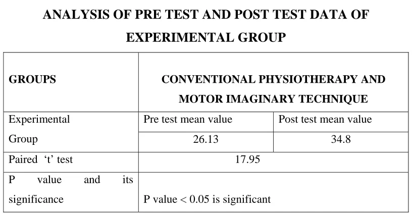
ANALYSIS OF PRE TEST AND POST TEST DATA OF
EXPERIMENTAL GROUP
GROUPS CONVENTIONAL PHYSIOTHERAPY AND MOTOR IMAGINARY TECHNIQUE
Experimental Group
Pre test mean value Post test mean value
26.13 34.8 Paired ‘t’ test 17.95
P value and its
significance P value < 0.05 is significant
28
GRAPH - IV
PRE TEST AND POST TEST VALUES OF EXPERIMENTAL
GROUP
26.1
34.8
0
10
20
30
Pre Test
Post Test
Pre Test
Post Test
F
29
VI.RESULTS
Effectiveness of control Group (conventional physiotherapy) is elicited by comparing the pre test and post test values of experimental group using paired ‘t’ test; the calculated value is 13.72 , whereas the critical value is 1.761. Since the calculated value is greater than the critical value, there exists a significant difference between the pretest and post test values of control group .
Effectiveness of Experimental group (conventional physiotherapy with motor imaginary technique ) is elicited by comparing the pretest and post test values of Experimental group using paired ‘t’ test, the calculated value is 17.95, whereas the critical value is 1.761. Since the calculated value is greater than the critical value, there exists a significant difference between the pretest and post test values of Experimental group
While comparing the post test values of control group and Experimental group using independent ‘t’ test, the calculated value is , 2.88 whereas the critical value is 1.761, which shows that there exists a significant difference between the post test values of two groups.
When comparing the mean values of both, the post test mean value of control group 29.33 is lesser than the post test mean value of Experimental group 34.8 which confirms that Experimental group shows a significant improvement than control group .
Rehabilitation of stroke patients is a complex and difficult procedure. Various physiotherapy strategies evolved over the years for the rehabilitation of stroke patients.
Motor imaginary technique is the therapeutic programme which aims at the optimization of function by training patients in various tasks related to the daily activities.
30
assessment scale (upper limb component). Motor imaginary technique in hemparetic patients with middle cerebral artery involvement.
Result obtained from statistical analysis between pretest and post test values of experimental group and control group at 5% level of significance showed significant improvement in Fugl Meyer Assessment Scale by Motor imaginary technique following 4weeks of exercise programme.
Analysis of results shows that there in an increase of 24% in outcome measure of Fugl Meyer Assessment scale.
31
VII DISCUSSION
Rehabilitation of the hemiplegic patients is essential for improving functional activities.stroke affects patient’s normal activities of daily living and make them dependent to others .
The purpose of this study is to synthesize the relavant literature about motor imaginary technique in order to facilitate its integration in to physical therapist practice .
SUSAN B.O’ SULLIVAN described occlusion of the proximal middle cerebral artery produces extensive neurological damage with significant cerebral oedema .increased intracranial pressure typically lead to loss of consciousness ,brain herniation and possibly death.
The most common characteristics of middle cerebral artery syndrome or contralateral spastic hemiparesis and sensory loss of face , upper extremity and lower extremity, with the face and upper extremity more involved than the lower extremity .
MAGILL suggested that mental practice is effective because it augments existing motor schema .at the level pretest the patient had limited ability to use the affected wrist and fingers but a greater ability to perform gross movements with the affected arm , as indicated by his scores on items on fugl-meyer scale.
After participating in mental practice intervention targeting grasping , reaching and gripping behaviours the patient maintained his gross motor score while improving on the fine motor components of fugl-meyer scale , at the post test the specificity of the changes in the areas targeting suggests enhancement of the existing motor plan as a possible mechanism.
32
musculature and in the appropriate neuro motor pathways. this correlative neuro motor activity is similar to the activity that we hypothesize occurs with repetitive physical practice and is responsible for motor performance improvements that individuals exhibit after mental practice .
we believed that the patient improvements between the pretest and the post test occurred because the patient, through mental practice, was provided with additional practice of functional tasks using the affected arm.
On a physiological level we believed that this practice caused priming of the motor cortex and appropriate activation of the neuro motor pathways, which resulted in the patient’s improvements. we believed that correlating changes in motor behaviour with changes in cortical organization using functional magnetic resonance imaging might substantiate this claim.
Mental image of movement can be generated independent of behavioural output of paretic limb.as patients motor function began to improve daily activities using the affected limb were implemented . outcome measures were grip strength shoulder flexibility and time to complete common daily activities such as dressing and inserting a key in lock with greater precision and ease of movement .
The functional activities of stroke patients is measured by fugl-meyer scale. it is an impairment based scale test items organized by sequential recovery stage (BRUNNSTORM 2007)
Thus, motor imaginary technique may provide a valuable tool to access the motor network and improve outcome after stroke .
Hence , it thought this form of technique can prove useful in stroke patients who have lost movements.
33
VIII.SUMMARY AND CONCLUSION
This is study finds out the efficacy of Motor imaginary technique in improving upper limb function in middle cerebral artery stroke.
In 30 patients the upper limb function was measured by Fugl Meyer assessment.
The control group subjects were given conventional physiotherapy and experimental group were given conventional physiotherapy and motor imaginary technique. For both groups various exercise through peg board, pronation- supination board, threading board.
The subjects were strictly instructed not to sleep during the treatment Initially patient feel difficult in imnagine and also feels bore . Samples were given conventional physiotherapy for lower extremity.
The duration of the treatment program was 4 weeks treatment motor performance was done through Fugl Meyer assessment scale.
34
IX.LIMITATIONS AND SUGGESTION
• This study was very short term and therefore to make it more valid long term is necessary.
• Since the study has been done with smaller number of subjects further studies should be conducted with large group of population.
• Motor imaginary technique is not well applicable for stroke patients who are having cognitive and sensory defect
• Though the Fugl Meyer Assessment and were administered objectively bias is possible, further study can be done other reliable assessment tools.
35
CHAPTER X
BIBLIOGRAPHY
1. A.B.Taby, Neurorehabilitation, Principles and practice, 2/e, 2001, Ahuja Publications.
2. Berta Bobath, Adult hemiplegia, Evaluation and treatment, 3/e, 1990, Butterworth- Heinemann page89-182.
3. Carole.B.Lewis, Geriatric Physical Therapy and clinical approach, page 379-396.
4. Catherine.A.Trombly, Neurophysical and development of treatment approaches, page 91-103.
5. Darcy Ann Umphred, Neurological Rehabilitation, 2/e, 1990, Mosby, page 772-776.
6. Glady Samuel Raj, Physical Therapy in Neuro conditions, 1/e, 2006, Jaypee Brothers, page 28-30.
7. Janet H. Carr, Neurological Rehabilitation, 1998, Elsevier, page 220-260. 8. Janet H. Carr, Stroke Rehabilitation optimizing motor performance, 1/e ,
2003, Butterworth- Heinemann. Page129-205.
9. Janet M. Howle Neuro-developmental treatment approach: theoretical foundations , Page 319
10. Joel A Delisa – Editor, Physical Medicine and Rehabilitation Principles and Practice,4 /e, 1998, Lippincott , page 986-990.
11. John Gilroy, Basic neurology, 3 /e, 2000, McGraw- Hill, page 225-227. 12. John Walton- Editor, Briain’s disease of Nervous system, Oxford.
13. Kenneth W. Lindsay, Neurology and Neuro surgery illustrated, 4/e, 2004, Elsevier. page 239-269.
36
15. Patrica.A.Downie, Cash’s Text book of Neurology for physiotherapists, 4 /e, 2007, Jaypee, page 194,213-254.
16. Raine S. The current theoretical assumptions of the Bobath Concept as determined by the members of BBTA. Physio Therapeutical Practice.
2007; 23: page137–152.
17. Raymond D.Adams, Principles of Neurology, 6 /e, 1997, McGraw hill. 18. Sethi P.K. Stroke: incidence in India and management of ischaemic stroke.
Neurosciences Today, 2002 6 (3). page. 139-143
19. Stuart.B.Porter, Tidy’s physiotherapy, 13/e, 2003, Butterworth and Heinemann, page446.
20. Susan B.O’ Sullivan, Physical Rehabilitation, 5/e, 2006, JaypeeBrothers, page 388-755.
37
CHAPTER XI
REFERENCE:
• INTERNATIONAL JOURNAL OF PHYSICAL THERAPY:
ISSN 2079-9209
• JOURNAL OF NEUROLOGICAL PHYSICAL THERAPY
WOLTER KLUVER
• THE JOURNAL OF NEUROLOGIC PHYSICAL THERAPY EDELLE C. PT.Phd
• INDIAN JOURNAL OF PHYSIOTHERAPY AND OCCUPATIONALTHERAPY
ISSN 0973 5674
• INTERNATION JOURNAL OF THERAPY AND REHABILITATION
LEVIN.S CARDOSA
• JOURNAL OF PHYSICAL THERAPY EDUCATION
HUNGIKO
• JOURNAL OF PHYSICAL THERAPY SCIENCE
ALEXANDRA VA23313
• JOURNAL OF PHYSIOTHERAPY RESEARCH AND PRACTICE
KLUHNKI
• JOURNAL OF NEROREHA
NADINE.DECK
• JOURNAL OF SMOKING AND RISK OF STROKE HANKEY GJ
• AMERICAN JOURNAL OF CLINICAL NUTRITION
LARSSON S.C
38
GORELICK PB
• JOURNAL OF INTERNATIONAL SOCIETY OF PATHOPHYSIOLOGY OF STROKE
DEB P SHARMA
• THE NEW ENGLAND JOURNAL OF MEDICINE
LEES K.R
• JOURNAL OF THROMBOSIS AND HAEMOSTASIS
39
CHAPTER XII
APPENDIX – I
CASE ASSESSMENT PROFORMA
CASE NO :
NAME :
SEX :
ADDRESS :
DATE OF ADMISSION : DATE OF EVALUATION :
HISTORY :
ON OBSERVATION :
ON EXAMINATION :
TREATMENT : Motor imaginary technique MEASUREMENT TOOL : fugl-meyer scale
S.NO. PRE TEST POST TEST
40
APPENDIX – II
PATIENT CONSENT FORM
TITLE: “A COMPARATIVE STUDY ON EFFECTIVENESS OF MOTOR IMAGINARY TECHNIQUE ON IMPROVING UPPER LIMB FUNCTION IN MIDDLE CEREBRAL ARTERY STROKE .”
INVESTIGATOR: _ _ _ _ _ _ _ _ _ _ _
PURPOSE OF THE STUDY:
I_ _ _ _ _ _ _ _ _ _ _ _ _ _ _ _ ,have been informed that this study will work towards achieving on the functional activities of daily living in post-stroke conditions for me and other patients.
PROCEDURE:
Each term of the study protocol has been explained to me in detail. I understand that during the procedure, I will be receiving the treatment for one time a day. I understand that I will have to take this treatment for four weeks. I understand that this will be done under investigator, _ _ _ _ _ _ _ _ _ _ _ _ _ _ _ supervision. I am aware also that I have to follow therapist’s instructions as has been told to me.
CONFIDENTIALITY:
41
SRISK AND DISCOMFORT:
I understand that there are no potential risks associated with this procedure, and understand that investigator will accompany me during this procedure. There are no known hazards associated with this procedure.
REFUSAL OR WITHDRAWL OF PARICIPATION:
I understand that the decision my participation is wholly voluntary and I may refuse participate, may withdraw consent at any time during the study.
I also understand that the investigator may terminate my participation in the study at anytime after researcher has explained me the reasons to do so.
I _ _ _ _ _ _ _ _ _ _ _ _ _ _ have explained to ………. the purpose of the research, the procedures required and the possible risks and benefits,to the best of my ability.
……… ……… investigator Date
I ………. Confirm that researcher has explained me the purpose of the research, the study procedure and the possible risks and benefits that I may experience. I have read and I have understood this consent to participate as a subject in this research project.
……….. ……… Subject Date
43
APPENDIX - III
FUGL MEYER ASSESSMENT SCALE
Area Test Scoring
Maximum Possible Score
Attained Score Motor
I Reflex
a. biceps---
b. triceps---
0 - No reflex activity can
sbe elicited
2 - Reflex activity can
be elicited
4
II. Flexor Synergy Elevation---
Shoulder retraction-
Abduction (at least 90°) -- External rotation ----
Elbow extension ----
0 - Cannot be performed
at all
1 - Performed partly
44 Forearm supination---
III. Extensor Synergy
Shoulder adduction/internal rotation----
Elbow extension ----
Forearm pronation –
0 - Cannot be performed
at all
1 - Performed partly
2 - Performed faultlessly 6
IV. Movement combining synergies
a. Hand to lumbar spine ---
b. Shoulder flexion to 90° elbow at 0 ---
c. Pronation/supination of forearm with
elbow at 90° and shoulder at 0
1 - No specific action performed
2 - Hand must pass anterior superior iliac
spine
3 - Action is performed faultlessly
0- Arm is immediately abducted or elbow
flexes at start of motion.
1- Abduction or elbow flexion occurs in later
phase of motion
2- Faultless motion
45
cannot be attained, and/ or pronation or
supination can not be performed at all
1- Active pronation or supination can be
performed even within a limited range of
motion, and at the same time the shoulder
and elbow are collectly positioned.
2- Complete pronation and supination with
correct positions at elbow and shoulder.
V. Movement out of synergy
a. Shoulder abduction to 90° elbow at 0 and foream pronated-
b. Shoulder flexion, 90°–180° elbow at 0 and forearm in mid position
0- Initial elbow flexion occurs or any deviation
from pronated forearm occurs.
1- Motion can be performed partly, or if during
motion, elbow is flexed or forearm cannot be
kept in pronation.
2- Faultless motion
0- Initial flexion elbow occurs or shoulder
abduction occurs
1- Elbow flexion or shoulder abduction, occurs
46 c. Pronation/supination of forearm elbow at 0
and shoulder between 30° – 90° of flexion----
during shoulder flexion
2- Faultless motion
0- Supination and pronation cannot be
performed at all or elbow and shoulder position
cannot be attained
1- Elbow and shoulder properly positioned and
pronation and supination performed in a
limited range.
2- Faultless motion
VI. Normal reflex activity
Biceps and/or finger flexors and triceps---
(This stage, which can render the score of two,
is included only if the patient has a score of 6
in stage V).
0- At least 2 of the 3 phase reflexes are
markedly hyperactive
1- One reflex markedly hyperactive or at least
2 reflex are lively.
2- No more than one reflex is lively and none
47
are hyperactive.
VII.
a. Stability, elbow at 90°, shoulder at 0
b. Flexion/extens
c. ion, elbow at 90°, shoulder at 0
c. Stability, elbow at 0, shoulder at 30°
d. Flexion/extension, elbow at 0, shoulder at
30°
a) 0- Patient cannot dorsiflex wrist to required
15
1- Dorsiflex is accomplished, but no resistance
is taken
0- Position can be maintained with some
(slight) resistance.
b) 0- Volitional movement does not occur
1- Patient cannot actively move the wrist joint
throughout the total ROM.
2- Faultless, smooth movement
c) Scoring is the same as for item a
d) Scoring is the same as for item b
e) 0- Cannot be performed
48
e. Cirumduction 1- Jerky motion or incomplete circumduction
2- Complete motion with smoothness
VIII.
a. Finger Mass Flexion
b. Finger Mass Extension
c. Grasp #1- MP joints extended and PIPS &
DIPS are flexed. Grasp is tested against
resistance
a) 0- No flexion occurs.
1- Some flexion, but not full motion
2- Complete active flexion ( compared with
unaffected hand)
b) 0- No extension occurs
1- Patient can release an active mass flexion
grasp
2- Full active extension.
c) 0- Required position cannot be acquired
1- Grasp is well
2. Grasp can be maintained against relatively
great resistance
d) 0- Function cannot be performed
49 d. Grasp #2 – patient is instructed to adduct
thumb, 1st carpometacarpophalangeal and interphalangeal joint at 0
e. Grasp #3 – patient opposes the thumb pad
against the pad of index finger. A pencil is
interposed.
f. Grasp #4- the patients should grasp a
cylinder shaped object (small can), the
volar surface of the 1st and 2nd finger against each other
g. Grasp #5 – a spherical grasp
1- Scrap of paper interposed between the thumb
and index finger can be kept in place, but not
against a slight a tug.
2- Paper is held firmly against a tug.
e) Scoring procedures are the same as for Grasp
#2.
f) Scoring procedures are the same as for Grasp
#2 and #3
g) Scoring procedures are the same as for grasp
#2, 3 and #4.
IX. Coordination/ speed – finger to nose (five
repetitions in rapid succession)
a) 0- Marked tremor
1- Slight tremor
2- No tremor
50 a) Tremor----
b) Dysmetria
c) Speed
b) 0- Pronounced or unsystematic dysmetria
1- Slight or systematic dysmetria
2- Do dysmetria
c) 0- Activity is more than 6 seconds
longer than unaffected hand
1- 2 to 5 seconds longer than
unaffected hand
2 Less than 2 seconds difference.
CONTROL GROUP
S.NO. AGE
SEX
AFFECTED
SIDE
PRE
TEST
POST
TEST
1 47 M
Right
2 52 M
Right
3 54 F
Left
4 45 M
Right
5 51 F
Left
6 52 F
Right
7 50 M
Left
8 49 M
Right
9 51 M
Left
10 54
F
Left
11 48 M
Right
12 55
F
Right
13 47
F
Right
14 53
F
Left
EXPERIMENTAL GROUP
S.NO. AGE
SEX
AFFECTED
SIDE
PRE
TEST
POST
TEST
1 51 F
Left
2 49 M
Left
3 47 F
Right
4 50 F
Right
5 54 M
Right
6 48 M
Right
7 42 F
Left
8 53 F
Left
9 54 M
Left
10 47 M
Left
11 51
F
Left
12 54
F
Right
13 52 M
Right
14 49 M
Right
GRAPH - I
PRE –TEST VALUES OF CONTROL GROUP AND EXPERIMENTAL
GROUP
26.6
26.13
0
5
10
15
20
25
30
Pre Test
Pre Test
F
U
G
L-
M
E
Y
E
R
S
C
A
L
E
Pre Test
GRAPH - II
POST –TEST VALUES OF CONTROL GROUP AND EXPERIMENTAL
GROUP
29.33
34.8
0
10
20
30
40
Post Test
Post Test
Pre Test
Post Test
GRAPH - III
PRE TEST AND POST TEST VALUES OF CONTROL GROUP
26.6
29.33
0
10
20
30
40
Pre Test
Post Test
Pre Test
Post Test
GRAPH - IV
PRE TEST AND POST TEST VALUES OF EXPERIMENTAL GROUP
26.1
34.8
0
10
20
30
Pre Test
Post Test
Pre Test
Post Test
F