JOURNAL OFVIROLOGY, July 1995, p. 4299–4307 Vol. 69, No. 7 0022-538X/95/$04.0010
Copyrightq1995, American Society for Microbiology
Effect of Single-Base Substitutions in the Central Domain of
Virus-Associated RNA I on Its Function
ARSHAD RAHMAN,1† PAWAN MALHOTRA,1‡ RAVI DHAR,2TARUN KEWALRAMANI,1 ANDBAYAR THIMMAPAYA1*
Lurie Cancer Center and Microbiology and Immunology Department, Northwestern University Medical School, Chicago,
Illinois 60611,1and Laboratory of Molecular Virology, National Cancer Institute, Bethesda, Maryland 208922
Received 19 January 1995/Accepted 23 March 1995
Adenoviruses use virus-associated RNA I (VAI RNA) to counteract the cellular antiviral response mediated by the interferon-induced, double-stranded-RNA-activated protein kinase PKR. VAI RNA is a highly struc-tured small RNA which consists of two long duplex regions connected at the center by a complex, short stem-loop. This short stem-loop and the adjacent base-paired regions, referred to as the central domain, bind to PKR and inactivate it. Currently it is not known whether binding of VAI RNA to PKR is dependent solely on the secondary (and tertiary) structure of the central domain or whether nucleotide sequences in the central domain are also critical for this interaction. To address this question, 54 VAI mutants with single-base substitution mutations in the central domain of the RNA were constructed, and their capacities to inhibit the autophosphoryation of PKR in vitro were determined. It was found that although about half of the mutants inhibited PKR activity as efficiently as the wild type, a significant number of mutants lost the inhibitory activity substantially, without a perceptible change in their secondary structures. These results indicate that, in addition to secondary structure, at least some nucleotides in the central domain may be critical for the efficient function of VAI RNA.
The interferon-induced, double-stranded RNA (dsRNA)-activated protein kinase PKR (also referred to as p68 kinase, eukaryotic initiation factor 2 [eIF-2] alpha kinase, and DAI) is a ribosome-bound latent protein kinase that cells use as a defense against infection by viruses. PKR is activated upon infection of cells with viruses, and the activated enzyme phos-phorylates the alpha subunit of the protein synthesis initiation factor eIF-2. The phosphorylated eIF-2 is unable to recycle during protein synthesis; as a result, protein synthesis comes to a halt (reviewed in references 11 and 25). Viruses have devel-oped diverse strategies to counteract this cellular antiviral re-sponse; the best studied is that used by adenoviruses (Ad) (reviewed in references 16, 17, 27, and 30). Human Ad syn-thesize large amounts of an RNA polymerase III-directed, low-molecular-weight RNA designated virus-associated RNA I (VAI RNA). It is a 160-nucleotide (nt), highly structured RNA with two long duplex regions connected at the center by a complex, short stem-loop. The central short stem-loop and the adjacent base-paired regions are referred to as the central domain. Cells infected with Ad mutants with the VAI RNA gene deleted are defective in translation and have high levels of PKR activity and phosphorylated eIF-2 (26, 28, 29, 32). VAI RNA binds to and irreversibly inactivates PKR and thereby protects and maintains the eIF-2 activity (4, 8, 12, 14, 22). The structural requirements of VAI RNA for its function have been investigated by introducing deletions and linker-scan and base-compensatory substitutions into the VAI RNA and ana-lyzing such mutants in vivo in virus infections and transient
assays and also in vitro for their capacities to bind to and block the autophosphorylation activity of PKR (1, 4, 5, 7, 8, 19–21, 24). These studies showed that mutant molecules would retain function as long as the integrity of the central domain was maintained. Only those molecules which underwent even a small alteration in the structure of the central domain failed to function in vivo and in vitro, indicating that the central domain is the most critical part of the molecule.
The in vivo target of the VAI RNA, PKR, has also been examined extensively by several investigators (9, 13, 18, 23). These studies indicate that both the inhibitor VAI RNA and the activator dsRNA bind to the same region of PKR. This raises an important question as to how the VAI RNA, which has a structure dramatically different from that of dsRNA, interacts with PKR. Because binding of VAI RNA to PKR correlates with function and because the integrity of the central domain is critical for its function, we asked whether interaction of VAI RNA with PKR is based solely on the secondary struc-ture of the central domain or whether nucleotide sequences within the central domain are also critical for this interaction. In other words, is it possible to construct VAI mutants with mutations in the central domain that are compromised for function without detectable alterations in the secondary struc-ture of the central domain? Because there is very little detect-able sequence homology between the nucleotide sequences of VAI RNAs of different serotypes (15), the nucleotide se-quences of the VAI RNA have not been considered to be important for function. The role of nucleotide sequences could not be addressed in previous mutagenesis studies because the functionally defective mutants that have altered nucleotide sequences in the central domain also have an altered central domain structure. In this study, we introduced 54 single-base substitution mutations into the central domain of VAI RNA and analyzed the capacities of the mutants to function in vitro. Surprisingly, we found that a significant number of these sub-stitution mutants failed to function efficiently, even though their secondary structures were intact. These studies suggest * Corresponding author. Mailing address: Lurie Cancer Center and
Microbiology and Immunology Department, Northwestern University Medical School, 303 E. Chicago Ave., Chicago, IL 60611. Phone: (312) 503-5224. Fax: (312) 908-1372.
† Present address: Department of Pharmacology, Rush-Presbyterian St-Luke’s Medical Center, Chicago, IL 60612.
‡ Present address: International Center for Genetic Engineering and Biotechnology, UNIDO, New Delhi 110001, India.
4299
on November 9, 2019 by guest
http://jvi.asm.org/
that, in addition to recognizing the structure, PKR may also recognize certain nucleotide sequences within the context of the secondary structure of the central domain.
MATERIALS AND METHODS
Construction of the single-base substitution mutations in the central domain. pT7VA is a plasmid in which the DNA sequences coding for the Ad type 5 (Ad5) VAI gene were cloned downstream of the T7 promoter. At the 39end, the VAI coding sequences terminate with an EcoRI site (6). When the EcoRI-linearized pT7VA is transcribed in vitro with T7 RNA polymerase, it produces an RNA with an authentic 59end and four U residues at the 39end, followed by a G residue which is a part of the EcoRI recognition sequence. The VAI coding sequences contain CspI and BstEII sites at positions161 and199, respectively, and a newly introduced EcoRI site at position1160 (all numbering is based on a G start [31]). Nucleotide sequences between positions161 and199 were mutated by the substitution of DNA sequences of pT7VA between CspI and BstEII sites with the synthetic double-stranded oligonucleotides with single-base substitutions at the positions indicated in the names of the mutants. Similarly,
nucleotides between positions199 and1160 were mutated by the substitution of synthetic double-stranded oligonucleotides between the BstEII and EcoRI sites. The mutations were confirmed by DNA sequence analysis. All DNA manipula-tions were carried out by using standard recombinant DNA technology.
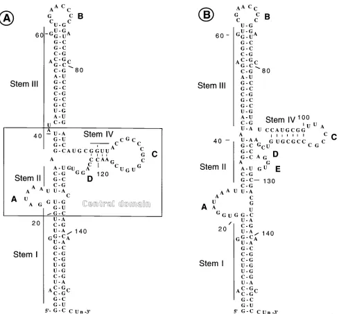
[image:2.612.65.554.67.520.2]Purification and in vitro phosphorylation of PKR.PKR was purified by sub-jecting the ribosomal salt wash to successive chromatographies on DEAE-cellu-lose, Mono S, and hexylamine agarose columns. Further details of this purifica-tion scheme can be obtained from reference 7. Autophosphorylapurifica-tion of PKR was carried out in a 50-ml reaction mixture consisting of 10ml of partially purified PKR obtained from the hexylamine agarose column fraction, 20 mM Tris-HCl (pH 7.5), 2 mM 2-mercaptoethanol, 4 mM magnesium acetate, 10 mCi of [g-32P]ATP (specific activity, 30 Ci/mmol; New England Nuclear Corp.), 2mg of reovirus RNA per ml as dsRNA, and various concentration of VAI RNAs, as indicated in the figures. The reaction mixture was incubated for 30 min at 308C, and the reaction was terminated by the addition of 50ml of 23sodium dodecyl sulfate (SDS) sample buffer (0.125 M Tris-HCl [pH 6.8], 4% SDS, 20% glycerol, 10% 2-mercaptoethanol). The reaction mixture was boiled for 5 min, and phos-phorylated polypeptides were analyzed on SDS–12.5% polyacrylamide gels with prestained molecular weight markers (Sigma catalog no. SDS-7B). Dearing strain reovirus type 3 was used for the preparation of reovirus RNA (7). FIG. 1. Secondary structure models of VAI RNA. (A) Structure originally proposed by Furtado et al. (6) and Mellits and Mathews (20); (B) revised secondary structure proposed by Clark et al. (4, 5). A to D refer to looped sequences.
on November 9, 2019 by guest
http://jvi.asm.org/
Preparation of mutant and WT VAI RNAs and in vitro PKR block assays. Plasmids containing wild-type (WT) or mutant VAI genes were transcribed in vitro by using T7 RNA polymerase as described previously (7) with an in vitro transcription kit (Riboprobe II core system; Promega catalog no. P2590). The RNA samples were gel purified by electrophoresis on a 6% native polyacryl-amide gel before use. The hexylamine agarose column fraction was preincubated at 308C for 10 min with in vitro-transcribed VAI RNA prior to the addition of dsRNA and [g-32P]ATP. The phosphorylated proteins were analyzed on SDS– 12.5% polyacrylamide gels (7).
Secondary structure analysis.The secondary structures of the mutant RNAs were analyzed exactly as described previously (6). WT and mutant VAI genes were transcribed in vitro, and the RNAs were end labeled at the 39end with 32P-labeled pCp, gel purified, and partially digested with single-strand-specific RNases T1, U2, and BC, such that the majority of the molecules were not digested and the rest were cleaved only once. The cleavage products were then resolved on 14% DNA sequencing gels.
RESULTS
Introduction of single-base substitution mutations in the central domain of VAI RNA.The experimentally derived sec-ondary structure model of VAI RNA consists of two long imperfectly base-paired regions (stems I and III) connected at the center by a perfectly base-paired short duplex region (stem II) (Fig. 1A). In this structure, nucleotide sequences in three regions form loops, the first from nt 23 to 30 (loop A), the second from nt 62 to 70 (loop B), and the third from nt 103 to 117 (loop C). Nucleotides between nt 80 and 117 fold to form a complex, short stem-loop structure, which includes stem IV and loop C, and a minor loop, loop D, from nt 122 to 127. The central portion of the molecule, which consists of the short stem-loop and the adjacent base-paired sequences, is referred to as the central domain. Because alternate base pairing is possible in the region between nt 91 and 128, the determina-tion of a precise structure for this region has been difficult and alternative secondary structures have been proposed (5, 15, 24). A structure recently proposed by Clark and coworkers is shown in Fig. 1B (4, 5). In this structure, the short stem-loop of
the central domain is shifted to a position slightly higher than that shown in Fig. 1A. This is because the nucleotide sequence ACCC between residues 118 and 123, which is conserved in VAI RNAs of different serotypes, is allowed to base pair with the nucleotide sequence GGGU between residues 36 and 41 (15). As a result, the nucleotides between nt 90 and 115 form a short stem-loop. In addition, a minor loop interrupts stem II at nt 122. Both structures are compatible with the single-strand-specific RNase digestion patterns (6, 20). Therefore, at present it is difficult to determine unambiguously which of the two structures is accurate. In addition, some of our mutational analyses are not consistent with the structure shown in Fig. 1B (7) (see Discussion). In this paper, we interpret our data on the basis of the secondary structure model shown in Fig. 1A. In Discussion, we also interpret our data on the basis of the secondary structure model shown in Fig. 1B.
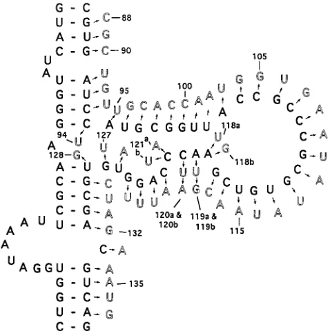
All previous mutational analyses of VAI RNA used large mutations. Such mutations always affected the precise folding of the molecule in the central domain. To assess the impor-tance of the nucleotide sequence in the function of the RNA, it was necessary to introduce single-base substitutions which, it was hoped, would not alter the secondary structures. There-fore, we introduced 54 single-base substitutions in the 39region of the central domain by an approach detailed in Materials and Methods. Of these, 12 mutations are transitions and the rest are transversions. The entire coding sequence of each mutant VAI gene was sequenced to confirm that the newly introduced base substitution was the only mutation in the entire coding sequence. Three of the four conserved nucleotides (nt 119, 120, and 121) were mutated twice. Figure 2 shows the locations of these mutations in the secondary structure of the central domain of the WT VAI RNA.
In vitro inhibition of autophosphorylation of PKR by mu-tant VAI RNAs.We recently showed that the in vivo properties of the VAI mutants can be faithfully reproduced in vitro (7). In this study, a considerably purified PKR preparation from HeLa cells was preincubated with in vitro-transcribed WT or mutant VAI RNAs that were previously tested for their function in vivo in the context of the viral chromosome. Reovirus RNA was then added as an activator along with [g-32P]ATP, to allow
the enzyme to autophosphorylate in vitro. Phosphorylation of PKR was then quantitated by subjecting the radiolabeled polypeptides to SDS-polyacrylamide gel electrophoresis. Un-der our assay conditions, we have found that at low concen-trations of VAI RNAs, mutants that failed to function in vivo uniformly failed to block the autophosphorylation (activation) of PKR in vitro, whereas mutants that were phenotypically WT in vivo inhibited the autophosphorylation of PKR in vitro ef-ficiently. We used these assay conditions to determine the capacities of the mutant VAI RNAs to inhibit the autophos-phorylation activity of PKR. All mutant VAI RNAs were as-sayed at least three times, and the majority of the mutants were tested four or five times. The average percent inhibitions of PKR activity (with error bars) for these mutants are shown in a bar diagram in Fig. 3.
[image:3.612.60.299.71.311.2]On the basis of their capacities to inhibit the activation of PKR in vitro, the mutants can be classified into three groups. The first group consists of those which inhibit PKR activity as efficiently as the WT VAI RNA. Most of these mutations are clustered between nt 90 and 118 (proximal part of stem III, stem IV, and loop C). Apparently, these mutations do not induce alterations in the secondary structures that would be detrimental to function. These results also suggest that these bases are not critical for the PKR-VAI RNA interactions. The second group of mutants show an intermediate phenotype in that the mutations in these VAI RNA molecules lead to a FIG. 2. Single-base substitutions in the central domain of VAI RNA. The
letters shown in the dark format are the WT sequence. Mutations are shown in light format. The bases numbered represent nucleotides from the 59end of the RNA, with G as the starting residue (31). Bases 118, 119, 120, and 121 are mutated twice.
VOL. 69, 1995 SINGLE-BASE SUBSTITUTIONS IN VAI RNA CENTRAL DOMAIN 4301
on November 9, 2019 by guest
http://jvi.asm.org/
moderate loss of activity, ranging from 20 to 50%. These mu-tations include nucleotides in the lower part of stem IV and in loop D and the bases that pair in the duplex regions of stem III and stem I. The third group of mutants are severely defective in vitro and include those with mutations at nt 91 (pm91), 111 (pm111), 132 (pm132), and 135 (pm135). These mutant RNAs, at a concentration of 0.1 mg/ml, lost about 60 to 100% of activity. With the exception of pm132, none of these mutations induce detectable alterations in the secondary structure of the central domain (see below). Thus, it seems that these residues
are critical for the binding of PKR to VAI RNA, and the results suggest that PKR-VAI RNA interaction may involve some degree of nucleotide sequence specificity.
Analysis of the secondary structures of the mutant RNAs.
[image:4.612.61.557.71.548.2]An important consequence of the mutations in an RNA is the loss of secondary structure, which would also potentially have an impact on the tertiary structure. Because previous studies have shown that even a minor alteration in the secondary structure of the central domain can lead to loss of function, we have analyzed the secondary structures of the mutant RNAs of FIG. 3. Bar diagrams showing the capacities of the mutant VAI RNAs to block the autophosphorylation of PKR activity in vitro. Mutant or WT VAI genes were transcribed in vitro with T7 RNA polymerase, gel purified with a native polyacrylamide gel, and incubated with a partially purified PKR from HeLa suspension cultures (7). The radiolabeled polypeptides were resolved in an SDS–12% polyacrylamide gel, and the radioactive PKR band was quantitated by a laser densitometer and plotted. The samples were assayed at least three times and in most cases four or five times. Values shown on the y axis correspond to the percent activity of PKR remaining compared with that in control experiments in which autophosphorylation was carried out without VAI RNA. Control values were taken as 100%. Error bars indicate deviations from average values.
on November 9, 2019 by guest
http://jvi.asm.org/
pm91, pm111, pm119b, pm120b, pm121b, pm132, and pm135.
The mutant VAI genes were transcribed in vitro, labeled at their 39 ends with [32P]pCp, and then digested with low
con-centrations of single-strand-specific RNases T1 (cleaves after
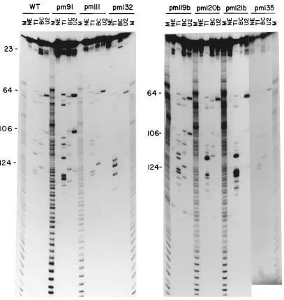
G), BC (cleaves after pyrimidines), and U2 (cleaves after A) such that less than 10% of the molecules were digested with RNases and each molecule in the digested portion was cleaved only once. The digests were then analyzed on 14% DNA se-quencing gels with ladder markers generated by treating the same RNAs with alkali (6). The nuclease digestion patterns of these RNAs are shown in Fig. 4. Some of the patterns are faint in this figure, but they have been repeated to confirm the cleavages. We and others have used this approach previously to predict the folding of the mutant VAI RNAs (6, 8, 21, 24). The results presented in Fig. 4 suggest that all of the RNAs, with the exception of that of pm132, show cleavage patterns
identical to that of WT RNA, including those specific for the central domain, suggesting that these RNAs retain their native secondary structures. For example, G (Fig. 4, lanes T1)-, py-rimidine (lanes BC)-, and A (lanes U2)-specific cleavages for the sequence CAGGUG between nt 121 and 128 are present in all mutants except pm132 and also in identical locations in the autoradiogram. Note that an additional strong G cleavage in the case of pm119b at nt 119 is due to the G substitution in this position. Similarly, cleavages specific for the sequence UACCG between nt 102 and 109 are also present in the RNAs of pm91,
pm111, pm119b, pm120b, pm121b, and pm135. The pattern
ob-tained for pm132 is somewhat difficult to interpret. A careful examination of this pattern indicates that the entire pattern is shifted to a slightly lower position in the gel including loop B-specific cleavages around nt 64. Clearly, this RNA has un-dergone some structural alterations which may account for the loss of activity. This result also suggests that even a minor alteration in the secondary structure can be detected in this gel system. In summary, these results suggest that several of these point mutants retain their native secondary structures yet lose the capacity to inhibit the autophosphorylation activity of PKR.
DISCUSSION
Currently, the only known function of VAI RNA in Ad infection is to block the autophosphorylation of PKR and thus protect the host-cell translation apparatus. The VAI RNA binds to PKR and inactivates it. To study the structural re-quirements for the VAI RNA to bind to PKR, our group and also that of Mathews have mutagenized the RNA extensively and analyzed it in vivo (both in the context of viral chromo-some and in transient assays) and also in vitro for its capacity to bind to and block the autophosphorylation of PKR (1, 6, 7, 19–21, 24). Although there are some minor differences be-tween the results of the two laboratories, in general these results indicate that the VAI RNA binding to PKR correlates with function and that the central domain and the proximal part of the apical stem are the critical parts of the molecule both for function and for binding. Studies have also shown that both VAI RNA and dsRNA bind to the N-terminal 171 amino acids of PKR, and mutational analysis of this region has indi-cated that amino acids critical for the binding of dsRNA are also critical for the binding of VAI RNA, suggesting that both the activator and the inhibitor bind to the same region in the protein (9, 13, 18, 23). These and other results suggest that PKR can be activated by any RNA as long as the RNA is perfectly double stranded in nature. On the other hand, bind-ing and inactivation of PKR would involve a specific secondary or tertiary structure in VAI RNA. This suggests that there may be subtle differences in the interactions of PKR with VAI RNA and with dsRNA. The purpose of this study was to determine whether interaction of VAI RNA with PKR is based solely on the secondary (and tertiary) structure in the central domain or whether there are nucleotide sequences within this central domain that play important roles in this interaction.
To evaluate the effect of the point mutations on the function of the RNAs, we used the in vitro autophosphorylation assay. We recently analyzed a series of VAI mutants in vitro for their capacities to inhibit the phosphorylation of partially purified PKR from HeLa cells (7). Previously, these same mutants were rebuilt into the viral chromosome and were examined for their phenotypes in a variety of assays, including viral polypeptide synthesis, growth yield, and PKR activation (1, 6). When a low concentration of these same mutant VAI RNAs was used in the PKR block assays in vitro, the mutants that failed to func-FIG. 3—Continued.
VOL. 69, 1995 SINGLE-BASE SUBSTITUTIONS IN VAI RNA CENTRAL DOMAIN 4303
on November 9, 2019 by guest
http://jvi.asm.org/
[image:5.612.62.297.70.535.2]tion in vivo uniformly failed to inhibit autophosphorylation of PKR (7), showing excellent correlation between in vivo and in vitro phenotypes. Thus, in our hands, the in vitro inhibitory activity of the mutant VAI RNAs is a very good predictor of the in vivo phenotype. In addition, in our in vitro assays, only VAI RNA can inhibit the activation of PKR at concentrations of 0.1 to 0.2mg/ml. Other small RNAs, such as Epstein-Barr virus-encoded small RNAs (EBERs), Tar RNA, and VAII RNA, require a much higher (50-fold more) concentration of RNA for the inhibition to be detected (8a), consistent with their poor capacities to complement for the function of VAI RNA in vivo (2, 3, 10, 16).
In the study reported here, we analyzed the 54 single-base substitution mutants in vitro by using the same-quality enzyme used in our previous studies (7). Thirty of the 54 single-base substitutions introduced into the central domain of the VAI RNA produce mutants that can block the autophosphorylation
of PKR as efficiently or nearly as efficiently as the WT RNA. On the basis of the secondary structure shown in Fig. 1A, these mutations mostly lie in the proximal part of the apical stem, the 59 portion of the short stem-loop, and loop C. We draw two conclusions from these results. First, these mutations did not induce structural alterations in the central domain to the ex-tent that the alterations would impinge on the function of the RNA. Second, these nucleotides are not critical for VAI RNA-PKR interactions. It is surprising that almost all nucleotides in loop C can be mutated, although one at a time, without causing loss of activity. This shows that these residues may not be directly involved in the PKR interactions. Perhaps these se-quences exist as a loop to allow the folding of the central domain in a configuration that is optimal for function.
Mutageneses of nt 91, 95, 96, and 111 and of most of the nucleotides between nt 118 and 138 affect function signifi-cantly, and in some cases the effect is quite severe. The loca-FIG. 4. Single-strand-specific RNase cleavage analysis of WT and point mutant VAI RNAs. In vitro-synthesized RNAs were labeled at the 39ends (as described in Materials and Methods) and digested with RNases T1, BC, and U2. The products were resolved in 14% DNA sequencing gels. M, ladders generated by partial digestion of the labeled RNA under alkaline conditions (6). Numbers 23, 64, 106, and 124 refer to nucleotides from the 59end of WT VAI RNA. Cleavages in regions 23, 64, and 106 to 124 belong to loop A, loop B, and the short stem-loop of the central domain, respectively.
on November 9, 2019 by guest
http://jvi.asm.org/
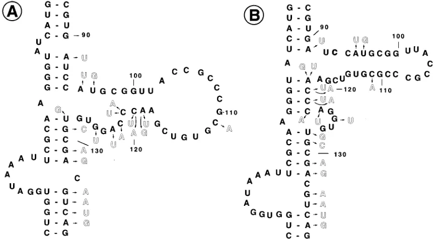
[image:6.612.104.511.67.497.2]tions of these mutations are shown in Fig. 5A. The severely defective mutants are pm91, pm111, and pm132, which retain less than 25% of the inhibitory activity of WT VAI RNA. Other defective mutants retain about 50 to 80% of the inhib-itory activity. One obvious consequence of introducing muta-tions, however small they are, is the alteration of the secondary structure. Even a small mutation can affect the secondary structure in the central domain and thus reduce or abolish activity. Surprisingly, we did not detect any alteration in the secondary structures for these defective mutants, except for
pm132. All cleavages specific to the central domain of the WT
RNA are detected in the mutant RNAs that we tested, and none of the cleavage patterns except that of pm132 warrants a new structure. Normally, this gel system is sensitive to even a minor change in the secondary structure, as evident from the results for pm132. The nucleotides between nt 127 and 133 belong to the perfectly base-paired duplex region (stem II) of the central domain. It is conceivable that this duplex region is very crucial for the PKR interaction with VAI RNA and that a single base mismatch in this region may be deleterious for its function (see below). It is also interesting to note here that we previously constructed and analyzed two nondefective VAI mutants (sub707 and sub708 [1, 6–8]) in which most of the loop A sequences are mutated without loss of activity, suggesting that nucleotide sequences in loop A may also not be critical for VAI RNA function. In summary, these point mutants collec-tively define a class of mutants which retain the secondary structure and yet lose biological activity, suggesting that per-haps there is some degree of nucleotide sequence specificity involved in the VAI RNA function.
Several secondary structures have been proposed for the central domain (4, 5, 15, 24); the one most recently proposed is shown in Fig. 1B (4, 5). In this structure, the short stem-loop of the central domain is positioned slightly higher than that proposed in the original structure (Fig. 1A). This is primarily because the conserved nucleotides ACCC between nt 118 and 123 are allowed to base pair with nucleotides between nt 36
and 41. While this structure is also compatible with nuclease sensitivity analysis, some of the mutational analysis is not con-sistent with this model structure (7). We recently described a phenotypically WT mutant, VAI CB, in which mismatches in the apical stem (stem III) were corrected to form a perfectly base-paired stem. In this mutant, as a result of mutations, the nucleotide sequence ACCC located between nt 118 and 123 of the WT sequence is duplicated between nt 90 and 95, which almost certainly would base pair with the nucleotides between nt 36 and 41. Thus, in view of these results, it is necessary to consider both structures as plausible at the present time. The elucidation of a precise structure for this region will require X-ray crystallographic data.
When the point mutants are considered in light of the re-vised structure shown in Fig. 1B, it appears that most of the nucleotides of the short stem-loop (stem IV and loop C) do not appear to be critical for function (Fig. 5B). Because mutations in stem IV lead to perturbations in the base-paired structure and because such mutations did not affect function, these re-sults suggest that function is not dependent on the base-paired region of stem IV. On the other hand, most of the mutations cluster in the duplex regions in the central domain that connect two long stems, stem I and stem III. These results are consis-tent with the interpretation that perfectly base-paired duplex regions in this region may be critical for PKR interaction with VAI RNA.
[image:7.612.90.525.72.310.2]Currently it is possible only to speculate on the precise mechanism by which VAI RNA intercedes in the autophos-phorylation of PKR. As stated above, studies have shown that both dsRNA and VAI RNA bind to the same region of PKR. It has been suggested that the duplex region of the apical stem proximal to the central domain and stem II (both structures) interact with PKR in a manner similar to the interaction of dsRNA with PKR, although the short stem-loop of the central domain would prevent activation of the kinase (4). Another possibility is that the PKR would interact with two stems but the PKR activation by VAI RNA would be prevented because FIG. 5. Locations of mutations in the central domain which result in significant loss of PKR-inhibitory activity. (A) Mutations shown in the context of the secondary structure shown in Fig. 1A. (B) Mutations shown in the context of the revised secondary structure shown in Fig. 1B.
VOL. 69, 1995 SINGLE-BASE SUBSTITUTIONS IN VAI RNA CENTRAL DOMAIN 4305
on November 9, 2019 by guest
http://jvi.asm.org/
the two stems do not lie on a single helical axis. Both mecha-nisms attach importance to the duplex regions in the central domain (4).
Why do many of the point mutations cause loss of activity, which in some cases is rather dramatic, even though the struc-ture of the central domain in these mutants remains intact? One possibility is that, in addition to involving the secondary structure, PKR interactions with VAI RNA involve some de-gree of sequence specificity. The enzyme would recognize these critical residues in the context of the secondary structure, and the bases that are substituted are not compatible with interaction with PKR. The question of whether these nucleo-tides are conserved between the VAI RNAs of different sero-types is important here but is difficult to address. The only nucleotide sequence homology identified between VAI RNAs of Ad2 (or Ad5), Ad7, and Ad12 is a tetranucleotide sequence, ACCC, that is presumed to base pair with nucleotides GGGU between nt 36 and 41. The nucleotides identified to be impor-tant for function in our study do not appear to be present in the same exact locations in VAI RNAs of other serotypes (15). Because Ad7 and Ad12 VAI RNAs can complement for the function of Ad2 VAI RNAs, at least in transient assays (15), it is likely that these residues, or residues similar to those iden-tified here, may exist in similar locations in other RNAs. These do not need to be the same nucleotide or nucleotides but could be purines or pyrimidines at specific locations in the central domain. However, it may not be possible to identify these nucleotides by looking at the linear sequences of different VAI RNAs because such nucleotides may be functionally important only when they are presented in the context of the secondary structure of the central domain. Because the secondary struc-tures of the VAI RNAs of other Ad serotypes have not been determined experimentally, it is difficult to identify the nucleo-tides that are critical for function in VAI RNAs of other serotypes which are also conserved. This situation becomes even more complex when the tertiary structure of this RNA is considered, as it is almost certain that many nucleotides of the central domain will contribute to its tertiary structure.
Another possibility that cannot be entirely ruled out at present is that these mutations induce perturbations in the secondary structure that are so small that they cannot be de-tected in our secondary structure analysis; however, such mi-nor perturbations would be sufficient to affect their activity. For example, perfectly base-paired stem II may be critical for the function of VAI RNA, and single-base mutations would perturb this duplex structure and hence compromise activity. However, this interpretation is not adequate to explain the loss of activity for mutants in which nt 95, 96, and 111 and nucleo-tides between nt 121 and 126 are mutated, as these nucleonucleo-tides are not expected to pair with other nucleotides in the model structure shown in Fig. 1A. Similarly, the loss of activity for mutants in which nt 123, 125, 134, and 135 are mutated is not explained, since these nucleotides are not expected to pair with other nucleotides in the structure shown in Fig. 1B. Further-more, the conserved nucleotides 119, 120, and 121 were mu-tated twice, and one of the two mutations (pm119a, pm120a, and pm121b; Fig. 2) should still maintain the duplex structure with the G-U pair. Thus, it seems that perturbation of the duplex regions may not be the only explanation for the loss of activity. Rather, PKR has some preference for the nucleotides at specific locations in the central domain. We also note that the actual secondary structure of the central domain in solu-tion may be slightly different from those presented in Fig. 1A and B, in which case some of the interpretation will need to be modified. A three-dimensional structure of this RNA will be essential to resolve many mechanistic questions, such as the
regions of the VAI RNA that bind to PKR and the critical residues necessary for this interaction, as well as the structural requirements of the N-terminal region of PKR for its interac-tion with both VAI RNA and dsRNA. Nonetheless, the avail-ability of a systematic collection of point mutants of the central domain of VAI RNA as described here will aid further in the unraveling of the intricate details of the RNA-protein interac-tions of this system.
ACKNOWLEDGMENTS
A.R. and P.M. contributed equally to this work.
We sincerely thank Ghanashyam Ghadge for help in purification of PKR, Lola Kwan for excellent technical assistance, and Swaminathan for reading the manuscript.
This work was supported by National Institutes of Health grants AI18029 and AI20156. T.K. was supported by the Medical Scientist Training Program of Northwestern University.
REFERENCES
1. Bhat, R. A., P. H. Domer, and B. Thimmapaya. 1985. Structural require-ments of adenovirus VAI RNA for its translation enhancement function. Mol. Cell. Biol. 5:187–196.
2. Bhat, R. A., and B. Thimmapaya. 1984. Adenovirus mutants with DNA sequence perturbations in the intragenic promoter of VAI RNA gene allow the enhanced transcription of VAII RNA gene in HeLa cells. Nucleic Acids Res. 12:7377–7388.
3. Bhat, R. A., and B. Thimmapaya. 1985. Construction and analysis of addi-tional adenovirus substitution mutants confirm the complementation of VAI RNA function by two small RNAs encoded by Epstein-Barr virus. J. Virol. 56:750–756.
4. Clark, P. A., and M. B. Mathews. Interactions between the double stranded RNA binding motif and RNA: definition of the binding site for the inter-feron-induced protein kinase DAI (PKR) on adenovirus VA RNA. RNA, in press.
5. Clark, P. A., P. Tsafira, and M. B. Mathews. 1994. Structural features of adenovirus 2 virus associated RNA required for binding to the protein kinase DAI. Nucleic Acids Res. 22:4364–4374.
6. Furtado, M. R., S. Subramanian, R. A. Bhat, D. M. Fowlkes, B. Safer, and B. Thimmapaya.1989. Functional dissection of adenovirus VAI RNA. J. Virol. 63:3423–3434.
7. Ghadge, G. D., P. M. Malhotra, M. R. Furtado, R. Dhar, and B. Thimma-paya.1994. In vitro analysis of virus-associated RNA I (VAI RNA): inhibi-tion of the double-stranded RNA-activated protein kinase PKR by VAI RNA mutants correlates with the in vivo phenotype and the structural integrity of the central domain. J. Virol. 68:4137–4151.
8. Ghadge, G. D., S. Swaminathan, M. G. Katze, and B. Thimmapaya. 1991. Binding of the adenovirus VAI RNA to the interferon-induced 68-kDa protein kinase correlates with function. Proc. Natl. Acad. Sci. USA 88:7140–7144. 8a.Ghadge, G. D., and B. Thimmapaya. Unpublished results.
9. Green, S. R., L. Manche, and M. B. Mathews. 1995. Two functionally distinct RNA-binding motifs in the regulatory domain of the protein kinase DAI. Mol. Cell. Biol. 15:358–364.
10. Gunnery, S., A. P. Rice, H. D. Robertson, and M. B. Mathews. 1990. Tat-responsive region RNA of human immunodeficiency virus I can prevent activation of the double-stranded RNA activated protein kinase. Proc. Natl. Acad. Sci. USA 87:8687–8691.
11. Hovanessian, A. G. 1989. The double-stranded RNA activated protein kinase induced by interferon: dsRNA-PK. J. Interferon Res. 9:641–647. 12. Katze, M. G., D. Decarato, B. Safer, J. Galabru, and A. G. Hovanessian.
1987. Adenovirus VAI RNA complexes with 68,000 Mr protein kinase to regulate its phosphorylation and activity. EMBO J. 6:689–697.
13. Katze, M. G., W. Wambach, M. L. Wong, M. Garfinkel, E. Meurs, K. Chong, B. R. G. Williams, A. G. Hovanessian, and G. N. Barber.1991. Functional expression and RNA binding analysis of the interferon-induced, double-stranded RNA-activated, 68,000-Mrprotein kinase in a cell-free system. Mol. Cell. Biol. 11:5497–5505.
14. Kitajewski, J. R., R. J. Schneider, B. Safer, S. Munemitsu, C. E. Samuel, B. Thimmapaya, and T. Shenk.1986. Adenovirus VAI RNA antagonizes the antiviral action of interferon by preventing activation of the interferon-induced eIF-2 alpha kinase. Cell 45:195–200.
15. Ma, Y., and M. B. Mathews. 1993. Comparative analysis of the structure and function of adenovirus virus-associated RNAs. J. Virol. 67:6605–6617. 16. Mathews, M. B. 1993. Viral evasion of cellular defense mechanisms:
regu-lation of the protein kinase DAI by RNA effectors. Semin. Virol. 4:247–257. 17. Mathews, M. B., and T. Shenk. 1991. Adenovirus virus-associated RNA and
translational control. J. Virol. 65:5657–5662.
18. McCormick, S. J., C. E. Thomas, and C. E. Samuel. 1992. Mechanism of interferon action: identification of a RNA binding domain within the
on November 9, 2019 by guest
http://jvi.asm.org/
terminal region of the human RNA-dependent P1/eIF-2 alpha kinase. Vi-rology 198:47–56.
19. Mellits, K. H., M. Kotsura, and M. B. Mathews. 1990. Interaction of ade-novirus VA RNAI with protein kinase DAI: nonequivalence of structure and function. Cell 61:843–852.
20. Mellits K. H., and M. B. Mathews. 1988. Effects of mutations in stem and loop regions on the structure and function of adenovirus VA RNAI. EMBO J. 7:2849–2859.
21. Mellits, K. H., T. Pe’ery, and M. B. Mathews. 1992. Role of the apical stem in maintaining the structure and function of adenovirus virus-associated RNA. J. Virol. 66:2369–2377.
22. O’Malley, R. P., T. M. Mariano, J. Siekierka, and M. B. Mathews. 1986. A mechanism for the control of protein synthesis by adenovirus VA RNAI. Cell 44:391–400.
23. Patel, R. C., and G. C. Sen. 1992. Identification of the double-stranded RNA binding domain of the human interferon-inducible protein kinase. J. Biol. Chem. 267:7671–7676.
24. Pe’ery, T., K. Mellits, and M. B. Mathews. 1993. Mutational analysis of the central domain of adenovirus virus-associated RNA mandates a revision of the proposed secondary structure. J. Virol. 67:3534–3543.
25. Samuel, C. E. 1993. The eIF-2 alpha kinases, regulators of translation in eukaryotes from yeast to humans. J. Biol. Chem. 268:7603–7606.
26. Schneider, R. J., B. Safer, S. M. Munemitsu, C. E. Samuel, and T. Shenk. 1985. Adenovirus VAI RNA prevents phosphorylation of the eukaryotic initiation factor 2 alpha subunit subsequent to infection. Proc. Natl. Acad. Sci. USA 82:4321–4324.
27. Schneider, R. J., and T. Shenk. 1987. Impact of virus infection on host cell protein synthesis. Ann. Rev. Biochem. 56:317–332.
28. Schneider, R. J., C. Weinberger, and T. Shenk. 1984. Adenovirus VAI RNA facilitates the initiation of translation in virus-infected cells. Cell 37:291–298. 29. Siekierka, J., T. M. Mariano, P. A. Reichel, and M. B. Mathews. 1985. Translational control by adenovirus: lack of virus associated RNA I during adenovirus infection resuts in phosphorylation of initiation factor eIF-2 and inhibition of protein synthesis. Proc. Natl. Acad. Sci. USA 82:1959–1963. 30. Thimmapaya, B., G. D. Ghadge, S. Swaminathan, and P. Rajan. 1993.
Translation control by adenovirus virus-associated RNA I, p. 203–225. In J. Elan (ed.), Translational regulation of gene expression 2. Plenum Press, New York.
31. Thimmapaya, B., N. C. Jones, and T. S. Shenk. 1979. A mutation which alters initiation of transcription by RNA polymerase III on the Ad5 chro-mosome. Cell 18:947–954.
32. Thimmapaya, B., C. Weinberger, R. J. Schneider, and T. Shenk. 1982. Adenovirus VAI RNA is required for efficient translation of viral mRNAs at late times after infection. Cell 31:543–551.
VOL. 69, 1995 SINGLE-BASE SUBSTITUTIONS IN VAI RNA CENTRAL DOMAIN 4307