0022-538X/95/$04.0010
Copyrightq1995, American Society for Microbiology
Functional and Structural Interactions between Measles Virus
Hemagglutinin and CD46
OFER NUSSBAUM,
1† CHRISTOPHER C. BRODER,
1BERNARD MOSS,
1LULI BAR-LEV STERN,
2SHMUEL ROZENBLATT,
1, 2AND
EDWARD A. BERGER
1*
Laboratory of Viral Diseases, National Institute of Allergy and Infectious Diseases, Bethesda, Maryland,
1and Department of Molecular Microbiology and Biotechnology, George S. Wise
Faculty of Life Science, University of Tel Aviv, Tel Aviv, Israel
2Received 21 December 1994/Accepted 26 February 1995
We analyzed the roles of the individual measles virus (MV) surface glycoproteins in mediating functional
and structural interactions with human CD46, the primary MV receptor. On one cell population, recombinant
vaccinia virus vectors were used to produce the MV hemagglutinin (H) and fusion (F) glycoproteins. As fusion
partner cells, various cell types were examined, without or with human CD46 (endogenous or recombinant
vaccinia virus encoded). Fusion between the two cell populations was monitored by a quantitative reporter gene
activation assay and by syncytium formation. MV glycoproteins promoted fusion with primate cells but not with
nonprimate cells; recombinant CD46 rendered nonprimate cells competent for MV glycoprotein-mediated
fusion. Markedly different fusion specificity was observed for another morbillivirus, canine distemper virus
(CDV): recombinant CDV glycoproteins promoted fusion with primate and nonprimate cells independently of
CD46. Fusion by the recombinant MV and CDV glycoproteins required coexpression of H plus F in either
homologous or heterologous combinations. To assess the role of H versus F in determining the CD46
depen-dence of MV fusion, we examined the fusion specificities of cells producing heterologous glycoprotein
combi-nations. The specificity of H
MVplus F
CDVparalleled that observed for the homologous MV glycoproteins:
fusion occurred with primate cells but not with nonprimate cells unless they produced recombinant CD46. By
contrast, the specificity of H
CDVplus F
MVparalleled that for the homologous CDV glycoproteins: fusion
occurred with either primate or nonprimate cells with no dependence on CD46. Thus, for both MV and CDV,
fusion specificity was determined by H. In particular, the results demonstrate a functional interaction between
H
MVand CD46. Flow cytometry and antibody coprecipitation studies provided a structural correlate to this
functional interaction: CD46 formed a molecular complex with H
MVbut not with F
MVor with either CDV
glycoprotein. These results highlight the critical role of the H glycoprotein in determining MV specificity for
CD46-positive cells.
Paramyxovirus virions are coated with surface projections
that mediate virion attachment to target cells displaying the
appropriate surface receptors. Fusion at neutral pH between
the virion and plasma membranes then ensues, resulting in
delivery of the nucleocapsid into the cytoplasm (reviewed in
references [ref.] 31, 40, and 48). By a related process, cells
displaying the viral glycoproteins at their surfaces can fuse with
receptor-bearing cells, resulting in the formation of
multinu-cleated giant cells (syncytia). In members of the morbillivirus
genus, such as measles virus (MV) (reviewed in ref. 8) and
canine distemper virus (CDV) (reviewed in ref. 6), the surface
projections are composed of two integral membrane
glycopro-teins: the hemagglutinin (H), which mediates cell attachment,
and the fusion glycoprotein (F), which promotes membrane
fusion (40, 48). Recently, the human CD46 antigen (membrane
cofactor protein), a type 1 integral membrane glycoprotein
found on nearly all human tissues and cell types (32), was
identified as the primary cellular receptor for MV. This
con-clusion derived from studies with a monoclonal antibody
(MAb) which inhibited MV binding (42) and was subsequently
shown to recognize CD46 (41). These findings, coupled with
the demonstration that recombinant human CD46 renders
ro-dent cells susceptible to MV binding, infection, and syncytium
formation (13, 41), provided definitive evidence of a receptor
function for CD46. Subsequent studies revealed that multiple
isoforms of human CD46 (produced by alternate RNA
splic-ing), as well as simian CD46, can serve as MV receptors (21,
30, 35, 38). The receptor functionality of a chimeric CD46
molecule with a glycosyl-phosphatidylinositol anchor indicates
that all essential sequences reside within the ectodomain (56).
In the present study, we used recombinant vaccinia
virus-based expression and assay systems to define the role of each
MV glycoprotein in determining fusion specificity for
CD46-positive cells. We also analyzed binding of the individual MV
glycoproteins to CD46. The results highlight the crucial role of
the MV H glycoprotein in functional and structural
interac-tions with the CD46 molecule.
MATERIALS AND METHODS
Cells and culture conditions.All of the cell types used in this study were obtained from the American Type Culture Collection, Rockville, Md. HeLa (human cervical carcinoma) and NIH 3T3 (mouse embryo) cells were grown in DMEM-10 (Dulbecco’s modified Eagle’s medium [Quality Biologicals, Rock-ville, Md.] supplemented with 10% fetal bovine serume [Sigma, St. Louis, Mo.], 2 mML-glutamine, and 50mg of gentamicin [Gibco BRL, Gaithersburg, Md.] per ml). RK13(rabbit kidney) and BS-C-1 (African green monkey kidney) cells were
grown in MEM-10 (Eagle’s minimal essential medium [Quality Biologicals] sup-plemented with 10% fetal bovine serum, 2 mM L-glutamine, and 50mg of
* Corresponding author. Mailing address: Laboratory of Viral Dis-eases, NIAID, National Institutes of Health, Building 4, Room 236, Bethesda, MD 20892. Phone: (301) 402-2481. Fax: (301) 480-1147. Electronic mail address: edward_berger@nih.gov.
† Present address: Laboratory of Vascular Surgery, Shiba Hospital, Tel Hashomer, Tel Aviv, Israel.
3341
on November 9, 2019 by guest
http://jvi.asm.org/
gentamicin per ml). Cultures were maintained in a humidified tissue culture incubator at 378C in 5% CO2.
Recombinant vaccinia viruses.Standard methods of homologous recombina-tion were used to introduce foreign genes into the thymidine kinase locus of the vaccinia virus Western Reserve (WR) strain (17). All recombinants were sub-jected to three rounds of plaque purification, and their titers were determined on BS-C-1 cells. For individual experiments, cells were infected at a multiplicity of infection of 10 PFU per cell with each designated vaccinia virus recombinant.
The F and H glycoproteins were from the Edmonston strain of MV and the Onderstepoort strain of CDV. Recombination plasmids (described in ref. 51) contained the bacteriophage T7 promoter, followed by a DNA copy of the 59 noncoding region of encephalomyocarditis virus RNA to enhance translation efficiency in the vaccinia virus system (18), followed by the morbillivirus glyco-protein open reading frame. The vaccinia virus recombinants and the plasmids from which they were derived are designated as follows: vT7-HMV, encoding H
of MV (HMV), from plasmid pTM-H/MV; vT7-FMV, encoding F of MV (FMV),
from plasmid pTM-F/MV; vT7-HCDV, encoding H of CDV (HCDV), from
plas-mid pTM-H/CDV; vT7-FCDV, encoding F of CDV (FCDV), from plasmid
pTM-F/CDV. Vaccinia virus recombinant containing the morbillivirus glycoprotein cDNAs were identified by picking thymidine kinase-negative plaques and screen-ing amplified samples by DNA slot blot hybridization and Western (immunoblot) analysis.
For production of recombinant human CD46, we used the cDNA encoding the MCP-BC2 isoform (GenBank accession number Y00651); nucleotide and amino acid numbering is as previously described (33). We prepared plasmids and corresponding recombinant vaccinia viruses encoding the full-length form, as well as a soluble secreted form (sCD46) representing the entire ectodomain. In each case, the cDNA was linked to a strong-early–strong-late synthetic vaccinia virus promoter (11a). A 1.5-kb cDNA fragment containing the entire CD46 coding sequence cloned into the EcoRI site of vector pSG5 (Stratagene, La Jolla, Calif.) was provided by M. K. Liszewski, Washington University, St. Louis, Mo. For full-length CD46, the 1.5-kb cDNA fragment was excised with EcoRI and cloned into EcoRI-digested plasmid pSC59 (11a). The resulting plasmid (pCB-48) was used to produce recombinant vaccinia virus vCB-48, encoding full-length CD46. For soluble CD46 (sCD46), a translation termination codon was inserted after the aspartic acid residue at amino acid 294 (nucleotide 1027), just prior to the predicted transmembrane domain, by using PCR technology (27). A PCR product was generated by using plasmid pCB-48 as the template, with one oligonucleotide primer (59GACACAATTGTCTGTGACAGTAACAGTACTT G-39) spanning the nucleotides from positions 839 to 870 and a second oligonu-cleotide primer (59-CCGGCACTAGTTAATCCAAACTGTCAAGTATTCCT TC-39) containing the translation termination codon as well as a new SpeI restriction endonuclease site. The resulting PCR fragment was first cloned into the pGEM-T vector system (Promega, Madison, Wis.) to generate plasmid pCB-50, and the nucleotide sequence of the cloned PCR fragment was verified. The 131-nucleotide SalI-SpeI fragment was excised from pCB-50 and ligated into pCB-48, which had been digested with SalI plus SpeI. The resulting plasmid, pCB-58, was used to construct recombinant vaccinia virus vCB-58, which encodes sCD46. These recombinant viruses were identified by picking thymidine kinase-negative plaques in conjunction with radioimmunoprecipitation analysis of met-abolically [35S]methionine-labeled lysates (full-length CD46) or culture
super-natants (sCD46) from amplified plaques.
For bacteriophage T7 RNA polymerase production, two vaccinia virus recom-binants were used. In vP11gene1 (1), the polymerase gene is linked to the natural late P11 vaccinia virus promoter; the advantages of using a late promoter in the cell fusion assay have already been described (45). In other experiments, we used vTF7-3 (19), which employs the natural early-late P7.5 vaccinia virus promoter.
Immunological reagents.The following antibodies against specific MV glyco-proteins were employed: for HMV, murine MAb 15 (ref. 24; provided by T. F.
Wild, Institut Pasteur de Lyon) and rabbit polyclonal anti-HMV serum 172
(provided by C. O¨ rvell, Karolinska Institutet, Stockholm, Sweden); for FMV,
murine MAb 186 (reference 36; provided by T. F. Wild) and rabbit polyclonal anti-FMVserum 180 (provided by C. O¨ rvell). The following murine MAbs against
specific CDV glycoproteins (ref. 47; obtained from C. O¨ rvell) were employed: for HCDV, MAb 1.347; for FCDV, MAbs 3.633 and 4.985. For analysis of CD46,
murine MAb J4-48 was purchased from Amac, Inc. (Westbrook, Maine). As negative controls, we employed murine MAbs D11 (16) and T4 (9), directed against the human immunodeficiency virus type 1 envelope glycoprotein.
Flow cytometry.For assay of surface-localized recombinant glycoproteins, NIH 3T3 cells were infected with the vaccinia viruses indicated in the legend to Fig. 1. At 3 h postinfection, the virus inoculum was removed and the cell monolayers were harvested by trypsinization, washed with complete medium, and incubated as suspension cultures overnight at 318C at a density of 33105
cells per ml. The cells were washed with phosphate-buffered saline (PBS)-fetal calf serum (FCS) (PBS containing 2.5% FCS), and aliquots containing 106
cells were incubated with the MAb indicated in the legend to Fig. 1 or control MAb T4 (2mg of purified immunoglobulin G1 [IgG1] or a 1:100 dilution of ascites fluid in a total volume of 100ml of PBS-FCS for 1 h at 48C in a microcentrifuge tube). The cells were washed twice with 1 ml of PBS-FCS, stained for 30 min at 48C with goat anti-mouse IgG-fluorescein isothiocyanate F(ab9)2(Boehringer
Mannheim, Indianapolis, Ind.; 1:20 dilution in PBS-FCS), and washed twice with 1 ml of PBS-FCS. Cell pellets were resuspended in 0.2 ml of PBS and fixed by
addition of an equal volume of PBS containing 4% paraformaldehyde. Samples were analyzed on a FACScan flow cytometer (Becton Dickinson).
For the sCD46 cell-binding assays, conditioned media were prepared by in-fecting 75-cm2flasks of NIH 3T3 cells with either vCB-58 or control WR vaccinia
virus. At 3 h postinfection, the monolayers were washed twice with 10 ml of serum-free OPTI-MEM medium (GIBCO, Grand Island, N.Y.), overlaid with 5 ml of OPTI-MEM, and incubated overnight at 378C. The conditioned media were harvested, clarified by centrifugation, and concentrated 10-fold in a 30-kDa exclusion Centricon microconcentrator (Amicon, Beverly, Mass.). NIH 3T3 cells producing the recombinant glycoproteins indicated in the legend to Fig. 5 were washed with PBS-FCS and resuspended at 107
/ml in PBS-FCS. Aliquots (100ml) of each lysate were mixed with conditioned media (50ml), incubated for 1 h on ice, and washed. Cells were stained with anti-CD46 MAb J4-48 (2mg in 100ml) followed by goat anti-mouse IgG-fluorescein isothiocyanate F(ab9)2and
pro-cessed for flow cytometry as described above.
Metabolic labeling, immunoprecipitation, and coprecipitation.NIH 3T3 cells (in six-well tissue culture plates, 23106cells per well) were infected with the
recombinant vaccinia viruses indicated in the figure legends. At 3 h postinfection, the inoculum was removed and the cell monolayers were washed once with 3 ml of methionine-free minimal essential medium and then overlaid with methi-onine-free minimal essential medium containing 5% dialyzed FCS and 100mCi of [35S]methionine per ml. The cultures were incubated overnight at 378C. For
analysis of cell-associated proteins, monolayers were washed with 2 ml of PBS and solubilized in 0.2 ml of lysis buffer containing 100 mM Tris-HCl (pH 8.0), 100 mM NaCl, and 0.5% (vol/vol) Triton X-100. After incubation on ice for 15 min, nuclei were removed by centrifugation. For analyses of sCD46, the supernatants of the infected and metabolically labeled cells were harvested and clarified by centrifugation. Direct immunoprecipitations of recombinant MV and CDV gly-coproteins were performed by mixing in a microcentrifuge tube 10ml of meta-bolically labeled cell lysate with the appropriate murine MAb ascities fluid (1ml) or polyclonal rabbit antiserum (1ml) in a total volume of 100ml of PBS con-taining 0.1% Triton X-100. Reaction mixtures were incubated for 4 h at 48C; when a murine MAb was used as the first antibody, 10mg of rabbit anti-mouse IgG (Calbiochem, La Jolla, Calif.) was added and the incubation was continued for 1 h. Protein A-Sepharose beads (100ml of a 20% suspension) were added, and the mixtures were incubated for 30 min with rocking. The Sepharose beads were centrifuged at 5003g for 5 min and washed twice with 1 ml of wash buffer
containing 50 mM Tris-HCl (pH 8.0), 300 mM NaCl, and 0.1% Triton X-100. For coimmunoprecipitations with CD46, an aliquot (50ml) of unlabeled cell lysate (107
cells per ml of lysis buffer) from NIH 3T3 cells infected with either vCB48 or control virus (WR) was mixed with the cell lysates indicated in the legend to Fig. 6, containing metabolically labeled MV and/or CDV glycoproteins. After incubation for 4 h at 48C, 2mg of anti-CD46 MAb J4-48 or a control murine MAb was added and the samples were incubated for an additional 1 h at 48C. Immunoprecipitates were collected with protein A-Sepharose beads as described above.
Immunoprecipitated proteins were eluted from the beads by heating for 5 min at 958C in sodium dodecyl sulfate-polyacrylamide gel electrophoresis (SDS-PAGE) sample buffer containing 5% (vol/vol) 2-mercaptoethanol. Proteins were separated by SDS-PAGE on 12.5% gels. Bands were visualized by fluorography.
Cell fusion assay.Cell-cell fusion mediated by the morbillivirus glycoproteins was measured by using a recently described vaccinia virus-based reporter gene activation assay (45). The transfection and infection protocols were performed in DMEM supplemented with 2.5% fetal bovine serum. Briefly, a monolayer of NIH 3T3 cells was coinfected with vP11gene1, encoding bacteriophage T7 RNA polymerase, and the indicated viruses encoding morbillivirus glycoproteins. NIH 3T3 cells were chosen as the glycoprotein-producing cells since of the cell types examined, these were the least permissive as fusion partners (see Results); cell fusion within the glycoprotein-expressing population was thus minimized. A second monolayer of the designated cell type was transfected with plasmid pG1NT7b-gal (39a); this plasmid contains the Escherichia coli lacZ gene linked to the bacteriophage T7 promoter and the 59untranslated region of encephalo-myocarditis virus. Transfections were performed with DOTAP transfection re-agent (Boehringer Mannheim) in accordance with the recommended protocol as previously described (45). After 3 to 3.5 h at 378C, the transfected cells were infected with either wild-type vaccinia virus WR or vCB-48, encoding full-length CD46. At 1.5 h postinfection, cells were detached by trypsinization, suspended to a density of 2.53105/ml in DMEM-2.5 medium in 50-ml conical polypropylene
centrifuge tubes (Falcon), and placed in a tissue culture incubator at 318C. Following incubation overnight (10 to 16 h) to allow accumulation of the vaccinia virus-encoded proteins, the cells were washed once by centrifugation and sus-pended in DMEM-2.5 medium. To initiate cell fusion, aliquots containing 105
cells of each of two designated cell populations were dispensed into duplicate wells of 96-well flat-bottom microtiter tissue culture plates; cytosine arabinoside was added to a final concentration of 40mg/ml to minimize fusion-independent
b-galactosidase production. The total volume of each well was adjusted to 0.2 ml with DMEM-2.5 medium. The plates were transferred to a 378C tissue culture incubator for 2.5 h, at the end of which time fusion was assessed.
For the quantitative reporter gene activation assay (45), Nonidet P-40 was added to each well to a final concentration of 0.5% (vol/vol).b-Galactosidase activity was quantified by colorimetric assay of aliquots of each lysate in 96-well flat-bottom microtiter plates with chlorphenol red-b-D-galactopyranoside
on November 9, 2019 by guest
http://jvi.asm.org/
(Boehringer Mannheim) as the substrate. The rates of substrate hydrolysis were determined at ambient temperature by measuring the increase in A570with a
microplate absorbance reader (Molecular Devices, Menlo Park, Calif.). Results are expressed as nanograms ofb-galactosidase per well, based on the rates obtained with parallel assays using a standard commercial preparation of E. coli
b-galactosidase (Boehringer Mannheim). As a complementary method of anal-ysis (45),b-galactosidase was detected in situ by fixing the cells with formalde-hyde plus glutaraldeformalde-hyde and staining them with 5-bromo-4-chloro-3-indolyl-b
-D-galactopyranoside (X-Gal; Boehringer Mannheim). Photographs were taken with an inverted tissue culture microscope with 203phase-contrast and 103
ocular objectives; images were generated with Adobe Photoshop.
In most experiments, syncytium formation was also monitored by direct mi-croscopic examination without staining forb-galactosidase. There was consistent agreement between the reporter gene activation assays and the syncytium anal-yses, although the former assays proved much more sensitive for detection of fusion events, as previously reported (45).
RESULTS
Synthesis of vaccinia virus-encoded recombinant
glycopro-teins.
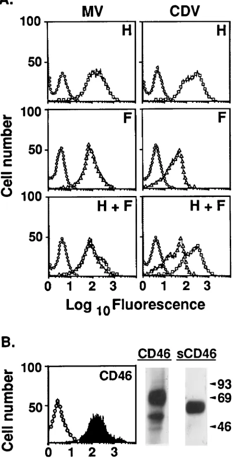
Figure 1A shows the flow cytometry analysis of cell
surface-localized recombinant vaccinia virus-encoded
glyco-proteins of MV (left panels) and CDV (right panels). With the
appropriate specific MAbs, discrete peaks were observed for
cells infected with vaccinia viruses encoding MV or CDV H
alone (top panels) or F alone (middle panels); in all cases, only
low-level background staining was observed with a control
MAb. Comparable levels of each glycoprotein were obtained
when cells were coinfected with vaccinia viruses encoding both
H and F of MV or CDV (bottom panels). Analysis of the
polypeptides produced upon infection with these recombinant
vaccinia viruses is described later.
Production of recombinant CD46 is shown in Fig. 1B. Flow
cytometry (graph, left panel) was used to analyze cell surface
CD46 produced upon infection with the vaccinia virus
encod-ing full-length CD46. Anti-CD46 MAb J4-48 yielded a discrete
peak, whereas only low-level background staining was
ob-served when a control MAb was used. Additional experiments
(data not shown) indicated that the amount of surface CD46
produced by the recombinant vaccinia virus system exceeded
the endogenous CD46 level observed in HeLa cells by about
fivefold. The autoradiogram (Fig. 1B, right panel) shows the
immunoprecipitation–SDS-PAGE analysis of metabolically
la-beled cells infected with recombinant vaccinia viruses encoding
full-length CD46 (left lane) or sCD46 (right lane). Full-length
CD46 immunoprecipitated from a detergent lysate migrated as
a broad band at around 67 kDa, consistent with the reported
migration of this isozyme (32). The secreted sCD46
immuno-precipitated from the medium migrated as a broad band at
approximately 62 kDa, consistent with the absence of the
trans-membrane and cytoplasmic domains (6.1 kDa).
Effect of recombinant CD46 on fusion mediated by MV
gly-coproteins.
Figure 2 shows the ability of the recombinant MV
H and F glycoproteins produced on one cell population to
mediate fusion with different types of partner cells. The
glyco-protein-producing cells also contained vaccinia virus-encoded
T7 RNA polymerase; the partner cells contained the
trans-fected lacZ-containing plasmid. Fusion was assessed both by in
situ staining (photomicrographs) and by measuring
b
-galacto-sidase activity in Nonidet P-40 cell lysates (numbers in insets);
similar conclusions were reached on the basis of direct
micro-scopic analysis of syncytium formation without staining for
b
-galactosidase (data not shown). The left-hand panels of Fig.
2 illustrate the fusion specificity of the recombinant MV
gly-coproteins. Extensive fusion occurred with human HeLa (top
panel) and simian BS-C-1 (data not shown) partners, as judged
by the presence of large, blue-stained syncytia and high levels
of
b
-galactosidase activity in the cell lysates. By contrast, fusion
[image:3.612.317.559.91.563.2]was not observed with either murine NIH 3T3 (middle panel)
FIG. 1. Production of MV and CDV glycoproteins and CD46 with vaccinia virus vectors. (A) Flow cytometry analysis of surface-localized recombinant MV (left panels) and CDV (right panels) glycoproteins. NIH 3T3 cells were coin-fected with vTF7-3, encoding T7 RNA polymerase, plus recombinant vaccinia virus vectors encoding the glycoproteins indicated (H, top panels; F, center panels; H and F, bottom panels). The following immunological reagents were employed: MAb 15 for HMV(h), MAb 186 for FMV(Ç), MAb 1.347 for HCDV
(h), and MAb 3.633 for FCDV(Ç). As background controls, the same cells were
incubated with MAb T4 (E). All samples were stained with goat anti-mouse IgG-fluorescein isothiocyanate and analyzed by flow cytometry. (B) Analysis of CD46. (Left panel) NIH 3T3 cells were infected with recombinant vaccinia virus vCB-48 encoding full-length CD46. Cells were incubated with anti-CD46 MAb J4-48 (filled curve) or with control MAb T4 (E) and then stained with goat anti-mouse IgG-fluorescein isothiocyanate. Samples were analyzed by flow cy-tometry. (Right panel) NIH 3T3 cells were infected with vCB-48 or vCB-58 encoding full-length CD46 or sCD46, respectively. After metabolic labeling, full-length CD46 was immunoprecipitated from a detergent cell lysate (left lane) and sCD46 was immunoprecipitated from the medium (right lane). The numbers on the right are molecular sizes in kilodaltons.
on November 9, 2019 by guest
http://jvi.asm.org/
or rabbit RK
13(bottom panel) partner cells, on the basis of the
absence of blue-stained cells and detection of only background
b
-galactosidase activity in the lysates. We conclude that the
fusion specificity of the recombinant MV glycoproteins
pro-duced and assayed by the vaccinia virus-based system was
con-sistent with the known restriction of MV infection for human
or simian cells (8).
In view of the recent reports that human CD46 functions as
an MV receptor (13, 21, 35, 38, 41, 42), we tested whether
vaccinia virus-encoded CD46 could confer fusion competence
on nonprimate cells which were otherwise nonpermissive
part-ners for MV glycoprotein-mediated fusion. In experiments
whose results are shown in the right-hand panels of Fig. 2, the
same MV glycoprotein-bearing cells were mixed with NIH 3T3
and RK
13partner cells containing the transfected
lacZ-con-taining plasmid and infected with the recombinant vaccinia
virus encoding CD46. The results indicate that recombinant
CD46 rendered both nonprimate cell types permissive for MV
FIG. 2. Cell type specificity of MV glycoprotein-mediated fusion. One population of NIH 3T3 cells was coinfected with vaccinia virus recombinants encoding T7 RNA polymerase plus the MV F and H glycoproteins. As partner cell populations, HeLa, NIH 3T3, and RK13cells were transfected with the lacZ-containing plasmid
and infected with wild-type vaccinia virus WR (left panels). Alternatively transfected NIH 3T3 and RK13cells were infected with the vaccinia virus recombinant
encoding CD46 (right panels). Mixtures of the glycoprotein-producing cells and the partner cells indicated were prepared. Fusion was scored at 2.5 h by in situ staining (photomicrographs) or by colorimetric assay of detergent cell lysates (insets; results are expressed as nanograms ofb-galactosidase per well6the sample standard deviation for duplicate samples). Photomicrograph images were generated from slides with Adobe Photoshop.
on November 9, 2019 by guest
http://jvi.asm.org/
glycoprotein-mediated fusion, as judged by both the
blue-stained cells and syncytia observed in situ and the 30- to
100-fold increase in
b
-galactosidase activity in the detergent cell
lysates. We consistently observed that the efficiency of fusion
with nonprimate cells producing recombinant CD46 was less
than that observed with primate cells displaying endogenous
CD46, on the basis of syncytium size and the amount of
b
-ga-lactosidase activity.
Fusion mediated by F and H glycoproteins of MV and CDV.
To assess the role of individual MV glycoproteins in mediating
functional interaction with CD46, we performed
complemen-tation analyses between the glycoproteins of MV and the
re-lated morbillivirus CDV (see below). It was necessary first to
characterize the fusion specificity of CDV glycoproteins. In
contrast to the results described above for MV, CDV
glyco-proteins mediated efficient fusion not only with primate cells
(HeLa) but also with nonprimate (NIH 3T3 and RK
13) cells.
This broad fusion specificity for the recombinant CDV
glyco-proteins is consistent with the known ability of CDV to
pro-ductively infect cultured cells of diverse nonprimate and
pri-mate species (6).
Complementation analysis also required characterization of
the fusion activities of various combinations of MV and CDV
glycoproteins (Fig. 3B). HeLa cells were chosen as fusion
part-ners in this experiment since they are permissive for both MV
and CDV glycoprotein-mediated fusion; similar results (not
shown) were obtained with simian BS-C-1 cells. The upper
panels in Fig. 3B compare the fusogenic activities observed
with individual glycoproteins versus pairs of glycoproteins. For
MV and CDV, fusion was observed when both H and F were
coproduced as judged by the high
b
-galactosidase activities;
only background
b
-galactosidase levels were observed with the
individual glycoproteins. In the lower panels of Fig. 3B, the
fusogenic activities of homologous versus heterologous
mix-tures of MV and CDV glycoproteins were examined; fusion
was observed with each heterologous mixture. These results
obtained by using the reporter gene assay to reveal functional
complementation between MV and CDV glycoproteins are
consistent with those of previous studies based on syncytium
analysis (51).
Roles of individual MV glycoproteins in mediating fusion
specificity for CD46-positive cells.
The markedly different
fu-sion specificities of MV and CDV glycoproteins, coupled with
the ability of heterologous mixtures of these glycoproteins to
support fusion, provided a means to assess the role of H and F
in determining the CD46 dependence of MV fusion. As shown
in Fig. 4, we compared the abilities of heterologous and
ho-mologous glycoprotein combinations to support fusion with
different cell types. The data presented are the ratios of
b
-ga-lactosidase levels obtained with pairs of partner cells chosen
because they highlight the specificity of MV
glycoprotein-me-diated fusion. Figure 4A shows the results obtained with NIH
3T3 partner cells plus or minus recombinant CD46. Fusion
mediated by the homologous MV glycoproteins was greatly
stimulated by CD46, consistent with the results shown in Fig. 2;
by contrast, CDV fusion was not stimulated by CD46. When
the heterologous glycoprotein combinations were examined,
CD46 stimulation correlated with the H rather than the F
glycoprotein of MV (i.e., a high fusion ratio occurred with
H
MVplus F
CDVbut not with H
CDVplus F
MV). Another
cor-relation is demonstrated in Fig. 4B, which shows the results of
an experiment which examined the fusion ratio obtained with
RK
13cells (poor partners for MV but good partners for CDV)
relative to that obtained with HeLa cells (good partners for
both). The nonpermissiveness of MV glycoproteins for RK
13 [image:5.612.317.555.86.519.2]cells was associated with the H rather than the F glycoprotein
FIG. 3. Fusion mediated by MV and CDV glycoproteins. (A) Cell type spec-ificity of CDV glycoprotein-mediated fusion. One population of NIH 3T3 cells was coinfected with vaccinia virus recombinants encoding T7 RNA polymerase plus the CDV F and H glycoproteins. As partner cell populations, HeLa, NIH 3T3, and RK13 cells were transfected with the lacZ-containing plasmid and
infected with wild-type vaccinia virus WR. Cell mixtures were prepared, and fusion was scored at 2.5 h by colorimetric assay of detergent cell lysates (error bars denote sample standard deviations for duplicate samples). (B) Fusion me-diated by individual MV and CDV glycoproteins versus combinations. The upper panels show requirements for coexpression of F and H. NIH 3T3 cells were coinfected with vaccinia virus recombinants encoding T7 RNA polymerase plus both (F plus H) or individual (H or F) glycoproteins of either MV or CDV, as indicated. When only one morbillivirus glycoprotein was expressed, wild-type vaccinia virus WR was used to maintain a constant multiplicity of infection. The lower panels show homologous versus heterologous glycoprotein combinations. NIH 3T3 cells were coinfected with vaccinia virus recombinants encoding T7 RNA polymerase plus homologous (left) or heterologous (right) combinations of MV and CDV glycoproteins, as indicated. As a negative control, cells expressed HCDVand no other morbillivirus glycoprotein (dashed line). In both the upper
and lower panels, the partner population was HeLa cells transfected with the
lacZ-containing plasmid and infected with wild-type vaccinia virus WR. Cell
mixtures were prepared, and fusion was scored at 2.5 h by colorimetric assay of detergent cell lysates (error bars denote sample standard deviations of duplicate samples).
on November 9, 2019 by guest
http://jvi.asm.org/
(i.e., a low RK
13/HeLa fusion ratio occurred with H
MVplus
F
CDVbut not with H
CDVplus F
MV). These results indicate that
the H rather than the F glycoprotein determines MV fusion
specificity; in particular, they directly demonstrate a functional
interaction between CD46 and H
MVin the fusion process.
The H glycoprotein also appeared to determine CDV fusion
specificity. In Fig. 4B, results obtained with the heterologous
combinations show that CDV H rather than F enabled CDV
fusion with RK
13cells (good RK
13/HeLa fusion ratios were
obtained with H
CDVplus F
MVbut not with H
MVplus F
CDV).
Similarly, the H glycoprotein was associated with the
non-responsiveness of CDV fusion to CD46 (no CD46
stimula-tion with H
CDVplus F
MV, good stimulation with H
MVplus
F
CDV). These findings obtained with the reporter gene assay,
highlighting the critical role of H in determining CDV fusion
specificity, are in agreement with previous findings based on
analysis of syncytium formation when the recombinant
glyco-proteins were produced in various cell types (51).
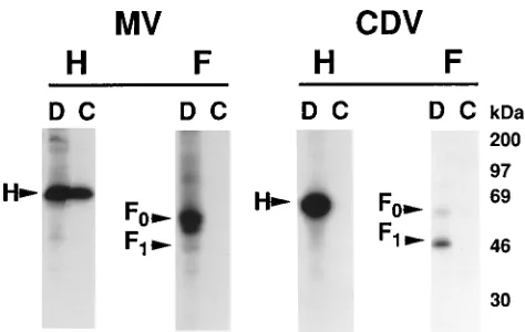
Binding of CD46 to paramyxovirus glycoproteins.
CD46
binding to individual MV and CDV glycoproteins was
mea-sured by two methods. The first involved flow cytometry
anal-ysis of the ability of intact cells displaying these surface
glyco-proteins to bind sCD46 (a genetically engineered secreted
form representing the entire ectodomain). Representative
pat-terns are shown in Fig. 5A, and the mean channel fluorescence
data are summarized in Fig. 5B. The results indicate that
sCD46 bound to cell surface H
MV, synthesized either alone or
in conjunction with F
MV. By contrast, sCD46 binding to
sur-face F
MV, or to either surface glycoprotein of CDV, was
in-distinguishable from the background obtained with control
cells not displaying morbillivirus glycoproteins.
The second approach involved coprecipitation analysis of
mixtures of detergent cell lysates (Fig. 6). Metabolically
35S-labeled cells synthesizing the vaccinia virus-encoded MV or
CDV glycoproteins indicated in Fig. 6 were solubilized with
Triton X-100. Direct immunoprecipitation from aliquots of
these labeled lysates confirmed the synthesis of each
radiola-beled protein, consistent with the flow cytometry analysis
pre-sented above. Thus, direct immunoprecipitation of H
MVand
H
CDVyielded predominant bands migrating at
;
79 to 80 kDa,
consistent with the known sizes of these glycoproteins (40, 48).
For the F glycoproteins, bands were observed at the expected
positions (40, 48) corresponding to the uncleaved F
0 [image:6.612.60.300.70.365.2]precur-sors (
;
60 kDa) and the cleaved F
1products (
;
40 kDa). Bands
FIG. 4. Roles of individual MV and CDV glycoproteins in mediating fusion specificity. One population of NIH 3T3 cells was coinfected with vaccinia virus recombinants encoding T7 RNA polymerase plus combinations of MV and CDV glycoproteins (homologous or heterologous). The following partner cell popu-lations were transfected with the lacZ-containing plasmid and infected with the vaccinia virus indicated. (A) One portion of NIH 3T3 cells was infected with the vaccinia virus encoding CD46 (1CD46), and the other portion was infected with wild-type vaccinia virus WR (2CD46). (B) RK13and HeLa cells were infected
with wild-type vaccinia virus WR. Cell mixtures were prepared, and fusion was scored at 2.5 h by colorimetric assay of detergent cell lysates. For each partner cell pair, the ratios of theb-galactosidase levels (fusion ratios) were calculated for the combinations of MV and CDV glycoproteins indicated.
FIG. 5. Flow cytometry analysis of sCD46 binding to surface MV and CDV glycoproteins. NIH 3T3 cells were infected with vaccinia viruses encoding the MV or CDV glycoprotein(s) indicated. Cells were incubated with concentrated conditioned media from cells infected either with a vaccinia virus encoding sCD46 (1sCD46; filled symbols) or with wild-type vaccinia virus WR (1control; open symbols). Cells were incubated with anti-CD46 MAb J4-48 and then stained with goat anti-mouse IgG-fluorescein isothiocyanate. Panels: A, repre-sentative flow cytofluorograms; B, summary of data for cells expressing individ-ual MV or CDV glycoproteins or combinations.
on November 9, 2019 by guest
http://jvi.asm.org/
[image:6.612.317.555.73.421.2]corresponding to the F
2products (
;
18 to 20 kDa) were also
detected (data not shown). In the case of F
MV, the broadness
of the F
0band and its inefficient cleavage are consistent with
reported results obtained with independently isolated
recom-binant vaccinia viruses (4, 60). The small amount of processed
protein was evidently sufficient to support the fusogenic
activ-ity observed for this recombinant F glycoprotein.
To test the ability of CD46 to interact with the MV and CDV
glycoproteins, equivalent aliquots of cell lysates containing the
35
S-labeled MV or CDV glycoproteins were mixed with an
unlabeled cell lysate containing vaccinia virus-encoded CD46.
Immunoprecipitates were obtained with MAb J4-48, which
binds to CD46 but does not block MV-induced syncytium or
rosette formation (13). The results indicate that the anti-CD46
MAb efficiently coprecipitated
35S-labeled H
MV
. By contrast to
these positive results obtained with H
MV, no coprecipitation
was observed when the analogous experiment was performed
with
35S-labeled F
MV
. Furthermore, no coprecipitation was
observed with either
35S-labeled CDV glycoprotein H or F.
Additional experiments (data not shown) were performed to
help interpret these results. First, the specificity of H
MVco-precipitation was verified by the absence of the H
MVband
when a control isotype-matched MAb was used in place of
J4-48 or when the coprecipitation was performed with a
con-trol unlabeled lysate lacking CD46 (i.e., cells infected with
control vaccinia virus WR). Second, we considered the
possi-bility that the failure to coprecipitate F
MVwas due not simply
to its inability to bind CD46 but instead to inactivation or
denaturation of this glycoprotein when synthesized and
solu-bilized in the absence of H
MV. This was ruled out by preparing
a lysate from metabolically labeled cells synthesizing both H
MVand F
MV; addition of the CD46-containing lysate resulted in
coprecipitation of H
MVwithout concomitant coprecipitation of
F
MV. Third, we examined whether the failure of H
CDVto
coprecipitate with CD46 was due not to absence of binding
between these molecules but instead to masking of the J4-48
epitope upon binding to H
CDV. This unlikely explanation was
ruled out by the finding that excess solubilized H
CDVdid not
interfere with the coprecipitation of H
MV.
Taken together, the flow cytometry and coprecipitation
ex-periments indicate direct binding of CD46 to H
MV; no binding
to F
MVwas observed. Under identical conditions, binding to
either glycoprotein of CDV did not occur. These results
pro-vide a structural correlate to the cell fusion experiments
indi-cating functional interaction between CD46 and H
MV.
DISCUSSION
The recent discovery that human CD46 (membrane cofactor
protein) serves as the primary MV receptor (13, 41) has
opened important directions for elucidating the mechanism of
MV entry into target cells. Experiments with MV virions have
highlighted the central role of CD46 in mediating MV binding
and infection (13, 21, 35, 38, 41). In the present study, we used
a vaccinia virus-based expression system and a quantitative
reporter gene activation assay to measure fusion between two
distinct cell populations. The results directly demonstrate that
recombinant CD46 serves as a fusion receptor for recombinant
MV glycoproteins in the absence of other MV components,
consistent with a previous analysis of syncytium formation (41).
We analyzed the relative contributions of each MV
glyco-protein in the specific interactions with CD46. It is generally
believed that the morbillivirus H and paramyxovirus HN
gly-coproteins function in the attachment of virions to target cells.
In the case of MV, the major evidence derives from findings
with monkey erythrocytes, including the ability of H
anti-bodies (polyclonal and monoclonal) to inhibit MV
virion-in-duced hemagglutination (7, 20, 23, 24, 34, 39, 44, 46, 54, 55),
the hemagglutinating activity of H isolated from MV virions (7,
10, 12, 20, 49), and the hemadsorption activity of recombinant
cell surface H produced by using various expression systems (2,
14, 57). Regarding interaction of H with CD46, indirect
evi-dence has been provided by the down-modulation of surface
CD46 upon production of vaccinia virus-encoded H (43, 56),
the CD46 dependence of purified H antigen presentation by
murine B-cell transfectants (22), and the binding to CD46 of
an uncharacterized MV glycoprotein fraction said to contain
predominantly H (35). In this report, we directly demonstrate
functional and structural interaction between H and CD46.
Experiments with heterologous combinations of MV and CDV
glycoproteins indicated that H
MVwas responsible for
specific-ity of fusion with CD46-bearing cells, thus providing clear
evidence of functional interaction. Complementary results
were obtained by flow cytometry and
radioimmunoprecipita-tion studies which demonstrated specific binding between H
MVand CD46. No interactions between F
MVand CD46 were
re-vealed in these experiments, although we cannot exclude the
possibility of subtle interactions not detected by the assays
employed.
We observed that for MV and CDV, production of both the
H and F glycoproteins was required for fusion. This is
consis-tent with most published results obtained with recombinant
MV and CDV glycoproteins suggesting that H is required for
(11, 51–53, 57, 60), or at least can stimulate (4, 5), fusion
mediated by F. However, some reports have concluded that
fusion can occur with F
MValone (3–5), in one case under the
nonphysiological condition of low pH (57). The basis for these
discrepant findings is unclear, although the differences in the
various methods used to produce and assay the recombinant
glycoproteins presumably play a major role. It should also be
noted that for the paramyxovirus genus, individual species vary
in their fusion dependence on HN and F coproduction
(re-viewed in ref. 31; see also ref. 26 and 29).
In analyzing the MV and CDV glycoprotein coproduction
requirements, we observed that fusion occurred with
heterol-FIG. 6. Coprecipitation analysis of CD46 binding to MV and CDV glyco-proteins. Triton X-100 lysates were prepared from metabolically labeled cells which had been infected with vaccinia viruses encoding the MV glycoproteins indicated. Equivalent aliquots of the lysates were analyzed by direct immuno-precipitation (D) to determine the total amount of each labeled glycoprotein and by coprecipitation (C) to determine the binding of each glycoprotein to CD46. The following antibodies were used for direct immunoprecipitation: HMV,
anti-serum 172; FMV, antiserum 180; HCDV, MAb 1.347; FCDV, MAb 4.985. For
coprecipitation analyses, a Triton X-100 lysate from cells infected with the vaccinia virus encoding CD46 was added and anti-CD46 MAb J4-48 was used for coprecipitation.
on November 9, 2019 by guest
http://jvi.asm.org/
[image:7.612.59.296.68.218.2]ogous combinations of H of one virus and F of the other, as has
been shown previously (51). A recent report, also using a
vaccinia virus expression system, made the contrary suggestion
that recombinant H
MVdoes not complement F
CDVfor fusion
(59). We point out that the F
CDVused in those experiments
was encoded by a cDNA clone (58) distinct from the one used
in our studies and that positive controls were not presented to
document the fusogenic activity of that recombinant F
CDVmolecule upon coproduction of homologous H
CDV. We
there-fore feel that those negative findings are outweighed by our
positive fusion results obtained with F
CDVcoexpressed with
either homologous or heterologous H; however, we do not rule
out the possibility that the alternative results might be due to
technical differences. Our finding that fusion does not require
homologous glycoprotein combinations should not be
inter-preted to imply the absence of specific functional interactions
between H and F. In a study of recombinant glycoproteins of
clinical MV isolates, efficient fusion was found to occur only
with certain H-F combinations, suggesting requirements for
specific functional interactions (11). Similar conclusions have
been reached with the paramyxovirus HN and F glycoproteins
(28, 50). Also relevant are the recent biochemical
demonstra-tions of molecular complexes between the MV H and F
gly-coproteins (37) and the human parainfluenza virus HN and F
glycoproteins (29). Whether the requirement for F and H
coproduction reflects essential functional interactions between
these molecules remains a critical question for future studies.
While studies in other laboratories have shown that
trans-fected CD46 DNA renders nonprimate cells susceptible to MV
infection (13, 21, 38), the resulting syncytia were found to be
less extensive than those obtained with primate cells displaying
endogenous CD46 (13). In those experiments using MV
viri-ons, suboptimal amounts of CD46 (13) or inefficiency of
postentry steps in the MV replicative cycle (41) could have
contributed to the results. Our studies quantitating cell fusion
with recombinant proteins provide direct evidence of lower
fusion efficiency of nonprimate than primate cells, despite the
high-level production of CD46 achieved with the vaccinia virus
vectors. It thus seems that additional factors contribute to the
greater ability of primate cells to undergo MV
glycoprotein-mediated fusion. In this regard, we note that surface proteins
other than CD46 have been suggested to participate in MV
entry into primate cells (15, 25, 61).
The cellular receptor mediating CDV infection has not been
identified. Several points should be noted regarding a possible
role for CD46. (i) We demonstrated that fusion mediated by
CDV glycoproteins was not stimulated by recombinant CD46
on the partner cells. While this might argue against a CDV
receptor function for CD46, interpretation is compromised by
the fact that all of the partner cell lines tested were somewhat
permissive for CDV glycoprotein-mediated fusion. It is
possi-ble that receptor levels were not limiting for fusion with these
cells, thereby masking the effects of exogenous receptor
pro-duction. (ii) We found that H
CDV(on the cell surface or in
detergent lysates) did not bind to CD46 under conditions in
which H
MVbound extensively. (iii) Others have reported that
surface CD46 is down-regulated upon infection with MV but
not upon infection with CDV (43). Taken together, these
re-sults suggest that a molecule(s) other than CD46 serves as the
CDV receptor. Furthermore, if CD46 is involved in CDV
in-fection, the functional determinant(s) must be present on
CD46 homologs of widely divergent species, since CDV
infec-tion and CDV glycoprotein-mediated fusion both display
broad specificity for nonprimate and primate cell types.
The role of the H glycoprotein in MV-CD46 interactions
suggests several directions for future research. These include
identification of regions of H and CD46 involved in binding,
analysis of the structural determinants involved in interactions
between H and F, and investigation of possible conformational
changes in viral and cellular components during the fusion
process.
ACKNOWLEDGMENTS
P. E. Kennedy provided expert technical assistance for these studies. We thank C. O¨ rvell and T. F. Wild for generously donating antibodies against MV and CDV glycoproteins and M. K. Liszewski and J. P. Atkinson for kindly providing the CD46 cDNA.
This study was supported in part by the NIH Intramural AIDS Targeted Antiviral Program and by an NIH intramural research train-ing award to C. C. Broder.
REFERENCES
1. Alexander, W. A., B. Moss, and T. R. Fuerst. 1992. Regulated expression of foreign genes in vaccinia virus under the control of bacteriophage T7 RNA polymerase and the Escherichia coli lac repressor. J. Virol. 66:2934–2942. 2. Alkhatib, G., and D. J. Briedis. 1988. High-level eucaryotic in vivo expression
of biologically active measles virus hemagglutinin by using an adenovirus type 5 helper-free vector system. J. Virol. 62:2718–2727.
3. Alkhatib, G., C. Richardson, and S. Shen. 1990. Intracellular processing, glycosylation, and cell-surface expression of the measles virus fusion protein (F) encoded by a recombinant adenovirus. Virology 175:262–270. 4. Alkhatib, G., J. Roder, C. Richardson, D. Briedis, R. Weinberg, D. Smith, J.
Taylor, E. Paoletti, and S. H. Shen.1994. Characterization of a cleavage mutant of the measles virus fusion protein defective in syncytium formation. J. Virol. 68:6770–6774.
5. Alkhatib, G., S. H. Shen, D. Briedis, C. Richardson, B. Massie, R. Weinberg,
D. Smith, J. Taylor, E. Paoletti, and J. Roder.1994. Functional analysis of N-linked glycosylation mutants of the measles virus fusion protein synthe-sized by recombinant vaccinia virus vectors. J. Virol. 68:1522–1531. 6. Appel, M. 1987. Canine distemper virus, p. 133–159. In M. J. Appel (ed.),
Virus infection of carnivores. Elsevier Science, Amsterdam.
7. Bellini, W. J., D. E. McFarlin, G. D. Silver, E. S. Mingioli, and H. F.
McFarland.1981. Immune reactivity of the purified hemagglutinin of mea-sles virus. Infect. Immun. 32:1051–1057.
8. Bellini, W. J., J. S. Rota, and P. A. Rota. 1994. Virology of measles virus. J. Infect. Dis. 170:S15–S23.
9. Broder, C. C., P. L. Earl, D. Long, S. T. Abedon, B. Moss, and R. W. Doms. 1994. Antigenic implications of human immunodeficiency virus type 1 enve-lope quaternary structure: oligomer-specific and -sensitive monoclonal anti-bodies. Proc. Natl. Acad. Sci. USA 91:11699–11703.
10. Casali, P., J. G. Sissons, R. S. Fujinami, and M. B. Oldstone. 1981. Purifi-cation of measles virus glycoproteins and their integration into artificial lipid membranes. J. Gen. Virol. 54:161–171.
11. Cattaneo, R., and J. K. Rose. 1993. Cell fusion by the envelope glycoproteins of persistent measles viruses which caused lethal human brain disease. J. Virol. 67:1493–1502.
11a.Chakrabarti, S., and B. Moss. Unpublished data.
12. Christie, M., C. Endresen, and G. Haukenes. 1981. Purification of measles virus H polypeptide and of F polypeptide. Arch. Virol. 69:177–187. 13. Dorig, R. E., A. Marcil, A. Chopra, and C. D. Richardson. 1993. The human
CD46 molecule is a receptor for measles virus (Edmonston strain). Cell
75:295–305.
14. Drillien, R., D. Spehner, A. Kirn, P. Giraudon, R. Buckland, F. Wild, and
J.-P. Lecocq.1988. Protection of mice from fatal measles encephalitis by vaccination with vaccinia virus recombinants encoding either the hemagglu-tinin or the fusion protein. Proc. Natl. Acad. Sci. USA 85:1252–1256. 15. Dunster, L. M., J. Schneider-Schaulies, S. Loffler, W. Lankes, R.
Schwartz-Albiez, F. Lottspeich, and V. ter Meulen.1994. Moesin: a cell membrane protein linked with susceptibility to measles virus infection. Virology 198: 265–274.
16. Earl, P. L., C. C. Broder, D. Long, S. A. Lee, J. Peterson, S. Chakrabarti,
R. W. Doms, and B. Moss.1994. Native oligomeric human immunodeficiency virus type 1 envelope glycoprotein elicits diverse monoclonal antibody reac-tivities. J. Virol. 68:3015–3026.
17. Earl, P. L., N. Cooper, and B. Moss. 1991. Expression of proteins in mam-malian cells using vaccinia viral vectors, p. 16.15.1–16.18.10. In F. M. Aus-ubel, R. Brent, R. E. Kingston, D. D. Moore, J. G. Seidman, J. A. Smith, and K. Struhl (ed.), Current protocols in molecular biology, vol. 2, suppl. 15. John Wiley & Sons, Inc., New York.
18. Elroy-Stein, O., T. R. Fuerst, and B. Moss. 1989. Cap-independent transla-tion of mRNA conferred by encephalomyocarditis virus 59 sequence im-proves the performance of the vaccinia virus/bacteriophage T7 hybrid ex-pression system. Proc. Natl. Acad. Sci. USA 86:6126–6130.
19. Fuerst, T. R., E. G. Niles, F. W. Studier, and B. Moss. 1986. Eukaryotic
on November 9, 2019 by guest
http://jvi.asm.org/
transient-expression system based on recombinant vaccinia virus that syn-thesizes bacteriophage T7 RNA polymerase. Proc. Natl. Acad. Sci. USA
83:8122–8126.
20. Gerlier, D., F. Garnier, and F. Forquet. 1988. Haemagglutinin of measles virus: purification and storage with preservation of biological and immuno-logical properties. J. Gen. Virol. 69:2061–2069.
21. Gerlier, D., B. Loveland, G. Varior-Krishnan, B. Thorley, I. F. C. Mckenzie,
and C. Rabourdin-Combe. 1994. Measles virus receptor properties are shared by several CD46 isoforms differing in extracellular regions and cyto-plasmic tails. J. Gen. Virol. 75:2163–2171.
22. Gerlier, D., M.-C. Trescol-Biemont, G. Varior-Krishnan, D. Naniche, I.
Fugier-Vivier, and C. Rabourdin-Combe.1994. Efficient major histocompat-ibility complex class II-restricted presentation of measles virus relies on hemagglutinin-mediated targeting to its cellular receptor human CD46 ex-pressed by murine B cells. J. Exp. Med. 179:353–358.
23. Gheuens, J., D. E. McFarlin, K. W. Rammohan, and W. J. Bellini. 1981. Idiotypes and biological activity of murine monoclonal antibodies against the hemagglutinin of measles virus. Infect. Immun. 200:207.
24. Giraudon, P., and T. F. Wild. 1985. Correlation between epitopes on hem-agglutinin of measles virus and biological activities: passive protection by monoclonal antibodies is related to their hemagglutination inhibiting activ-ity. Virology 144:46–58.
25. Harrowe, G., J. Sudduthklinger, and D. G. Payan. 1992. Measles virus-substance P receptor interactions: Jurkat lymphocytes transfected with sub-stance P receptor cDNA enhance measles virus fusion and replication. Cell. Mol. Neurobiol. 12:397–409.
26. Heminway, B. R., Y. Yu, and M. S. Galinski. 1994. Paramyxovirus mediated cell fusion requires co-expression of both the fusion and hemagglutinin-neuraminidase glycoproteins. Virus Res. 31:1–16.
27. Ho, S. N., H. D. Hunt, R. M. Horton, J. K. Pullen, and L. R. Pease. 1989. Site-directed mutagenesis by overlap extension using the polymerase chain reaction. Gene 77:51–59.
28. Hu, X., R. Ray, and R. W. Compans. 1992. Functional interactions between the fusion protein and hemagglutinin-neuraminidase of human parainflu-enza viruses. J. Virol. 66:1528–1534.
29. Hu, X., Q. Yao, O. Morozova, and R. W. Compans. Association of the parainfluenza fusion and hemagglutinin-neuraminidase glycoproteins on cell surface. Submitted for publication.
30. Iwata, K., T. Seya, S. Ueda, H. Ariga, and S. Nagasawa. 1994. Modulation of complement regulatory function and measles virus receptor function by the serine-threonine-rich domains of membrane cofactor protein (CD46). Bio-chem. J. 304:169–175.
31. Lamb, R. A. 1993. Paramyxovirus fusion: a hypothesis for changes. Virology
197:1–11.
32. Liszewski, M. K., and J. P. Atkinson. 1992. Membrane cofactor protein. Curr. Top. Microbiol. Immunol. 178:45–60.
33. Lublin, D. M., M. K. Liszewski, T. W. Post, M. A. Arce, M. M. Le Breau,
M. B. Rebentisch, R. S. Lemons, T. Seya, and J. P. Atkinson.1988. Molec-ular cloning and chromosomal localization of human membrane cofactor protein (MCP). J. Exp. Med. 168:181–194.
34. Lund, G. A., and A. A. Salmi. 1981. Purification and characterization of measles virus haemagglutinin protein G. J. Gen. Virol. 56:185–193. 35. Maisner, A., J. Schneider-Schaulies, M. K. Liszewski, J. P. Atkinson, and G.
Herrler.1994. Binding of measles virus to membrane cofactor protein (CD46): importance of disulfide bonds and N-glycans for the receptor func-tion. J. Virol. 68:6299–6304.
36. Malvoisin, E., and F. Wild. 1990. Contribution of measles virus fusion pro-tein in protective immunity: anti-F monoclonal antibodies neutralize virus infectivity and protect mice against challenge. J. Virol. 64:5160–5162. 37. Malvoisin, E., and T. F. Wild. 1993. Measles virus glycoproteins: studies on
the structure and interaction of the haemagglutinin and fusion proteins. J. Gen. Virol. 74:2365–2372.
38. Manchester, M., M. K. Liszewski, J. P. Atkinson, and M. B. A. Oldstone. 1994. Multiple isoforms of CD46 (membrane cofactor protein) serve as receptors for measles virus. Proc. Natl. Acad. Sci. USA 91:2161–2165. 39. McFarlin, D. E., W. J. Bellini, E. S. Mingioli, T. N. Behar, and A. Trudgett.
1980. Monospecific antibody to the haemagglutinin of measles virus. J. Gen. Virol. 48:425–429.
39a.Morgan, R. A. (National Center for Human Genome Research, Bethesda,
Md.).Personal communication.
40. Morrison, T., and A. Portner. 1991. Structure, function, and intracellular processing of the glycoproteins of Paramyxoviridae, p. 347–382. In D. W. Kingsbury (ed.), The paramyxoviruses. Plenum Press, New York.
41. Naniche, D., G. Varior-Krishnan, F. Cervoni, T. F. Wild, B. Rossi, C.
Ra-bourdin-Combe, and D. Gerlier.1993. Human membrane cofactor protein (CD46) acts as a cellular receptor for measles virus. J. Virol. 67:6025–6032. 42. Naniche, D., T. F. Wild, C. Rabourdin-Combe, and D. Gerlier. 1992. A monoclonal antibody recognizes a human cell surface glycoprotein involved in measles virus binding. J. Gen. Virol. 73:2617–2624.
43. Naniche, D., T. F. Wild, C. Rabourdin-Combe, and D. Gerlier. 1993. Measles virus haemagglutinin induces down-regulation of gp57/67, a molecule in-volved in virus binding. J. Gen. Virol. 74:1073–1079.
44. Norrby, E., and Y. Gollmar. 1975. Identification of measles virus-specific hemolysis-inhibiting antibodies separate from hemagglutination-inhibiting antibodies. Infect. Immun. 11:231–239.
45. Nussbaum, O., C. C. Broder, and E. A. Berger. 1994. Fusogenic mechanisms of enveloped-virus glycoproteins analyzed by a novel recombinant vaccinia virus-based assay quantitating cell fusion-dependent reporter gene activa-tion. J. Virol. 68:5411–5422.
46. O¨ rvell, C., and E. Norrby. 1980. Immunological relationships between
ho-mologous structural polypeptides of measles and canine distemper virus. J. Gen. Virol. 50:231–245.
47. O¨ rvell, C., H. Sheshberadaran, and E. Norrby. 1985. Preparation and
char-acterization of monoclonal antibodies directed against four structural com-ponents of canine distemper virus. J. Gen. Virol. 66:443–456.
48. Rima, B. K. 1983. The proteins of morbilliviruses. J. Gen. Virol. 64:1205– 1219.
49. Sato, T. A., M. Enami, and T. Kohama. 1995. Isolation of the measles virus hemagglutinin protein in a soluble form by protease digestion. J. Virol.
69:513–516.
50. Sergel, T., L. W. Mcginnes, M. E. Peeples, and T. G. Morrison. 1993. The attachment function of the Newcastle disease virus hemagglutinin-neuramin-idase protein can be separated from fusion promotion by mutation. Virology
193:717–726.
51. Stern, L. B.-L., M. Greenberg, J. M. Gershoni, and S. Rozenblatt. 1995. The hemagglutinin envelope protein of canine distemper virus (CDV) confers cell tropism as illustrated by CDV and measles virus complementation anal-ysis. J. Virol. 69:1661–1668.
52. Taylor, J., S. Pincus, J. Tartaglia, C. Richardson, G. Alkhatib, D. Briedis, M.
Appel, E. Norton, and E. Paoletti.1991. Vaccinia virus recombinants ex-pressing either the measles virus fusion or hemagglutinin glycoprotein pro-tect dogs against canine distemper virus challenge. J. Virol. 65:4263–4274. 53. Taylor, J., R. Weinberg, J. Tartaglia, C. Richardson, G. Alkhatib, D. Briedis,
M. Appel, E. Norton, and E. Paoletti.1992. Nonreplicating viral vectors as potential vaccines: recombinant canarypox virus expressing measles virus fusion (F) and hemagglutinin (HA) glycoproteins. Virology 187:321–328. 54. ter Meulen, V., S. Loffler, M. J. Carter, and J. R. Stephenson. 1981.
Anti-genic characterization of measles and SSPE virus haemagglutinin by mono-clonal antibodies. J. Gen. Virol. 57:357–364.
55. Togashi, T., C. O¨ rvell, F. Vartdal, and E. Norrby. 1981. Production of
antibodies against measles virions by use of the mouse hybridoma technique. Arch. Virol. 67:149–157.
56. Varior-Krishnan, G., M.-C. Trescol-Bie´mont, D. Naniche, C.
Rabourdin-Combe, and D. Gerlier.1994. Glycosyl-phosphatidylinositol-anchored and transmembrane forms of CD46 display similar measles virus receptor prop-erties: virus binding, fusion, and replication; down-regulation by hemagglu-tinin; and virus uptake and endocytosis for antigen presentation by major histocompatibility complex class II molecules. J. Virol. 68:7891–7899. 57. Vialard, J., M. Lalumie`re, T. Vernet, D. Briedis, G. Alkhatib, D. Henning, D.
Levin, and C. Richardson.1989. Synthesis of the membrane fusion and hemagglutinin proteins of measles virus, using a novel baculovirus vector containing theb-galactosidase gene. J. Virol. 64:37–50.
58. Wild, T. F., A. Bernard, D. Spehner, D. Villeval, and R. Drillien. 1993. Vaccination of mice against canine distemper virus-induced encephalitis with vaccinia virus recombinants encoding measles or canine distemper virus antigens. Vaccine 11:438–444.
59. Wild, T. F., J. Fayolle, P. Beauverger, and R. Buckland. 1994. Measles virus fusion: role of the cysteine-rich region of the fusion glycoprotein. J. Virol.
68:7546–7548.
60. Wild, T. F., E. Malvoisin, and R. Buckland. 1991. Measles virus: both the haemagglutinin and fusion glycoproteins are required for fusion. J. Gen. Virol. 72:439–442.
61. Yoshikawa, Y., K. Yamanouchi, T. Takasu, S. Rauf, and A. Ahmed. 1991. Structural homology between hemagglutinin (HA) of measles virus and the active site of long neurotoxins. Virus Genes 5:57–67.