“IMPACT OF PRE AND POST OPERATIVE BEDSIDE PULMONARY FUNCTION TEST ON THORACOTOMY PATIENT”
– A COMPARATIVE STUDY ”
A project submitted towards partial fulfilment of the
requirements of for the degree of
MASTER OF PHYSIOTHERAPY
Submitted by
Register number: 271730203
under the guidance of
PROF. B. SUBASHINI MPT (CARDIO)
Submitted to
THE TAMIL NADU Dr. M.G.R. MEDICAL UNIVERSITY
Chennai – 32
P.P.G. COLLEGE OF PHYSIOTHERAPY
9/1, Keeranatham road,
Saravanampatti ,
Coimbatore – 641035
“IMPACT OF PRE AND POST OPERATIVE BEDSIDE PULMONARY FUNCTION TEST ON THORACOTOMY PATIENT”
– A COMPARATIVE STUDY ”
INTERNAL EXAMINER :
EXTERNAL EXAMINER :
A project submitted in partial fulfilment
of the requirement for the degree of
MASTER OF PHYSIOTHERAPY
To
THE TAMILNADU Dr. M. G. R. MEDICAL UNIVERSITY,
CHENNAI-32
CERTIFICATE I
This is to certify that the dissertation entitled
“IMPACT OF PRE AND POST OPERATIVE BEDSIDE
PULMONARY FUNCTION TEST ON THORACOTOMY
PATIENT – A COMAPARATIVE STUDY ”
was carried out by
Reg. No. 271710203, P.P.G College of Physiotherapy,
Coimbatore-35,
affiliated to the
Tamilnadu Dr.M.G.R medical
university,
Chennai-32,
under
the
guidance
of
Prof. B. SUBASHINI MPT (CARDIO)
CERTIFICATE II
This
is
to
certify
that
the
dissertation
entitled
“IMPACT OF PRE AND POST OPERATIVE BEDSIDE
PULMONARY
FUNCTION
TEST
ON
THORACOTOMY
PATIENT – A COMAPARATIVE STUDY ” was carried out by Reg.
No. 271710203, P.P.G College of Physiotherapy, Coimbatore-35,
affiliated to the Tamilnadu Dr.M.G.R medical university, Chennai-32,
under my guidance and direct submission.
ACKNOWLEDGEMENT
I thank
GOD, the almighty who laid the foundation for
knowledge and wisdom and has always been my source of strength
and inspiration.
I thank my
PARENTS, who had given me the opportunity,
guidance, encouragement and support throughout the course of my
study.
I Express my sincere gratefulness to our Chairman
Dr L.P.THANGAVELU, M.S, F.R.C.S and our correspondent
MRS.SHANTHI
THANGAVELU,
M.A
P.P.G.
Group
of
Institutions, Coimbatore, for their encouragement and providing the
source for the successful of the study.
I
express
my
sincere
gratefulness
to
My
principal
Prof. Dr. C. SIVA KUMAR, MPT(Ortho), MIAP, Ph.D., who gave
me his precious time and his guidance and encouragement helped me
to complete this dissertation successfully.
I extend my sincere thanks to
Prof. B. SUBASHINI MPT
(CARDIO), for assisting me with valuable inputs and guiding me
through the course of my work.
The gratitude I have for each and every
SUBJECTS who
participated in this study is beyond expression. Their whole hearted
cooperation is easily the biggest reason of all, for this study to be what
it is.
LIST OF CONTENTS
CHAPTER
CONTENT
PAGE NO
I
1.1
1.2
1.3
1.4
1.5
INTRODUCTION
Introduction
Need of study
Aim of the study
Objective of the study
Hypothesis
1
7
8
8
9
II
REVIEW OF LITERATURE
10
III
3.1
3.2
3.3
3.4
3.5
3.6
3.7
3.8
3.9
3.10
3.11
MATERIALS AND METHODOLOGY
Stuudy design
Study Population
Sample size
Sampling technique
Study setting
Study duration
Selection Criteria
Materials Used
Parameter
Procedure of the study
Technique
15
15
15
15
15
15
15
16
16
16
17
IV
4.1
4.2
DATA ANALYSIS & RESULT
Statistical tools
Demographic data
21
23
V
DISCUSSION
44
VII
LIMITATIONS & SUGGESTIONS
49
VIII
REFERENCES
50
LIST OF TABLES
TABLE
NO
TOPIC
PAGE
NO
1.
2.
3.
4.
5.
6.
Percentage and frequency distribution according to age
Percentage and frequency distribution according to gender
Comparison in total lung capacity of patients between pre and post
surgery on day 1, day 3 and day 7
Comparison in breath-holding time among patients between pre
and post surgery on day 1, day 3 and day 7
Comparison in total lung capacity of patients between days 1 and 3,
1 and 7, and 3 and 7 post operatively
Comparison between days: 1 and 3, 1 and 7, and 3 and 7 in
breath-holding time of patients post operatively
23
25
27
31
35
LIST OF FIGURES
FIGURE
NO
TOPIC
PAGE
NO
1.
2.
3.
4.
5.
6.
Distribution of the age of
studied patients had
thoracotomy
surgery
Distribution of the gender of
studied patients had
Thoracotomy
surgery
.
Box and Whisker diagram depicting the distribution of total lung
capacity of patients at pre-operatively (baseline) and
post-operatively on days
1, 3 and 7.
The distribution of breath-holding time of patients at
pre-operatively (baseline) and post-pre-operatively on days
1, 3 and 7.
The distribution of total lung capacity of patients post-operatively
on days 1, 3 and 7.
Depicting the distribution of breath-holding time of patients
post-operatively on days 1, 3 and 7.
24
26
30
34
38
1
“IMPACT OF PRE AND POST OPERATIVE BEDSIDE
PULMONARY FUNCTION TEST ON THORACOTOMY
PATIENT – A COMPARATIVE STUDY ”
CHAPTER I
1.1 INTRODUCTION
Pulmonary function tests (PFTs) are breathing test to find out how well you move air in out of your lung and how well oxygen enters your body. The most common PFTs are Spirometry, diffusion study and body plesmethography. Sometime only one test is done, often on same day.(1)
Pulmonary function testing and monitoring plays an important role in respiratory evaluation. A complete bedside evaluation of the respiratory status is especially useful in preoperative assessment. Bedside traditional tool to assess pulmonary gas exchange such as arterial or transcutaneous blood gas analysis, pulse oxymetry and additional valuable information about global lung function is provided through measurement of pulmonary mechanics and volume. (2)
PFTs have been progressed from initially used water seal types to modern era electronic computerized version. They are patient friendly and easier to understand. Pulmonary function test can be carried out at bedside in critically ill patient with the help of portable spirometer in addition to the routine clinical test. If the PFTs are done with quality assurance,validation of equipment, proper technique, reference values and applying right ethnic correction factors, the data generated are most of the times accurate and reproducible. (3)
PFTs can be used to:
2
Spirometry is one of the most common lung function tests. The spirometer measure how much air you can breathe into your lung and how much air you quickly blow out of your lungs. To get the “best” test result, the test is repeated thrice. (1)
Author Hamid Hassanzadeh hypothesized that the use of incentive Spirometry by patients is less than the recommended level and is affected by patient related factor and type of surgery. To determine its post operative use the author prospectively surveyed all patients in their institutions general ward who had under gone thoracotomy Compliance with incentive spirometry use in the patient population was poor and was largely influenced by type of surgery performed and postoperative day. Because of postoperative complication such as atelectesis, fever and pneumonia continue to affect outcomes after thoracotomy, a need exists for improved preoperative and in hospital counseling strategies regarding incentive spirometery use and for objective measurements for monitoring incentive spirometry use and potential confounding variables. (4)
Pulmonary complication were defined as the development of 3 or more of 6 new findings: cough, phlegm, dyspnea, chest pain, temperature greater than 38 degree C, pulse rate more than 100 beats/min. (5)
Postoperative complication like fever and leuckocytosis are common after joint replacement. A study conducted by Borger JE et al. to determine the incidence of fever and leucocytosis after joint replacement. He found that after joint replacement more than half of patients develop leucocytosis and some develop fever. (6)
Pulmonary Complication occurs in 20% to 40% of patients following abdominal and thoracic operations making pulmonary complication the single largest cause of morbidity and mortality in the postoperative period. The prophylactic respiratory maneuver is one of many aspects of post-operative care that are intended to minimize pulmonary complications. Various respiratory maneuver and devices to encourage those maneuvers are used. (7)
3
lead to changes in the ventilation pattern resulting in the patient taking shallow breath which reduce the ability to clear sputum from the chest. (8)
INCENTIVE SPIROMETERY
Mechanical breathing device such as the incentive spirometery has been introduced into clinical practice. Incentive spirometry encourages the patient to take long, slow deep breath mimicking natural sighing and also provides a visual positive feedback. (8) Incentive spirometer is available either by volume of inspiration (volume oriented) or flow rate (flow oriented). The flow-oriented incentive spirometer (Triflow device) consists of three chambers in series, each of which contains a ball. When the patient effort generates a subatmosphere pressure above the ball. It rises in the chamber in series, each of which contains the ball. When the patient’s effort generates a subatmosphere pressure above the ball, it rises in the chamber. An inspiratory flow of 600mL/s is required to raise the first ball, an inspiratory flow of 900mL/s is required to elevate the first and second balls, and a flow of 1200 mL/s is required to elevate all three balls. (8)
The volume-oriented incentive spirometer is a compact device of 4000mL capacity and has a one-way valve to prevent exhalation into the unit. A sliding pointer indicates the prescribed inspiratory volume and an inspiratory flowguide coaches the subject to inhale slowly. (8)
Incentive spirometer is mechanical device that were originally introduce in surgical patient in an attempt to reduce postoperative pulmonary complications, by increasing inspiratory capacity. The device is activated by patient inspiratory effort. When a slow deep inspiration is performed, with the lips sealed around the mouthpiece, the ongoing inspiration is motivated by visual feedback, for example a ball is rising to a preset marker. A patient aim to predetermine flow or to achieve a preset volume and is encourage to hold at full inspiration for 2-3 sec. (9)
4
impaired respiratory muscle function. Spirometer with a low imposed work of breathing should be considered, if appropriate for these groups, and in some postoperative patient. (9)
5
BREATH HOLDING TIME:
This is a semi-objective measure that generally shows a direct relationship between the maximum breath holding time (BHT) and the resting PaCO2 (short breath holding time is usually associated with a low or unstable resting PaCO2). BHT tends to increase as the breathing pattern becomes more regular and stable. The patient is given a description of the method and then asked to take a normal breath out and to hold the breath in the resting phase until it becomes too uncomfortable to hold any longer. This breath-holding after a full inspiration and a full expiration. Whichever method is used, it is essential to use a similar process and similar posture each time. (9) Standardization of the procedure would be helpful. The average BHT is suggested to be approximately 30 seconds. The patient will hope to increase the BHT if it is low but it is not helpful to know the norm at this time, as it may cause a forced hold time, which could damaging to the process. If breath-holding time is recorded at regular intervals it could be use as a semi-objective outcome measure. (9)
6 Normal :Greater than 25sec
Borderline :15-25 sec Severe :Less than 15sec
The preoperative physical function of a patient is an independent predictor of postoperative morbidity and mortality. The advent of surgical stress often leads to a substantial decrease in physical functioning through different pathways. In addition, prolonged periods of physical inactivity in the post- operative phase induce loss of muscle mass, cardiopulmonary deconditioning, pulmonary complications, and psychological distress. These phenomena may result in a decreased quality of life postoperatively, increased morbidity, and occasionally premature de
Several studies have shown that the pre operative aerobic capacity of lung patient correlates with the rate of post operative pulmonary complication. Some study finding imply that optimal preoperative physical functioning of patient can be important determinant of postoperative functioning, complication rate and length of hospital stay. (11)
7
1.2 NEED OF THE STUDY
8
1.3 AIM OF STUDY
AIM: The aim of the study was to find the impact of Pre and Post operative bedside
Pulmonary function test on Thoracotomy patient.
1.4 OBJECTIVES OF THE STUDY
To find out the pre operative bedside pulmonary function test on the patient undergone Thoracotomy .
To find out the post operative bedside pulmonary function test at days 1, 3 and 7 on patient undergone Thoracotomy .
9
1.5 HYPOTHESIS
NULL HYPOTHESIS: there will be no significant effect on pulmonary function before and after Thoracotomy
10
CHAPTER II
REVIEW OF LITERATUREHeo HM et al (2018) found that 46 patients (44 men, 2 women) were randomized to the combination group (ISE plus CE; n=23) or the CE group (n=23). The CE regimen of both groups consisted of 20 exercises performed for 30 min once a day. The ISE was performed once a day for 30 min. The trial duration was 16 weeks. Patients were assessed before and at the end of treatment by measuring the Bath Ankylosing Spondylitis Disease Activity Index, Bath Ankylosing Spondylitis Functional Index (BASFI), chest expansion, finger to floor distance, pulmonary function measures, and 6-min walk distance. Both groups improved significantly in terms of chest expansion (p<0.01), finger to floor distance (p<0.01), and BASFI (p<0.05) after completing the exercise program. However, only the combination group showed significant improvements in the forced vital capacity (p<0.05), total lung capacity (p<0.01), and vital capacity (p<0.05).
Peter O. Newton et al (2018) found that some patients with adolescent idiopathic scoliosis may have clinically relevant pulmonary impairment that is out of proportion with the severity of the scoliosis, and this may alter the decision-making process regarding which fusion technique will produce an acceptable clinical result with the least additional effect on pulmonary function.
11
use was correlated with surgery type, postoperative day/time, and patient’s age and sex. Student’s t test, Spearman test, and one-way analysis of variance were used to compare differences (P,.05). Spirometry use averaged 4.1 times per hour (range, 0-10 times). No statistical correlations were found between spirometry use and age. Sex did not influence spirometry use. The arthroplasty group reported significantly higher use than did the spine group: 4.3 and 3.5 times per hour, respectively. Mean use increased significantly between postoperative days 1, 2, and 3.
D'Lima, Darryl et al (2017) found that the effects of preoperative physical therapy or general cardiovascular conditioning exercises with the routine procedure of no preoperative physical therapy on patients undergoing primary thoracotomy. Thirty patients were randomly assigned to 1 of 3 groups. Group 1 was the control group. Group 2 participated in a physical therapy program designed to strengthen the upper and lower limbs. Group 3 participated in a cardiovascular conditioning program, consisting of arm ergometry, cycle ergometry, aquatic exercises, and aerobic activity. All patients were evaluated preoperatively and postoperatively, the Arthritis Impact Measurement Scale, and the Quality of Well Being instrument. Both experimental groups tolerated their respective exercise protocols extremely well. All 3 groups showed significant improvement postoperatively
12
patient from the United kingdom was significantly year and two year follow up examination (p=0.025), the difference was not clinically important.
N.D.Clement, R.Burnett et al (2016) found that patient with a worse post operative generic physical health, and those with a sub-clinical improvement, will have a greater rate of dissatisfaction with thoracotomy. Prospectively complied data for ,primary thoracotomy were used. Patient demographics, comorbidity, and pre- and post-operative (1 year). Patient satisfaction was also assessed 1 year post-operatively. The satisfaction rate of patients with a poor post-operative SF-12 physical component summary (PCS score) (B40 points) and those with a subclinical improvement (4 points) in the score were compared to those with a score of more than and a clinically significant improvement, respectively.
Karin Valkenet, Ingrid GL van de Port (2016) found that Preoperative exercise therapy can be effective for reducing postoperative complication rates and length of hospital stay after cardiac or abdominal surgery. More research on the utility of preoperative exercise therapy and its long-term effects is needed as well as insight in the benefits of using risk models. Twelve studies of patients undergoing joint replacement, cardiac or abdominal surgery were included. The PEDro scores ranged from 4 to 8 points.Preoperative exercise therapy consisting of inspiratory muscle training or exercise training prior to cardiac or abdominal surgery led to a shorter hospital stay and reduced postoperative complication rates. By contrast, length of hospital stay and complication rates of patients after joint replacement surgery were not significantly affected by preoperative exercise therapy consisting of strength and/or mobility training.
M.Wong (2016) found that Hip fracture is a common injury among the elderly. Although patients who receive hip fracture surgery carry the best functional recovery
compared to other treatment modalities, the presence of postoperative pulmonary
complications, such as atelectasis, pneumonia, and pulmonary thromboembolism,
may contribute to increased length of hospital stay, perioperative morbidity, and
mortality. This review aims to provide evidence-based recommendations for
preoperative assessment and perioperative strategies to reduce the risk of pulmonary
complications after hip fracture surgery. Clinical assessment and basic laboratory
13
Well documented risk factors for pulmonary complications include advanced age,
poor general health status, current infections, pre-existing cardiopulmonary diseases,
hypoalbuminemia, and impaired renal function. Apart from optimizing the patient's
medical conditions, interventions such as lung expansion maneuvers and he risk
ofpulmonary complications after hip fracture surgery.
Daniel P. Lemanu, Primal P. Singh et al (2016) found the extent to which preoperative conditioning (PREHAB) improves physiologic function and whether it correlates Eight low- to medium-quality RCTs were included in the final analysis. The patients were elderly (mean age [60 years), and the exercise programs were significantly varied. Adherence to PREHAB was low. Only one study found that PREHAB led to significant improvement in physiologic function correlating with improved clinical outcomes.
Scott Ritterman, Lee E. Rubin et al (2016) Thoracotomy is the most common and successful elective surgeries performed in the United States each year. Preoperative medical preparation and postoperative rehabilitation are equally important to a successful outcome. Physical deconditioning, tobacco use, obesity and medical co-morbidities can adversely affect outcomes and should be addressed before any elective procedure. Formal postoperative therapy is geared towards the specific surgery and is aimed at returning the patient to independent activity.
14
according to type of surgery and type of PET. Twenty-one reviews on PET with a low risk of bias were included. Seven reviews investigated PET in multiple surgical fields’ and14 in just a single surgical field. PET was studied before cardiac surgery (n=9), orthopaedic surgery (n=8), abdominal surgery(n=8), thoracic surgery (n=8), vascular surgery(n=3), and urologic surgery (n=1).
15
CHAPTER III
MATERIALS AND METHODOLOGY
3.1 STUDY DESIGN :Observational, cross-sectional research design.
3.2 STUDY POPULATION
Thoracotomy patients
3.3 SAMPLE SIZE:
20 Patients
3.4 SAMPLING TECHNIQUE:
Purposive sampling method used to select the desired samples from the population of patients had Thoracotomy surgery. Samples that met inclusion-exclusion criterion had chosen by using purposive sampling technique.
3.5 STUDY SETTING:
Ashwin Multispecialty Hospital
3.6 STUDY DURATION :
6 Months
3.7 SELECTION CRITERIA:
INCLUSION CRITERIA:
Patient who had undergone Thoracotomy Age of individual lies between 40-80 yrs.
Both males and females are included in the study.
16
EXCLUSION CRITERIA:
Individual suffering from breathing disorder are excluded. Malignancy of lung
Reluctance to participate
3.8 MATERIALS USED
Incentive Spirometer Stop watch
Paper Pen Nose clip
3.9 PARAMETER
3.10 PROCEDURE OF THE STUDY:
Patient both males and females who were admitted for Thoracotomy surgery in Ashwin Hospital and who were medically fit for the surgery according to surgeon were participated in the study.
The subjects were explained about the importance and procedure of the study. An informed consent was obtained.
Pre operative bedside pulmonary function test explained to the patient that how to perform the exercise and readings were taken.
After that post operative bedside pulmonary function test reading will be taken and ask the patient to continue the exercise. The post operative reading were taken every day till the day of discharge.
The difference between the readings will be compared and it will be checked that it reaches to the same level as before the surgery.
17
3.11 TECHNIQUE:
1) INCENTIVE SPIROMETER:
Patients have to be in a sitting position in a quiet room.
Patient must be in comfortable position & in loose fitting gowns which may not restrict the chest movements and abdominal expansion.
Data gathered prior to testing includes patient age, weight, and any chief complaint.
Transmission of infection should be avoided by strictly adhering to hygiene and infection control manner.
Unobstructed mouth piece: remove loose dentures if any, put mouth piece over tongue.
Therapist explains the procedure to the patient by his own spirometer. Therapist asks the patient to perform the same as it done by him. Patient is asking to make a maximal inspiratory effort.
Patient repeat the exercise 5-6 times till the ball is held in max inspiratory position and maximum reading will be considered.
Proper handling and hygiene of instrument were explained. Patient have to perform exercise every 2 hourly.
2) BREATH HOLDING TEST:
Patient to be in comfortable sitting position in a quiet room. Procedure is explained to the patient by the therapist.
Patient is asked to take a deep breath as much as the patient can and hold the breath till he can.
The nose clip is used to prevent any air leak.
Patient starts holding the breath at the count of three. Therapist note the time by the stopwatch.
Best of three reading is considered.
18
Patient both males and females who are admitted for thoracotomy surgery in Ashwin Hospital and medically fit for the surgery according to surgeon met the inclusion-exclusion criteria were participated in the study during specified schedule. Twenty patients had Thoracotomy surgery recruited using purposive sampling (non-probability sampling) method from Ashwin Hospital.
Twenty subjects who had thoracotomy surgery were available for the study group had observed bedside pulmonary function test. The lung capacity (cubic centimeter/second) using incentive spirometer and breath holding time using breath holding test had collected at sampling stage one prior to Thoracotomy surgery designated as baseline observations.
At sampling stage two to four, the lung capacity and breath holding time had re-collected after Thoracotomy surgery on days 1, 3 and 7 which stored as post-intervention observations for further statistical analysis. After necessary instructions and information about the study, the subjects had explained about the complete study procedure in his/her own language and his/her willingness to participate in the study had recorded in a consent form dually signed by him/her.
The patients had analyzed before and after Thoracotomy surgery in order to evaluate the pre and post-operative bedside pulmonary function test to assess the compromised pulmonary function after surgery and to measure the improvement in pulmonary function, and may be due to administered incentive spirometer among patients had
19
DATA ANALYSIS
The responses of frequencies were calculated and analyzed by using the raw data of
20 subjects. Microsoft excels sheet and statistical software, SPSS version 17.0 trials
used for analysis. The statistical analysis of the gathered data subjected to descriptive
and inferential statistics with respect to objectives of the present study.
The descriptive statistics had used to identify the features and the characteristic of the
subjects while inferential statistics used to test the hypotheses in order to make a
comparison in lung capacity (cubic centimeter/second) and breath holding time (second) between pre-operatively (baseline) and post-operatively on days one, third and seventh among Thoracotomy surgery patients from the gathered data.
Results on continuous measurements presented on Mean ± SD (Min-Max) while the results on categorical measurements presented in numbers or percentage. This was assumed that the observations recorded had followed a normal distribution. Therefore, a parametric test, Paired t-test used to identify the significance of mean differences in total lung capacity and breath holding time pre-operatively (baseline) and
post-operatively on days 1st, 3rd and 7th among Thoracotomy surgery patients.
Paired t-test is also applied to know the difference in lung capacity and breathe
holding time post-operatively on days 1st, 3rd and 7th betweenday 1 and day 3, day 1
and day 7, and day 3 and day 7.
The probability value, p>0.05 was considered as statistically insignificant but the
probability value from p<0.06 to p<0.09 was considered as suggestively or poorly
significant. The probability value from p<0.05 to p<0.02 was considered as
statistically significant while from p<0.01 to p<0.001 was considered as statistically
20
CRITICAL VALUES AND NOTATIONS:
Following are the notations used to present the significance of observed probability
value for lung capacity and breathe holding time before and after Thoracotomy
surgery.
Insignificant/Not Significant (p value: p>0.05)
Suggestively/Poorly Significant (p value: p<0.09 to p<0.06)
21
CHAPTER IV
DATA ANALYSIS AND RESULTS
4.1 STATISTICAL TOOLS:USED FORMULAE: n x M ean n 1 i i
,
1 n X x . D . S n 1 i 2 i
(If n<30) and
n X x . D . S n 1 i 2 i
(If n>30)
where
n
1 i
i
x =Sum of all observations and
n=Number of subjects included for study according to inclusion criteria.
n 1 i 2 i Xx =Sum of squares of the deviations from the mean
The probability value, t-value for paired t-test had calculated by the given formula
) X ( . E . S X
t (Degree of freedom=n-1).
Wherever, the standard error of difference between means of paired samples calculated by-
n . D . S ) X . E .22
The organization and analysis of the findings is determined under the following tables and legends. The present chapter is dedicated to the tabulated and statistically analyzed data.
The present study entitled “IMPACT OF PRE AND POST OPERATIVE BEDSIDE PULMONARY FUNCTION TEST ON THORACOTOMY PATIENT" is carried out in Ashwin Hospital
Twenty cases (N=20) of Thoracotomy patients treated as study elements were
purposively selected as subjects for the present study. More than three-fourth (16, 80.0%) of the patients were female selected from a total of twenty Thoracotomy (total) patients while rests (4, 20.0%) of the patients were male.
The age of all studied Thoracotomy patients obtained in the ranges from 50 to 75 years. The scatter of mean age (mean ± SD) for all patients (N=20) with surgery found to be 60.65±6.49 years.
At sampling stage one, the data for pulmonary function pre-operatively had collected prior to surgery. At second to fourth sampling stages, the data for pulmonary function had re-collected after surgery on days 1, 3 and 7 and were utilized for further statistical analysis as post-operative observations.
23
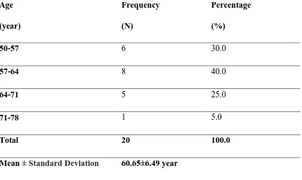
[image:32.595.105.535.165.422.2]4.2DEMOGRAPHIC DATA TABLE 1:-
PERCENTAGE AND FREQUENCY DISTRIBUTION ACCORDING TO AGE Age
(year)
Frequency (N)
Percentage (%)
50-57 6 30.0
57-64 8 40.0
64-71 5 25.0
71-78 1 5.0
Total 20 100.0
Mean ± Standard Deviation 60.65±6.49 year
Table 1 presents the distribution of age of studied patients had Thoracotomy surgery. Table showed that more than one-third (40.0%) of the patients had more frequently in the age group of 57-64 years.
Second most common age group of 50-57 years included six (30.0%) patients while five (25.0%) patients had Thoracotomy surgery noted within age group of 64-71 years.
The highest age group of 71-78 years for this study included only 1 (5.0%) patient
24
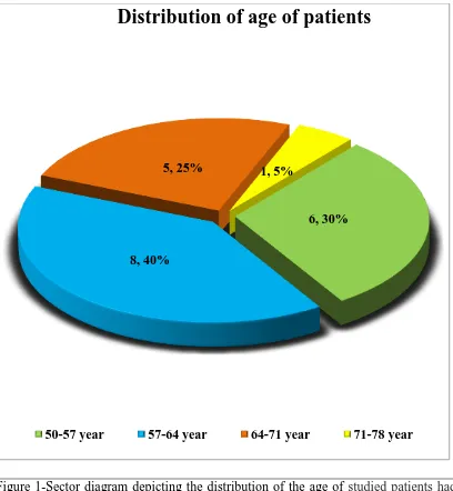
Figure 1-Sector diagram depicting the distribution of the age of studied patients had
Thoracotomy surgery.
6, 30%
8, 40%
5, 25% 1, 5%
Distribution of age of patients
25
TABLE 2:-
PERCENTAGE AND FREQUENCY DISTRIBUTION ACCORDING TO GENDER
Gender
Frequency (N)
Percentage (%)
Male 4 20.0
Female 16 80.0
Total 20 100.0
The distribution of gender of studied Thoracotomy surgery patients reports in table 2.
Table indicated that more than three-fourth (80.0%) of the patients were most
commonly female had selected from the population of patients had Thoracotomy
surgery.
Four (20.0%) of the patients had Thoracotomy surgery found to be male had
26
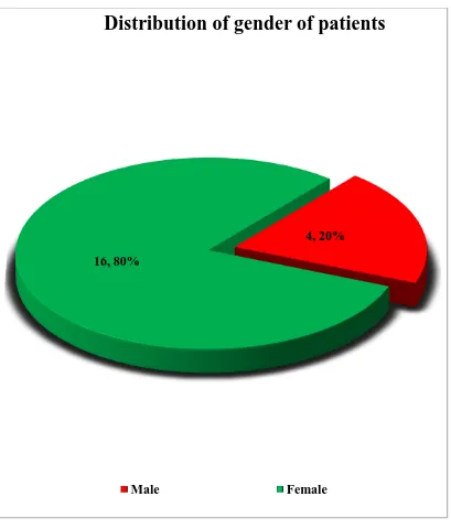
Figure 2- Sector diagram depicting the distribution of the gender of studied patients
had Thoracotomy surgery.
4, 20% 16, 80%
Distribution of gender of patients
[image:35.595.110.519.65.545.2]27
TABLE 3:-
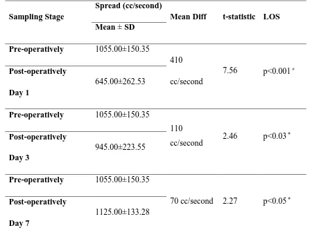
COMPARISON IN TOTAL LUNG CAPACITY OF PATIENTS BETWEEN PRE AND POST SURGERY ON DAY 1, DAY 3 AND DAY 7
Sampling Stage
Spread (cc/second)
Mean Diff t-statistic LOS Mean ± SD
Pre-operatively 1055.00±150.35
410 cc/second
7.56 p<0.001# Post-operatively
Day 1
645.00±262.53
Pre-operatively 1055.00±150.35
110
cc/second 2.46 p<0.03
Post-operatively Day 3
945.00±223.55
Pre-operatively 1055.00±150.35
70 cc/second 2.27 p<0.05
Post-operatively Day 7
1125.00±133.28
#
The mean differences are highly significant at the 0.001 level of significance. The mean differences are highly significant at the 0.03 and 0.05 levels of significance. [Mean Diff-Mean Difference; LOS-Level of Significance]
Table 3 highlights that the total lung capacity pre-operatively of patients undergoing
Thoracotomy surgery in bedside pulmonary function test found to be significantly
28
The post operatively lung capacity of patients had Thoracotomy surgery on day 1 found to be compromised significantly as compared to pre-operatively. Significant improvements noted in the total lung capacity of the patients on day 3 post operatively after use of Incentive spirometer, and on day 7 the mean total lung capacity found to be greater as compared to baseline total lung capacity.
Average (Mean ± SD) total lung capacity (1055.00±150.35 cubic centimeter/second) before surgery at sampling stage one found to be higher as compared to post-operatively total lung capacity on day 1 (645.00±262.53 cubic centimeter/second). These differences in mean total lung capacity between pre-operatively (baseline) and post-operatively (410 cubic centimeter/second) among Thoracotomy surgery patients were large and thus statistically reached at highly significant (p<0.001) level of significance.
After Thoracotomy surgery of studied patients, the average total lung capacity on day three (945.00±223.55 cubic centimeter/second) was increased due to administration of incentive spirometer but found to be remain little smaller and lowered as compared to pre-operative total lung capacity (1055.00±150.35 cubic centimeter/second). These differences in mean total lung capacity among patients between baseline and post-surgery on day third (110 cubic centimeter/second) were statistically significant (p<0.03).
29
Moreover, this was inference statistically that Thoracotomy surgery patients had significantly improved total lung capacity post-surgery on day 7 as compared to pre surgery and post-surgery on days 1 and 3. Furthermore, this study reported that incentive spirometer is the effective treatment protocol may be considered as a better tool to combat the weak total lung capacity after Thoracotomy
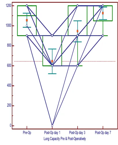
Figure 3 presented the Box and whisker diagram showing the distribution and comparison of the total lung capacity of patients undergone thoracotomy at pre-operatively (baseline) and post-pre-operatively on days 1, 3 and 7 using median, quartiles and error bars (95% confidence interval of mean).
30
Figure 3-Box and Whisker diagram depicting the distribution of total lung capacity of patients at pre-operatively (baseline) and post-operatively on days 1, 3 and 7.
0
200
400
600
800
1000
1200
Lung Capacity Pre & Post-Operatively
C
u
b
ic
C
e
n
t
im
e
t
e
r
/
S
e
c
o
n
d
31
TABLE 4:-
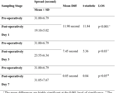
COMPARISON IN BREATH-HOLDING TIME AMONG PATIENTS BETWEEN PRE AND POST SURGERY ON DAY 1, DAY 3 AND DAY 7
Sampling Stage
Spread (second)
Mean Diff t-statistic LOS Mean ± SD
Pre-operatively 31.00±6.79
11.90 second 11.84 p<0.001# Post-operatively
Day 1
19.10±5.02
Pre-operatively 31.00±6.79
7.45 second 5.36 p<0.03
Post-operatively Day 3
23.55±6.34
Pre-operatively 31.00±6.79
0.05 second 0.04 p>0.05
Post-operatively Day 7
31.05±7.67
#
The mean differences are highly significant at the 0.001 level of significance. The mean differences are significant at the 0.03 level of significance. The mean differences aren’t significant (insignificant) at the 0.05 level of significance. [Mean Diff-Mean Difference; LOS-Level of Significance]
32
Pre-operatively breath-holding time of patients undergone Thoracotomy surgery at bedside pulmonary function test found to be significantly differed and higher as compared to breath-holding time post operatively at day 1 and day 3. The post-operative breath-holding time of patients on day 1 had Thoracotomy surgery found to be compromised significantly as compared to baseline stage. Significant improvements noted in breath-holding time of the patients post-surgery on days 3, and 7. Post-surgery on day 7, the mean breath-holding time found to be little greater as compared to baseline breath-holding time.
Average (Mean ± SD) breath-holding time (31.00±6.79 second) before surgery at sampling stage one found to be higher as compared to post-surgery breath-holding time on day 1 (19.10±5.02 second). The differences in mean breath-holding time between baseline and post-surgery on day 1 (11.90 second) among patients were higher and thus confirmed statistically highly significant (p<0.001).
After Thoracotomy surgery, the average breath-holding time of patients on day three (23.55±6.34 second) was increased due to administration of incentive spirometer but found remain smaller and lowered as compared to pre-operative breath-holding time (31.00±6.79 second). The statistical agreement indicated that these differences in mean breath-holding time among patients between baseline and post-surgery on day third (7.45 second) were statistically significant (p<0.03).
Average breath-holding time on day seven (31.05±7.67 second) of studied patients after Thoracotomy surgery found to be little increased and improved as compared to pre-operative holding time (31.00±6.79 second). Differences in mean breath-holding time among patients between baseline and post-surgery on day seven (0.05 second) were very little and thus couldn’t reach at statistically significant (p>0.05) level of significance.
33
Furthermore, this study reported that incentive spirometer is the effective treatment protocol may be considered as a better tool to combat the weak breath-holding time after Thoracotomy.
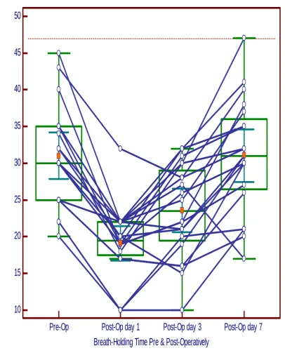
Figure 4 presented the Box and whisker diagram showing the distribution and comparison of the breath-holding time of patients undergoing Thoracotomy surgery at pre-operatively (baseline) and post-operatively on days 1, 3 and 7 using median, quartiles and error bars (95% confidence interval of mean).
34
Figure 4-Box and Whisker diagram depicting the distribution of breath-holding time of patients at pre-operatively (baseline) and post-operatively on days 1, 3 and 7.
10
15
20
25
30
35
40
45
50
Breath-Holding Time Pre & Post-Operatively
T
im
e
(
S
e
c
o
n
d
)
35
TABLE 5:-
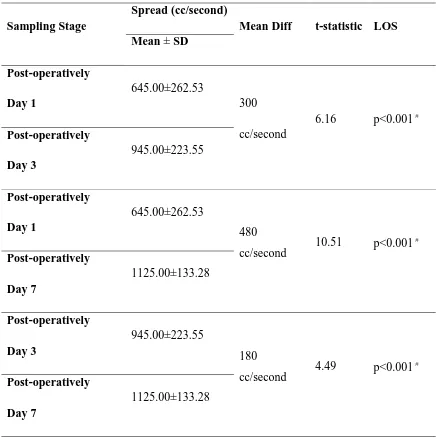
COMPARISON IN TOTAL LUNG CAPACITY OF PATIENTS BETWEEN DAYS 1 AND 3, 1 AND 7, AND 3 AND 7 POST OPERATIVELY
Sampling Stage
Spread (cc/second)
Mean Diff t-statistic LOS Mean ± SD
Post-operatively Day 1
645.00±262.53
300 cc/second
6.16 p<0.001# Post-operatively Day 3 945.00±223.55 Post-operatively Day 1 645.00±262.53 480
cc/second 10.51 p<0.001 # Post-operatively Day 7 1125.00±133.28 Post-operatively Day 3 945.00±223.55 180
cc/second 4.49 p<0.001 #
Post-operatively Day 7
1125.00±133.28
#
The mean differences are highly significant at the 0.001 level of significance. [Mean Diff-Mean Difference; LOS-Level of Significance]
36
to be improved successively measured on day 3 and day 7 which can be easily seen in table 5.
Post-operatively total lung capacity of patients had Thoracotomy surgery on day 7 found to be significantly differed and higher as compared to total lung capacity post operatively on day 1 and day 3.
Post-operatively on day 3, the average (Mean ± SD) total lung capacity (945.00±223.55 cubic centimeter/second) of patients found to be increased as compared to post-operatively total lung capacity on day 1 (645.00±262.53 cubic centimeter/second). These differences in mean total lung capacity between day 1 and day 3 (300 cubic centimeter/second) post-operatively among patients had Thoracotomy surgery were statistically highly significant (p<0.001).
Average total lung capacity on day seven (1125.00±133.28 cubic centimeter/second) of studied patients after Thoracotomy surgery was increased at large as compared to total lung capacity on day 1 (645.00±262.53 cubic centimeter/second) post-operatively. At post-surgery, these differences in mean total lung capacity among patients between day seven and day one (480 cubic centimeter/second) were statistically highly significant (p<0.001).
37
Henceforth, the statistical agreement indicated that on day seven the Thoracotomy surgery patients noted with significantly improved total lung capacity post-operatively as compared to day one and day three. Furthermore, results showed that incentive spirometer is the effective treatment protocol may be considered as a better tool to combat the weak total lung capacity after Thoracotomy
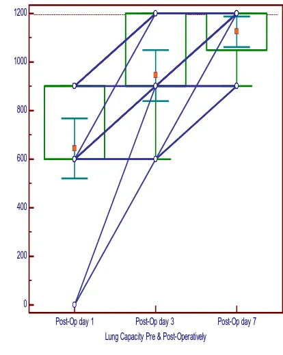
Figure 5 presented the Box and whisker diagram showing the distribution and comparison of the total lung capacity of patients undergone Thoracotomy surgery post-operatively on days 1, 3 and 7 using median, quartiles and error bars (95% confidence interval of mean).
38
Figure 5-Box and Whisker diagram depicting the distribution of total lung capacity of patients post-operatively on days 1, 3 and 7.
0
200
400
600
800
1000
1200
Lung Capacity Pre & Post-Operatively
C
u
b
ic
C
e
n
t
im
e
t
e
r
/
S
e
c
o
n
d
39
TABLE 6:-
COMPARISON BETWEEN DAYS: 1 AND 3, 1 AND 7, AND 3 AND 7 IN BREATH-HOLDING TIME OF PATIENTSPOST OPERATIVELY
Sampling Stage
Spread (second)
Mean Diff t-statistic LOS Mean ± SD
Post-operatively Day 1
19.10±5.02
4.45 second
3.66 p<0.001# Post-operatively Day 3 23.55±6.34 Post-operatively Day 1 19.10±5.02
11.95 second 8.01 p<0.001# Post-operatively Day 7 31.05±7.67 Post-operatively Day 3 23.55±6.34
7.50 second 5.29 p<0.001# Post-operatively
Day 7
31.05±7.67
#
The mean differences are highly significant at the 0.001 level of significance. [Mean Diff-Mean Difference; LOS-Level of Significance]
40
administration of incentive spirometer found to be rose and improved successively recorded on day 3 and day 7.
Breath-holding time of patients had Thoracotomy found to be significantly differed and higher post-operatively on day 7 as compared to day 1 and day 3.
Post-operatively on day 3, the average (Mean ± SD) breath-holding time (23.55±6.34 second) among patients was rose as compared to post-operatively breath-holding time on day 1 (19.10±5.02 second). These differences in mean breath-holding time between day 1 and day 3 (4.45 second) post-operatively among patients had Thoracotomy were statistically highly significant (p<0.001).
Average breath-holding time on day seven (31.05±7.67 second) of patients after Thoracotomy was increased at large as compared to breath-holding time on day 1 (19.10±5.02 second) post-operatively. At post-surgery, these differences in mean breath-holding time among patients between day seven and day one (11.95 second) were statistically highly significant (p<0.001).
Increases and improved average breath-holding time of studied patients noted on day seven (31.05±7.67 second) post-surgery as compared to post-surgery breath-holding time on day three (23.55±6.34 second). Post-surgery differences in mean breath-holding time of patients between day seven and day three (7.50 second) were highly significant (p<0.001).
41
Overall, results of present research documented that incentive spirometer is the effective treatment protocol may be considered as a better tool to combat the weak breath-holding time after Thoracotomy.
Figure 6 presented the Box and whisker diagram showing the distribution and comparison of the breath-holding time of patients undergoing Thoracotomy post-operatively on days 1, 3 and 7 using median, quartiles and error bars (95% confidence interval of mean).
42
Figure 6-Box and Whisker diagram depicting the distribution of breath-holding time of patients post-operatively on days 1, 3 and 7.
Furthermore, this was concreted statistically that patients had Thoracotomy found with compromised total lung capacity and breath-holding time. Therefore, administration of incentive spirometer after Thoracotomy found to be advantageous
10
15
20
25
30
35
40
45
50
Breath-Holding Time Pre & Post-Operatively
T
im
e
(
S
e
c
o
n
d
)
43
among patients and the patients had significantly improved total lung capacity and breath-holding time to overcome post-surgery difficulties in pulmonary functioning.
Overall, present research reported that the incentive spirometer is an effective conservative treatment protocol after Thoracotomy survivors to improve lung capacity and breath-holding time.
Finally, the above all statements and inferences from all the tables indicated the rejection of null hypothesis. Therefore, the alternative hypothesis is accepted which stated as “There will we significant effect on pulmonary function before and after the Thoracotomy” and that impacted the achievement of the entire selected objectives followed with fulfillment of the aim of the proposed research entitled “IMPACT OF PRE AND POST OPERATIVE BEDSIDE PULMONARY FUNCTION TEST ON THORACOTOMY PATIENT”.
44
CHAPTER V
DISCUSSIONThe purpose of the study is to provide beneficial effects of bedside pulmonary function in a variety of surgical fields. Overall, it may seems that PFT is beneficial both pre operatively and post operatively.
Twenty subjects who had thoracotomy were available for the study group had observed bedside pulmonary function test. The measurement like lung capacity (cubic centimeter/second) using incentive spirometer and breath holding time using breath holding test were conducted prior to thoracotomy and these data obtained were designated as baseline observations.
The lung capacity and breath holding time were re-collected after thoracotomy on days 1, 3 and 7.There data were stored as post-intervention observations for further statistical analysis.
Moreover, this was infered statistically that thoracotomy patients had significantly improved total lung capacity post-surgery on day 7 as compared to pre surgery and post-surgery on days 1 and 3.
Thus, results showed that incentive spirometer is an effective treatment protocol which may be considered as a better tool to combat the weak total lung capacity after thoracotomy
45
.Patient suffering from pain in other joints were also more likely to have a poor post-operative physical well being and subclinical improvement.(16)
Guide line recommended that preoperative spirometry is useful. The newer generation spirometer are simpler to use and both patient and therapist friendly.(18)
To the author’s knowledge, no studies in orthopaedic literature analysed the extent of incentive spirometer use in orthopaedic patient population or the factor that are correlated with patient compliance in using the incentive spirometry device. The author hypothesized that use of such devices by orthopaedic patient is far less than the recommended level and is affected by patient age and sex, surgery type and postoperative day and time.(19)
In study by Mesfin A.Lemma et al consecutive patient undergone thoracotomy incentive spirometry use was far less than what is recommended: only one patient in 10 met the recommended usage level, only 1 in 6 patient used the device > 7 times per hour and approximately one third of patient used the device <2 times per hour or not at all. Multiple reasons were hypothesized for the poor compliance observed in the study.(20)
Age and sex were not correlated with incentive spirometry use, and a distinct subpopulation of patients (32% of all patients) stayed noncompliant (<2 uses of the incentive spirometry device per hour) during their entire hospital stay. Several reasons may exist for this finding: (1) they may have had higher levels of pain or fatigue than their compliant peers; (2) they may not have received an adequate understanding of the benefits of incentive spirometry use during their preoperative education session; and (3) they may not have received adequate in-hospital encouragement to use the incentive spirometry device provided.(15)
Breath-holding time of patients had thoracotomy found to be significantly differed and higher post-operatively on day 7 as compared to day 1 and day 3.
46
to be advantageous among patients as they had significantly improved total lung capacity and breath-holding time to overcome post-surgery difficulties in pulmonary functioning.
In some study by Lingard et al comparing the patient from a centre in Montreal, Quebec, Canada and a centre in Boston, Massachutts, important difference between two centre were identified with respect to the preoperative status of patients. Preoperative PFT was the strongest determinant of functional outcome at six month and two years following thoracotomy. They hypothesized that there would be a significant difference in pain and functional status at the time of the preoperative assessment among the patient after adjusting for independent covarities.(15)
In study by Larsen et al. also found that although a thoracotomy seems to restore a patient’s quality of life, it does not restore their general physical wellbeing to that of the accepted population norm. This has been acknowledge by Becker et al. Starting that “the success of thoracotomy does not rely soley on thoracotomy(16)
There is no doubt that pulmonary function test plays an important role in diagnosis, management and followup.(14)
The average (Mean ± SD) total lung capacity (1055.00±150.35 cubic centimeter/second) before surgery found to be higher as compared to post-operatively total lung capacity on day 1(645.00±262.53 cubic centimeter/second) and 3(945.00±223.55 cubic centimeter/second). On day 7 the TLC (1125.00±133.28 cubic centimeter/second) of studied patients after thoracotomy found to be increased and improved as compared to pre-operative total lung capacity .
The (p value) of post-operative total lung capacity on day 1, 3 and 7 is statistically significant (p<0.001) (p<0.03) and (p<0.05) respectively.
47
Average breath-holding time on day seven (31.05±7.67 second) of studied patients after thoracotomy found to be little increased and improved as compared to pre-operative breath-holding time (31.00±6.79 second). However couldn’t reach at statistically significant (p>0.05) level of significance.
In present study it has been seen that bedside PFT is helpful in enhancing the quality of life of the patient and reduce the days of stay in hospital. It imply that increase in vital capacity of the lungs and increase in holding capacity helps the patient to recover faster.
Boysen and Blok AJ Suggested found that those patients who undergo lobectomy may transiently lose considerably more pulmonary function than would certainly be anticipated from the amount of tissue resected.
While J Richardson S Sabanathan found that Pulmonary function was better preserved in the paravertebral group who had higher oxygen saturation and less postoperative respiratory morbidity
Chin-Tung Lau Kenneth found that long term PFT result following thoracoscopic lobectomy is better than open lobectomy, This may be due to impaired respiratory musculature after thoracotomy
48
CHAPTER VI
SUMMARYAlso, the incentive spirometer is an effective conservative treatment protocol after thoracotomy survivors to improve lung capacity and breath-holding time.
CONCLUSION
49
CHAPTER VII
LIMITATIONS
1. The present study had a few limitations, like; the sample study was small.
2. Difference in intra-operative anesthesia medication as well as amount of postoperative analgesia used may have suppressed lung function.
3. Patient with postoperative delirium was not included in the study.
SUGGESTIONS
1. Future study can be based upon a relatively larger sample that is more representative of the population.
2. Further study can be done on patients with other surgical Procedures. 3. It would be better if clinical spirometery is used for evaluation in this study
50
CHAPTER VIII
REFERENCES
1. Efficacy of incentive spirometer exercise on pulmonary functions of patients with ankylosing spondylitis stabilized by tumor necrosis factor inhibi... PubMed 2012 Sep;39(9):18548.
2. Lawrence G. Lenke .Results of preoperative pulmonary function testing of adolescents with idiopathic scoliosis: A study of six hundred and thirty-one patients Peter O. Newton Children's Hospital San Diego Frances D. Faro Children's Hospital San DiegoSohrabGolloglyChildren's Hospital San Diego Randal R. Betz Shriner's Hospital for Children – Philadelphia
3. D’Lima, Darryl D The effect of preoperative exercise on thoracotomy May 2016 :326:174-182
4. Simon JP Devos Welters H, Jansen IJ a restrospective study of 70 patients 2015 :Vol 68 :Issue -3 235-41
5. Hamid Hassanzadeh, MD Amit jain, BS Eric W Tan MD Benjamin E Stein MD Megam L Van Hoy RN Nadine N Stewart RN Mesfin aA Lemma MD post operative Incentive spirometry June 2015 : 35:6
6. Sjaak pouwels, David Hageman,Lindy N M Gommans, Edith M Willigendael Simon W Nienhuijs , Marc R Schelitinga, Joep A,W Teijink. Preoperative exercise therapy in surgical care : a scoping review :journal of Clinical anesthesia June 2016 :33, 476-490
7. Karin Valkenet Ingrid GL van de Port The effects of preoperative exercise therapy on postoperative thoracotomy :a systematic review: clinical rehabilitation 2015:25:99-111
8. Choudhary sumer pulmonary function test in clinical practice: Importance requirement and limitations January – June 2013 3:1 7-2
9. Lingard, Elizabeth A katz jeffrey N Write Eilzabeth A Sledge Clement B Predicting the outcome of thoracotomy Oct 2015 :86 10:2179
51
11. Iverson LI Ecker RR Fox HE A Comparative study of IPPB the incentive spirometer and blow bottles the prevention of atelectasis following cardic surgery March 2016 25: 3 197-200
12. Robert H Bartlett, MD alan B Gazzaniga MD Tamar R Geraghty RN Respiratory Maneuver to prevent Post operative pulmonary complications A Critical review JAMA may 2016 224 :7 : 1017- 1021
13. Peter O Newton Results of Preoperative pulmonary function testing of adolescences with idiopathic scoliosis : A study of six hundred and thirty one patients , Children Hospital Diego 2016 :1073 : 1937- 1946
14. Lippincott Williams & Wilkins ,Incentive spirometry does not enhance recovery after thoracic surgery :Critical care Medicine :2002: 28:3
15. Czaplicki AP Borger JE Politi JR Chambers BT Taylor BC J Arthroplasty Evaluation of postoperative fever and leukocytosis in patients after thoracotomy Dec 2015 : 26; 8 :1386-9
16. Schoenfeld AJ Ochoa LM Bader JO Belmont PJ jr Am Risk Factor for immediate post operative complications following spine surgery National surgical quality improvement program Sep 2014 93: 17 :1577 -82
17. Weindler J Kiefer RT the efficacy of postoperative incentive spirometry is influenced by devise specific imposed breathing June :119:67: 1858-64
18. Overend TJ Anderson CM Lucy SD C Bhatia C Jonsson BI Timmernans C The effect of the incentive spirometry on Postoperative pulmonary complications a systematic review Sep 2015 :120:3 971-8
19. Galal Ghaly MD,Mohmend Kamel MD, Abu Nasar MS,The Annals of thoracic surgery, Vol 101, issue 2, Feb 2016, pg 465-472.
20. Rodrigo B, Interiano, Sue C Kaste, Chenghong , Journal of cancer survivorship Oct 2017, VOl 11, Issue 5,PP 553-561.
21. Daniel almquist Nabin Khanal, International journal of cancer research and treatment May 2018vol 38, no 5, 2903-2907
22. Fransis Ali Osaman, Alisia Mangram, Joseph Sucher, The American journal of Surgery, vol 216 issue 1, july 2018 page 46-51.
23. Ran Macke , Mathew j Schuchert , David d odell, journal of cardiothoracic surgery 2015, 10:49
52
25. Sue Jenkins chapter 10 patients problem management and outcome may 2016 26. James C latrids, James kang Rita Kandel, Journal of orthpedic research 35,1,(5-7)
2017
27. Colin F Mackenzie Chapter 8 Chest Physiotherapy for special patient “ Physiotherapy in the intense care unit – Second edition
28. David A Lipson Chapter 245 Aproach to the patient with disease of the respiratory system Harrison’Principle of Internal medicine –Seventh edition
29. W Darlus Reid Chapter 7 Pulmonary Function test
30. Donna Frownfelter Chapter 8 Pulmonary function test Principles and practice of cardiao pulmonary therapy third edition
31. Zara Cohen Chapter 20 Pulmonary function testing Egans Fundamental of respiratory care –Eleventh edition
32. Richard H Kallet Chapter 16 Bedside assessment of patient Egans Fundamentals of respiratory care Eleventh edition
33. Karl T Weber Kenin P Neuman Chapter 34 Principles and application of cardiopulmonary exercise testing Fishman’s Pulmonary disease and disorder Fifth edition
34. Carolyn Kishner Lymn Allen Colby Chapter 25 Management of Pulmonary Condition Fifth edition
35. Richardson Sabanathan BJA British journal of Anaesthesia, Vol 83, Issue 3,1 September 2016 ,Pages 387-392
36. Chin –Tung Lau Kenneth Journal of Pediatric surgery Volume 53, Issue 12, December 2018, Pages 2383-2385
53
CHAPTER-IX
ANNEXURE-I
INFORMED CONSENT FORM
TITLE: “IMPACT OF PRE AND POST OPERATIVE BEDSIDE
PULMONARY FUNCTION TEST ON THORACOTOMY
PATIENT – A COMPARATIVE STUDY ”
INVESTIGATOR:
CO-INVESTIGATOR:
PURPOSE OF THE STUDY:
I --- have been informed that is study will help clinicians and therapists to find out the effectiveness of floor and aquatic exercise on improving functional capacity and forced expiratory volume.
PROCEDURE:
I --- understand that I will undergo experiments with --- Under the direct supervision of coach. I am aware that I have to follow therapist’s instruction as has been told to me.
RISK AND DISCOMFORT:
I --- understand that there are no potential risks associated with this procedure, and understand that --- will accompany me during this procedure.
CONFIDENTIALITY :
54
REQUEST FOR MORE INFORMATION:
I --- understand that I may ask any question about the study at any times……… are available to answer my question. Copy of this concern form will be given to me keep for my careful reading.
REFUSAL OR WITHDRAWAL OF PARTICIPATION:
I ………understand that my participation is voluntary and I may withdraw consent and discontinue participation at any time after he has explained the reasons for doing so.
INJURY STATEMENT:
I ………..Understand that the diagnosis/ treatment procedure, under the guidance of my therapist, is likely to cause any/ no injury. In such case medical attention will be provide, but in compensation will be provide.
I understand my agreement to participation in this study and I am not waiving any of my legal rights. I ………… confirm that ……… have explained me the purpose of the study procedure and possible risk that it may experience. I have read and I have understood this concern to participate as a subject in this study………
………. ………
SUBJECT DATE
………. ………
55
I have explained………. The purpose of the research, the procedure required and the possible risks and benefits, to the best of my ability.
Investigator’s Signature
57
ANNEXURE –II
RESPIRATORY ASSESSMENT FORM
SUBJECTIVE EVALUATION
OBJECTIVE ASSESSMENT A. DYSPNOEA
ON STERNOUS ACTIVITY ON ORDINARY ACTIVITY ON MILD EXERTION AT REST
NAME AGE GENDER HEIGHT WEIGHT BMI
OCCUPATION
CHIEF COMPLAINTS
DURATION OF CONDITION PAST MEDICAL HISTORY PRESENT MEDICAL HISTORY FAMILY HISTORY
58
B.WHEEZE
DIURNAL VARIATIONS AGGERVATING FACTORS POSTURAL VARIATIONS
C.COUGH
DRY/PRODUCTIVE
D.SPUTUM
COLOUR
CONSISTANCY
ON OBSERVATION
BODY BUILT
BREATHING PATTERN
USE OF ACCESSORY MUSCLE
CYANOSIS
VITAL SIGNS
TEMPERATURE
RESPIRATORY RATE
BLOOD PRESSURE
59
ON EXAMINATION
A.BREATHING PATTERN
RATE
DEPTH
RHYTHUM
CHEST WALL EXPANSION
B.CHEST WALL CONFIGURATION
PECTUS EXCAVATUM
PECTUS CARINATUM
FLIAL CHEST
C. ON PALPATION
SYMMETRY OF CHEST
MOVEMENT
MUSCLE SPASM
TACTILE FREMITUS
TRACHEALDEVIATION
D.ON PERCUSSION
RESONANT
HYPER RESONANT
DULL
FLAT
E.ON AUSCULTATION
A. BREATH SOUNDS
NORMAL
ABNORMAL
ADVENTITOUS
60
INVESTIGATION
PULMONARY FUNCTION TEST FVC
FEV 1 FEV 1 / FVC PEFR
ARTERIAL BLOOD GAS
ANALYSIS
CHEST X RAY
DIAGNOSIS
AIM OF TREATMENT
MEANS OF TREATMENT
WHEATHER THE PATIENT IS
SELECTED YES NO
FOR STUDY
61
ANNEXURE –III
Sr. no.
Age
(y) Sex Incentive Spirometry cc/sec BHT(sec)
PRE Post-Op 1 Post-Op 3
Post-Op 7 PRE
Post-Op1 Post-Op3 Post-op7
1 61 F 1200 900 1200 1200 30 20 32 41
2 54 F 1000 600 900 900 22 10 10 21
3 75 M 900 600 600 1200 25 22 28 32
4 65 F 900 600 900 1200 25 17 16 26
5 67 F 1200 600 900 1200 25 17 16 20
6 61 F 900 900 1200 1200 43 32 28 40 <