JOURNAL OF VIROLOGY, Nov. 1975,p.1236-1247
Copyright ©1975 AmericanSocietyforMicrobiology Printed inU.S.A.
Simian
Virus
40-Specific
Proteins in HeLa Cells
Infected
with
Nondefective Adenovirus 2-Simian Virus
40
Hybrid Viruses
GERNOT WALTER* AND HAWLEY MARTIN
Tumor Virology Laboratory, The Salk Institute, Post OfficeBox 1809,San Diego, California92112 Receivedforpublication 17June 1975
Thesynthesis of simian virus 40(SV40)-specificproteins in HeLa cells infected
with thenondefective adenovirus2(Ad2)-SV40hybrid viruses, Ad2+ ND2, Ad2+
ND3, Ad2+ ND4, and Ad2+ ND5, was investigated. Infected-cell proteins were
labeled with radioactive amino acids late after infection, when host protein
synthesiswasshutoff,andanalyzed bypolyacrylamide gelelectrophoresis inthe
presence of sodium dodecyl sulfate. All polypeptides normally seen in Ad2-infected cellswerefound in cells infected by the hybrid viruses. In additiontothe
Ad2-specific proteins, cells infected with Ad2+ ND2 contain two SV40-specific
proteins withapparentmolecular weights of 42,000 and 56,000, cells infected with
Ad2+ ND4containoneprotein withanapparentmolecular weight of 56,000, and
cells infected with Ad2+ ND5 contain one protein with an apparent molecular
weight of 42,000. Cells infected with Ad2+ ND3 do not contain detectable
amounts of proteins not seen during Ad2 infection. Pulse-chase experiments
demonstrate thattheSV40-specific proteins induced by Ad2+ ND2, Ad2+ ND4,
and Ad2+ ND5 are metabolically unstable. These proteins are not present in
purified virions. Two nonstructural Ad2-specific proteins have been
demon-strated in Ad2 and hybrid virus-infected cells which have a smaller apparent
molecular weight afterashortpulse than afterapulse followed by achase. The
molecular weight increase during the chase may be caused by the addition of
carbohydratetoapolypeptide backbone.
Theexpression of the simian virus 40 (SV40)
genome during productive infection occurs in
twophases. The early phaseischaracterizedby the induction of the SV40-specific antigens, T (13), tumor-specific transplantation antigen
(TSTA)
(8), and U (26), andby the inductionofcellularDNAsynthesis (10,40). Thelate phase
begins with the synthesis of viral DNA and
structural proteins.
Little isknown aboutthe chemical and
bio-logical properties of the early antigens, T,
TSTA, and U. They could be either proteins
coded by the SV40 genome (early proteins) or,
alternatively,cellular products induced or
mod-ified by the early viral proteins. The major
obstacle to thedetection andcharacterizationof
early SV40 proteins and early antigens is the
continuation of host protein synthesis in
SV40-infected cells. Atpresent, no methods are
avail-able forselectively inhibiting host protein
syn-thesis and making the clear detection of early
proteins possible.
The nondefective adenovirus 2 (Ad2)-SV40
hybrid viruses, isolated by Lewis and
co-work-ers, are a useful tool forthe study of earlySV40
proteins. They have the following properties:
each of the five nondefective
hybrid
viruses,
Ad2+ ND1
(24),
Ad2+ND2,
Ad2+ND3,
Ad2+ ND4, and Ad2+ ND5(25),
contains asingle
continuous segment of
SV40
DNAcovalently
insertedat thesame site in the Ad2genome(3,
12,
16, 21,32).
TheSV40
DNAsegments differin size: Ad2+ ND1 contains 17 to
20%;
Ad2+ ND2, 32 to36.5%;
Ad2+ND3, 7%;
Ad2+ND4,
43 to 48%; and
Ad2+
ND5,
28% of theSV40
genome (16,
20, 31,
32).
Thesegments
arecompletely
overlapping
andcolinear withSV40
DNA(16,
20)
(Fig. 1).
They
haveacommonendpoint which is located in the Haemophilus influenzaerestriction endonuclease fragment G ofSV40 DNAand is0.11 mapunits of theSV40
genome away from the
cleavage
site of EcoRlrestriction endonuclease
(16,
20,
31,
32).
AllSV40 DNA segments contain at their common
end point a major fraction of the Hin G
frag-ment (20) which is located in the late region of
theSV40 genome. During productive infection
withthe nondefective hybridviruses, the SV40
DNAsegment containedineachhybridvirus is
fully transcribed (18, 23). The transcripts are
complementary to the minus (early) strand of
SV40 DNA and consist of both early and
1236
on November 10, 2019 by guest
http://jvi.asm.org/
SV40-SPECIFIC PROTEINS 1237
Ad2+ND
U .Ad2+ND2 U TSTA
Ad2+ND3
-U TSTA T
Ad2 ND5
Hin Restriction Endonuclease Cleavage Map
F J G B H A C D E K F
| I I I I
Late Regi Early Region Late Region
0 0.1 0.2 0.3 0.4 0.5 0.6 0.7 0.8 0.9 1.0
FRACTIONAL LENGTH OF SV40 DNA
FIG. 1. SV40 DNA segments in nondefective Ad2-SV40 hybrid viruses in relation to the Haemophilus influenza restriction endonuclease cleavage map ofSV40. The position of the early and late gene regions in SV40 DNAisderived from the results of Khoury et al. (17) and Dhar et al. (5). Arrows indicate the direction of transcription. The dotted lines indicate the degree of variation in the length of theSV40DNA segments as determinedbydifferentinvestigators(16, 20, 31, 32).TheHaemophilusinfluenza cleavage map is derived from Danna et al. (4).
antilate sequences (18).
Ad2+ ND4, containing
the largest segment of the SV40 genome,
in-duces the synthesis ofthree
SV40-specific
anti-gens: T,
TSTA,
and U(25, 27).
Ad2+ ND2induces TSTA and U, and Ad2+ ND1 induces onlyU antigen (25-27). The SV40DNAregions
in these hybrid
viruses,
which determine theexpression of the
early
SV40
antigens,
havebeen mapped onthe
SV40
genome(16, 20).
TheexpressionoftheSV40 information in the
nondefective
hybrid
viruses iscontrolledby
theAd2 genome and takes
place
at a late timeduring productive
infection,
thatis,
afterhybrid
viral DNA
synthesis
hasbeen initiated(23).
It isthis property of the
hybrid
viruseswhichfacili-tates the identification and characterization of
early
SV40proteins,
since lateduring
infectionhostprotein
synthesis
isshut offby
theadenovi-rus genome. As a consequence, it may be
possible
to detectearly SV40
proteins
orSV40-specific
antigens
inhybrid
virus-infectedcells
by
labeling
with radioactive amino acidslate in the infection
cycle
and thenanalyzing
the labeled
proteins
by
gel
electrophoresis
andautoradiography.
It hasrecently
been shownthat HeLa cells infected with Ad2+ND1
synthe-size a polypeptide of molecularweight 28,000
which is not present in Ad2-infected cells
(9,
28). Inthisreport, wedemonstratethe presence
of specific proteins in cells infected
by
Ad2+ND2, Ad2+
ND4,
and Ad2+ ND5. We haveprovisionally designated these
proteins
as"SV40
specific,"since theirexpressioniscausedby the SV40 information contained in the
hy-brid virus genomes.
MATERIALS AND METHODS
Cells. HeLa S3 cellswere growninsuspension in Eagle minimum essential medium supplemented with 5% calf serum or in monolayer in Dulbecco's modification of Eagle medium (DMEM) with 10% calfserum. HeLa S3 cells were only used for labeling experiments. Foradenovirus plaque assays, a differ-enttype ofHeLacell with a flattermorphology was used.These cells wereobtained from the Cold Spring Harbor Laboratory and grown in DMEM supple-mented with 10% calf serum. CV1 cells, a line of African green monkey kidney cells (AGMK), were obtained from the American Type Culture Collection and grown in DMEM supplemented with 10% fetal calf serum. Primary AGMK cells were purchased
from Microbiological Associates, Inc., and grown in
DMEMwith 10% fetal calf serum.
Viruses. Seed stocks of nondefective Ad2-SV40
hybrid viruses Ad2+ ND1, Ad2+ ND2, Ad2+ ND3,
Ad2+ND4, and Ad2+ND5 wereobtained from A. M. Lewis, Jr.Stocks of Ad2+ ND1, Ad2+ ND2, and Ad2+ ND4wereprepared in
CV,
and HeLa S3 cells. StocksofAd2, Ad2+ ND3, and Ad2+ ND5 were prepared in HeLaS3 cells.
Hybrid stocks of Ad2+ ND1, Ad2+ ND2, Ad2+
ND3, and Ad2+ ND5 were shown to be free of adeno-associated viruses by cesium chloride density centrifugation, high multiplicity infections, and heat sensitivity assays which will bedescribed elsewhere. All stocksofAd2+ND4,however, were contaminated withadeno-associated virus.
Plaque titrations were routinely performed on HeLa and CVl cells. In one experiment, primary AGMK cells were used to assay Ad2, Ad2+ ND1, Ad2+ ND2, and Ad2+ ND4. The assays wereperformed as describedby Williams (41). Theoverlay medium for HeLacellscontained 0.6% agar (Difco)and0.9%agar
for
CV,
and AGMK cells. After incubationfor3to4on November 10, 2019 by guest
http://jvi.asm.org/
[image:2.498.101.385.71.246.2]1238 WALTER AND MARTIN
days, 2ml ofadditionaloverlay mediumwasadded.
Overlaymediumcontainingneutral redand 0.9%agar was added 6to 7days after infection of HeLa cells,
and 8 to 10days after infection ofCV, and AGMK cells. MgCl2 was omitted from the overlay medium
when plaque assays were performed on CVl and
AGMK cells. The plaque titers of virus stocks
pre-pared in HeLa or CV, cells are showh in Table 1.
Plaque formation inCV,cellswasless efficient than
inAGMK cells. Whereas Ad2+ ND1 and Ad2+ND4
formed plaqueswiththesameefficiencyonHeLaand
AGMK cells, Ad2+ ND2 formed 10-fold less plaques
on AGMK cells thanon HeLa cells. Ad2showed at least a 1,000-fold difference in plaque efficiency
be-tweenHeLaandAGMK cells(Table 1).
Thehigh titerstocksofAd2+ ND3andAd2+ ND5
up to dilutions of 10-3 produced strong cytopathic effect (CPE)in theentireCV,monolayer,preventing
the detection of plaques. Neither CPE nor plaques wereobservedatdilutionsof10-'. Ad2alsoproduced
totalCPEonCV1 cellsuptoadilution of10-', buta
small number ofplaques at a dilution of 10'. This
observedCPEispresumablycausedbyahigh
concen-tration of freepentons inthesehightiterstocks and
can beeliminated by trypsintreatment (9). Since in
our experiments trypsin treatment of the high titer stockswasnotperformed, the titers of10' PFU/mlof
Ad2+ND3and Ad2+ND5onCV,cellsonlyrepresent maximumestimates.Asimilar CPEwasproduced by
the high titer Ad2 stock when assayed on AGMK
cells. In thiscase,CPEwasobserveduptoadilution of10-'. It is likely, therefore,that theAd2 titerof 106
PFU/ml on AGMJK cells represents an
overestima-tion. The titers of Ad2+ ND3 and Ad2+ ND5stocks
werenotdeterminedonAGMK.
Infectionandlabelingof cells. Forlabeling
exper-iments, 2 x 10⁸ HeLa S3 cellswere seededon 3-cm
plastic petridishes (NUNC, Denmark) and infected 24 h later with 0.2 ml of virus. After an adsorption
period of 1 h, 2 ml of growth medium, DMEM
containing 10% calfserum, wasadded perdish. For
labeling cells, the culture medium was changed to labeling mediumcontaining 2.5%of the normallevel
ofaminoacids, 5% calfserum, and 20 ACiof "4C-la-beled L-amino acid mixtureperml. A0.6-ml amount oflabelingmediumwasaddedperdish. Afterlabeling,
the cell layer was washed three times withDMEM. Thecellswerelysedonthedish with0.2ml of
electro-phoresis sample buffer (0.0625 M Tris, pH 6.8; 3% sodium dodecyl sulfate [SDS]; 5%
2-mercaptoetha-nol; 10% glycerol; 0.001% bromphenol blue; 2 mM
phenylmethylsulfonyl fluoride). Glycerol and brom-phenol blue were omitted until after boiling. The
lysatesweresonically treatedfor 10swithaBranson
sonifieratposition4andheated for 2mininaboiling
waterbath.
Polyacrylamide gel electrophoresis and scintil-lation autography (fluorography). The
polyacryl-amidegelsystemofLaemmli and Maizelasdescribed
by Laemmli (19) wasemployed, exceptthat the gels
wereformedasslabs 1 mmthickand 9.5cmlong
be-tween glass plates. Thegeneral apparatusand
meth-odology for gel electrophoresis have been described elsewhere(29).Theratioofacrylamideto
bisacrylam-ide was 30 to 0.8. A 10-ul volume of cell lysate in
electrophoresis sample buffer, containing5to10,gof
protein and approximately 20,000to 100,000counts/
min, were loaded per slot. Electrophoresis was
per-formed at a constant current of 10 mA. After
elec-trophoresis, the gels wereprepared forfluorography, which is 10 times more sensitive than conventional
autoradiography, asdescribedby Bonner andLaskey
(2). Briefly, the gels were dehydrated in dimethyl
sulfoxide, soaked inasolution of2,5-diphenyloxazole
indimethyl sulfoxide, immersed in water toremove
dimethyl sulfoxide, dried and exposed to medical X-ray film (Kodak, RP Royal X-omat, RP/R54) at
-70 C.
Thepurityofthechemicals usedfor gel
electropho-resis can influence the relative mobility of proteins
and thequalityof separations of complex mixtures of
proteins (36). Therefore, we list the source of the
reagents used in this study: acrylamide (Eastman
5521)wasrecrystallizedfromchloroform; bisacrylam-ide (Eastman8383);
N,N,N',N'-tetramethylethylene-diamine (Eastman 8178); glycine (Eastman 445); SDS(BDH, speciallypure,30176); Tris (Trizma base,
reagentgrade, T-1503);ammonium persulfate (Baker
0762); and2-mercaptoethanol (Eastman4196). The molecularweight ofproteins was determined
by comparing their mobilityon SDS polyacrylamide gels with proteins of known molecular weight. The following markerproteinswereused: Ad2 proteinsas
characterized by Maizel et al. (30), Ishibashi and Maizel (14), andEverittetal.(7); ,B-galactosidaseof
Escherichia coli (135,000 daltons); bovine serum
al-bumin (68,000 daltons);ovalbumin (43,000daltons); chymotrypsinogen (25,700 daltons); and cytochrome
cfrom horse heart(12,270daltons).
[image:3.498.69.463.564.660.2]Labeling and purification of virions. HeLa S3 TABLE 1. Plaque-forming ability ofAd2-SV40 hybrid viruses
Infectivity(PFU/ml) Stockprepared
Virus
inc
reaeHeLa CV, AGMK in
Ad2+ ND1 9.2 x 10⁶ 7.0 x 10' 1.1 X 10⁷
Cv1
Ad2+ ND2 2.4 x 10' 1.2 x 10² 2.5 x10⁵ CVI
Ad2+ND3 4.5x 10⁹ <103 NDa HeLa
Ad2+ND4 6.9 x 10⁶ 2.2 x 10' 3.0 x 10⁶ CV1
Ad2+ND5 1.7 x10¹⁰ <103 ND HeLa
Ad2 3.5 x10⁹ 8.0 x 10' <106 HeLa
aND, Not done.
on November 10, 2019 by guest
http://jvi.asm.org/
SV40-SPECIFIC PROTEINS 1239
cells, 2 x 101per 10-cm dish, were seeded in DMEM
containing 10% calf serum. After 24 h, parallel cul-tures wereinfectedwith Ad2, Ad2+ND1,Ad2+ND2,
Ad2+ ND3, Ad2- ND4, and Ad2+ ND5. The multi-plicity of infection was approximately 1to2PFU/cell.
After 21 h of infection, the growth medium was replaced bylabelingmedium containing one-tenthof
the normal level of aminoacid,5%calf serum, and 6.5
M(Ciof "4C-labeledL-amino acidmixture perml. Five milliliters of labeling medium was added per dish. After 52 h ofinfection, the labelingmedium was
re-moved,and the cells werewashedoncewith DMEM and scraped from the culture dishes in 0.01 MTris,
pH 8.0. The virus purification was performed as
described previously (30). After centrifugation in CsCl, the purified virionsweredialyzed firstagainst
0.01 M Tris, pH 7.4, at 4C and then against 0.1%
SDS at room temperature. They were concentrated by lyophilization and dissolved in electrophoresis
sample buffer. Thisprocedureforconcentratingvirus
was preferred since precipitation of virus with tri-chloroacetic acid in some instances resulted in a
selective loss of the virion polypeptide VII. The reasonforthis loss isunknown.
RESULTS
Induction of
SV40-specific
proteins by
Ad2+
ND1,
Ad2+ ND2, Ad2+ND4,
andAd2+
ND5. Monolayers of HeLa cells were infected with Ad2 and the non-defective
hybrid viruses,
Ad2+ND1, Ad2+
ND2,
Ad2+ND3,
Ad2+ND4, and Ad2+ND5, and labeled from38 to 40h aftertheinfection with "4C-labeledaminoacids. The
infected cells were lysed in SDS containing buffer(Materials and
Methods).
Theradioactive polypeptides of the lysates wereanalyzed
by
SDS acrylamidegel
electrophoresis
andvisual-ized
by
fluorography
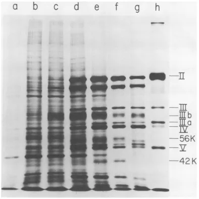
(2) as described above.Figure 2 shows a fluorogram of a 7.5 to 30%
acrylamide
gradientgel.
The patterns ofAd2-specific proteins from Ad2 and
hybrid
virus-in-fected cells were similar to thosepreviously
described (1, 14,
39).
In addition totheadeno-virus-specificproteins, Ad2+ ND1-infected cells containeda
polypeptide
withamolecularweight
of 28,000 (28 K protein) which has beende-scribed
previously
(9, 28). Itmigrated
very closeto twoadenovirus-specific proteins, Vaand
Vb,
andin ourhandswas
separable
fromtheseonly
in 7.5 to 30%gradient
gels.
Since itwaspresentonly insmall quantity, a
long
exposure time ofthe fluorogram was required for its detection.
Ad2+ ND2 induced twoSV40-specific
polypep-tides with molecular
weights
of 42,000 (42 Kprotein) and 56,000 (56 K protein). Ad2+ ND4 inducedoneSV40-specific proteinwitha
molec-ular
weight
of56,000. In severalAd2+ ND4in-fections this protein was
consistently
detectedin smaller quantity than the 56 K
protein
inAd2+ ND2-infected cells. A protein of
42,000
molecular weight was not detectable in Ad2+
ND4-infected cells, using a variety of different
gel conditions and after long exposure of the
gel to theX-rayfilm.
Threeotherproteins with apparent molecular
weights of 96,000, 85,000, and 65,000 were
synthesized only in Ad2+ ND4-infected cells.
They were structural proteins of adeno-associated virus (15, 35) present as a contami-nant in our Ad2+ ND4 stock. The 96,000 dalton
AAV protein was clearly detectable in a 7.5 to 30% acrylamide gradient gel (Fig. 2), whereas the 85,000 and the 65,000 dalton AAV proteins
comigrated with Ad2 protein Ill and IIIa/IV,
respectively. Under alternate gel conditions, these AAV proteins have been separated from
theAd2 proteins, the 85,000 dalton AAV protein
from Ad2 protein III by prolonged gel electro-phoresis and the 65,000 dalton AAV protein
from Ad2 proteinsIIIaand IV byraising the pH
ofthepolyacrylamide gel buffer from 8.8 to 9.3.
Under theseconditions, the 96,000, 85,000, and 65,000 molecular weight proteins from Ad2+
ND4-infected cells were shown to comigrate with the three structural proteins of purified
AAVisolated fromAd2+ ND4-infected CV, and
HeLa cells (manuscript in preparation).
Although cells infected with Ad2+ ND3 and
Ad2+ ND5 contain SV40-specific RNA
se-quences (23), they do not synthesize detectable
amounts of SV40-specific antigens (25, 27).
Interestingly, Ad2+ ND5-infected cells
synthe-sized a polypeptide late in infection that was not detectable in Ad2-infected cells and that
comigrated with the Ad2+ ND2-induced 42 K protein (Fig. 2). No additional proteins in Ad2+
ND3-infected cellscould be detected by any of
theanalytical gel conditions that wetested.
Time of synthesis of SV40-specific proteins. To determine the time course of the
appearance oftheSV40-specificproteins, HeLa
cellswere infectedwith Ad2+ ND2, Ad2+ ND4,
and Ad2+ ND5 and labeled with
"4C-labeled
amino acids at different times after infection.
Figure3 shows that the56Kand 42K proteins
induced by Ad2+ ND2 can firstbe detected at
23hafter infectionsimultaneously with the
ap-pearance ofstructural Ad2 proteins. Their
syn-thesis continues late in infection when host
proteinsynthesis isshutoffalmostcompletely.
Asimilar time course was observed for the
syn-thesis of the 42 K protein in cells infected by Ad2+ ND5 (data not shown). Thebeginning of
synthesis ofthe 56,000 dalton protein in cells
infected by Ad2+ ND4 could not be measured
because this protein was present in very small quantity.
Metabolic
stability
of theSV40
proteins.
Pulse-chaseexperiments were
performed
toex-amine the metabolic
stabilitv
of theon November 10, 2019 by guest
http://jvi.asm.org/
1240 WALTER AND MARTIN
a
b
c
d
e
f
g
h
rn- -~
~---ms amommw"o4i- C
mm
-~~~~~~~~~~~~~~~~~~.
...-
Tu
10-m wmm
4w
-c
28k
-w-z0.M Y
~ ._ _ ..
_
_4sno
FIG. 2. SDS polyacrylamide gel fluorogram of 'IC polypeptides from cells infected by nondefective Ad2-SV40hybrid viruses. Parallel cultures of HeLaS3cellswereinfectedwith the five nondefective Ad2-SV40
hybrid viruses and labeled with 14C amino acids. The multiplicity ofinfectionwas approximately 10to50 PFU/cell. Thetime of labelingwasfrom38through40h afterinfection.A mock-infectedcontrolwassimilarly
treated.Samples of10
Al
wereappliedtoa7.5to30%acrylamide gradientgel. Aconstant currentof10mAwasapplied for5hand45min. The sample order is: mock-infectedcells (a), cells infected by Ad2 (b),Ad2+ND1
(c),Ad2+ ND2(d),Ad2+ ND3(e), Ad2+ND4 (f),Ad2+ ND5 (g), purified Ad2 viripns (h). The fluorographic
exposuretimefor (a)to(h)was40h; (i)and(j)showexposuretime of6days of samples(b) and (c). Procedures
indetailaredescribed intext.Ad2 polypeptidesweredesignated with Roman numerals accordingtoMaizelet
al. (30) and Ishibashiand Maizel (14). Polypeptides VIa and VIbarenotseparated under the gel conditions used here. They areseparable, however, on a7.5 to30%gradient slabgel of 20-cm length (datanotshown).
PolypeptidesIIIband Vcaredescribed in "Metabolic stability oftheSV40-specificproteins". No distinction was made between the designation of Ad2 virion proteins and Ad2-specific proteins in infectedcells. The
samples ofAd2+ ND3- and Ad2+ ND5-infected cells contained about twice as much protein as the other
infected-cell samples. This resulted in incomplete separation of polypeptidesII andIIain samples(e) and (g).
SV40-specific proteins. Ad2-, Ad2+ ND2-, and
Ad2+ ND4-infected cells were labeled with
radioactive amino acids from 39 to 41 h after
infection and compared with cells labeledatthe
same time and chased with unlabeled amino
acids for 12 h. Figure 4 demonstrates that
within a period of 12 h, the 56 K and 42 K
proteins induced by Ad2+ ND2 and the 56 K
protein induced by Ad2+ ND4 disappeared
almost completely. Further experiments with
Ad2+ ND2-infected cells labeled for 2 h and
chased foreither 2, 4,6, or 10hsuggestthat the
half-life of the 56 K and 42 K proteins is less
than4 h (Fig. 5). Cells labeled foronly 15 min
showrelativelymorelabel incorporatedinthese
two proteins than cells labeledfor 2 h(Fig. 5),
again indicating their metabolic instability.
Pulse-chase experiments with Ad2+ ND5
sug-gestthat the half-life of the Ad2+ND5-induced
42Kprotein isasshortasthat of thetwoAd2+
ND2-induced proteins (data not shown). It
should be pointed out that the degradation of
both Ad2+ ND2-specific proteins during the
chase didnotresult in theformation of discrete
and stable smaller polypeptides of molecular
weights between 42,000 and 12,000, since the
polypeptide patterns of Ad2 and Ad2+
ND2-infected cells aftera12h chase looked
indistin-guishable in this molecular weight range.
Two characteristic changes which occurred
during the chaseofbothAd2 andhybrid
virus-infected cells (Fig.4and5) have been described
recently. Andersonetal. (1) demonstrated that
thepolypeptide probably correspondingtoVIa/
VIb in this paper is the precursor of virion
polypeptide VII. Ishibashi and Maizel (14) have
shown that thepolypeptides Va, Vb, VIa, and
VIb are presentin "young" virions and absent
in"aged" virions. They have suggested that Va,
Vb, and VIa could be theprecursorstoVI, VIII,
andVII, respectively.
Twootherchangeswere observedtobe
com-mon to both adenovirus and hybrid
virus-infected cells. A polypeptide designated Vc in
Fig.4hasaslightly smallerapparentmolecular
weight after a 2 h pulse than after a 2h pulse
6
r
K-4.o',<
J. VIROL.
IJ
on November 10, 2019 by guest
http://jvi.asm.org/
[image:5.498.76.461.81.277.2]cd
e
f
g
h
-_
'4
I!Ej
V 4U. g8
.
a
_e
40Om
41W.1
----mb
--m b
56K
42K
FIG. 3. SDSpolyacrylamide gel fluorogram of "4C-labeled polypeptides from cells infected by Ad2+ ND2; timecourseofthesynthesis ofthe 56 K and42 Kproteins.Parallel culturesofHeLaS3 cellswereinfected with
Ad2+ ND2 and labeledfor2 h with "4C-labeled amino acids(see text) atdifferent timesafter infection. The
multiplicity of infectionwasapproximately 100PFUIcell.A 7.5%acrylamide gelwas runfor6hataconstant
currentof10 mA. Thesampleorder is:(a) mock-infected cells, (b)Ad2+ND2-infectedcellslabeled 9through11 h, (c)15through 17h, (d)21through23h, (e)27through29h, (f)34through36hafter infection,(g)Ad2-infected
cells, (h) purifiedAd2 virions.
a
b
a0
on November 10, 2019 by guest
http://jvi.asm.org/
[image:6.498.44.438.131.525.2]1242 WALTER AND MARTIN
a
b
c
d
e
f
..I
56K--42K
III
_
m-1122-v
.
Vc
[-ii-
*.--
*_
Sn_E
m
ama
c-_
Sb
-. 1-:'.i_pkw
_
-
ZVa /Vb
..V.
aI
_~~~~4 -._m
~~-
zmm
FIG. 4. SDSpolyacrylamide gel fluorogram of "4C-labeledpolypeptides from cells infected withAd2,Ad2+
ND2, and Ad2+ ND4. Comparison of cells pulse-labeled for 2 h (39 through 41hpostinfection) with cells pulse-labeled for2 handchasedfor12h (41through 53hpostinfection). The infection is described in legend to Fig. 2. The cells were labeledasdescribed in text. Before chasing with DMEM plus 10% calf serum, the cell layer was rinsed three times with DMEM. Samples (a) through (f) were applied to an 11.5% gel. The running timewas 5hat 10mA.(a) Ad2 pulse, (b) Ad2 chase, (c) Ad2+ ND2 pulse, (d) Ad2+ ND2 chase, (e) Ad2+ ND4 pulse, (f) Ad2+ ND4 chase.
followedbya12h chase. A similar phenomenon
occurredwithaprotein in the regionbetween III
and IIIa, designated IIlb. Incells labeled for 15
min, proteins Vc and
IlIb
appeared slightlysmaller in size than after a 2 h label (Fig. 5). During the chase of 2, 4, and 6 h, they were
replaced in a stepwise fashion by proteins of
larger apparent molecular weight. No further
increase in sizeoccurred after 6 h. The stepwise
increase in size of protein
IlIb
isperturbed bythe presence of a large amount of serum
al-bumininthe 2hpulsesample. Neither Vc nor
IIIbseem tobepresentinpurified virions (Fig.
5). The apparent molecular weights of Vc and
on November 10, 2019 by guest
http://jvi.asm.org/
[image:7.498.68.460.72.507.2]IIIb after a 15 min pulse are30,500 and
74,000,
respectively, and 34,000 and 79,000,
respec-tively,after a 2 h pulse followed by a 10 h chase.
These results, combined with the observation
thatthese proteins have a more diffuse
appear-ance on gels after a chase than after a short
pulse of 15 min (Fig. 5), suggest that the increase in molecular weight during the chase
might be the result of a sequential addition of
carbohydrate
to apolypeptide backbone. Figure3 demonstrates that IIIb is synthesized in
in-fected cells earlier than the major structural
Ad2proteins. Therefore, it is likely thatIIIbis
a
b
c
d
identical to an Ad2-induced early protein of
similar molecular weight: El described by Walter and Maizel (39), the 71 K
protein
described by Anderson et al. (1), and the DNA-binding proteindescribed
by
Levineetal. (22).Analysis of virion proteins. It was
impor-tant to determine whether the new
SV40-specific proteins present in
hybrid
virus-infected cells were
structural
components of hybrid virions. To investigate this possibility, Ad2 and the fivehybrid
viruses were labeledwith
"4C-labeled
amino acids, purified, ande
f
9
IT
as
_
n_
-_
_a_
-56K
42K
_m
EZ
c
Va/M
b
IL-SSS-S
FIG. 5. SDS polyacrylamide gel fluorogram of "4C-labeled polypeptides from Ad2+ ND2-infected cells; metabolicstabilityoftheAd2+ ND2-induced 56Kand 42 Kproteins.Parallel culturesofHeLa S3 cellswere
in-fectedwith Ad2+ ND2atamultiplicity of100PFU/cell. Oneculturewaslabeledfor15min30 hafter infection withlabelingmediumcontainingneither unlabeledamino acidsnor serumand 30 gCiof "4C-labeledaminoacids
perml. The other cultureswerelabeled 30 hafter infection for2 hasdescribed in text. Thechasewasperformed
asdescribedinlegendtoFig.4. Thesampleswereappliedtoa7.5%gelandrunfor5 hat 10 mA. Thesample order is:(a) purifiedAd2 virions. Ad2+ND2-infectedcells:(b) 15-min label, (c) 2-hlabel,(d)2-hlabelfollowed bya2-hchase,(e)2-hlabelfollowed bya4-hchase,(f)2-h labelfollowed bya6-hchase,(g)2-hlabelfollowed bya
10-h chase.
lLa
--Ez
.V...
on November 10, 2019 by guest
http://jvi.asm.org/
[image:8.498.46.432.216.573.2]b
c
d
ef
~
__ *iv;
_.
"_~~~~~~~~~~~~~~~
...I *.__ __ __ v __ ...,,....111~~I
tT #a
_@ --~~~~~~~~~~~~~~~~~~~~~~~~~~~~~~~~~~~~~~~~~L
\14
'L
-_
V-m_
s o sfem_ .
_S,,,..~~~~~~~~~~~
..,I;
il-tgVITi
--LX---FIG. 6. SDS polyacrylamide gel autoradiogram of "4C-labeled polypeptides of purified virions. The pro-ceduresfor labeling and purification ofvirionsweredescribed intext.Sampleswereappliedtoa 12.5%gel.The running time was6h and 15minat 10 mA. (a)Ad2+ ND1, (b)Ad2+ND2, (c)Ad2+ ND3, (d)Ad2+ND4,(e)
Ad2+ND5,(f)Ad2.
1244
a
on November 10, 2019 by guest
http://jvi.asm.org/
[image:9.498.71.461.44.607.2]analyzedonaslab gel. Figure6shows thatall six
virus preparations have indistinguishable
poly-peptidepatternscharacteristicofAd2virions (1,
7, 14, 30). No proteins of 56,000, 42,000, and
28,000 molecular weight were detectable in the
hybrid virions, suggesting that these proteins
are not part of the virion structure. Several
minorpolypeptidesmigratingbetween
polypep-tidesIIand III, IV and V, V and VI, werefound
in all virus preparations. They may correspond
to components of similar mobility described previously(1, 7, 14). Aprotein ofslightlyhigher
mobility thanprotein IIIwasfound
only
in viruspreparations which had beendialyzed, but not
in trichloroacetic acid-precipitated virus (see above). It is likely that this
protein
is derivedfromprotein II or IIIby proteolytic degradation
which may haveoccurred during
dialysis (34).
DISCUSSION
We have demonstrated in cells infected
by
nondefective Ad2-SV40
hybrid
viruses thesyn-thesis of
SV40-specific proteins
notpresent
inAd2-infected
cells. Theexpression
of thesepro-teins takes
place
late in the infectioncycle.
A [image:10.498.41.235.376.611.2]common propertyof the SV40-specific
proteins
TABLE 2. Relationship between the size of the SV40 DNAsegments in nondefective AD2-SV40 hybrid
viruses, the estimated coding capacity of these segments, and thesizeof the SV40-specific proteins in
hybrid virus-infected cellsa Fraction Mol wt of protein
Hybridvirus ofSV40 (x 10-')
Refer-genome Ex- ence
(%) e Found
pected
Ad2+ ND1 17.2 31 28 31
18 32.5 16
20 36 20
AD2+ ND2 32 57.5 56; 42 16
36.5 64 20
Ad2+ ND3 6.4 11.5 None 20
7 12.5 Noe 16
Ad2+ ND4 43 77 56 16
46.3 83 20
48 86 32
Ad2+ ND5 27.3 49 42 20
28 50 16
aThe coding capacity estimate was based on a
valueof3.25x 106molecularweightforthesize ofthe SV40 genome (11). The values for the expected
molecular weights would be smaller if the antilate RNAtranscribed from theHaemophilusinfluenzae G fragmentisnottranslated.
induced by Ad2+ ND2, Ad2+ ND4, and Ad2+
ND5 is their metabolic instability. It seems
likely that the 56K protein induced by Ad2+
ND4 and the 42 K protein induced by Ad2+
ND5 are identical with the proteins of
corre-sponding molecular weights in Ad2+
ND2-infected cells.
InTable2,the apparent molecularweightsof
these proteins are compared with the coding capacity ofthe SV40 DNAsegments contained
inthehybrid viruses. It is conceivable that the
proteins of molecular weights 28,000, 56,000,
and 42,000 induced by Ad2+ ND1, Ad2+ ND2,
and Ad2+ ND5, respectively, may represent
the gene products of the complete SV40
DNA fragments contained in these viruses.
Although thispossibilityisappealing, it should
bepointed outthatseveral complicating factors
permit only an approximate estimation of the coding capacityoftheSV40segmentscontained
in the hybrid viruses. First, the values for the
fractionallengthsofthesesegments, as reported
from several laboratories, vary (16, 20, 31, 32).
Second, the published valuesfor the molecular weight ofSV40 DNA show considerable
varia-tions ranging from 2.5 x 10⁶to 3.6 x 10⁶ (38).
Tworecentestimates of 3.25 x 10'(11) and 3.6
x 106 daltons
(37)
arepresumably
more accu-rate than some of the earlier values. Third, all nondefective hybrid viruses contain the sameamount, approximately6%oftheSV40genome,
of late SV40 DNA which is transcribed in
infectedcells into antilate RNA (18, 23)(Fig. 1).
It is not known whether or not this antilate
RNAistranslated.Fourth,atpresent we cannot
ruleoutthat the SV40-specificproteinsinduced by nondefectivehybridviruses arehybrid mole-cules coded for by both Ad2 and SV40 DNA.
Recent experiments
by
Oxman et al. (33) sug-gestthatafractionoftheSV40-specific RNAincells infectedby Ad2+ND4 is covalently linked
toAd2RNA.However, it is notknown whether ornot the Ad2-specific segments in thehybrid RNA molecules are translated. If the SV40-specific proteins in
hybrid
virus-infected cells do represent SV40 gene products, theymustcontainoverlappingaminoacidsequences
since the
early
region ofthe SV40genome cancode only for approximately 100,000 daltons of protein. We expect that the isolation of these
proteins and the comparison of their tryptic peptides will clarify thispoint.
Considering the coding capacity of Ad2+
ND4, onemighthaveexpectedtofind aprotein
of approximately 80,000 molecular weight in
Ad2+ ND4-infected cells (Table 2). It seems
likely that such a protein is present in Ad2+
ND4-infected cells but was not detectedeither
on November 10, 2019 by guest
http://jvi.asm.org/
1246 WALTERAND MARTIN
because it comigrated with an Ad2- or
AAV-specific protein or because it is very unstable.
On the other hand, itcannotbeexcludedatthe
moment that AAV interferes with the
expres-sion of SV40-specific proteins in Ad2+
ND4-infected cells. Future experiments with Ad2+ ND4 free of AAV should clarify this point.
An interesting point to be considered is the
possiblerelationship between the SV40-specific
antigens induced by Ad2+ ND1, Ad2+ ND2, and
Ad2+ ND4 (Fig. 1) and the SV40-specific
pro-teins in cells infected by these viruses. The
molecular weight of SV40 T antigen has been
reported to be 70,000 (6). Therefore, it is
un-likely that the 56 K protein in Ad2+ ND4-infected cells is T antigen. However, the 56 K
proteins induced by Ad2+ ND4 and Ad2+ ND2
may represent TSTA or contain the antigenic
determinants of both TSTA and U in one
polypeptide chain, whereasthe 42 K and 28 K
proteins induced by Ad2+ND2 andAd2+ND1,
respectively, mayonly have Uantigen
specific-ity. Ad2+ ND5 does not induce SV40-specific
antigens, although cells infected by this virus
synthesizea proteinof42,000daltons. Thelack
of detectable antigens in Ad2+ ND5-infected
cells couldbe explained bythefindingthat the
42 K protein, a possible candidate for U
anti-gen, is metabolically unstable and therefore
neveraccumulatesinquantitiessufficienttobe
detectable by immunological techniques.
ACKNOWLEDGMENTS
We thank A. M. Lewis, Jr., for providingus with seed stocks of the nondefectiveAd2-SV40 hybridviruses.Weare
grateful to Kristine Mann for advice in performing Ad2
plaque assays. We thank Bart Sefton, Wade Gibson, and Inder Verma for many helpful suggestions regarding the
preparationof themanuscript.
Thisinvestigationwassupported byPublic Health Service grant5-ROl CA-15365-OlA1 from theNational Cancer Insti-tute,Grant GB-41879fromtheNational ScienceFoundation, andSpecial Grant 688 from the American CancerSociety,
California Division.
LITERATURE CITED
1. Anderson, C. W.,P.R.Baum, and R.F.Gesteland.1973. The processing of adenovirus 2-induced proteins. J.
Virol. 12:241-252.
2. Bonner, W. M., and R.A.Laskey. 1974. Afilmdetection methodfortritium-labelled proteins and nucleic acids
inpolyacrylamidegels. Eur. J. Biochem. 46:83-88. 3. Crumpacker, C. S., P. H. Henry, T. Kadefuda, W. P.
Rowe,M. J.Levin,andA.M.Lewis, Jr.1971.Studies
of nondefective adenovirus 2-simian virus 40 hybrid
viruses.III. Basecomposition, molecular weight, and conformation of the Ad2+ NDl genome. J. Virol. 7:352-358.
4. Danna, K. J., G. H. Sack,Jr., and D. Nathans. 1973. Studies of simianvirus40DNA.VII.Acleavagemapof
theSV40genome.J.Mol.Biol. 78:363-376.
5. Dhar, R., S. Zain, S. M. Weissman, J. Pan, and K. Subramanian. 1974.NucleotidesequenceofRNA
tran-scribed in infected cells andby Escherichia coli RNA polymerase from a segment of simian virus 40DNA. Proc.Natl. Acad. Sci.U.S.A. 71:371-375.
6. DelVillano, B.C.,and V. Defendi.1973.Characterization ofSV40 T antigen. Virology 51:34-46.
7. Everitt,E., B. Sundquist, U. Petterson, and L. Philipson. 1973.Structural proteins of adenoviruses. X. Isolation and topographyoflowmolecular weight antigens from the virion of adenovirus type2.Virology 52:130-147. 8. Girardi, A.J., and V. Defendi. 1970. Induction of SV40
transplantation antigen(Tr Ag) during the lyticcycle. Virology 42:688-698.
9. Grodzicker, T.,C.Anderson, P. A.Sharp, and J. Sam-brook. 1974. Conditional lethal mutants of adenovirus 2-simian virus 40 hybrids. I. Host range mutants of Ad2+ ND1. J. Virol. 13:1237-1244.
10. Hatanaka,M., and R. Dulbecco. 1966. Induction of DNA synthesis by SV40. Proc. Natl. Acad. Sci. U.S.A. 56:736-740.
11. Helling, R. B., H. M.Goodman, and H. W. Boyer. 1974. Analysis of endonuclease R EcoRl fragments of DNA from lambdoid bacteriophages and other viruses by agarose-gel electrophoresis. J. Virol. 14:1235-1244.
12. Henry, P. H., L. E. Schnipper, R. J. Samaha, C. S. Crumpacker, A. M.Lewis, Jr., and A.S. Levin. 1973. Studies of nondefective adenovirus 2-simian virus 40 hybridviruses.VI.Characterization of the DNA from fivenondefective hybrid viruses. J. Virol. 11:665-671.
13. Hoggan, M. D., W. P. Rowe, P. H. Black, and R. J. Huebner. 1965. Productionof tumorspecific antigens by oncogenic viruses during acute cytolytic infections. Proc. Natl. Acad.Sci. U.S.A. 53:12-19.
14. Ishibashi, M., and J. V. Maizel, Jr. 1974. The polypep-tidesofadenovirus. V.Young virions, structural inter-mediate between top components and aged virions. Virology 57:409-424.
15. Johnson, F. B., H. L. Ozer, and D. H. Hoggan. 1971. Structural proteins of adenovirus-associated virus type 3.J.Virol. 8:860-863.
16. Kelly, T. J., Jr., and A. M. Lewis, Jr. 1973. Use of nondefective adenovirus-simian virus 40 hybrids for mapping the simian virus 40 genome. J. Virol. 12:643-652.
17. Khoury, G., P. Howley, D. Nathans, and M. Martin. 1975. Post-transcriptional selection of simian virus 40-specific RNA. J. Virol. 15:433-437.
18. Khoury, G., A. M. Lewis, Jr., M. N. Oxman, and A. S. Levine. 1973.Strand orientation of SV40 transcription in cells infected by non-defective adenovirus 2-SV40 hybrid viruses. Nature (London) New Biol. 246:202-205.
19. Laemmli, U. K. 1970. Cleavage of structural proteins during theassembly of the head of bacteriophage T4. Nature(London)227:680-685.
20. Lebowitz,P., T. J. Kelly, Jr., D. Nathans, T. N. H. Lee, and A. M. Lewis, Jr. 1974. A colinear map relating the
simianvirus 40(SV40) DNA segments of six adenovi-rus-SV40hybrids to the DNA fragments produced by restrictionendonuclease cleavage ofSV40 DNA. Proc. Natl.Acad. Sci. U.S.A. 71:441-445.
21. Levin, M. J., C. S. Crumpacker, A. M. Lewis, Jr., M. N. Oxman,P. H.Henry, and W. P. Rowe. 1971. Studies of nondefective adenovirus 2-simian virus 40 hybrid
vi-ruses.II.Relationship of adenovirus 2 deoxyribonucleic acid and simian virus 40 deoxyribonucleic acid in the Ad2+ND1genome. J. Virol.7:343-351.
22. Levine, A. J., P. C. Van der Vliet, B. Rosenwirth, J. Rabek,G. Frenkel, and M. Ensinger. 1975. Adenovirus-infected, cell-specific, DNA-binding proteins. Cold Spring Harbor Symp. Quant. Biol. 39:559-566.
23. Levine, A.S., M. J. Levin, M. N. Oxman, and A. M. Lewis, Jr. 1973. Studies of nondefective adenovirus
on November 10, 2019 by guest
http://jvi.asm.org/
2-simianvirus40hybrid viruses.VII.Characterization
of the simian virus 40RNA species induced by five
nondefective hybrid viruses.J.Virol. 11:672-681. 24. Lewis, A. M., Jr., M. J. Levin, W. H. Wiese, C. S.
Crumpacker, and P. H.Henry. 1969. Anon-defective (competent)adenovirus-SV40hybridisolated from the
Ad2-SV40 hybrid population. Proc. Natl. Acad. Sci. U.S.A. 63:1128-1135.
25. Lewis,A.M.,Jr., A. S. Levine, C. S. Crumpacker,M. J.
Levin, R. J.Samaha, and P.H.Henry.1973. Studies of
nondefective adenovirus 2-simian virus 40hybrid
vi-ruses.V. Isolation ofadditionalhybridswhich differ in
theirsimianvirus40-specific biological properties.J.
Virol. 11:655-664.
26. Lewis, A. M., Jr., and W. P. Rowe. 1971. Studies on
nondefective adenovirus-simian virus 40 hybrid
vi-ruses.I. Anewly characterized simianvirus 40antigen
induced by the Ad2+ND1virus. J. Virol. 7:189-197. 27. Lewis, A. M., Jr., and W. P. Rowe. 1973. Studies of
nondefective adenovirus 2-simian virus 40hybrid
vi-ruses.VIII. Associationofsimian virus 40
transplanta-tion antigen with aspecific region of the early viral
genome.J. Virol. 12:836-840.
28. Lopez-Revilla, R., and G. Walter. 1973. Polypeptide specific for cells with adenovirus 2-SV40 hybridAd2+
ND1. Nature(London) New Biol. 244:165-167. 29. Maizel,J.V.,Jr. 1971.Polyacrylamide gel electrophoresis
of viralproteins, p.179-246. In K. Maramorosh and H.
Koprowski (ed.), Methodsinvirology, vol.5.Academic Press Inc.,New York.
30. Maizel,J.V.,Jr.,D.0.White,and M. D. Scharff. 1968.
Thepolypeptidesofadenovirus.I.Evidence for
multi-ple proteincomponentsinthe virion andacomparison
oftypes2,7aand 12.Virology36:115-125.
31. Morrow,J.F.,and P.Berg.1972.Cleavageof simian virus 40 DNA at a unique site by a bacterial restriction
enzyme.Proc. Nat. Acad. Sci. U.S.A. 69:3365-3369. 32. Morrow,J.F.,P.Berg,T. J.Kelly, Jr.,and A. M.Lewis,
Jr.1973.Mapping of simian virus40early functionson
theviralchromosome. J. Virol. 12:653-658.
33. Oxman, M. N., M. J. Levin, andA.M. Lewis, Jr.1974.
Controlofsimianvirus 40geneexpression in
adenovi-rus-simian virus 40hybridviruses.Synthesisofhybrid adenovirus 2-simian virus 40RNA molecules in cells
infected withanondefectiveadenovirus 2-simianvirus
40hybrid virus. J. Virol. 13:322-330.
34. Pereira,H.G., and J. J. Skehel. 1971.Spontaneousand tryptic degradation of virus particles and structural componentsofadenoviruses. J.Gen. Virol. 12:13-24.
35. Rose, J. A., J. V. Maizel, Jr.,J. K. Inman, and A. J. Shatkin. 1971. Structural proteins of adenovirus-associatedviruses.J. Virol.8:766-770.
36. Swaney, J. B., G. F. Vande Woude,and H. L. Bachrach. 1974. Sodium dodecylsulfate-dependent anomalies in
gelelectrophoresis: alterationsinthebandingpatterns
of foot-and-mouth disease virus polypeptides. Anal.
Biochem. 58:337-346.
37. Tai, H.T., C. A. Smith, P. A. Sharp, andJ.Vinograd.
1972. Sequence heterogeneityin closedSV40DNA.J.
Virol. 9:317-325.
38. Tooze, J. (ed). 1973. The molecular biology of tumor
viruses, p. 274-275. Cold Spring Harbor Laboratory, ColdSpring Harbor, New York.
39. Walter, G., and J. V.Maizel, Jr. 1974. Thepolypeptides
ofadenovirus. IV. Detection ofearly and late
virus-induced polypeptides and theirdistributionin subcel-lular fractions. Virology57:402-408.
40. Werchau,H., H.Westphal,G. Maass,and R. Hass. 1966.
Untersuchungen uiber den Nukleinsaurestoffwechsel
von Affennierengewebe-Kulturzellen nach Infektion
mit SV40: II. Einfluss der Infektion auf den
DNA-Stoffwechsel der Zelle. Arch. Gesamte Virusforsch. 19:351-361.
41. Williams, J.F. 1970.Enhancementofadenovirusplaque formation on HeLa cells by magnesium chloride. J.
Gen.Virol. 9:251-258.