CLINICAL PERFORMANCE OF RESIN-MODIFIED GLASS
IONOMER CEMENT AND A FLOWABLE LIGHT CURE
COMPOSITE IN CLASS V NON-CARIOUS CERVICAL
LESIONS: AN IN-VIVO STUDY
Dissertation submitted to
THE TAMILNADU Dr. M.G.R. MEDICAL UNIVERSITY
In partial fulfillment for the degree of
MASTER OF DENTAL SURGERY
MASTER OF DENTAL SURGERY
BRANCH IV
CONSERVATIVE DENTISTRY AND ENDODONTICS
CERTIFICATE
This is to certify that this dissertation titled “CLINICAL PERFORMANCE
OF RESIN-MODIFIED GLASS IONOMER CEMENT AND A FLOWABLE LIGHT CURE COMPOSITE IN CLASS V NON-CARIOUS CERVICAL
LESIONS: AN IN-VIVO STUDY” is a bonafide record of work done by
Dr. R. Mohamed Noorul Hasan under my guidance and to my satisfaction during his Post Graduation study period between 2016 – 2019. This dissertation is submitted to THE TAMILNADU Dr. M. G. R. MEDICAL UNIVERSITY, in partial fulfilment for the award of the degree of Master of Dental Surgery in Conservative Dentistry and Endodontics, Branch IV. It has not been submitted (partial or full) for the award of any other degree or diploma.
__________________________ ____________________________
Dr. Subha Anirudhan. M.D.S., Dr. Minu Koshy M.D.S.,
__________________________ _____________________________ Dr. V. PRABHAKAR. M.D.S., Dr. V. PRABHAKAR. M.D.S.,
Date:
Place: Coimbatore Guide and professor
Department of Conservative Dentistry and Endodontics, Sri Ramakrishna Dental College and Hospital, Coimbatore
Co-guide and Professor
Department of Conservative Dentistry and Endodontics,
Sri Ramakrishna Dental College and Hospital, Coimbatore
Principal
Sri Ramakrishna Dental College and Hospital, Coimbatore
Professor and Head
Department of Conservative Dentistry and Endodontics,
DECLARATION
I hereby declare that no part of the dissertation will be utilized for gaining financial assistance for research or other promotions without getting prior permission from the principal, Sri Ramakrishna Dental College and Hospital. In addition, I declare that no part of this work will be published either in print or in electronics without permission from the guide who has been actively involved in the dissertation. The author solely has rights for publishing this work with prior permission from principal, Sri Ramakrishna Dental College and Hospital, Coimbatore.
____________________ _______________________ _________________________
Signature of H.O.D Signature of the Guide Signature of the Candidate
Name of the Candidate Dr. R. Mohamed Noorul Hasan
Title of the Study CLINICAL PERFORMANCE OF
RESIN-MODIFIED GLASS IONOMER CEMENT AND A FLOWABLE LIGHT CURE COMPOSITE IN CLASS V NON-CARIOUS CERVICAL LESIONS: AN IN-VIVO STUDY
Place of the Study Sri Ramakrishna Dental College and Hospital
Duration of the course 2016 – 2019
Name of the guide Dr. Subha Anirudhan
CERTIFICATE - II
This is to certify that this dissertation titled “CLINICAL PERFORMANCE
OF RESIN-MODIFIED GLASS IONOMER CEMENT AND A FLOWABLE
LIGHT CURE COMPOSITE IN CLASS V NON-CARIOUS CERVICAL
LESIONS: AN IN-VIVO STUDY” of the candidate Dr. R. Mohamed Noorul Hasan
with registration number 241617302 for the award of Master of Dental Surgery in the
branch of Conservative Dentistry and Endodontics. I personally verified the
urkund.com website for the purpose of plagiarism check. I found that the uploaded thesis file contains from introduction to conclusion pages and result shows 0 percentage of plagiarism in the dissertation.
__________________________________
ACKNOWLEDGEMENT
This thesis is the result of work done with immense support from many people and it is with immense pleasure that I express my heartfelt gratitude to all of them.
I owe an immense debt of gratitude to Dr. V. Prabhakar, MDS, Principal & Head of Department, Department of Conservative Dentistry and Endodontics, Sri Ramakrishna Dental College and Hospital, for his unwavering guidance, immeasurable encouragement and constant support during my post graduate tenure.
I would like to thank and acknowledge my guide Dr. Subha Anirudhan,MDS, Professor, Department of Conservative Dentistry and Endodontics, Sri Ramakrishna Dental College and Hospital and my co- guide Dr. Minu Koshy, MDS, Professor,
Department of Conservative Dentistry and Endodontics, Sri Ramakrishna Dental College and Hospital who have always been a source of support and encouragement at any moment, in and out of the Department. I am also grateful to them for their innovative ideas, constructive suggestions, valuable criticism and constant encouragement.
My list of acknowledgement would become meaningless without dedicating all my efforts to my parents Dr. R. Mohamed Rafique, MBBS and Mrs. Shereen Nawaz
and my elder brother Dr. R. Mohamed Yaseen, MDS. No amount of words can justify or express their love, support, sacrifice, encouragement and inspiration they have given me and made me the person of what I am today. I am blessed to have the support of my family, friends and relatives.For all the people I have acknowledged here, above everyone I bow my head to the Almighty.
CONTENTS
S.NO. TITLE PAGE NO
1 INTRODUCTION 1
2 AIM AND OBJECTIVE 4
3 REVIEW OF LITERATURE 5
4 MATERIAL AND METHODS 20
5 RESULTS 31
6 DISCUSSION 43
7 SUMMARY AND CONCLUSION 49
1
INTRODUCTION
Non-carious cervical lesion (NCCL), is defined as a loss of dental hard tissue leading to sensitivity. They are a commonly encountered clinical conditions in dental
practice1,2,3. Restoration of non-carious cervical lesions presents some inconvenience,
mainly concerning the location of their margins, because the cervical margin is determined in cementum and/or dentine. This characteristic marks the cervical margin more susceptible to micro leakage, causing cavosurface stains and postoperative sensitivity and the incidence of secondary carious lesions.
In cases when the patient experiences tooth hypersensitivity or if pulp vitality is affected or when plaque retention is promoted then direct restorative treatment of
NCCLs may become necessary4. In these situations, methacrylate-based composites are
considered the gold-standard for direct restorative procedures due to their better aesthetic as well as mechanical properties as compared to glass ionomer cements or
hybrid ionomers 5.
The ideal restorative material should be adhesive, tooth coloured and abrasion resistant. Especially for cervical restorations, the restorative materials should have a low modulus of elasticity so that it allows some flexibility under flexural forces during function. Some studies have reported the superior performance of resin composites while some others have reported that the best material for restoring NCCL is Resin Modified Glass Ionomer Cement (RMGIC). Some investigators have also reported the similar performance between the two materials. Poor colour stability of RMGIC has
been reported by some of the investigators in their study 6.
2
are relatively unesthetic and have reduced mechanical properties than the composite resins. To overcome the shortcomings of Glass Ionomer Cement, resin-modified GIC has been developed. In these materials, the accumulation to light-curing resin components, and in some systems, additional self-curing resin components, have managed to a greater resistance to early moisture contact and desiccation, better
mechanical properties, and equivalent fluoride release.7
More recently, composites have been forced themselves as restorative substitutes to Glass ionomer cements for the restoration of Non-carious cervical lesion due to their better esthetic properties, better mechanical properties and better
adhesiveness due to modern dentin adhesives.7 Shortcomings of composite restorations
in cervical areas have been linked with stress generation on the tooth restoration interface, as a consequence of polymerization shrinkage, and tensile stress caused by load in oblique occlusal direction.
To address the problem of debonding as a consequence of polymerization shrinkage, flowable composites were proposed as a restorative replacement, due to their low‑elasticity module. For restoration of NCCLs, especially the use of flowable composites seems to be rational, as their modulus of elasticity is substantially lower as compared to packable composites, which has been proposed to result in an increased
absorption of polymerization shrinkage and flexural stress 8,9
A-3
glycidyl methacrylate (Bis-GMA) from dental resins along with pigments, additives and catalyst. Filler content is 65% of weight, 38% of volume. The available literature does not show any studies comparing 3-in-1 flowable composite with RMGIC.
4
AIM & OBJECTIVE
The purpose of this study was
1. To evaluate the clinical performance of 3 in 1 flowable composite (Constic)
and Resin Modified Glass Ionomer Cement (Ketac N100) in Non-Carious Cervical Lesion for a time period of 6 months.
2. To compare the clinical performance of both 3 in1 flowable composite
5
REVIEW OF LITERATURE
Clinical performance of dentinal adhesives in 7 various materials and
techniques combinations was evaluate by Heyman et al (1991)10. Three different
6
A review article was published by Levitch et al (1994)1 stating that the
evidence for each of the aetiological factors such as erosion, abrasion and tooth flexure as they relate to the development of non-carious cervical lesions. He further discussed morphology, location, prevalence and distribution by age and sex of Non carious cervical lesion. The author concludes that non carious cervical lesions are caused by multifactorial aetiology.
Clinical performance of glass ionomer cement evaluated by Barnes et
al (1995)11 and reported that data which collected up to 12 months after placement
of both compomer restorations and liners which was a part of an ongoing study evaluating the performance of those materials. Based on that data, the authors concluded that this new generation of light-activated glass-ionomer restoratives provided clinical results comparable to those recorded for composite resins at 12 months.
Bond strength between dentin and three adhesive systems, was
evaluated by Cardoso et al (1998)9 by means of micro tensile, shear and tensile
tests. Extracted human molars were embedded in acrylic resin and had the dentin exposed on three of their smooth surfaces. The shear test was performed with a chisel. Tensile testing was made by pulling the resin cone via a metallic clamp. For micro-tensile testing, composite approximately 5 mm high was placed over the entire exposed dentin. Then, using a diamond disk perpendicular to the bonding
interface, ‘sticks’ with 0.25 mm2 rectangular cross sectional area were obtained and
7
The coefficient of variation was lower for the micro-tensile group, compared with values found for the shear and tensile tests.
Clinical retention of three adhesive systems viz a one-bottle resin bonding agent (One-Step/Pertac Hybrid), three-step (EBS/Pertac Hybrid) and a
resin modified glass ionomer cement (Fuji II LC) was evaluated by Dijken (1999)12
in NCCL during a three year period. He concluded that, the 1-bottle adhesive showed more failures. The five lost EBS restorations were found to be in non-sclerotic lesions, while the rest three lost Fuji II LC restorations were placed in sclerotic lesions. For the 1-Step material the loss of frequency for non-sclerotic versus sclerotic lesions was 31.8 and 65.2%, respectively. He also stated that slight roughening of sclerotic dentin surfaces with a diamond bur did not increase retention of the restorations.
Dentin bonding performance of eight adhesive systems was compared
by Bouillaguet et al (2001)13 using a microtensile bond strength test. Two
conventional adhesive systems (Scotchbond Multipurpose Plus, OptiBond FL), four one-step adhesive systems (Scotchbond 1, Asba S.A.C., Prime and Bond NT, Excite) and two self-etching adhesive materials (Clear Liner Bond 2 V and Prompt L-Pop) were evaluated. They concluded that Scotchbond Multipurpose Plus exhibited significantly higher bond strength values than all other materials. They also found that the fracture modes were mostly adhesive.
The clinical performance of RMGIC and a composite resin restorative
material was evaluated by Ozgunaltay and Onen (2002)14 in non-carious class V
8
were clinically assessed after six, twelve, twenty-four and thirty-six months with the US Public Health Service criteria. At three years, 88 teeth in 21 patients were evaluated. Then restorations were assessed and scored clinically satisfactory for colour match, marginal discoloration, marginal adaptation and anatomical form. Restoration retention for both groups was more without statistically significant difference. However, Vitremer restorations showed a lesser incidence of Alfa scores for colour match and marginal discoloration than Z100 restorations
The resin bonding to cervical sclerotic dentine was reviewed by Tay and
Pashley (2004)15. The purpose of this review was to examine what is already known
about the structure of this type of dentine. Micro-tensile bond strengths towards the gingival, occlusal and deepest portions of the wedge-shaped lesions were expressively lower than similar areas artificially prepared in normal teeth. They concluded that, when resin bonds to sclerotic dentine are extended to include peripheral sound dentine, their bond strengths are probably high enough to permit retention of class V restorations by adhesion, without additional retention.
Five year clinical performance of a self-etching primer systems including selective enamel-etching with phosphoric acid and 1-bottle adhesive
system was evaluated by Kubo et al (2005)16. According to them, 100% retention
rates were observed for both restorative groups. No caries was found in association with any restorations. Superficial and localized marginal discoloration was found around 18% of the restorations, and mainly at the dentinal margin.
Clinical retention of a new RMGIC based adhesive combined with a hybrid resin composite or a poly-acid modified resin composite was evaluated by
9
evaluated with slightly modified USPHS criteria every six months during a 6-year period. He concluded that, the RMGIC adhesive showed a superior clinical retention combined with the resin composite material, with 2% annual failure rate.
Three-year clinical performance of Class V restorations made of a polyacid-modified resin composite, Dyract were clinically evaluated by Demirci et
al (2005)18 . Restorations were clinically evaluated at baseline, first, second, and
third year recall visits, according to the modified Ryge criteria by two experienced, calibrated examiners. Retention rate after three years in Class V carious restorations was 92.4%, with only seven failed restorations. Colour change and marginal discoloration in restorations were found to be statistically significant at the end of third year, but none of the affected restorations required replacement. They established that Dyract showed good clinical success rate but significant colour change and marginal discoloration in carious Class V lesions.
5-year clinical performance of a 1-bottle adhesive and resin composite system with a resin-modified glass ionomer restorative was comparatively
evaluated by Franco et al (2006)19 in non-carious cervical lesions. The restorations
were assessed by two independent examiners, using the modified USPHS criteria at baseline and six, twelve, twenty-four and sixty months. They concluded that after 5 years of evaluation, the clinical performance of RMGIC restorations was superior to the resin composite restorations.
Dijken et al (2007) 20 evaluated the clinical long term retention to dentin
10
evaluated at baseline and then every six months during a thirteen years follow-up. Dentin bonding efficiency was determined by the percentage of restorations lost. The total loss rate at thirteen years was 60.3%, with a significant different failures rates for the different systems varying between 26.3 and 94.7%. 3 systems showed already at 18 months or earlier catastrophical debonding. The yearly failure rates for the three-step etch-and-rinse systems were: Clearfil LB 2.0%, Allbond 2 4.1%and Denthesive 7.3%. For two-step etch-and-rinse Gluma 2000 6.5%, and for self-etch systems ART 3.2%, Denthesive 2 5.7% and PUB 3 4.5%. They conclude that a continuous deprivation of the resin–dentin bond was observed for all bonding systems during recall conveyed by the growing loss rates. A wide variation of dentin bonding effectiveness was seen between the systems independent to adhesion strategy.
The retention of a self-etching adhesive, Clearfil SE Bond with Clearfil ST resin composite (SE), along with the phosphoric acid-etch single bottle adhesive Single Bond with A110 resin composite (SB) and a RMGIC, Fuji II LC, (FJ) were
compared by Burrow and Tyas (2007)21. They concluded that RMGIC performed
best over the three years of the study, with a cumulative retention rate of 97%. SE Bond showed a retention rate of 90%, and Single Bond showed the poorest rate of retention of only 77% at 3 years. Marginal staining was evident around one restoration each of Single Bond and Fuji II LC at two years, and one restoration of Single Bond and two of SE Bond at three years.
Retention and marginal staining of restorations placed in NCCL using the new all-in-one adhesive, G-Bond, with Gradia Direct resin composite was
evaluated by Burrow and Tyas (2007)22 evaluated the. They did 47 restorations in
11
were retained at the 1-year recall. In addition, there was only one restoration exhibiting marginal staining. The colour match of all restorations was also excellent according to the authors. The one-year results of G-Bond shows similar outcomes to other recent studies that have investigated the use of self-etching priming adhesives for the restoration of NCCL.
A 2-year clinical performance of one micro-hybrid and 3 different types of flowable resins (flowable ormocer, flowable compomer and flowable composite)
was evaluated by Çelik et al (2007)23 evaluated the in non-carious cervical lesions.
He stated that, there was a significant difference statistically between Dyract Flow and the other materials in colour match at twelve and eighteen months, no significant difference was found among all of the materials tested at twenty-four months. Significant dissimilarities were revealed between Filtek Z250 and the other materials in marginal adaptation at eighteen and twenty-four months. With respect to the marginal discoloration, secondary caries, surface texture and anatomic form, no substantial dissimilarities were found between the resin materials. It was determined that different types of resin materials validated acceptable clinical performance in non-carious cervical lesions, except for the retention rates of the Dyract Flow restorations.
Peumans et al (2007)24 tested the hypothesis that a greater composite
12
‘golden standard’ three-step etch-and-rinse adhesive Optibond FL applied with micro-hybrid composite Prodigy (OFL/ Pro, Kerr). The recall rate at seven years was 80%. 11% of the O-FL/Pro restorations were clinically unacceptable due to retention loss (5.5%) and severe marginal discoloration (5.5%). In the PMQ-group, twenty-two percent of the PMQ/A-Mi restorations (8% loss of retention, 5% severe enamel margin defects, 3% severe dentin margin defects, 6% severe marginal discoloration, 3% extreme sensitivity) and 19% of the PMQ/A-Hy restorations (13% loss of retention, 3% severe enamel margin defects, 3% severe marginal discoloration) required repair or replacement. They concluded that composite stiffness does not affect the clinical longevity of the cervical composite restorations.
clinical performance of an all-in-one adhesive (iBond) applied in sclerotic and non-sclerotic non-carious cervical lesions with a three-step etch prime- bond adhesive (Gluma Solid Bond, SB) was compared by Ritter et al
(2008)25. The restorations were evaluated for colour match, marginal adaptation,
retention, anatomic form, cavosurface margin discoloration, secondary caries, pre- and post-operative sensitivity, surface texture and fracture at insertion (baseline), six, eighteen months and at three years using modified USPHS evaluation criteria. They established that there were dissimilarities between the clinical performance of the all-in-one adhesive and that of the three-step etch-prime-bond adhesive when applied to NCCL with changed degrees of dentin sclerosis.
Dzakovich and Oslak (2008)26 tried to reproduce the NCCL invitro in
13
high) or with water only, on mounted human teeth with and without simulated gingival tissues i.e. 6 toothbrushes x 4 brushing solutions (L, M, H, dentifrices, or water only) x 2 gingival mask conditions = 48 test/control groups of 4 teeth each = 192. The control sets, brushed in water, revealed no visible loss of tooth structure. Each group brushed with toothpaste, regardless of the degree of abrasiveness or toothbrush bristle firmness, demonstrated visible wear at the level of the CEJ. The authors concluded that significant NCCL were created due to horizontal brushing of teeth with commercially available toothpaste, while brushing with water only did not generate these cervical lesions.
Two-year clinical performance of S3 Bond (S3) and G-Bond (GB) was
evaluated by Kubo et al (2009)27 in hundred and eight NCCL. The restorations were
blindly by two examiners at baseline, six months, one and two years using modified USPHS criteria. They specified that, the only negligible clinical problem was the integrity of the enamel margin. Slight marginal staining occurred adjacent to few restorations of both S3 and GB. There was no significant difference in the clinical performance between S3 bond and G-Bond for each variable.
The three-year clinical performance of a flowable (Clearfil Flow FX)
and (hybrid (Clearfil AP-X) resin composite was evaluated by Kubo et al (2010)28
14
The performance of Giomer (Beautifil II) and Resin modified glass
ionomer (Fuji II LC) was evaluated and compared by Jyothi et al (2011)6 in
non-carious cervical lesions. Each pair of lesion was restored with giomer or RMGIC assigned randomly. Clinical assessment of restorations was done using USPHS criteria. He concluded that Giomer showed better surface finish compared to resin modified glass ionomer. Both Giomer and resin modified glass ionomer showed equal retention ability.
The current aspects on bonding effectiveness and stability in dentistry
was reviewed by Cardoso et al (2011)29. They observed the evolution of bonding
methods by manufacturers to reduce the number of steps required for reducing technique sensitivity to increase in efficiency of bonding to the tooth surface. They also discuss about the role of bonding adhesives in minimal invasive dentistry. They have analysed the current theoretical and clinical aspects of adhesion to enamel and dentine, and discuss the diverse possibilities to overcome problems which nowadays still challenge clinicians in their achievement of a more stable and effective bond to tooth enamel and dentine.
Restorative options and their performance in a particular environment of
non-carious cervical lesions was reviewed by Pecie et al8. He researched for
15
lesion. He also states that consistent improvement of the materials are still required in other aspects.
Perez et al (2011) 4 reviewed the article which describes when , why and
how to restore a non-carious cervical lesion. He explains various methods that can be utilized to restore NCCL. He concludes the study saying, different approaches to be used according to specific situation.
The clinical performance of three different adhesive aesthetic materials
was compared by Stojanac et al (2013)30 . The restoration of non-carious cervical
lesions was done with either a micro filled composite, a Nano hybrid composite or a compomer. Results showed that most of the restorations were clinically satisfactory after 12 and 24 months, with no statistically significant differences among the three groups. Therefore they concluded that, treatment of non-carious cervical lesions using composite and compomer materials, combined with the appropriate adhesive systems and properly implemented restorative procedures, gives satisfactory results after a two-year evaluation period.
3-year clinical performance of two one-step self-etch adhesives was
evaluated by Moretto et al (2013)31. At 3 years, the retention rate was 93.8% for
Clearfil Tri-S Bond and 98.8% for G- bond. After 3 years, both adhesives presented an increase in the percentage of clinically acceptable marginal discoloration. The overall 3-year clinical success rate was 92.6% for Clearfil Tri-S Bond and 97.6% for G- bond.
Micro-leakage in class V lesions restored with composite resin with / without liner and injectable nano-hybrid composite resin was compared and
16
Nano hybrid composite resin Tetric N-Ceram using etch and rinse adhesive system. (ii) In Group B, 1mm of flowable composite Tetric N-Flow applied as liner prior to the composite restoration. (iii) And in Group C: Restored with injectable composite G-aenial Universal Flo. They concluded that, all the 3 materials presented more micro-leakage at gingival margins compared to occlusal margins. Among all the groups G-aenial Flo showed the least micro leakage at the gingival wall.
The effect of different restoration techniques on the formation of internal microgaps between materials and dentin was evaluated by Dionysopoulos et al
(2014)33 in class V restorations. The cavities were randomly divided into 5 groups
of 10 cavities each and restored: Group 1: preheating (55∘C) conventional
composite (Filtek Z250), Group 2: flowable composite (Filtek Flow), Group 3: Filtek Flow + Filtek Z250 light-cured separately, Group 4: Filtek Flow + Filtek Z250 light-cured simultaneously, and Group 5 (control): Filtek Z250 at room
temperature (23∘C). The combination of Filtek Flow with Filtek Z250 which was
light-cured separately exhibited better internal adaptation than control group. They concluded that different restoration techniques exhibit different behaviour regarding internal adaptation to dentin after photo polymerization.
The effect of different adhesive systems and tooth preparation on the retention of tooth-coloured restorative materials placed in non-carious cervical
lesions was assessed by Santos et al (2014)34 in their systematic review article of
17
ionomer cement has a significantly less risk of loss of a NCCL restoration compared to either a three-step etch-and rinse or a two-step etch-and-rinse adhesive system; a three step etch-and-rinse adhesive system has a considerably lower risk of loss of a NCCL restoration compared to a two-step etch-and-rinse adhesive system. No significant difference could be observed in the risk of loss of a tooth-coloured NCCL restoration between a three-step etch-and-rinse adhesive system and either a two-step self-etch / a one-step self-etch adhesive system.
EQUIA with a resin-modified glass ionomer cement (RMGIC; GC Gold Label glass ionomer light cured universal restorative cement) and a nano-hybrid composite (Tetric N-Ceram) in non-carious cervical lesions was compared by Vaid
et al (2015)35 using USPHS criteria. According to their study, EQUIA, RMGIC,
and nano-hybrid composite performed alike at 1-month, 6 months, and 1-year follow-up periods.
Clinical performance of two different micro hybrid resin composites
(TPH Spectrum or Filtek Z250) was evaluated by Tuncer et al (2015)36 in
non-carious cervical lesions (NCCLs) after 24 months. After 24 months they concluded that , retention rates was good for both, TPH showed significant difference in marginal discolouration and both TPH and Z250 showed significant differences in marginal adaptation, over the 24‑month period, both micro hybrid resin composites demonstrated acceptable clinical results in NCCLs.
1 year clinical performance of a one-step, self-etch adhesive combined with a composite to restore NCCLs with / without prior acid etching was assessed
18
acceptable clinical performance level for restoration of Non carious cervical lesions after 12 months especially after acid etching.
Clinical performance of two flowable composites for restoration of Class-V non-carious cervical lesions (NCCLs), one with novel (N’Durance® Dimer Flow, Septodont; ND) and one with modified conventional matrix composition (FiltekTM Supreme XTE Flow, 3M-ESPE; FS) was evaluated by
Cieplik et al (2017)5. At 60 months 94.7% of ND and 84.2% of FS restorations
were rated clinically acceptable. He concluded that no significant differences for all selected FDI criteria were recorded among ND and FS at each examination time point except for the criteria surface lustre at 60 months, where FS showed significantly better results. No significant dissimilarities over time could be noticed for both materials. There was a trend for more deterioration along the enamel margins than along the dentine margins.
various restorative options and significance in the managing of
non-carious cervical lesions was evaluated by Alwadai (2018)38. He said that, the
19
A scoping review to explore the use of FDI criteria 10 years after their
introduction was conducted by Marquillier et al (2018)39 conducted. He compared
20
MATERIALS AND METHODS
Inclusion criteria:
1. Vital teeth
2. With occluding opposing tooth
3. With interproximal contact
4. Fair oral hygiene
5. Age: 20-70 yrs.
6. Depth of cavity : 1-3mm
Exclusion criteria:
1. Parafunctional tooth wear
2. Dental caries
3. Discoloured teeth
4. Poor oral hygiene
Methodology:
This research was conducted in Department of Conservative Dentistry and Endodontics, Sri Ramakrishna Dental College and Hospital, Coimbatore, Tamil Nadu after obtaining Ethical Clearance from the institution. The Sample size was valued
using n-master software (which was made by the Department of Epidemiology,
Christian medical college, Vellore). The following data were incorporated into the software
α (allowable type I error) (5%), β (allowable type II error) (20%),
21
The clinically significant minimum difference was assumed as 0.30.
Study Design:
22
Procedure:
23
Source : http://dx.doi.org/10.14219/jada.archive.1995.0359
RYGE’S CRITERIA/ MODIFIED USPHS CRITERIA 11
Alfa Bravo Charlie
Colour match
(Visual
inspection)
The restoration
appears to match the
shade and
translucency of
adjacent tooth
tissues.
The restoration does not
match the shade and
translucency of adjacent
tooth tissues, but the
mismatch is within the
normal range of tooth
shades. (Within normal
range: Similar to silicate
cement restorations for
which the dentist did not quite succeed in matching tooth colour by his choice
among available silicate
cement shades.)
The restoration does not match the shade and translucency of the
adjacent tooth
structure, and the
mismatch is outside the normal range of
tooth shades and
translucency.
Anatomic
contour
(Visual
inspection and
explorer)
The restoration is a
continuation of
existing anatomic
form or is slightly flattened. It may be
over contoured.
When the side of the explorer is placed
A surface concavity is evident. When the side of the
explorer is placed
tangentially across the
restoration, it does not touch two opposing cavosurface line angles at the same time,
24
Source : http://dx.doi.org/10.14219/jada.archive.1995.0359
tangentially across
the restoration, it does not touch two opposing
cavosurface line
angles at the same time.
but the dentin or base is not exposed.
CAVOSURFAC
E MARGINAL
DISCOLORATI
ON
(Visual
inspection)
There is no visual evidence of marginal discoloration
different from the
colour of the
restorative material and from the colour of the adjacent tooth structure.
There is visual evidence of marginal discoloration at the
junction of the tooth
structure and the restoration, but the discoloration has not
penetrated along the
restoration in a pulpal
direction.
There is visual
evidence of marginal discoloration at the junction of the tooth
structure and the
restoration that has penetrated along the restoration in a pulpal direction.
Marginal
integrity
(Visual
inspection and
explorer)
The explorer does not catch when drawn across the surface of
the restoration
toward the tooth, or, if the explorer does not catch, there is no visible crevice along
The explorer catches and there is visible evidence of a crevice, which the explorer penetrates, indicating that the edge of the restoration does not adapt closely to the tooth structure. The dentin and/or the base is not
The explorer
penetrates crevice
defect extended to the dento-enamel
25
Source : http://dx.doi.org/10.14219/jada.archive.1995.0359 the periphery of the
restoration.
exposed, and the restoration is not mobile.
Secondary
caries
(Visual
inspection)
The restoration is a
continuation of
existing anatomic
form adjacent to the restoration.
There is visual evidence of
dark keep discoloration
adjacent to the restoration (but not directly associated with cavosurface margins).
No Data
Surface texture
(explorer)
Surface texture
similar to polished
enamel as
determined by means of a sharp explorer.
Surface texture gritty or similar to a surface subjects to a white stone or similar to
a composite containing
supramicron-sized particles.
Surface pitting is
sufficiently coarse to inhibit the continuous
movement of an
explorer across the surface.
Gross fracture
(Visual
inspection and
explorer)
Restoration is intact and fully retained.
Restoration is partially
retained with some portion of the
restoration still intact
Restoration is
26
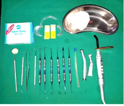
[image:37.595.109.533.118.480.2]ARMAMENTARIUM
27
28
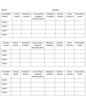
[image:39.595.156.508.174.568.2]SCORE FORM
29
30
[image:41.595.208.422.117.528.2]CLINICAL VIEW
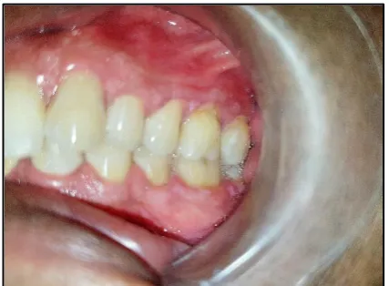
[image:41.595.209.423.134.291.2]Figure 5: Preoperative view
Figure 6: 15 days review
[image:41.595.210.420.552.714.2]31
RESULTS
The data collected were compiled and transferred to version 20 SPSS software for statistical analysis. For survival analysis, Kaplan Meier test was done and the statistical analysis was completed with chi-square test along with Pearson correlation test for relation of age to gross fracture of material and Spearman’s correlation test for relation of sex with gross fracture of the material.
80 cervical restorations were to be evaluated in 22 patients at baseline, 15 days and 6 months. However, one patient was lost to follow up on account of him moving out of station. Hence, an overall of 76 restorations could be evaluated. Of the 21 patients evaluated 15 were males and the remaining 6 were females belonging to the age group between 20-70 years. Out of the 76 restorations 48 was performed in maxillary teeth and the remaining 28 were performed in the mandibular teeth. 40 restorations were done in Group 1 and 36 restorations were done in Group 2.
At the end of 6 months, 14 restorations of the total 76 restorations exhibited loss of retention of the restoration from the class V cavity.
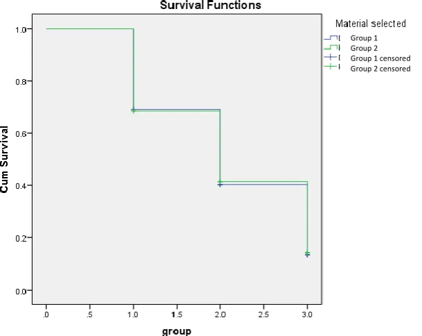
Survival analysis based on materials used using Kaplan Meier analysis was done. According to this test, both the materials have very similar survival in oral cavity. Group 1 had 4% loss of survival, whereas Group 2 had 5.3% loss of survival, however
both did not show any significant difference when compared with each other. (TABLE
1)
When comparing the materials based on Modified USPHS Criteria/ Ryge’s Criteria, in each parameter both materials showed significant decline with respect to time.
32
same colour match after 15 days and at 6 months 30 restorations had retained alpha score which shows 75% had good colour match .In Group 2, of the 36 restorations done, the colour match immediately after restoration was poor , as only 20 (55.55%) restorations had Alpha score and 16 (44.44%) restorations had bravo score and when compared but when comparing them with 6 month review , in the Group 2 out of 20 alpha restorations 9 (45%) restorations retained Alpha with a loss of restoration in 7 teeth. Therefore in colour match Group 1 has performed well with significant p-value
with respect to time (p = 0.024). (TABLE 2)
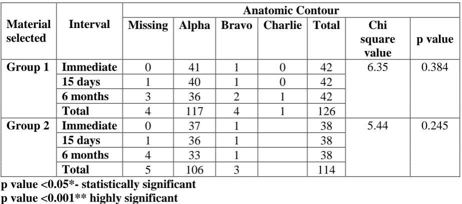
On comparing them with anatomic contour, Group 1 declined from 41 samples to 36 samples after 6 months , 87 % of samples retained the alpha score (p= 0.384) . Group 2 showed better retention of anatomic contour with 33 out of 37 samples (89.1%) retaining the alpha score after 6 months (p= 0.245). Hence Group 2 is better than Group
1 in retaining anatomic contour. (TABLE 3)
On comparison of cavosurface marginal discolouration, both Group 1 retained 35 restorations out of 42 restorations with alpha score in 6 month period (83.33%) with a significant p-value (0.016). Group 2 retained 33 of 38 samples (86.84%) with the alpha score with a non-significant p-value (p= 0.108). But comparing both materials,
Group 2 performs marginally better than Group 1. (TABLE 4)
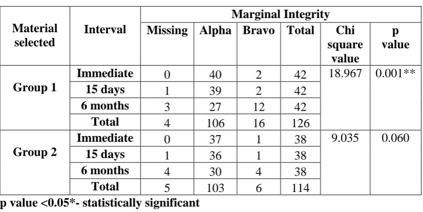
On comparison of marginal integrity, Group 1 has 27 of 40 samples (42.5%) retaining alpha scores at 6 months with highly significant result (p= 0.001). Group 2 retained 30 of 37 samples (81.08%) with alpha score (p= 0.060). However, both when
33
[image:45.595.163.468.456.698.2]Intergroup comparison
Table 01: Survival analysis based on materials used using Kaplan Meier analysis
M ate rial se le ct ed T otal N N of E ve n ts
Censored Mean 95%
Confidence Interval
Median 95%
Confidence Interval N Pe rc en t E stim ate S td. E rr or L owe r B ou n d Upp er B ou n d E stim ate S td. E rr or L owe r B ou n d Upp er B ou n d Group 1
126 121 5 4% 2.036 0.074 1.891 2.181 2 0.137 1.731 2.269
Group 2
114 108 6 5.3% 2.049 0.79 1.895 2.203 2 0.151 1.705 2.295
Overall 240 229 11 4.6% 2.042 0.054 1.937 2.148 2 0.101 1.801 2.199
Chi-Square (log rank mantel cox) 0.025
p value 0.873
Graph 01: Survival analysis based on materials used using Kaplan Meier analysis Group 1
34
DESCRIPTIVE AND INFERENTIAL STATISTICS
[image:46.595.112.544.162.372.2]INTRAGROUP COMPARISON (BETWEEN TIME INTERVALS)
Table 02: Comparison based on colour match within the group between time intervals using chi square test.
Group Interval Time
Colour Match
Missing Alpha Bravo Charlie Total Chi
square value
p value
1 Immediate 0 40 2 42 11.198 .024*
15 days 1 38 3 42
6 months 3 30 9 42
Total 4 108 14 126
2 Immediate 0 20 16 2 38 12.511 0.051
15 days 1 19 16 2 38
6 months 4 9 24 1 38
Total 5 48 56 5 114
p value <0.05*- statistically significant p value <0.001** highly significant
Table 03: Comparison based on anatomic contour match within the group between time intervals using chi square test.
Material selected
Interval
Anatomic Contour
Missing Alpha Bravo Charlie Total Chi
square value
p value
Group 1 Immediate 0 41 1 0 42 6.35 0.384
15 days 1 40 1 0 42
6 months 3 36 2 1 42
Total 4 117 4 1 126
Group 2 Immediate 0 37 1 38 5.44 0.245
15 days 1 36 1 38
6 months 4 33 1 38
Total 5 106 3 114
[image:46.595.107.569.487.691.2]35
Table 04: Comparison based on cavosurface marginal discolouration within the group between time intervals using chi square test.
Material selected
Interval
Cavosurface Marginal Discolouration
Missing Alpha Bravo Total Chi
square value
p value
Group 1 Immediate 0 42 0 42 12.229 0.016*
15days 1 41 0 42
6months 3 35 4 42
Total 4 118 4 126
Group 2 Immediate 0 38 0 38 7.589 0.108
15 days 1 37 0 38
6 months 4 33 1 38
Total 5 108 1 114
p value <0.05*- statistically significant p value <0.001** highly significant
Table 05: Comparison based on marginal integrity within the group between time intervals using chi square test.
Material selected
Interval
Marginal Integrity
Missing Alpha Bravo Total Chi
square value
p value
Group 1
Immediate 0 40 2 42 18.967 0.001**
15 days 1 39 2 42
6 months 3 27 12 42
Total 4 106 16 126
Group 2
Immediate 0 37 1 38 9.035 0.060
15 days 1 36 1 38
6 months 4 30 4 38
Total 5 103 6 114
[image:47.595.108.521.477.684.2]36
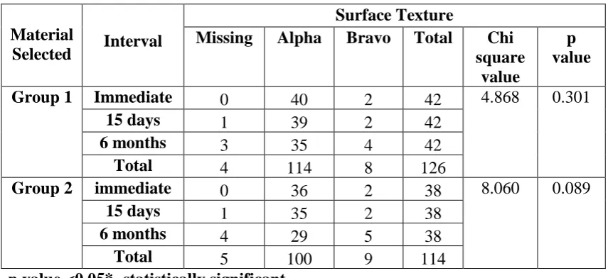
On comparison of surface texture, Group 1 retained 35 of 40 samples (87.5%) with alpha scores at 6 months (p= 0.301). Group 2 retained 29 of 36 samples (80.55 %) with alpha score (p= 0.089). however, both when compared with each other shows not
much significance in result. (TABLE 6)
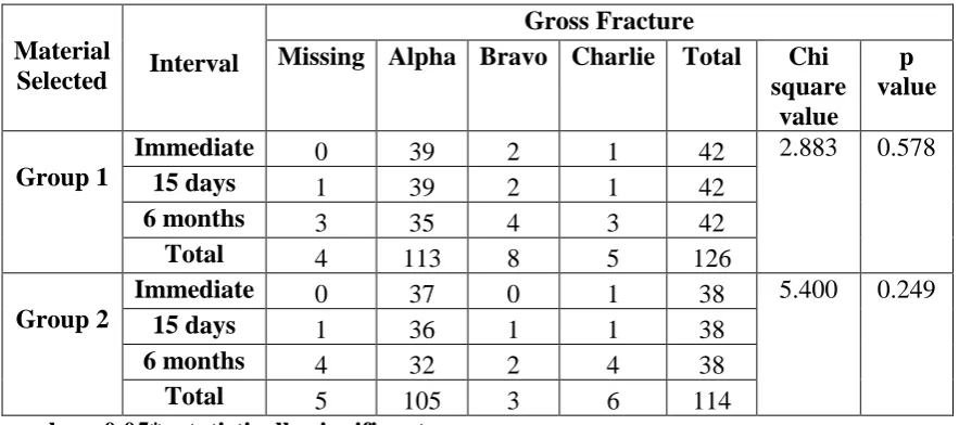
On comparison of gross fracture, Group 1 retained 35 of 39 samples (89.7%)
with alpha scores at 6 months (p= 0.578). Group2 retained 32 of 37 samples (86.4%)
retaining the alpha score (p= 0.249). however, both when compared with each other
shows not much significance in result. (TABLE 7)
On comparison of secondary caries, both Group1 and Group 2 have shown no
[image:48.595.102.530.430.626.2]secondary caries over 6 months. (TABLE 8)
Table 06: Comparison based on surface texture within the group between time intervals using chi square test.
Material
Selected Interval
Surface Texture
Missing Alpha Bravo Total Chi
square value
p value
Group 1 Immediate 0 40 2 42 4.868 0.301
15 days 1 39 2 42
6 months 3 35 4 42
Total 4 114 8 126
Group 2 immediate 0 36 2 38 8.060 0.089
15 days 1 35 2 38
6 months 4 29 5 38
Total 5 100 9 114
37
Table 07: Comparison based on gross fracture within the group between time intervals using chi square test.
Material
Selected Interval
Gross Fracture
Missing Alpha Bravo Charlie Total Chi
square value
p value
Group 1
Immediate 0 39 2 1 42 2.883 0.578
15 days 1 39 2 1 42
6 months 3 35 4 3 42
Total 4 113 8 5 126
Group 2
Immediate 0 37 0 1 38 5.400 0.249
15 days 1 36 1 1 38
6 months 4 32 2 4 38
Total 5 105 3 6 114
[image:49.595.107.549.108.304.2]p value <0.05*- statistically significant p value <0.001** highly significant
Table 08: Comparison based on secondary caries within the group between time intervals using chi square test.
Material
Selected Interval
Secondary Caries
Missing Alpha Total Chi
square value
p value
Group 1
Immediate 0 42 42 3.615 0.164
15 days 1 41 42
6 months 3 39 42
Total 4 122 126
Group 2
Immediate 0 38 38 5.439 0.066
15 days 1 37 38
6 months 4 34 38
[image:49.595.108.526.456.645.2]38
[image:50.595.82.550.127.399.2]INTERGROUP COMPARISON (BETWEEN TWO MATERIALS)
Table 09: Comparison based on colour match between the groups during different time interval using chi square test.
Time
Interval Group
Colour Match Chi
square value
p value
Missing Alpha Bravo Charlie Total
Immediate Group
1 40 2 0 42
19.404 0.000*
Group
2 20 16 2 38
Total 60 18 2 80
15 days Group
1 1 38 3 0 42
17.071 0.012**
Group
2 1 19 16 2 38
Total 2 57 19 2 80
6 months Group
1 3 30 9 0 42
19.117 0.000**
Group
2 4 9 24 1 38
Total 7 39 33 1 80
Table 10: Comparison based on anatomic contour between the groups during different time interval using chi square test.
Time
Interval Group
anatomic contour Chi
square value
p value
Missing Alpha Bravo Charlie Total
Immediate Group
1 41 1 42
0.005 0.943
Group
2 37 1 38
Total 78 2 80
15 days Group
1 1 40 1 42
.011 0.995
Group
2 1 36 1 38
Total 2 76 2 80
6 months Group
1 3 36 2 1 42
1.410 0.703
Group
2 4 33 1 0 38
[image:50.595.108.545.499.761.2]39
Table 11: Comparison based on cavosurface marginal discolouration between the groups during different time interval using chi square test.
Time Interval Group Cavosurface Marginal Discolouration Chi square value p value
Missing Alpha Bravo Total
Immediate Group 1 42 42 0 1
Group 2 38 38
Total 80 80
15 days Group 1 1 41 42 .005 0.943
Group 2 1 37 38
Total 2 78 80
6 months Group 1 3 35 4 42 1.806 0.405
Group 2 4 33 1 38
Total 7 68 5 80
Table 12: Comparison based on marginal integrity between the groups during different time interval using chi square test.
Time
Interval Group
marginal integrity Chi
square value
p value
Missing Alpha Bravo Total
Immediate Group 1 40 2 42 0.251 0.616
Group 2 37 1 38
Total 77 3 80
15 days Group 1 1 39 2 42 .254 0.881
Group 2 1 36 1 38
Total 2 75 3 80
6 months Group 1 3 27 12 42 4.111 0.128
Group 2 4 30 4 38
40
Table 13: Comparison based on surface texture between the groups during different time interval using chi square test.
Time
Interval Group
Surface Texture Chi
square value
p value Missing Alpha Bravo Total
Immediate Group 1 40 2 42 0.011 0.918
Group 2 36 2 38
Total 76 4 80
15 days Group 1 1 39 2 42 .016 0.992
Group 2 1 35 2 38
Total 2 74 4 80
6 months Group 1 3 35 4 42 .618 0.734
Group 2 4 29 5 38
[image:52.595.107.539.408.668.2]Total 7 64 9 80
Table 14: Comparison based on gross fracture between the groups during different time interval using chi square test.
Time
Interval Group
gross fracture Chi
square value
p value Missing Alpha Bravo Charlie Total
Immediate Group
1 39 2 1 42
1.857 0.395
Group
2 37 0 1 38
Total 76 2 2 80
15 days Group
1 39 2 1 42
.254 0.881
Group
2 36 1 1 38
Total 75 3 2 80
6 months Group
1 35 4 3 42
0.746 0.689
Group
2 32 2 4 38
41
Table 15: Comparison based on secondary caries between the groups during different time interval using chi square test.
Time
Interval Group
Secondary caries Chi
square value
p value
Missing Alpha Total
Immediate Group 1 42 42 0 1
Group 2 38 38
Total 80 80
15 days Group 1 1 41 42 .005 0.943
Group 2 1 37 38
Total 2 78 80
6 months Group 1 3 39 42 .286 0 .593
Group 2 4 34 38
Total 7 73 80
Based on Pearson correlation test, Group 1 shows less significance with increase in age with Pearson correlation value of 0.12 (p = 0.178) .Group 2 also shows less significance of fracture of restorations over increase with age with Pearson correlation
value of 0.04 (p=0.613). (TABLE 16)
According to Spearman’s correlation test, with Group 1, there is a negative correlation for females than males with an r value (Spearman’s correlation co-efficient) of -.227 for Group 2 which is statistically highly significant with a (p-value = 0.001). According to Spearman’s correlation test, with Group 2, there is a negative correlation for females than males with an r value (Spearman’s correlation co-efficient) of -.275 which is statistically significant (p-value = 0.012). Which means females are marginally
42
Table 16: Correlation test between materials used and Age and between Materials used and sex.
Material Selected Age Gross
Fracture
Material Selected Sex Gross
Fracture Group
1
Pearson Correlation
1 0.12 Group
1
Spearman's Correlation Coefficient
1 -.227
P value 0.178 P value . 0.011
N 126 126 N 126 126
Group 2
Pearson Correlation
1 0.04 Group
2
Spearman's Correlation Coefficient
1 -.236
P value 0.346 P value . 0.011
43
DISCUSSION
Abrasion is the outcome of friction amid a tooth and an exogenous substance
such as friction caused by toothbrush26,40,41. Until recently, the primary restorative
option for NCCL was Glass Ionomer Cement, either alone or as a liner, due to its high
retention rate12,42 and the choice between other restorative materials were determined
by the erosive/abrasive or abfraction nature of the lesions.8 More recently, due to their
better aesthetic properties, their improved adhesive capacity due to modern dentin adhesives and their better mechanical properties, resin composites imposed themselves as an alternative restoration to GIC, expanding their variety of indications to nearly
every cervical lesions.8
Within their limits, the existing clinical studies report excellent aesthetics and
better longevity as well as outstanding mechanical properties for resin composites,21–
23,24 Clinical assessments over several periods of time, including long-term
studies,24,16,20,43 have standard good results in clinical performance regarding all
parameters, therefore placing these materials in a more favourable position compared to compomers and resin-modified GIC. Surface texture, anatomical form, marginal integrity and colour match proved by far to be superior in most of the clinical
evaluations.8,14
Nevertheless, some disadvantages have been reported for composite materials.8
Despite their general acceptance, resin composites still shows drawbacks such as marginal sealing deficiency and especially degradation of adhesion over time. Failures
due to retention loss have also been stated,19 as a consequence of combined factors like
on-going cervical stress,19,44 flexural strength, rigidity, adhesive layer characteristics
44
Therefore, choosing a reliable adhesive system and a well-tested composite material is of primary importance, as clinical results vary significantly with the product
employed.8 Restorations in this area are not only incapable of halting tooth flexure but
they are also affected by the continuing action of flexural stress.44,45
In clinical studies, the success of a material is specified by its longevity in the oral cavity, which makes retention rates the most important evaluation criteria. Self-etch adhesive systems and composite resins allows restorations with insignificantly invasive preparation and satisfactory aesthetic appearance. Most of the previous studies were performed in vitro, hence it is difficult to forecast the clinical performance of those
adhesives and resins. NCCLs are usually evaluated for a period of 3 to 5 years 37.
According to Blunck et al., a period of 6 months to 1 year could be enough to accurately
predict the clinical behaviour of an adhesive37.
This study has evaluated and compared the clinical performance of Group 1 and Group 2 in class V non-carious lesions using Modified USPHS criteria/Ryge’s criteria which is a frequently used for assessing the restoration in long-term, and is considered legal for the purpose of comparison among studies at different periods. American Dental Association (ADA) (2001) guidelines for submitted dentin and enamel adhesive materials require provisional acceptance, meaning that no more than 5% of the restorations have been lost at the six-month of recall and, in order to get full recognition, the aggregate incidence of clinical failures in each of two independent clinical studies
has to be less than 10% of restorations lost after 18 months.23
45
Materials.15 When applied to non-retentive cervical lesions, the clinical performance of
restorations relies on the bond strength values of adhesive resins. The forces created by compression of the restoration are localized at the bulk of the resin composite as compressive stress and less as shear stress at the adhesive interface. Therefore, the adhesive bond is preserved, while marginal adaptation is adversely affected, which is only valid when the adhesive bond is sufficiently strong.
It has been recommended that bevelling of the enamel margins of NCCL may
provide greater retention rates of restorations.23,46–48 While routine mechanical
preparation may not be currently required, it was considered a necessity before the
development of new adhesive resins.23 According to Tay and Pashley 15,49, grinding of
the surface hyper mineralized layer prior to adhesive bonding may not result in any increase in bond strength since the underlying sclerotic dentin may still contain crystallites that may impede resin infiltration into the dentinal tubules. In this study, bevelling procedure was not done.
According to Marquillier et al (2018) and others, most of the studies that were done since 2007 have been using Modified USPHS criteria and have validated their results .This criteria provides better detailed result hence most of the studies were already using this study. Therefore we decided to use this same criteria for comparing
our results with other studies also bringing standardization in this study.14,39
There was a statistically significant difference in colour match of Group 1 after 6 months (p =0.024) when compared to Group 2 (p-value = 0.051). The colour change was statistically significant from baseline to first, then to second, and then third recall visits among both material with respect to time period with p-value of 0.000 at baseline,
46
to the high hydrophilic content of monomer in hybrid materials, there was a greater water sorption rate which apparently led to the colour change. Thereby water absorbed during the weeks after photo polymerization initiates an acid-base reaction which
results in the setting mechanism18 .
There was a statistically significant difference in cavosurface marginal discolouration where Group 1 (16.7%) showed more discolouration (p-value = 0.016) than Group 2 (13.16%) (P-value = 0.108). This might be explained by the bond failure
that allowed ingress of exogenous stain14.
The marginal integrity of Group 1 was lost significantly (32.5%) (p-value = 0.001) when compared with Group 2 (18.92) (p-value = 0.060). The major key cause for micro-leakage is the polymerisation shrinkage, poor marginal adaptation as well as poor retention rates.
Flowable composites have less quantities of filler with low modulus of
elasticity as well as highly flexibility to dislodging forces.38,50,51 The reasoning for using
flowable composites in this study is its low elasticity that displays increased flexibility when under occlusal stress especially in the cervical areas. They are said to absorb the stresses during polymerization shrinkage, its elastic properties allows the material to be flexible to occlusal stress and hence preventing dislodgement on comparison with conventional and hybrid composites as they are more vulnerable to dislodgement when under flexure stresses.
47
abrasion resistance present in flowable composites. The benefit of flowable composite
over hybrid as well as micro-filled composite is the absence for sculpturing38,50,51 .
According to Franco et al (2006)19 only anatomic form has lost up to 3.4% and
rest other parameters such as marginal integrity , marginal discolouration, secondary caries , colour stability remains 100 % but the author had excluded the 14.28% restorations with loss of retention.
According to Hussainy et al (2018)7 only 5.9% restorations lost the colour
stability in flowable composites and no loss with RMGIC. 11.8% of restoration of flowable composite had cavosurface marginal discolouration and flowable composite had 14.7% loss in marginal integrity compared to 9.1 % loss in RMGIC.
All the other parameters of Ryge’s [Modified USPHS] criteria such as anatomic contour, surface texture, gross fracture and secondary caries had some differences but nothing significant over the period of 6 months. Hence performance of both were similar in all other categories.
LIMITATIONS
Studies with longer periods are required for more accurate results. The existing clinical studies report excellent aesthetics and better longevity and outstanding
mechanical characteristics for resin composites21–24. Many clinical studies16,20,24,43 have
48
and compared. A long-term clinical study could provide the most precise results regarding the toughness of these adhesive restorations, but such type of study would need several years with regular recall visits and a high rate of recall for clinical
49
SUMMARY & CONCLUSION
The current study evaluated and compared the clinical performance of 3 in 1 flowable composite with a RMGIC in Non-Carious Cervical Lesion for a time period of 6 months.
80 restorations with non-carious cervical lesions were treated with either one of the above mentioned products after randomization of allocation. Patients were then recalled for follow up at baseline, 15 days and 6 months. Clinical Evaluation was done using Ryge criteria (Modified USPHS) for direct clinical evaluation of restoration.
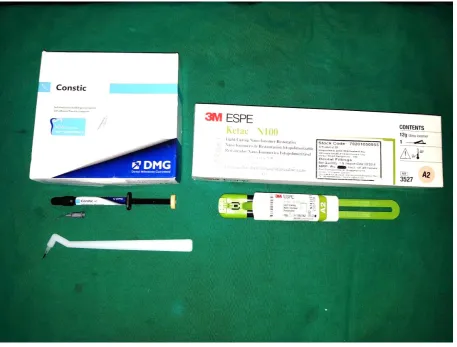
The groups allocated in this study were: Group 1: DMG Constic Flowable Composite Group 2: 3M ESPE Ketac N100
50
Conclusion:
Thereby Within the limitations of this current study it was concluded that:
51
BIBLIOGRAPHY
1. Levitch LC, Bader JD, Shugars DA, Heymann HO. Non-carious cervical
lesions. J Dent. 1994;22(4):195–207.
2. Kolak V, Pešić D, Melih I, Lalović M, Nikitović A, Jakovljević A.
Epidemiological investigation of non-carious cervical lesions and possible etiological factors. J Clin Exp Dent. 2018;10(7):e648–56.
3. Venkatesan K, Kuzhanchinathan M, Prakash P. Critical review of noncarious
cervical lesions. SRM J Res Dent Sci [Internet]. 2018;9(2):74. Available from:
http://www.srmjrds.in/article.asp?issn=0976-433X;year=2018;volume=9;issue=2;spage=74;epage=78;aulast=Venkatesan
4. Perez CDR, Gonzalez MR, Prado NAS, De Miranda MSF, MacÊdo MDA,
Fernandes BMP. Restoration of noncarious cervical lesions: When, why, and how. Int J Dent. 2012;2012.
5. Cieplik F, Scholz KJ, Tabenski I, May S, Hiller KA, Schmalz G, et al.
Flowable composites for restoration of non-carious cervical lesions: Results after five years. Dent Mater [Internet]. 2017;33(12):e428–37. Available from: http://dx.doi.org/10.1016/j.dental.2017.09.012
6. Jyothi K, Kumar A, Jayashankara C, Annapurna S, Venugopal P. Clinical
evaluation of giomer- and resin-modified glass ionomer cement in class V noncarious cervical lesions: An in vivo study. J Conserv Dent [Internet]. 2011;14(4):409. Available from: