0022-538X/05/$08.00⫹0 doi:10.1128/JVI.79.19.12296–12303.2005
Copyright © 2005, American Society for Microbiology. All Rights Reserved.
V3 Loop-Determined Coreceptor Preference Dictates the Dynamics of
CD4
⫹
-T-Cell Loss in Simian-Human Immunodeficiency
Virus-Infected Macaques
Siu-hong Ho,
1Lili Shek,
1Agegnehu Gettie,
1James Blanchard,
2and Cecilia Cheng-Mayer
1*
Aaron Diamond AIDS Research Center, The Rockefeller University, 455 First Ave., 7th Floor, New York, New York 10016,1
and Tulane National Primate Research Center, Tulane University Medical Center, 18702 Three Rivers Road, Covington, Louisiana 704332
Received 27 May 2005/Accepted 6 July 2005
We used experimental infection of rhesus macaques with envelope gp120 V3 loop isogenic simian-human immunodeficiency virus (SHIV) molecular clones to more clearly define the impact of human
immunodefi-ciency virus type 1 coreceptor usage in target cell selectivity and the rates of CD4ⴙ-T-cell depletion. Functional
assays demonstrate that substitution of the V3 loop of the pathogenic CXCR4-tropic (X4) SHIVSF33A2
molec-ular clone with the corresponding sequences from the CCR5-tropic (R5) SHIVSF162P3 isolate resulted in a
switch of coreceptor usage from CXCR4 to CCR5. The resultant R5 clone, designated SHIVSF33A2(V3), is
replication competent in vivo, infecting two of two macaques by intravenous inoculation with peak viremia that
is comparable to that seen in monkeys infected with X4-SHIVSF33A2. But while primary infection with the X4
clone was accompanied by rapid and significant loss of peripheral and secondary lymphoid CD4ⴙT
lympho-cytes, infection with R5-SHIVSF33A2(V3)led to only a modest and transient loss. However, substantial depletion
of intestinal CD4ⴙT cells was observed in R5-SHIVSF33A2(V3)-infected macaques. Moreover, naı¨ve T cells that
expressed high levels of CXCR4 were rapidly depleted in X4-SHIVSF33A2-infected macaques, whereas
R5-SHIVSF33A2(V3)infection mainly affected memory T cells that expressed CCR5. These findings in a unique
isogenic system illustrate that coreceptor usage is the principal determinant of tissue and target cell specificity
of the virus in vivo and dictates the dynamics of CD4ⴙ-T-cell depletion during SHIV infection.
Since the discovery of the chemokine receptors CCR5 and CXCR4 as the major coreceptors (CoRs) for entry of human immunodeficiency virus type 1 (HIV-1), mounting evidence indicates that coreceptor expression and usage play crucial roles in viral transmission, persistence, and pathogenesis (28). Most HIV-1 strains transmitted between humans use CCR5 as their coreceptor (R5 strains). With progression to disease, variants that use CXCR4 (X4 viruses) emerge in about 50% of infected individuals and are associated with a more rapid rate of peripheral CD4⫹-T-cell loss (33). We and others have used infection of nonhuman primates with simian-human immuno-deficiency virus (SHIV) expressing the envelopes of R5 and X4 HIV-1 strains as a model system to study the impact of core-ceptor usage in HIV-1 infection and AIDS pathogenesis (4). In infection of rhesus macaques (RM) with R5 and X4 patho-genic SHIVs, SHIVSF162P3 and SHIVSF33A, respectively, we
previously reported a slower rate of peripheral CD4⫹-T-cell depletion in R5-SHIVSF162P3-infected RM that recapitulated
the progression of infection seen in humans (13, 15). In con-trast, infection with pathogenic X4-SHIVSF33A resulted in
rapid and precipitous decline in peripheral and lymphoid CD4⫹T cells, mirroring infection with X4 isolates (15, 16, 22). However, analysis of lamina propria lymphocytes (LPLs) pu-rified from jejunal tissues revealed that R5-SHIVSF162P3
caused severe depletion of mucosal CD4⫹ T cells within 2
weeks of infection, whereas the loss of mucosal CD4⫹T cells in X4-SHIVSF33A-infected macaques was more gradual (15).
Different tissue sites of replication and CD4⫹-T-cell depletion patterns in macaques infected with CXCR4- and CCR5-tropic viruses have also been reported previously by others (30, 31), leading to the suggestion that target cell availability as influ-enced by differential coreceptor expression and viral tropism as dictated by coreceptor preference have profound conse-quences on clinical disease. Nevertheless, the strains used in all these studies were independent isolates, with variants that dif-fered in genetic sequences and, hence, in properties other than host cell tropism which might have contributed to the differ-ences seen in the pathogenic sequela. For instance, a major (7 out of 10 clones) and a minor (1 out of 10 clones) variant, as determined by V1 to V5envsequences, that differed in their entry efficiency and neutralization susceptibility were present in the R5-SHIVSF162P3 isolate we used (17). Furthermore,
while both X4-SHIVSF33Aand R5-SHIVSF162P3showed
neu-tralization resistance, the X4 virus appeared to be more fuso-genic and cytopathic in vitro (6, 7, 18). A confirmation that early targeting of different T-cell populations and different tissues by X4 and R5 viruses is due solely to the coreceptors used could greatly benefit from infection of RM with variants that differ only in their coreceptor binding site sequences.
For HIV-1, differential usage of coreceptor in vitro has been shown to depend on the charge and/or structure of the V3 loop of envelope gp120 (2, 8). Towards the objective of constructing isogenic SHIVs that differ only in coreceptor preference, the V3 loop of SHIVSF33A2, a molecular clone that displays the
infection characteristics of the parental pathogenic
X4-* Corresponding author. Mailing address: Rockefeller University, Aaron Diamond AIDS Research Center, 455 First Ave., 7th Floor, New York, NY 10016. Phone: (212) 448-5080. Fax: (212) 448-5158. E-mail: cmayer@adarc.org.
12296
on November 8, 2019 by guest
http://jvi.asm.org/
SHIVSF33Aisolate in RM (14), was replaced with the
corre-sponding region of the R5-SHIVSF162P3isolate (18). The
func-tion and coreceptor choice of the resulting chimeric envelope glycoprotein were determined. Furthermore, infection of RM with virus expressing the chimeric V3 envelope, designated SHIVSF33A2(V3), was characterized and compared to infection
with the X4 isogenic SHIVSF33A2molecular clone.
MATERIALS AND METHODS
Cells. Human osteosarcoma cells expressing CD4 and either CCR5 (HOS.CD4.CCR5) or CXCR4 (HOS.CD4.CXCR4) were kind gifts from N. Landau (Salk Institute, La Jolla, CA) and were maintained in Dulbecco’s mod-ified Eagle’s medium supplemented with 10% fetal bovine serum (FBS), 0.5
g/ml of puromycin (Sigma-Aldrich, St. Louis, MO), and
penicillin-streptomy-cin. 293T cells used for transfection were cultured in Dulbecco’s modified Eagle’s medium with 10% FBS and penicillin-streptomycin. Human peripheral blood mononuclear cells (PBMCs) were obtained by Ficoll-Hypaque gradient
purifi-cation followed by stimulation with 3g/ml of phytohemagglutinin A
(Sigma-Aldrich) in RPMI 1640 medium supplemented with 10% FBS, 2 mM glutamine,
100 U/ml penicillin, 100g/ml streptomycin, and 20 U/ml of interleukin-2
(Chi-ron Corp., Emeryville, CA).
Plasmid constructs and virus production.PCR-based overlapping extension
methodology was employed to replace the V3 loop of X4-SHIVSF33A2envgp120
with that of R5-SHIVSF162P3. Briefly, a HincII-to-HindIII fragment of Env33A2
(6) (nucleotides 1249 to 2386 [GenBank accession number M38427]) was
sub-cloned into pBlueScript KS II(⫹) vector to serve as the template for PCR
amplifications. The inner amplification primers used were SH5 (5⬘-GGG GAA
AGC ATT TTA TGC AAC AGG AGA CAT AAT AGG AGA TAT AAG ACA
AGC ACA TTG TAA CAT TAG TAG AGC-3⬘) and SH6 (5⬘-GCA TAA AAT
GCT TTC CCC GGT CCT ATA GGT ATA CTT TTT CTT GTA TTG TTA TTG
GGT CTT GTA C-3⬘), which encompassed the envgp120 V3 sequence of
R5-SHIVSF162P3(in italics, with overlapping regions underlined) flanked by the
C2 and C3 sequences of X4-SHIVSF33A2. The outer primers used were T3 and
T7. The amplified fragment was completely sequenced for verification, and an
envelope expression vector containing the V3 chimericenv, Env33A2(V3), was
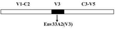
constructed by subcloning the HincII-to-HindIII-amplified fragment back into Env33A2 (Fig. 1). The same fragment was ligated into the corresponding regions
of SHIVSF33A23⬘DNA (14) to generate the 3⬘genome of the V3 isogenic clone
SHIVSF33A2(V3). Env162P3 (18)-, Env33A2-, and Env33A2(V3)-pseudotyped
lu-ciferase reporter viruses were prepared by cotransfecting 293T cells with the
NL4.3-Luc-E⫺R⫺vector and the corresponding Env expression plasmid using
the DMRIE-C reagent (Invitrogen). Reporter viruses were harvested 72 h
post-transfection and quantified for p24gagantigen content (Beckman Coulter,
Ful-lerton, CA). SHIV was obtained by cotransfection of 293T cells with SIVmac239
5⬘and SHIVSF33A2or SHIVSF33A2(V3)3⬘hemigenomes followed by cocultivation
with phytohemagglutinin A-stimulated PBMCs as previously described (23). Recovered viruses were amplified and propagated in human PBMCs, and culture
supernatants were collected for viral p27gag
antigen determination (Coulter Corporation, Miami, FL) and for assessment of the 50% tissue culture infectious dose in human PBMCs.
Entry and blocking assays. A total of 5 ⫻ 103 HOS.CD4.CCR5 or
HOS.CD4.CXCR4 cells were seeded in 96-well plates 24 h before use. Cells were infected with 5 ng p24 equivalent of the indicated pseudotyped viruses in the
presence of 2g/ml of polybrene followed by incubation for 72 h at 37°C. At the
end of the incubation period, cells were harvested, lysed, and processed
accord-ing to the manufacturer’s instructions (Promega, Madison, WI). Entry, as quan-tified by luciferase activity, was measured with an MLX microtiter plate
lumi-nometer (Dynex Technologies, Inc., Chantilly, VA). For the blocking assay, 5⫻
105
PBMCs were pretreated with various concentrations of AOP-RANTES (a generous gift of Oliver Hartley, University of Zurich) for 1 h before infection. Percent blocking was calculated by the amount of entry in the presence of AOP-RANTES relative to that in the absence of AOP-RANTES.
Animal infections.All infections were carried out in adult RM (Macaca mu-latta) individually housed at the Tulane National Primate Research Center in
compliance with theGuide for the Care and use of Laboratory Animals(28a).
Animals were confirmed to be serologically negative for simian type D retrovirus, simian immunodeficiency virus (SIV), and simian T-cell lymphotrophic virus prior to infection. Whole blood lymphocytes were sampled at designated time intervals. Plasma viremia was quantified by branched DNA analysis (Bayer
Di-agnotics, Emeryville, CA), and absolute CD4⫹and CD8⫹ cell counts were
monitored by TruCount (BD Biosciences, Palo Alto, CA). Biopsy samples were
obtained by surgery at 2 and 8 weeks postinfection (wpi) for the SHIVSF33A2(V3)
-infected animals and at 2 and 9 wpi for the SHIVSF33A2-inoculated animals. The
percentages of CD4⫹T cells in the biopsy samples were analyzed by flow
cytometry (FACScalibur) using CD3-fluorescein isothiocyanate (FITC), CD4-phycoerythrin (PE), and CD8-peridinin chlorophyll protein antibodies. Further
phenotyping of peripheral and lymphoid CD4⫹T cells was performed by using
CD28-allophycocyanin, CD95-PE or -FITC, CCR5-PE, and CXCR4-PE. Except for CD3-FITC (BioSource, Camarillo, CA), all antibodies were obtained from BD Biosciences.
RESULTS
The V3 loop of X4-SHIVSF33A2 dictates CoR usage. The
35-amino-acid V3 domains of the X4-SHIVSF33A2molecular
clone and the R5-SHIVSF162P3isolate differ in 10 amino acids
(Fig. 1), with the overall charge of this region being⫹7 for SHIVSF33A2and⫹5 for SHIVSF162P3, values that are
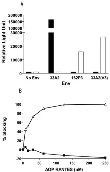
consis-tent with those reported previously for other X4 and R5 HIV-1 strains (10). In single-round infectivity assays, we found that contrary to Env33A2, the chimeric V3 envelope Env33A2(V3) was not able to mediate entry into HOS.CD4 cells expressing CXCR4. Instead, Env33A2(V3), similar to Env162P3, used CCR5 as the CoR (Fig. 2A). To confirm the coreceptor usage of Env33A2(V3), blocking of entry into human PBMCs with AOP-RANTES, an amino terminus-modified form of the CCR5 natural ligand RANTES (36), was assessed. Results showed that entry of virus expressing Env33A2(V3) was effi-ciently blocked by AOP-RANTES in a dose-dependent man-ner, while virus expressing Env33A2 was not affected, even at a concentration of 250 nM (Fig. 2B). In fact, a slight increase in Env33A2-mediated infection in the presence of high con-centrations of AOP-RANTES (⬎50 nM) was observed, con-sistent with reports of stimulation of X4 virus replication by CC chemokines (9, 19). Taken together, the data showed that Env33A2(V3) is functional and that the substitution of the V3 loop of Env33A2 with that of Env162P3 resulted in a switch in coreceptor usage from CXCR4 to CCR5.
SHIVSF33A2(V3) is replication competent in RM. Env33A2(V3) was then used to replace the corresponding re-gion of X4-SHIVSF33A2 to construct the chimeric virus
SHIVSF33A2(V3). The two clones differ only in the V3 loop, with
an overall identity of 99.88%. Two macaques each were inoc-ulated intravenously with 103 50% tissue culture infectious
doses of R5-SHIVSF33A2(V3) (macaques BT35 and CF17) or
X4-SHIVSF33A2(macaques CA05 and CA06). All four animals
were infected, with peak viremia of 106to 108RNA copies/ml
[image:2.585.67.259.67.121.2]plasma at 2 to 3 wpi and with kinetics of virus replication slightly faster in the X4-SHIVSF33A2-infected macaques than in
FIG. 1. Comparison of the V3 loop sequence of Env33A2 and Env33A2(V3). Chimeric Env was constructed by replacing the V3 loop of X4-SHIVSF33A2gp120 (white) with the corresponding sequences of R5-SHIVSF162P3(black) as described in Materials and Methods.
VOL. 79, 2005 HIV-1 CORECEPTOR AND CD4⫹-T-CELL LOSS 12297
on November 8, 2019 by guest
http://jvi.asm.org/
the R5-SHIVSF33A2(V3)-infected macaques (Fig. 3A and B).
Replication of both viruses declined thereafter, with a post-peak viral load of 102to 103RNA copies/ml plasma at 20 wpi.
The dynamics of CD4ⴙ-T-cell loss differ in
R5-SHIVSF33A2(V3)- and X4-SHIVSF33A2-infected macaques. We next examined the impact of X4 and R5 clone infection on the CD4⫹-T-cell compartment. A drop in peripheral CD4⫹ T cells, a feature that is characteristic of infection with the patho-genic isolate R5-SHIVSF162P3and with SIV, was observed in
both R5-SHIVSF33A2(V3)-infected animals during peak viremia
(2 to 3 wpi). The levels of circulating CD4⫹T lymphocytes fluctuated thereafter but rebounded close to preinfection val-ues by 8 wpi (Fig. 3B). In contrast, X4-SHIVSF33A2infection
led to a rapid and more sustained loss of peripheral CD4⫹T cells (Fig. 3A).
To further examine the impact of CoR usage on the dynam-ics of the CD4⫹-T-cell compartment, the percentages of CD4⫹ T cells in various lymphoid and nonlymphoid tissues during peak (2 to 3 wpi) and postpeak (8 to 9 wpi) infection were analyzed (Fig. 4). No significant loss in CD4⫹T cells was noted in the draining and peripheral lymph nodes (LNs) of the two R5-SHIVSF33A2(V3)-infected animals during the peak or
post-peak infection period (Fig. 4B). Despite a viral load that is
comparable to that of the R5-SHIVSF33A2(V3)-infected
ani-mals, however, a 30 to 50% loss of CD4⫹T cells was found in all lymphoid tissues of CA05 examined at 9 wpi (Fig. 4A). In CA06, the X4-SHIVSF33A2-infected animal with a log higher
peak viremia, massive depletion of CD4⫹T cells was seen in both the draining and systemic LNs within 2 weeks of infection, with some degree of restoration noted in these secondary LNs by 9 wpi.
Contrary to what was observed in the secondary lymphoid compartments, a 60% and 55% drop in CD4⫹T cells in the bone marrow (BM) and mucosal tissues such as the lamina propria of the gut (LPL), respectively, in CF17 was present at 8 wpi (Fig. 4B). A similar degree of postpeak CD4⫹-T-cell loss was also noted in the BM and LPL in the other R5-SHIVSF33A2(V3)-infected monkey, BT35. For X4-SHIVSF33A2
infection, a decrease in BM CD4⫹T cells (25%) was seen in CA05 at 2 wpi, but dramatic depletion was already present in CA06. While the gut remained relatively intact in both X4 clone-infected macaques at 2 wpi, substantial CD4⫹-T-cell de-pletion (60%) was detected in CA06 by 9 wpi (Fig. 4A), while CA05 displayed a more modest loss (25%). The targeting of BM by the X4 and R5 isogenic SHIV molecular clones is in agreement with reports that CD34⫹progenitor cells could be infected by HIV-1 variants with different phenotypes (34, 41). Furthermore, although the dynamics of CD4⫹-T-cell loss among the two X4-SHIVSF33A2-infected macaques varies due
[image:3.585.67.259.68.369.2]to a difference in viral load, the data show that by switching the coreceptor usage of this clone virus through replacement of its V3 loop, the site of early virus replication changes. While the X4 clone preferentially targets and depletes cells in peripheral blood and secondary LNs within the first few weeks of infec-tion, the R5 clone targets mucosal tissue sites. The preferential depletion of CD4⫹T cells in the gut and not the LNs of the R5 clone-infected animals during primary infection is consistent with observations made in HIV-1-infected humans (5, 11, 27),
[image:3.585.303.543.70.256.2]FIG. 2. In vitro properties of Env33A2(V3). (A) Relative entry of luciferase reporter viruses expressing Env33A2, Env162P3, and Env33A2(V3) into HOS.CD4.CXCR4 (black bars) and HOS.CD4. CCR5 (white bars) cells. (B) CCR5 usage of reporter viruses express-ing Env33A2(V3) (‚) was verified by blocking entry into human PB-MCs with the modified CCR5 ligand AOP-RANTES. Viruses pseudotyped with Env33A2 (■) served as negative controls. All data were the means of triplicates.
FIG. 3. Virologic and immunologic measurements in SHIV-in-fected RM. (A) Animals CA05 and CA06 inoculated with X4-SHIVSF33A2 and (B) animals BT35 and CF17 inoculated with R5-SHIVSF33A2(V3). Viremia (RNA copies/ml [solid lines]) and absolute CD4⫹-T-cell counts (dashed lines) of peripheral blood were monitored during the first 20 weeks of infection.
on November 8, 2019 by guest
http://jvi.asm.org/
SIV-infected macaques (21, 25, 26, 37, 39), and animals in-fected with the pathogenic R5-SHIVSF162P3isolate (15).
T-cell-subset distribution and CoR expression varied in
lymphoid and mucosal tissues of RM.CXCR4 and CCR5 are
differentially expressed on naı¨ve and memory CD4⫹T cells, the two major T-cell subpopulations that can be distinguished in macaque cells by surface staining for the CD28 and CD95 antigens (32). To determine whether the differential compart-mentalization of the X4 and R5 clones during primary infec-tion is influenced by the availability and susceptibility of their target cells, the frequency of the two T-cell subsets in various lymphoid and mucosal tissues and CoR expression on their surface were determined (Fig. 5). Consistent with previous reports (1, 3, 20, 35, 38, 40), naı¨ve (CD95lowCD28high) CD4⫹ T lymphocytes were found in abundance in the periphery and
LNs but were rare in the gut. More than 95% of cells in this latter compartment were memory cells (CD95highCD28highor
CD95highCD28low) (Fig. 5A). Virtually all the naı¨ve cells were
[image:4.585.101.488.70.285.2]positive for CXCR4 and negative for CCR5 (Fig. 5B), while cells of the memory phenotype expressed both receptors, with higher CCR5 expression on memory cells in the gut than in the periphery and secondary lymphoid compartments (Fig. 5C). Within the macaque host, therefore, there is greater represen-tation of CXCR4-positive cells in peripheral blood and LNs, while the majority of CCR5-expressing cells reside in mucosal effector sites. This, then, explains the early preferential target-ing of the X4 clone to the periphery and LN compartments and the R5 clone to the gut-associated lymphoid tissue since the greatest number of their respective target cells reside in these compartments.
FIG. 4. Percentage of CD4⫹ T lymphocytes in various lymphoid and nonlymphoid compartments of (A) X4-SHIVSF33A2- and (B) R5-SHIVSF33A2(V3)-infected RM. The percentages of CD4⫹T cells in the inguinal (Ing), iliac (Ili), mesenteric (Mes), and colonic (Col) lymph nodes, BM, and LPL from the jejunum were analyzed before (white bars), during peak (2 to 3 wpi [gray bars]), and postpeak (8 to 9 wpi [black bar]) infection. Note that the mesenteric LN of CA06 was not available for analyses. Dashed bars show percentages of CD4⫹T cells in the same compartments of uninfected control macaques for reference when preinfection values for the infected macaques were not available. Error bars show standard errors of values from two to three uninfected animals.
FIG. 5. T-cell subset distribution (A) and CoR expression in naı¨ve (B) and memory (C) T cells in various lymphoid and nonlymphoid tissues of uninfected control RM. Lymphocytes from peripheral blood (PBMC), lymphoid (inguinal [Ing] and mesenteric [Mes]) and mucosal (LPL) tissues, and BM were prepared from two to three uninfected macaques, and a CD4⫹small lymphocyte gate was used for phenotyping of CD28 and CD95 surface expression. CXCR4 and CCR5 expression levels on naı¨ve and memory T cells, the two major CD4⫹-T-lymphocyte subsets defined by CD28/CD95 costaining, were further determined. Error bars show standard errors of data.
VOL. 79, 2005 HIV-1 CORECEPTOR AND CD4⫹-T-CELL LOSS 12299
on November 8, 2019 by guest
http://jvi.asm.org/
[image:4.585.76.507.558.675.2]Differential target cell selectivity in R5-SHIVSF33A2(V3)- and
X4-SHIVSF33A2-infected macaques.To further establish CoR
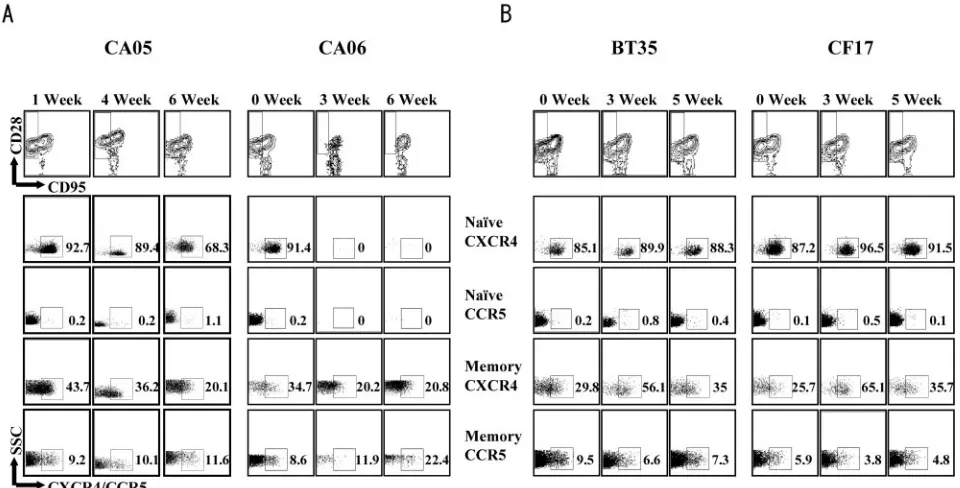
specificity of the two clones in vivo and to assess the impact of CoR preference on target cell selectivity, changes in the phe-notype (Fig. 6) as well as chemokine receptor expression (Fig. 7) in circulating and lymphoid CD4⫹T cells were determined. As anticipated, the X4 clone selectively targeted and depleted naı¨ve CD4⫹T cells in peripheral blood. The percentage of this T-cell subpopulation dropped from a baseline value of 63.8% to 42% within 2 to 4 weeks of infection in CA05 (Fig. 6A) and further declined by 6 wpi (Fig. 6B). A more drastic loss of naı¨ve T cells, from 57.4% to 1.5% within 2 weeks of infection, was seen in CA06, the monkey with the higher peak viremia (Fig. 6A). By 6 wpi,⬎99.9% of circulating CD4⫹T cells in CA06 were cells of the memory phenotype (Fig. 6B). Similar prefer-ential early targeting of naı¨ve T cells was also seen in the LNs, where loss of this T-cell subset accounted for the total drop in percentage of CD4⫹T cells (Fig. 6C). For animals infected with the R5-SHIVSF33A2(V3) clone, a rather modest (⬃15%)
but specific loss of memory cells was observed in CF17 at 2 to 4 wpi (Fig. 6A), and this further decreased at 5 wpi before rebounding close to preinfection values by 6 wpi (Fig. 6B). In BT35, an increase in the memory T-cell subset, similar to that
reported for SIV infection (25), was seen at 1 wpi, and this was followed by a gradual decline.
When changes in chemokine receptor expression on circu-lating CD4⫹ T lymphocytes were examined, results showed that naı¨ve cells expressing high levels of CXCR4 were selec-tively eliminated in early X4-SHIVSF33A2infection, followed or
accompanied by depletion of CXCR4⫹ memory cells (Fig. 7A). The difference in the tempo of depletion of the two CXCR4⫹-T-cell subpopulations was more evident in CA06, where a complete loss of naı¨ve CXCR4⫹cells compared to a 40% drop in CXCR4⫹memory cells was present at 3 wpi. In contrast, a transient drop in CCR5⫹memory T lymphocytes was seen in both R5-SHIVSF33A2(V3)-infected macaques at 3 wpi, and
[image:5.585.71.519.68.373.2]this was accompanied by a corresponding increase in the percent-age of CXCR4⫹memory T cells (Fig. 7B). By 5 wpi, however, a rebound in the percentage of CCR5⫹memory cells was noted. The selective destruction of CXCR4⫹lymphocytes by the X4 clone and CCR5⫹lymphocytes by the R5 clone further demon-strates their CoR specificity in vivo and shows that in addition to the extent of virus replication, the dynamics of CD4⫹-T-cell loss are influenced by the CoR usage of the inoculating virus, the level of CoR expression on target cells, and the representation of these cells in the various tissue compartments.
FIG. 6. Different subsets of CD4⫹T cells are targeted by X4-SHIVSF33Aand R5-SHIVSF33A2(V3). PBMCs and lymph node cells collected at the indicated times postinfection from SHIVSF33A2- and SHIVSF33A2(V3)-infected RM were phenotyped for CD3, CD4, CD28, and CD95 expression. (A) Contour plots depict differential depletion of naı¨ve (CD95lowCD28high) and memory (CD95highCD28low/high) CD4⫹T cells in early X4 (CA05 and CA06) and R5 (BT35 and CF17) SHIV clone infections, respectively. (B) Changes in the levels of naı¨ve (F) and memory (䊐) CD4⫹T cells in blood during the first 6 weeks of virus infection with X4 and R5 SHIV molecular clones. (C) Changes in total percentages of CD4⫹T cells as well as percentages of naı¨ve cells in various lymphoid tissues of macaque CA05.
on November 8, 2019 by guest
http://jvi.asm.org/
DISCUSSION
The use of SHIV isogenic clones that differ only in their CoR binding sequences for infection of nonhuman primates provides an ideally suited model to study the impact of HIV-1 coreceptor usage on the composition and dynamics of the CD4⫹-T-cell compartment. We report here the generation and characterization of such viruses. We show that the substitution of the V3 loop of the X4-SHIVSF33A2molecular clone with the
corresponding sequences of the R5-SHIVSF162P3isolate alone
was sufficient to alter its coreceptor preference in vitro and its replication characteristics in vivo. Differences in the sites of early virus replication as well as the rates of CD4⫹-T cell-loss in the periphery, LNs, and mucosal tissue compartments were seen in RM infected with the X4 compared to the R5 isogenic SHIV molecular clones. Infection with X4-SHIVSF33A2
re-sulted in a rapid drop, massively in the case of CA06, in CD4⫹-T-cell numbers in peripheral blood and secondary LNs during the period of peak virus production (Fig. 3 and 4). This loss of CD4⫹T cells in the peripheral tissues preceded deple-tion of CD4⫹T cells in the intestinal mucosa of CA06, the X4-SHIVSF33A2-infected macaque with the highest viral
bur-den. In contrast, loss of CD4⫹ T cells in the blood of both R5-SHIVSF33A2(V3)-infected macaques was modest and
tran-sient, with little or no depletion seen in the secondary lymph nodes during primary infection. However, lower percentages of CD4⫹T cells were seen in the gut-associated lymphoid tissue, consistent with preferential depletion in mucosal effector sites by the R5 clone. These observations of differential sites of early virus replication in animals infected with the X4 and R5 iso-genic clones are consistent with previous findings in animals
infected with the pathogenic X4-SHIVSF33A and
R5-SHIVSF162P3 isolates (15) and demonstrate that coreceptor
utilization, and not other undetermined strain-related differ-ences, is the major determinant for this difference.
We showed not only differential early targeting of various tissue compartments in X4 and R5 clone-infected macaques but also preferential depletion of particular CD4⫹-T-cell sub-sets within these compartments. Naı¨ve T cells were dramati-cally depleted in X4-SHIVSF33A2-infected macaques, whereas
the memory T cells were targeted by R5-SHIVSF33A2(V3)(Fig.
6 and 7). These findings of differences in tissue and T-cell targeting by X4 and R5 viruses can, to a large extent, be explained by differential expression of their cognate receptors on the two major CD4⫹-T-cell subsets and the relative tissue distribution of these cell populations in vivo. CCR5 expression is largely restricted to memory T cells that are infrequent in peripheral blood and LNs but prevalent in tissue effector sites such as intestinal lamina propria (Fig. 5). Accordingly, CD4⫹ -T-cell loss occurs predominantly in the gut of R5-SHIVSF33A2(V3)-infected macaques, with only modest
[image:6.585.54.534.71.315.2]deple-tion seen in peripheral blood (Fig. 3) and no depledeple-tion seen in the secondary lymphoid tissue compartments (Fig. 4). In con-trast, CXCR4 is expressed on memory as well as naı¨ve CD4⫹ T cells in peripheral blood and all lymphoid tissues examined (Fig. 5). In this regard, the observation that the loss of CD4⫹ T cells in the peripheral tissues was followed by elimination of CD4⫹T cells in the intestinal mucosa of CA06 indicates that there is no absolute restriction in the sites of CD4⫹-T-cell depletion induced by SHIV infection but that the rate and extent of T-cell loss in these various tissue compartments are
FIG. 7. CXCR4- and CCR5-positive cells are selectively depleted by X4-SHIVSF33A2and R5-SHIVSF33A2(V3), respectively. Data were generated by gating through lymphocytes and then gating through CD4⫹cells. After initial gating on naı¨ve (CD95lowCD28high) and memory (CD95high CD28low/high) CD4⫹lymphocytes, individual samples were analyzed for CXCR4 and CCR5 expression. Percentages of gated naı¨ve and memory CD4⫹T cells in (A) X4 and (B) R5 clone-infected RM that express CXCR4 or CCR5 are shown.
VOL. 79, 2005 HIV-1 CORECEPTOR AND CD4⫹-T-CELL LOSS 12301
on November 8, 2019 by guest
http://jvi.asm.org/
influenced by the total number of susceptible target cells that are available within these sites. Conceivably, the faster kinetics of replication seen for the X4 isogenic clone are due to the larger pool of susceptible target cells available for this virus (Fig. 3A and 5). Furthermore, the greater frequency and abun-dance of CXCR4 expression on naı¨ve cells compared to mem-ory cells can explain the early targeting of this T-cell subset by X4-SHIVSF33A2. The effects of infection by CXCR4- and
CCR5-using viruses on naı¨ve and memory CD4 subsets have recently been described using X4-SHIV and SIV (29–31). Our use of an isogenic system, showing early depletion of CXCR4⫹ naı¨ve cells by the X4-SHIVSF33A2molecular clone and CCR5⫹
memory cells by the R5-SHIVSF33A2(V3) clone (Fig. 7),
con-firms and extends findings in those other reports, demonstrat-ing that coreceptor preference is the principal basis of this selectivity.
In a prior study, one of two macaques inoculated intrave-nously with a cell-free X4-SHIVSF33A2 molecular clone
suf-fered precipitous peripheral CD4⫹-T-cell loss and progression to simian AIDS within 30 wpi, while the other displayed a less severe but sustained depletion of CD4⫹T cells and controlled its infection (14). The variability in the level of viremia and kinetics and extent of cell destruction seen in these and the present two macaques infected with the X4-SHIVSF33A2
mo-lecular clone contrasts with the patterns observed in animals infected with the uncloned X4-SHIVSF33Astrain, suggesting
that the clonal virus is less pathogenic than the viral isolate. But the possibility of individual host variability cannot be ex-cluded. Additional studies in a larger group of animals are required to address this. Despite these variations, however, the data show that the pattern of CD4⫹-T-cell loss induced by X4-SHIVSF33A2clone infection is dramatically altered through
modifications of its V3 loop. Furthermore, the availability of these X4 and R5 isogenic SHIV molecular clones for infection of macaques is expected to enhance our understanding of the role of tropism in transmission, persistence, and pathogenesis of HIV-1 infection. For example, coinfection of RM with these viruses should allow us to determine whether cell tropism alone dictates the R5 dominance we observed in macaques dually infected with the pathogenic isolates X4-SHIVSF33Aand
R5-SHIVSF162P3(12). Additionally, since the sites of
replica-tion and immune cells targeted by the clones differ, quesreplica-tions of interest will be whether the type and/or extent of selective immune pressures exerted by the shared host on the two vi-ruses will differ as well. An examination of sequence changes in the envelope and/or other genomic regions of viruses recov-ered from isogenic X4- and R5-SHIV-infected macaques over time, coupled with antiviral immune measurements, may pro-vide critical insights into these questions. Last, although core-ceptor switch has rarely been reported in SIV-infected ma-caques (24, 31), infection with the R5-SHIVSF33A2(V3)clone, a
virus generated on the backbone of an X4 clone with only 10 amino acid changes in the V3 loop, may provide a unique opportunity to investigate whether reversion to an X4 pheno-type can occur in infection of RM.
In summary, although the number of animals used is limited, this proof-of-concept study using an isogenic system clearly shows that the course of infection with the X4-SHIVSF33A2
molecular clone is markedly altered by changing its coreceptor choice through replacement of its V3 loop sequences with
those of an R5 strain. Coreceptor targeting of specific CD4⫹ -T-cell subpopulations coupled with the susceptibility and dis-tribution of the target cells contribute to the differing rates and sites of CD4⫹-T-cell depletion seen in X4-SHIVSF33A2- and
R5-SHIVSF33A2(V3)-infected macaques. Further studies with
these two clones in larger, more systematic studies are ex-pected to provide important information on the role of tropism in HIV-1 infection and disease induction.
ACKNOWLEDGMENTS
We thank Lisa Chakrabarti and Viviana Simon for critical comments and Peter Lopez for help with fluorescence-activated cell sorter anal-yses.
This research was supported by grants from the U.S. National In-stitutes of Health (AI46980, AI41945, and CA72822).
REFERENCES
1.Anton, P. A., J. Elliott, M. A. Poles, I. M. McGowan, J. Matud, L. E. Hultin, K. Grovit-Ferbas, C. R. Mackay, I. S. Y. Chen, and J. V. Giorgi.2000. Enhanced levels of functional HIV-1 co-receptors on human mucosal T cells
demonstrated using intestinal biopsy tissue. AIDS14:1761–1765.
2.Berger, E. A., P. M. Murphy, and J. M. Farber.1999. Chemokine receptors as HIV-1 coreceptors: roles in viral entry, tropism, and disease. Annu. Rev.
Immunol.17:657–700.
3.Bleul, C., L. Wu, J. A. Hoxie, T. A. Springer, and C. R. Mackay.1997. The HIV coreceptors CXCR4 and CCR5 are differentially expressed and
regu-lated on human T lymphocytes. Proc. Natl. Acad. Sci. USA94:1925–1930.
4.Bogers, W. M., C. Cheng-Mayer, and R. C. Montelaro.2000. Developments
in preclinical AIDS vaccine efficacy models. AIDS14(Suppl. 3):S141–S151.
5.Brenchley, J. M., T. W. Schacker, L. E. Ruff, D. A. Price, J. H. Taylor, G. J. Beilman, P. L. Nguyen, A. Khoruts, M. Larson, A. T. Haase, and D. C. Douek.2004. CD4⫹T cell depletion during all stages of HIV disease occurs
predominantly in the gastrointestinal tract. J. Exp. Med.200:749–759.
6.Chakrabarti, L. A., T. Ivanovic, and C. Cheng-Mayer.2002. Properties of the surface envelope glycoprotein associated with virulence of simian-human
immunodeficiency virus SHIVSF33A molecular clones. J. Virol. 76:
1588–1599.
7.Cheng-Mayer, C., A. Brown, J. Harouse, P. A. Luciw, and A. J. Mayer.1999. Selection for neutralization resistance of the simian/human
immunodefi-ciency virus SHIVSF33Avariant in vivo by virtue of sequence changes in the
extracellular envelope glycoprotein that modify N-linked glycosylation. J.
Vi-rol.73:5294–5300.
8.Cocchi, F., A. L. DeVico, A. Garzino-Demo, A. Cara, R. C. Gallo, and P. Lusso.1996. The V3 domain of the HIV-1 gp120 envelope glycoprotein is
critical for chemokine-mediated blockade of infection. Nat. Med.2:1244–
1247.
9.Dolei, A., A. Biolchini, C. Serra, S. Curreli, E. Gomes, and F. Dianzani.1998. Increased replication of T-cell-tropic HIV strains and CXC-chemokine re-ceptor-4 induction in T cells treated with macrophage inflammatory protein
(MIP)-1alpha, MIP-1beta and RANTES beta-chemokines. AIDS12:183–
190.
10.Fouchier, R. M., M. Groenink, A. Kootstra, M. Tersmette, H. G. Huisman, F. Miedema, and H. Schuitemaker.1992. Phenotype-associated sequence variation in the third variable domain of the human immunodeficiency virus
type 1 gp120 molecule. J. Virol.66:3183–3187.
11.Guadalupe, M., E. Reay, S. Sankaran, T. Prindiville, J. Flamm, A. McNeil, and S. Dandekar.2003. Severe CD4⫹T-cell depletion in gut lymphoid tissue during primary human immunodeficiency virus type 1 infection and substan-tial delay in restoration following highly active antiretroviral therapy. J.
Vi-rol.77:11708–11717.
12.Harouse, J. M., C. Buckner, A. Gettie, R. Fuller, R. Bohm, J. Blanchard, and C. Cheng-Mayer.2003. CD8⫹T cell-mediated CXC chemokine receptor 4-simian/human immunodeficiency virus suppression in dually infected
rhe-sus macaques. Proc. Natl. Acad. Sci. USA100:10977–10982.
13.Harouse, J. M., A. Gettie, T. Eshetu, R. C. Tan, R. Bohm, J. Blanchard, G. Baskin, and C. Cheng-Mayer.2001. Mucosal transmission and induction of simian AIDS by CCR5-specific simian/human immunodeficiency virus SHIVSF162P3. J. Virol.75:1990–1995.
14.Harouse, J. M., A. Gettie, R. C. Tan, T. Eshetu, M. Ratterree, J. Blanchard, and C. Cheng-Mayer.2001. Pathogenic determinants of the mucosally
trans-missible CXCR4-specific SHIVSF33A2map to env region. J. Acquir. Immune
Defic. Syndr.27:222–228.
15.Harouse, J. M., A. Gettie, R. C. H. Tan, J. Blanchard, and C. Cheng-Mayer.
1999. Distinct pathogenic sequela in rhesus macaques infected with CCR5 or
CXCR4 utilizing SHIVs. Science284:816–819.
16.Harouse, J. M., R. C. Tan, A. Gettie, P. Dailey, P. A. Marx, P. A. Luciw, and C. Cheng-Mayer.1998. Mucosal transmission of pathogenic
CXCR4-utiliz-ing SHIVSF33Avariants in rhesus macaques. Virology248:95–107.
on November 8, 2019 by guest
http://jvi.asm.org/
17.Hsu, M., C. Buckner, J. Harouse, A. Gettie, J. Blanchard, J. E. Robinson, and C. Cheng-Mayer.2003. Antigenic variations in the CD4 induced sites of
the CCR5-tropic, pathogenic SHIVSF162P3gp120 variants. J. Med. Primatol.
32:211–217.
18.Hsu, M., J. M. Harouse, A. Gettie, C. Buckner, J. Blanchard, and C. Cheng-Mayer.2003. Increased mucosal transmission but not enhanced pathogenic-ity of the CCR5-tropic, simian AIDS-inducing simian/human
immunodefi-ciency virus SHIVSF162P3maps to envelope gp120. J. Virol.77:989–998.
19.Kinter, A., A. Catanzaro, J. Monaco, M. Ruiz, J. Justement, S. Moir, J. Arthos, A. Oliva, L. Ehler, S. Mizell, R. Jackson, M. Ostrowski, J. Hoxie, R. Offord, and A. S. Fauci.1998. CC-chemokines enhance the replication of
T-tropic strains of HIV-1 in CD4(⫹) T cells: role of signal transduction.
Proc. Natl. Acad. Sci. USA95:11880–11885.
20.Kunkel, E. J., J. Boisvert, K. Murphy, M. A. Vierra, M. C. Genovese, A. J. Wardlaw, H. B. Greenberg, M. R. Hodge, L. Wu, E. C. Butcher, and J. J. Campbell.2002. Expression of the chemokine receptors CCR4, CCR5, and
CXCR3 by human tissue-infiltrating lymphocytes. Am. J. Pathol.160:347–
355.
21.Li, Q., L. Duan, J. D. Estes, Z. M. Ma, T. Rourke, Y. Wang, C. Reilly, J. Carlis, C. J. Miller, and A. T. Haase.2005. Peak SIV replication in resting
memory CD4⫹T cells depletes gut lamina propria CD4⫹T cells. Nature
434:1148–1152.
22.Luciw, P., C. Mandell, S. Himathongkham, J. Li, T. Low, K. Schmidt, K. Shaw, and C. Cheng-Mayer.1999. Fatal immunopathogenesis by SIV/HIV-1
(SHIV) containing a variant form of the HIV-1SF33envgene in juvenile and
newborn rhesus macaques. Virology263:112–127.
23.Luciw, P. A., E. Pratt-Lowe, K. E. S. Shaw, J. A. Levy, and C. Cheng-Mayer.
1995. Persistent infection of rhesus macaques with T-cell-line-tropic and macrophage-tropic clones of simian/human immunodeficiency viruses
(SHIV). Proc. Natl. Acad. Sci. USA92:7490–7494.
24.Marx, P. A., and Z. Chen.1998. The function of simian chemokine receptors
in the replication of SIV. Semin. Immunol.10:215–223.
25.Mattapallil, J. J., D. C. Douek, B. Hill, Y. Nishimura, M. Martin, and M. Roederer.2005. Massive infection and loss of memory CD4⫹T cells in
multiple tissues during acute SIV infection. Nature434:1093–1097.
26.Mattapallil, J. J., Z. Smit-McBride, M. McChesney, and S. Dandekar.1998. Intestinal intraepithelial lymphocytes are primed for gamma interferon and
MIP-1 expression and display antiviral cytotoxic activity despite severe
CD4⫹T-cell depletion in primary SIV infection. J. Virol.72:6421–6429.
27.Mehandru, S., M. A. Poles, K. Tenner-Racz, A. Horowitz, A. Hurley, C. Hogan, D. Boden, P. Racz, and M. Markowitz.2004. Primary HIV-1
infec-tion is associated with preferential depleinfec-tion of CD4⫹T lymphocytes from
effector sites in the gastrointestinal tract. J. Exp. Med.200:761–770.
28.Moore, J. P., S. G. Kitchen, P. Pugach, and J. A. Zack.2004. The CCR5 and CXCR4 coreceptors—central to understanding the transmission and patho-genesis of human immunodeficiency virus type 1 infection. AIDS Res. Hum.
Retrovir.20:111–126.
28a.National Research Council.1996. Guide for the care and use of laboratory animals. National Academy Press, Washington, D.C.
29.Nishimura, Y., C. R. Brown, J. J. Mattapallil, T. Igarashi, A. Buckler-White,
B. A. Lafont, V. M. Hirsch, M. Roederer, and M. A. Martin.2005. Resting
naive CD4⫹T cells are massively infected and eliminated by X4-tropic
simian-human immunodeficiency viruses in macaques. Proc. Natl. Acad. Sci.
USA102:8000–8005.
30.Nishimura, Y., T. Igarashi, O. K. Donau, A. Buckler-White, C. Buckler, B. A. Lafont, R. M. Goeken, S. Goldstein, V. M. Hirsch, and M. A. Martin.2004.
Highly pathogenic SHIVs and SIVs target different CD4⫹T cell subsets in
rhesus monkeys, explaining their divergent clinical courses. Proc. Natl. Acad.
Sci. USA101:12324–12329.
31.Picker, L. J., S. I. Hagen, R. Lum, E. F. Reed-Inderbitzin, L. M. Daly, A. W. Sylwester, J. M. Walker, D. C. Siess, M. Piatak, Jr., C. Wang, D. B. Allison, V. C. Maino, J. D. Lifson, T. Kodama, and M. K. Axthelm.2004. Insufficient
production and tissue delivery of CD4⫹memory T cells in rapidly
progres-sive simian immunodeficiency virus infection. J. Exp. Med.200:1299–1314.
32.Pitcher, C. J., S. I. Hagen, J. M. Walker, R. Lum, B. L. Mitchell, V. C. Maino, M. K. Axthelm, and L. J. Picker.2002. Development and homeostasis of T
cell memory in rhesus macaque. J. Immunol.168:29–43.
33.Rowland-Jones, S. L.2003. Timeline: AIDS pathogenesis: what have two
decades of HIV research taught us? Nat. Rev. Immunol.3:343–348.
34.Ruiz, M. E., C. Cicala, J. Arthos, A. Kinter, A. T. Catanzaro, J. Adelsberger, K. L. Holmes, O. J. Cohen, and A. S. Fauci.1998. Peripheral blood-derived
CD34⫹progenitor cells: CXC chemokine receptor 4 and CC chemokine
receptor 5 expression and infection by HIV. J. Immunol.161:4169–4176.
35.Sallusto, F., D. Lenig, C. R. Mackay, and A. Lanzavecchia.1998. Flexible programs of chemokine receptor expression on human polarized T helper 1
and 2 lymphocytes. J. Exp. Med.187:875–883.
36.Simmons, G., P. R. Clapham, L. Picard, R. E. Offord, M. M. Rosenkilde, T. W. Schwartz, R. Buser, T. N. C. Wells, and A. E. Proudfoot.1997. Potent inhibition of HIV-1 infectivity in macrophages and lymphocytes by a novel
CCR5 antagonist. Science276:276–279.
37.Smit-McBride, Z., J. J. Mattapallil, M. McChesney, D. Ferrick, and S. Dandekar.1998. Gastrointestinal T lymphocytes retain high potential for
cytokine responses but have severe CD4⫹T-cell depletion at all stages of
simian immunodeficiency virus infection compared to peripheral
lympho-cytes. J. Virol.72:6646–6656.
38.Vajdy, M., R. Veazey, I. Tham, C. deBakker, S. Westmoreland, M. Neutra, and A. Lackner.2001. Early immunologic events in mucosal and systemic lymphoid tissues after intrarectal inoculation with simian immunodeficiency
virus. J. Infect. Dis.184:1007–1014.
39.Veazey, R. S., M. DeMaria, L. V. Chalifoux, D. E. Shvetz, D. R. Pauley, H. L. Knight, M. Rosenzweig, R. P. Johnson, R. C. Desrosiers, and A. A. Lackner.
1998. Gastrointestinal tract as a major site of CD4⫹T cell depletion and
viral replication in SIV infection. Science280:427–431.
40.Veazey, R. S., K. G. Mansfield, I. C. Tham, A. C. Carville, D. E. Shvetz, A. E. Forand, and A. A. Lackner.2000. Dynamics of CCR5 expression by CD4⫹T cells in lymphoid tissues during simian immunodeficiency virus infection.
J. Virol.74:11001–11007.
41.Weiser, B., H. Burger, P. Campbell, S. Donelan, and J. Mladenovic.1996. HIV type 1 RNA expression in bone marrows of patients with a spectrum of
disease. AIDS Res. Hum. Retrovir.12:1551–1558.
VOL. 79, 2005 HIV-1 CORECEPTOR AND CD4⫹-T-CELL LOSS 12303