COMPARATIVE ANALYSIS OF
CYTOMORPHOLOGIC FEATURES OF THYROID
LESIONS USING VARIOUS CYTOLOGICAL
STAINING TECHNIQUES
DISSERTATION
SUBMITTED TO TAMILNADU DR.M.G.R. MEDICAL UNIVERSITY, CHENNAI
in partial fulfilment of
the requirements for the degree of
M.D. (PATHOLOGY)
BRANCH - III
TIRUNELVELI MEDICAL COLLEGE HOSPITAL,
TIRUNELVELI- 627011
CERTIFICATE
I hereby certify that this dissertation entitled “COMPARATIVE ANALYSIS
OF CYTOMORPHOLOGIC FEATURES OF THYROID LESIONS USING
VARIOUS CYTOLOGICAL STAINING TECHNIQUES” is a record of work
done by Dr.N.MEENAKSHI, in the Department of Pathology, Tirunelveli Medical
College, Tirunelveli, during her postgraduate degree course period from 2015- 2018.
This work has not formed the basis for previous award of any degree.
Prof.Dr. K. SITHYATHIYAMUNAVARAH M.D., DEAN
CERTIFICATE
I hereby certify that this dissertation entitled “COMPARATIVE ANALYSIS OF
CYTOMORPHOLOGIC FEATURES OF THYROID LESIONS USING
VARIOUS CYTOLOGICAL STAINING TECHNIQUES” is a record of work
done by Dr.N.MEENAKSHI, in the Department of Pathology, Tirunelveli Medical
College, Tirunelveli, during her postgraduate degree course period from
2015-2018,under my guidance and supervision, in the Department of Pathology Tirunelveli
Medical College & Hospital, Tirunelveli, in partial fulfilment of the requirement for
M.D., (Branch III) in Pathology examination of the Tamilnadu Dr. M.G.R Medical
University will be held in MAY 2018. This work has not formed the basis for
previous award of any degree.
Prof. Dr. J. SURESH DURAI,M.D.,
Department of pathology, Tirunelveli Medical College,
Prof.Dr.K. SHANTARAMAN.MD.,
Professor and Head, Department of Pathology, Tirunelveli Medical College Tirunelveli- 627011.
DECLARATION
I solemnly declare that this dissertation titled “COMPARATIVE ANALYSIS
OF CYTOMORPHOLOGIC FEATURES OF THYROID LESIONS USING
VARIOUS CYTOLOGICAL STAINING TECHNIQUES” submitted by me for
the degree of M.D, is the record work carried out by me during the period of
2015-2017 under the guidance of Prof.Dr. J. SURESH DURAI, M.D, Professor of
Pathology, Department of Pathology, Tirunelveli Medical College, Tirunelveli. The
dissertation is submitted to The Tamilnadu Dr. M.G.R. Medical University, Chennai,
towards the partial fulfilment of requirements for the award of M.D. Degree (Branch
III) Pathology examination to be held in May 2018.
Place: Tirunelveli Dr.N.MEENAKSHI,
Date: Department of Pathology,
Tirunelveli Medical College,
ACKNOWLEDGEMENT
This dissertation has come to your hands as a result of the combined effort of
lot of people. I am thankful to all these people for helping me in bringing out this
work.
I thank the DEAN Prof.Dr. SITHY ATHIYA MUNAVARAH, M.D for
permitting me to conduct this study and to avail the resources of the hospital.
I am also thankful to my Professor and Head, Department of Pathology
Prof.Dr.K.SHANTARAMAN M.D, for his support and guidance in doing this
dissertation.
I express my heartfelt gratitude to my guide Prof. Dr. J. SURESH DURAI
M.D, Professor, Department of Pathology, who has guided me through all the steps in
my study right from the beginning till the end.
I am extremely thankful to the respected Professors of my department
Prof.Dr.SWAMINATHAN.K M.D, Prof.Dr.ARASI RAJESH M.D,
Prof.Dr.VASUKI MUTHURAMAN M.D, and Associate Professor;
Dr.V.BAGIYALAKSHMI M.D Assistant Professors; Dr.HIDAYA FATHIMA
M.D., Dr.JOHNSY MERLA M.D., Dr.MAHALAKSHMI M.D., Dr. SINDHUJA
M.D., and Dr.DINA MARY M.D., for their concern, valuable suggestions, and
support during the study.
I also thank the lab technicians MRS.VEERALAKSHMI,
MR.SANKARANARAYANAN, MR.BALAMURUGAN, MRS.AMUTHA,
I also thank my fellow postgraduates and my friends without whom this study
would not have been possible. A special word of thanks to my family who have
helped me in this journey.
I thank the almighty, for blessing me not only in this study, but in all
CONTENTS
S.NO TITLE PAGE.NO
1.
2.
3.
4.
5.
6.
7.
INTRODUCTION
AIM OF THE STUDY
REVIEW OF LITERATURE
MATERIALS AND METHODS
OBSERVATION AND RESULTS
DISCUSSION
SUMMARY & CONCLUSION
BIBILIOGRAPHY
MASTER CHART
1
3
4
59
67
78
CERTIFICATE - II
This is certify that this dissertation work title “COMPARATIVE ANALYSIS
OF CYTOMORPHOLOGIC FEATURES OF THYROID LESIONS USING
VARIOUS CYTOLOGICAL STAINING TECHNIQUES” of the candidate
Dr.N.MEENAKSHI, with registration Number 201513306 for the award of
M.D. Degree in the branch of PATHOLOGY (III). I personally verified the
urkund.com website for the purpose of plagiarism check. I found that the uploaded
thesis file contains from introduction to conclusion page and result shows
4 PERCENTAGE of plagiarism in the dissertation.
ABBREVIATIONS
AUS–Atypia of Undetermined Significance
ATC–Anaplastic thyroid carcinoma
BFN–Benign follicular nodule
C cells–clear cells
CDX2–Cluster of Differentiation X2
CK-19–Cytokeratin 19
EA–Eosin Azure
FA–Follicular adenoma
FC–Follicular carcinoma
FN–Follicular neoplasm
FNA- Fine needle aspiration
FNAC–Fine needle aspiration cytology
FV-PTC–Follicular variant of papillary thyroid carcinoma
GD– Graves’ disease
H&E–Hematoxylin and eosin stain
HT–Hashimotos thyroiditis
HTT–Hyalinizing trabecular tumour
IHC–Immunohistochemistry
INCI–Intranuclear cytoplasmic inclusion
LBP–Liquid based preparation
MC–Medullary carcinoma
ND/UNS–Nondiagnostic or unsatisfactory
NG–Nodular goiter
OG 6–Orange G 6
MGG–May Grunwalds Giemsa
Pap–Papaicolou stain
PAX8–Paired box 8
PC–Papillary carcinoma
PCT–Papillary carcinoma of thyroid
PTDC–Poorly Differentiated Thyroid Carcinoma
RAS–Rat Associated Sarcoma
T3–Tri-iodothyronine
T4–Tetra- iodothyronine or thyroxine
TCV–Tall cell variant
TSH–thyroid stimulating hormone
TTF 1–Thyroid Transcription Factor 1
US–Ultrasound
INTRODUCTION
Thyroid lesions are a common clinical problem. Fine-needle
aspiration (FNA) for cytologic evaluation of thyroid lesion was originally
used by Martin and Ellis at New York Memorial Hospital for Cancer and
Allied Diseases in 1930 (1,22). FNA of thyroid is now practiced worldwide
and proves to be the most economical and reliable minimally invasive
diagnostic procedure.
The incidence of malignancy in a solitary thyroid nodule or in a
multinodular goiter is equal and about 5% in non-endemic areas(1).
Therefore preoperative distinction of benign lesion is important to avoid
unnecessary surgery. FNAC of thyroid lesions show sensitivity as high as
93.4% with a positive predictive value of malignancy 98.6 % and 74.9 %
specificity(2). Dignostic accuracy is important in thyroid lesions since it
decides the type of thyroidectomy performed on the patient.
Two fundamentally different methods of fixation and staining are
used in FNAC: air-drying followed by a Romanowsky stain such as May
Grunwalds Gimsa (MGG), Jenner-Giemsa, Wright’s stain or Diff-Quik; and alcohol-fixation followed by Papanicolaou (Pap) or hematoxylin and
eosin (H&E) staining. Both methods have their advantages and
deficiencies. Both the methods are useful since some features of cells,
Hence combining the morphological features of various stains can improve
the diagnostic accuracy.
Ultrasonography (US), thyroid function tests, antibody profiles and
FNA, used in conjunction in selected cases, complement one another(2). In
recent years a number of immunocytochemical and molecular markers
have been applied to cytological material from thyroid to improve
AIM OF THE STUDY
To analyse the cytomorphologic features of thyroid lesions using
REVIEW OF LITERATURE
THYROID GLAND
It is a butterfly-shaped organ located anteriorly to the trachea
at the level of the second and third tracheal rings. It has two lobes
connected by the isthmus in the midline. Its bilaterality is significant
because the presence of malignant cells on one or both sides can
significantly alter the management of the patient, e.g., requiring more
extensive surgery, such as bilateral neck dissections if there is local
extension of the tumour(3).
HISTOLOGY AND FUNCTION
The thyroid gland is covered by thin capsule of connective tissue.
It extends into the glandular parenchyma and divides each lobe into
irregularly shaped and variably sized lobules. Follicles are the functional
units of the thyroid gland. Each lobule consist of 20 to 40 follicles.
Follicles are spherical and cyst like, between 0.02 and 0.9 mm in diameter.
Follicles consist of a central colloid core surrounded by a single layered
epithelium resting on a basal lamina. Follicular epithelial cells produce
colloid. Colloid consists of an iodinated glycoprotein, inactive
iodothyroglobulin, stored form of the active thyroid hormones,
tri-iodothyronine (T3) and tetra-tri-iodothyronine or thyroxine (T4). Sufficient
metabolic activity of the body for up to three months. Follicles are
surrounded by a delicate connective tissue stroma, containing dense plexus
of fenestrated capillaries, extensive lymphatic networks and sympathetic
nerve fibres which supply the arterioles and capillaries. Some nerve fibres
end close to the follicular epithelial cells.
Follicular cells may vary from squamous or low cuboidal to
columnar cells, depending on their level of activity, which is controlled by
circulating thyroid stimulating hormone (TSH). Resting follicles are large
and lined by squamous or low cuboidal epithelium with abundant luminal
colloid. Apical microvilli are short in resting cells, but elongate and often
branch on stimulation by TSH. Follicles showing different levels of
activity may co-exist.
Active follicular cells are highly polarized functionally. The
secretion of TSH leads to endocytosis of colloidal droplets at the luminal
epithelium. The hormone provokes the cells to extend cytoplasmic
processes into the luminal colloid and sequester droplets of colloid. The
iodinated thyroglobulin in the intracellular colloid droplets is degraded by
follicular cell lysosomes, liberating T3 and T 4, which passes to the base of
the cell and they are released.
The epithelial cells lining the acini are unique, in that they are
interior of the acini or into the vascular channels(4). As Boyd so well puts it,
"They have both a back and a front door". Secretion into the acinus may be
considered as a process of storage, a provision for a time of greater need;
while secretion in the reverse direction may be looked upon as meeting the
current needs of the tissues.
In the colloid which is secreted into the acini there are two
protein bodies: one is a nucleoprotein which contains no iodine but is high
in phosphorus. This fraction has no known function. The other one is
thyroglobulin, which is rich in iodine and from the molecule of which the
essential thyroid hormone is probably split off as required(4).
The thyroid builds thyroglobulin when it is supplied with
iodine and essential amino-acids. Under normal circumstances there is an
approximate balance between the formation and release of hormone. The
demand of the tissues for thyroid hormone varies with age, sex and season,
and is increased by many physiological and pathological processes such as
pregnancy, menstruation and infections.
C cells: thyroid parenchyma also contains C (clear) cells, so called
because they have pale staining cytoplasm. C cells are members of the
amine precursor uptake and decarboxylation (APUD) system of dispersed
neuroendocrine cells. They produce peptide hormone calcitonin, which
from renal tubule ultrafiltrate. C cells populate the middle third of each
lateral lobe of thyroid and are typically found scattered within thyroid
follicles, lying inside the basal lumina but not reaching the follicle lumen.
Occasionally they occur in clusters in the interfollicular stroma. Therefore
it is also called as parafollicular cells.
FINE NEEDLE ASPIRATION CYTOLOGY (FNAC) OF THYROID LESIONS
INDICATION
The main indications of FNA in thyroid lesions are the following:
1. Evaluation of solitary thyroid nodules to distinguish benign from
malignant neoplasm.
2. Evaluation of diffuse thyroid lesion to distinguish inflammatory or
autoimmune lesions from nodular goiter
3. Confirmation and categorization of clinically obvious thyroid
malignancy for example in case of anaplastic carcinomas
preoperative palliative treatment may be required, and lymphoma
and metastatic malignancy where surgery is usually not indicated.
4. To obtain material for ancillary tests
FNA has been shown to be the safest and most accurate of diagnostic
tools in thyroid . Its use has simultaneously diminished the number of
surgeries done for benign lesions and increased the proportion of
malignancies in surgically resected thyroids
Generally in thyroiditis, usual type of papillary carcinoma, medullary
carcinoma, anaplastic carcinoma and high-grade lymphoma cytologic
diagnosis is accurate. False negative reporting occur in cystic lesions
harboring malignancy, low-grade or intermediate-grade lymphomas
occurring in a background of Hashimoto’s thyroiditis , anaplastic
carcinoma with necrosis, focal involvement of the gland by thyroiditis
and in cases with dual pathology where the dominant non-neoplastic lesion
overlies or obscures a small carcinoma. False negatives have been shown
to be minimized by using ultrasound guided fine needle aspiration. In fine
needle aspiration cytology of thyroid, the term follicular neoplasm is used
for the spectrum of follicular nodules because the diagnostic criteria for
follicular carcinoma is based on capsular or vascular invasion which
cannot be assessed on cytology and distant metastases.
The false positive rate can be reduced further by excluding
indeterminate follicular lesions. Even though there is slight increase in
accuracy achieved by core needle biopsy, follicular adenomas and
follicular carcinomas cannot be distinguished, also fine needle aspiration is
COMPLICATIONS OF THYROID FNAC
There are no contraindications to thyroid FNA. Local hemorrhage may be
caused by needling, occasionally causing a hematoma in the anterior neck
may lead to airway compression. Carotid hematoma is a very rare
complication. Transient vocal cord paralysis, acute transient goiter, acute
suppurative thyroiditis and chemical neuritis occur occasionally. Puncture
of the trachea during needling usually causes coughing. Small amounts of
blood may be coughed up but recovery is rapid. Needling may convert a
hot nodule to a cold one and vice versa, therefore scans should be done
before FNA. Post-FNA infarction is an uncommon complication and most
reported cases have been Hurthle cell nodules, followed by PC and FNs.
Hemorrhage, necrosis or infarction caused by needling may occasionally
obscure the histological pattern of thyroid neoplasms. Cellular and
vascular granulation tissue of organising hematoma or necrosis can mimic
sarcoma or angiomatous tumors.
Fibrosis, papillary hyperplasia, calcification, cholesterol clefts,
vascular thrombosis and capsular distortion simulating invasion are other
worrisome histological alterations that occasionally follow FNAC(2).
Changes are, in general, proportionate to the size of the needle used and
the number of needle passes. Post-needling alterations are generally less
with the fine needle capillary sampling technique. Aggressive and
avoided. In cases where needling is to be repeated for inadequate or
inconclusive cytology, it is wise to allow an interval of a week to 10 days
for any artifacts of initial needling to minimize. Cases of tumor
implantation along the needle track have been documented rarely.
TECHNICAL CONSIDERATIONS
First examine the patient in sitting position. Then the patient should
be made to lie supine with a pillow behind the neck for hyperextension, to
make lesion more obvious. The fine needle capillary sampling technique is
suitable in vascular structures like thyroid as it provides cellular material
with minimal dilution by blood. Instruct the patient to not to swallow when
needling. The lesion is needled with a fine needle , quickly and gently at
different angles and points of entry. Conclude needling before or as soon
as material appears at the hub of the needle, the needle then attached to an
air-filled syringe, and material deposited and smeared on to clean glass
slides. Half of the smears can be air-dried for Romanowsky staining,
while the rest should be wet-fixed in alcohol for Papanicolaou (PAP) stain
or H&E.
If the aspirate is scanty, air-drying with Diff-Quik or MGG stain is
better as it ensures retention of 100% of cells on the slide(2). Rapid
smearing is important in bloody samples, as clotting of blood will entangle
diagnostic cells and distort morphology. Slow drying of wet samples
hair-dryer can be used for rapid drying but should be avoided in samples that
may be infectious, to avoid aerosols. Despite cost and compensation issues,
bedside evaluation of a Diff-Quik or ultrafast PAP-stained smear is
advantageous to ascertain adequate cellularity and representative sampling
and to select cases for ancillary studies.
ULTRASOUND GUIDED FNAC
Published data indicate reduced non-diagnostic and false-negative
rates with ultrasound (US) evaluation and US guidance(2). Real-time
ultrasound examination allows visualization of the needle within the
lesion, hence facilitating accurate biopsy of small nonpalpable nodules(5)
Indications of US guided FNAC
Traditionally, the main indication for FNA biopsy of the thyroid
has been the presence of a solitary nodule. The Society of Radiologists in
Ultrasound suggested that FNA should be considered for a nodule 1.0 cm
or more at the largest diameter if microcalcifications are present and for a
nodule 1.5 cm or larger if the nodule is solid or if there are coarse
calcifications within the nodule (6). The American Association of Clinical
Endocrinologists recommended FNA even for nodules smaller than 10 mm
whenever clinical information or US features arouse suspicion about the
The US features that are suggestive of malignancy include
microcalcifications, marked hypoechogenicity, an irregular or
microlobulated margin, a longitudinal dimension larger than the
cross-sectional dimension, intrinsic vascularity, direct tumor invasion of adjacent
soft tissue, and metastasis to one or more lymph nodes(8) .
In palpable nodules of nodular goiter, the cytologic sampling should
be focussed on lesions characterized by suspicious US findings rather
than on larger or clinically dominant nodules(6,70).
The presence of abnormal lymph nodes or thyroid extracapsular
extension overrides the ultrasound features of thyroid nodule and should
prompt US-FNA or biopsy of lymph node and or ipsilateral nodule(9 )
FIXATION
Two most commonly used methods of fixation used in cytology are air
dryring and wet fixation .
DRY FIXATION
Fixation by air-drying causes increase in size of the cell, because it
causes both cytoplasm and nucleus, to flatten on the slide . It therefore
appears larger than a cell fixed in ethanol. Nuclear enlargement and
variation in nuclear size are exaggerated in air-dried smears. This enhances
the difference between normal and abnormal cells . Optimal fixation of
air-dried smears depends on rapid drying. This can be enhanced by using a
produce artifacts, in particular shrinkage of cells and nuclei, which may
render diagnosis impossible.
WET FIXATION:
In smears that are wet fixed with alcohol the cells maintains its
three-dimensional rounded shape . In wet fixed smears the size of the cell
and nucleus is comparable to that of tissue sections. The smears should be
immersed in alcohol immediately to prevent focal drying artifacts but there
will be considerable cell loss during alcohol fixation. The main problem
with wet-fixed smears is that highly cellular smears dry so quickly that
drying artifacts can be difficult to avoid which may lead to difficulties in
diagnosis.
STAINING METHODS USED IN CYTOPATHOLOGY:
Three staining methods are used routinely in thyroid cytopathology.
Papanicolou stain is most popularly used. Romanowsky stains are used
for rapid assessment of adequacy of aspirate. Hematoxylin and eosin
stains are used for cell blocks as well as for staining smears.(10)
ROMANOWSKY STAINS
Romanowsky stains allow better estimation of relative cell and
nuclear sizes, and superior visualization of cytoplasmic details, smear
background elements and intercellular matrix components(11)
In Romanowsky stains, air drying leads to an apparent
to an increase in their apparent size proportional to the volume of the
nuclei. The degree of perceived nuclear and cytoplasmic size increase due
to air drying depends on the type of cell; it is smallest in mature squamous
cells.
The apparent enlargement of cells and nuclei in air dried
Romanowsky-stain preparations, compared to the cell and nuclear sizes in
wet fixed Papanicolaou stained preparations, amplifies the cell and nuclear
size differences within the specimen and permits more accurate evaluation
of relative cell size as well as nuclear size and shape . In addition, uniform
air drying of well prepared, evenly smeared cytological preparations
stained with Romanowsky stains, makes it preferable for morphometry (13),
since the unintentional focal air drying of wet fixed smears lead to nuclear
size variability. Air drying also influences the structure of the nuclear
chromatin, which becomes condensed and thus more hyperchromatic (14).
The accentuation of size and chromaticity differences between normal and
malignant nuclei produced by Romanowsky stains is a useful feature for
evaluating fine needle aspirates, especially when dealing with well
differentiated malignancies (15). Accurate exfoliative cytological diagnosis
is dependent on high power evaluation of nuclear chromatin changes. By
contrast, because they usually are very cellular and show many small true
tissue fragments, fine needle aspirates frequently can be diagnosed under
Romanowsky stains are essential for low power pattern-based diagnosis
(16) owing to their better definition of cell cytoplasm and their accentuation
of enlarged tumor cell nuclei.
Romanowsky stains enhance cytoplasmic detail, a useful feature for
determining differentiation of neoplastic cells. Clearer cytoplasmic
definition also enables better appreciation of the plasmacytoid appearance
of certain neoplastic cells. Such plasmacytoid cells, defined by their
eccentrically placed nuclei, are characteristic not only of plasma cell
tumors , but also of neuroendocrine tumors of the pancreas, carcinoid
tumour, medullary carcinoma of the thyroid(17)
Improved visualization of cytoplasmic granules may be valuable for
fine needle aspirates of the thyroid and other organs. A common finding in
fine needle aspirates of the thyroid is the presence of paravacuolar
granules, which represent lysosomes containing hemosiderin or lipofuscin
pigments (18) . Paravacuolar granules are more common in samples from
normal thyroid than in those from colloid nodules (19) , but are found across
the spectrum of thyroid pathology and thus are not specific for any
pathology. Their presence, however, identifies the aspirate as coming from
the thyroid and helps exclude aspirates of similar appearing cells from the
parathyroids . Other types of granules potentially encountered in thyroid
fine needle aspirates include the red cytoplasmic granules occasionally
granules can be helpful for cytological diagnosis of this otherwise difficult
to diagnose malignancy.
Occasionally, fine needle aspirates of the thyroid may show
follicular epithelial cells with marginal vacuoles, a phenomenon known as
a “ fire-flare ” appearance (21) . Initially it was thought as a distinctive feature of hyperthyroidism but now it is considered as a nonspecific
finding in both neoplastic and non-neoplastic disorders that could be
identified only with Romanowsky stains (22) . In case of metastasis, the
fire-flare appearance is helpful for identifying the primary tumor, usually
follicular carcinoma of thyroid(23).
In Romanowsky staining, amyloid stains deep blue with possible focal
metachromasia (24,25) . Amyloid detection could be important for
diagnosing medullary carcinomas of the thyroid (26).
Colloid is a noncellular element whose identification is important
for diagnostic purpose of thyroid FNAC. Both the amount of colloid and
its quality are important in thyroid fine needle aspirates; the ratio of
colloid to cells is one of the most important diagnostic criteria for
differentiating between colloid nodules and potentially malignant thyroid
neoplasms. While thick colloid can be identified easily on both
Romanowsky and Papanicolaou stains, thin colloid may be extremely
difficult to visualize in Papanicolaou stained smears. In Romanowsky
and cracking patterns, which imparts a mosaic like crackling (9) cracked
glass or crazy pavement appearance (27)
Pathologists would be familiar with the Romanowsky stained
appearance of hematolymphoid cells, so Romanowsky stains are better to
wet fixed preparations for diagnosing hematolymphoid neoplasms. Certain
characteristic features of hematolymphoid cells are not seen or are more
difficult to recognize in Papanicolaou or hematoxylin and eosin stains.
Although Romanowsky stains are mandatory for accurate interpretation of
fine needle aspirates of primary lymph node conditions, many
cytopathologists also use Papanicolaou stained smears for subtyping of
lymphomas, because the better nuclear chromatin definition afforded by
this stain aids in differentiating centroblasts from centrocytes.
The contrast between nuclear chromatin and nucleoli is variable,
nucleoli are not always demonstrated well in Romanowsky stains. Wet
fixed variations of Romanowsky stains have been recommended to
overcome this shortcoming (20); these offer nuclear chromatin and nucleolar
detail similar to wet fixed Papanicolaou or hematoxylin and eosin stained
preparations. As expected, however, the advantages of increased nuclear
size and accentuation of nuclear size differences brought about by air
drying are lost. In addition to the differences in the nuclear chromatin
detail, wet fixed stains exhibit sharper nuclear outlines, a feature that is
neoplastic cells. Nuclear grooves and pseudo-inclusions are more distinct
in wet fixed preparations and unusual chromatin patterns, e.g., the “ salt
and pepper ” chromatin in neuroendocrine neoplasms, are appreciated
more easily. Many of these nuclear features, such as nuclear grooves in
papillary carcinomas of the thyroid (29) and nuclear inclusions in both
thyroid tumors and melanoma, also can be demonstrated in Romanowsky
stained preparations, but they may be more difficult to identify
Further constraint is that Romanowsky stains, including the
Diff-Quik stain commonly used in cytopathology, were created and intended for
use with very thin, evenly smeared preparations. Typical fine needle
aspirates commonly contain thicker tissue fragments and a thin smear may
be impossible to make. Thicker tissue fragments, which may consist of
three-dimensional cell groups or partially necrotic tissue fragments,
frequently are poorly stained and difficult to visualize. In such cases,
Papanicolaou or hematoxylin and eosin stains may reveal the cytological
features of the individual cells within the three-dimensional cell clusters
and may show well defined, individual, intact, non-necrotic cells. The
technique for making good air dried smears from fine needle aspiration
material is important. The optimal smearing technique usually requires
some practice. Smears should be air dried relatively quickly (within 5 min)
noticeable artifact formation to shorten the air drying time from about 3
min to about 1 min per smear in case of rapid on site evaluation(30).
Romanowsky stained slides can be used for immunostains of certain
lymphoid markers.
PAPANICOLOU STAIN
Papanicolaou (PAP) staining was first described by George
Papanicolaou, the father of Cytopathology, in 1943(31) .
Papanicolaou stain ( PAP stain) is the most important stain utilized
in the practice of Cytopathology. It is a polychromatic stain containing
multiple dyes to differentially stain various components of the cells. The
major components include hematoxylin, a basic dye which is a nuclear
stain and two cytoplasmic stains EA 50 and OG 6 containing three acid
dyes –light green, eosin and orange G.
With papanicolou stain cytoplasm stains eosinophilic, cyanophilic
or orange. Keratin stains deep orange. Nucleus stains deep blue and
nucleolus stains red.(32)
The colloid appear as fine film of variable colour , from gray-green
to rose with PAP stain(9) . Colloid is cyanophilic to eosinophilic and
orangish when mixed with blood. (10)
In PAP-stained smears, thin colloid stains pale green or orange, with
cracking artifacts seen. Thick colloid appears as clumps of dark green or
Dependence on smearing technique is less when compared to air
dried smears. Cellular architecture is very well maintained in wet fixed
smears.(10) . Smears should be wet fixed rapidly. Immediate fixation is
necessary since even slightest air drying lead to significant artifactual
changes. But there will be a considerable loss of cells while fixing in
alcohol.
Individual cells are usually clearly seen in Pap stained smears. Cell
and nuclear size are comparable to tissue sections in wet fixed smears.
Cytoplasmic details are poorly demonstrated but nuclear details are
excellently demonstrated(2) . Wet fixed stains exhibit sharper nuclear
outlines, a feature that is particularly useful for highlighting the nuclear
contour abnormalities of neoplastic cells. Nuclear grooves and
pseudo-inclusions are more distinct in wet fixed preparations in case of papillary
carcinoma of thyroid and unusual chromatin patterns, e.g., the “ salt and pepper ” chromatin in neuroendocrine neoplasms, are appreciated more
easily(27)
Most cytopathologists believe that nuclear chromatin is better
defined in wet fixed than in air dried preparations. Pap stained smears are
very useful in differentiating follicular and hurthle cell lesions.
Psammoma bodies are well visualised and presents variable morphology
with multiple colours. Pap stained slides can be selected for cellularity,
HEMATOXYLIN AND EOSIN STAIN
Hematoxylin and eosin stain can be used in both air dried and wet
fixed smears. It is used as a progressive stain for cytology.
This stain gives excellent nuclear details(10) . Mostly it is similar to
papanicolou stain, in most of the parameters. Dependence on smearing
technique is moderate. Immediate fixation is needed to prevent drying
artifacts. There will be considerable cell loss during wet fixation.
Celullar architectural features are well visualised. Hematoxylin and eosin
stain provides crisp and excellent nuclear features such as chromatin,
nuclear membrane and nucleoli. This stain demonstrates cytoplasmic
details as well. Keratinisation is well demonstrated and oncocytic
changes are readily identified using hematoxylin and eosin stain.
Psammoma bodies are well visualised and appear as basophilic,
concentric lamallated structure. Colloid will be eosinophilic.(10) . H&E
stained smears are very useful in differentiating follicular and hurthle cell
lesions. But its use in hematologic malignancies and lymphoproliferative
diseases is not as specific as Romanowsky stains and its application in
immunostains is not known.
PROCEDURE FOR CYST FLUIDS
The method for cytopreparation is determined by the gross quality of
If the specimen is clear , it should be centrifuged at 2500 rpm for 10
minutes . if the sediment is visible make a smear or wet film preparation. If
the specimen is poorly cellular , a cytospin preparation is useful(10).
- If the specimen or sediment is grossly bloody, the saponin technique
may be used to hemolyse red cells.
Saponin technique :
Saponin is an enzyme that lyses red blood cells. A saponin solution is
useful in processing grossly bloody specimens. Excess saponin may
destroy the cellular component of the specimen . hence it should be used
cautiously.
The bloody specimen should be suspended in 30 ml of balanced salt
solution. Then 5 drops of saponin solution is added and agitated gently for
1 minute. 15 drops of calcium gluconate solution is added to stop the
action of saponin enzyme and mixed well. Centrifuge for 10 minutes at
2500 rpm . Prepare smears from sediment and used used for staining(10).
Technique for toluidine blue wet-film preparation
The wet –film preparation of sediment from the centrifuged specimen allows rapid assessment of its cellularity, as well as its cell type,
that is differentiating between benign and malignant. This examination
guides cytotechnologists to follow a suitable method for processing the
To make Toluidine Blue Wet-Film Preparation place a drop of
centrifuged sediment on the slide and place one drop of toluidne blue
solution on the slide and mix. Coverslip and examine under microscope.(10)
RAPID ON SITE EVALUATION OF SMEARS
The advent of rapid Romanowsky-type stains, such as the Diff-Quik
stain, is a marked improvement in the efficiency of fine needle aspiration
and core biopsy procedures. Pathologists, radiologists and clinicians
frequently use various types of imaging guidance, such as computerized
tomography and ultrasound, to obtain fine needle aspirate and core biopsy
specimens from deep lesions. Most of such specimens were sent to the
pathology laboratory for time-consuming cytological processing and
evaluation. Now these specimens can be evaluated on site using
Romanowsky-type stains that take 30 seconds or less to perform. Other
rapid stains occasionally are used including the ultrafast Papanicolaou
stain (33,34) and the toluidine blue stain , but none has achieved the
widespread acceptance of the Diff-Quik stain. Rapid on site evaluation of
specimens has reduced significantly the number of unsatisfactory
procedures, which has decreased the need for the patient to undergo a
repeat biopsy. Further, because of the pathologist ’ s ability to carry out
all of the duties involved in this process, from performing the procedure,
as cultures and flow cytometry, a new specialist, the “ interventional
cytopathologist, ” has been forged. ANCILLARY STUDIES :
Ancillary testing is not required for the majority of thyroid
aspirates and is not recommended as part of routine practice. When
appropriate, an adequate specimen should be available before proceeding
to ancillary studies. Cell blocks are the most suitable for histochemistry
and immunohistochemistry (IHC). Molecular testing can be performed on
aspiration fluid, cyst fluid, cell blocks and liquid based preparations
(LBP).(35)
The practice of using ancillary tests is essentially based on
availability of resources and expertise as well as the clinical demand.
1. Characterisation of cells in the aspirate Cysts
A panel of cytokeratin stains may be useful to confirm the presence of
epithelial cells. This exercise may be valuable when assessing specimens
that may be deemed non-diagnostic due to paucity of cells.(36, 37)
TTF-1 and thyroglobulin are useful to establish thyroid origin. TTF1
is considered a sensitive marker for thyroid and lung carcinomas. However
TTF1 does not have perfect specificity and rare neoplasms from other
express TTF-1.(38 , 39 )Thyroglobulin is more specific but interpretation of
cytoplasmic positivity may be difficult due to background staining.
-PAX8 is another marker that shows consistent positivity in thyroid
epithelial cells but is also expressed in renal, ovarian and pancreatic
epithelium.(40)
-CDX2 is known to be positive in columnar cell variant of PTC.(41)
-Mucin production is not a feature of primary thyroid neoplasms
with the exception of rare tumours such as mucoepidermoid
carcinomas.
Confirmation and classification of a specific malignancies like Papillary,
medullary and anaplastic carcinomas can be done with a very high degree
of accuracy on cytomorphology alone. There are times when the cells are
cytologically malignant but the subtyping is uncertain. In other situations
the full complement of cytological criteria may be lacking leading to a
suspicious rather than a definitive report. Selective use of IHC may be
valuable to arrive at a conclusive diagnosis.
- CK19, HBME1 and Galactin combinations have been shown to be
reliable in the diagnosis of classical PTC(42,43).
- The V600E mutation of the BRAF kinase (BRAF) gene is a
cell variants (80%).(44,71) Detection of this mutation in aspirates
should virtually confirm the diagnosis of PTC therefore can be used
to confirm a diagnosis in suspicious cases.
- RET ⁄ PTC and PAX8 ⁄ PPARc gene abnormalities have been
described in several thyroid neoplasms. However their use in thyroid
cytology is not currently established.
- IHC for calcitonin, CEA and neuroendocrine markers in cell block
preparations may confirm medullary thyroid carcinoma.
-Generally ATC does not show any specific IHC pattern. However
metastatic malignancies would be the primary differential diagnosis and
a negative reaction to site specific antibodies may be of help.
2. To detect genetic or molecular characteristics and provide prognostic
information, a panel of mutations including those in the BRAF V600E and
RAS genes and rearrangements involving RET ⁄ PTC and PAX8 ⁄ PPARc
have been recommended in the management of thyroid nodules. Of these
the BRAF mutation is the most evaluated marker for prognosis in relation
to PTC as it appears to play a key role in the development and progression
of this disease. PTCs with the BRAF V600E mutation are believed to be
associated with advanced stage and aggressive biological behaviour
although this notion is being questioned lately.(45,46) The recent introduction
melanomas harbouring the T1799A point mutation has renewed interest in
the identification of BRAF mutated thyroid carcinomas as possible targets
for alternative therapy for otherwise treatment - resistant BRAF
T1799A-mutated PTC.
3.Flow cytometry : Cell surface marker analysis by flow cytometry is
indicated in suspected lymphoproliferative disorders.
The decision to submit material for flow cytometry may have to be made at
the time of aspiration with on site evaluation. This decision may be guided
by the clinical circumstances in addition to the presence of suspicious or
concerning cytological features. Flow cytometry enables characterisation
of the lymphoid population with confirmation of clonality. It should be
noted, however, monoclonality may occur in Hashimoto thyroiditis.(47,48) .
The results should be interpreted in conjunction with the clinical and
cytological findings.
- Thyroglobulin assay in aspirates of suspected metastatic papillary
thyroid carcinoma can be helpful in establishing a diagnosis.
Aspiration samples, the supernatant of cyst fluid or a needle rinse
may be tested for thyroglobulin levels.(49,50)
- Parathyroid hormone assay in lesions suspected of being
parathyroid origin. The material can be sent for parathyroid hormone
NOMENCLATURE USED IN REPORTING
Reporting of thyroid FNA specimens should follow a standard
format which should be clinically relevant and helps to direct
management. At the National Cancer Institute sponsored thyroid state of
the science conference in Bethesda in October, 2007, consensus was
reached regarding indications, pre-FNA requirements, FNA techniques,
diagnostic terminology, etc (51,69).
The Bethesda System for reporting thyroid cytopathology includes
six categories. Each category has an implied cancer risk, which ranges
from 0% to 3% for the “Benign” category to virtually 100% for the
“Malignant” category (51,69). As a function of these risk associations, each category is linked to evidence-based clinical management guidelines.
THE BETHESDA SYSTEM FOR REPORTING THYROID CYTOPATHOLOGY(51)
The recommended diagnostic categories are as follows:
I. Nondiagnostic or Unsatisfactory
Cyst fluid only
Virtually acellular specimen
Other (obscuring blood, clotting artifact, etc.)
II. Benign
Consistent with a benign follicular nodule (includes
Consistent with lymphocytic (Hashimoto) thyroiditis in the
proper clinical context
Consistent with granulomatous (subacute) thyroiditis Other
III. Atypia of Undetermined Significance or Follicular Lesion of
Undetermined Significance
IV. Follicular Neoplasm or Suspicious for a Follicular Neoplasm
specify if Hürthle cell (oncocytic) type
V. Suspicious for Malignancy
Suspicious for papillary carcinoma
Suspicious for medullary carcinoma
Suspicious for metastatic carcinoma
Suspicious for lymphoma
Other
VI. Malignant
Papillary thyroid carcinoma
Poorly differentiated carcinoma
Medullary thyroid carcinoma
Undifferentiated (anaplastic) carcinoma
Squamous cell carcinoma
Carcinoma with mixed features (specify)
Metastatic carcinoma
Category I : Nondiagnostic or Unsatisfactory
A specimen is considered “Nondiagnostic” or “Unsatisfactory” if it fails to meet the following adequacy criteria.
Criteria for Adequacy
A thyroid FNA sample is considered adequate for
evaluation if it contains a minimum of six groups of well-visualized (i.e.,
well-stained, undistorted, and unobstructed) follicular cells. Each group
should contain at least ten cells , usually on a single slide.
Exceptions to this requirement apply to the following special
circumstances:
1. Solid nodules with cytologic atypia
A sample that contains significant cytologic atypia is never
considered ND/UNS. It is mandatory to report any significant atypia; a
minimum number of follicular cells is not required
2. Solid nodules with inflammation
Nodules in patients with lymphocytic (Hashimoto)
thyroiditis, thyroid abscess, or granulomatous thyroiditis may contain
only numerous inflammatory cells. Such cases are interpreted as
Benign and not as ND/UNS. A minimum number of follicular cells is
3. Colloid nodules
Specimens that consist of abundant thick colloid are
considered Benign and satisfactory for evaluation. A minimum
number of follicular cells is not required if easily-identifiable colloid
predominates.
Nodules with an initial ND/UNS result should be re-aspirated, after 3
months ; the 3-month interval is recommended to prevent falsepositive
interpretations due to reactive/reparative changes.(52)
Ultrasound guidance with immediate, on-site adequacy evaluation is
preferred for repeat aspiration after an initial ND/UNS specimen,
especially for solid nodules. Repeating the FNA results in a diagnostic
interpretation in up to 60% of cases.(53) Most of the nodules which falls
under ND/ UNS category are often benign.
Category II : Benign follicular nodule
The term “benign follicular nodule” applies to a cytologic sample that
is adequate for evaluation and consists predominantly of colloid and
benign-appearing follicular cells in varying proportions. The general term
BFN may be utilized in reporting; alternatively, a more specific term like
colloid nodule, nodular goiter, hyperplastic/adenomatoid nodule, or
Graves’ disease may be used, depending on the associated clinical
It is the most commonly encountered entity in thyroid cytopathology
and includes a group of benign lesions with similar cytologic features that
are classified histologically as nodules in nodular goiter (NG), hyperplastic
(adenomatoid) nodules, colloid nodules, nodules in Graves’ disease, and a
subset of follicular adenomas such as macrofollicular type . The
distinction among these lesions is not possible by FNA, and can be
managed in a similar, conservative manner. BFNs are characterized by
variable amounts of colloid, benign-appearing follicular cells, Hürthle
cells, and macrophages.
Patients with benign thyroid cytology are generally followed clinically
with periodic physical examination at 6 to 18 months interval and
ultrasonography if needed. Repeat FNA is recommended for nodules
showing significant growth or developing US abnormalities, such as
irregular margins, microcalcifications, intra-nodular hypervascularity, and
hypoechogenicity in solid areas.(54)
The risk of cancer associated with cytologically benign thyroid
nodules is difficult to calculate because only a few cases (approximately 10
%) undergo surgery.
Category III : Atypia of Undetermined Significance
The term “Atypia of Undetermined Significance” (AUS) is used
only for specimens that contain cells (follicular, lymphoid, or other) with
suspicious for a follicular neoplasm, suspicious for malignancy, or
malignant.
AUS is a cytologic diagnosis of last resort and should not be used
indiscriminately
The recommended management for an initial AUS interpretation is the
clinical correlation and, for most cases, a repeat FNA at an appropriate
interval.(54, 55) Usually a repeat FNA results in a more definitive diagnosis
The risk of malignancy in this category is closer to 5–15%.
Category IV: Follicular neoplasm” or “Suspicious for a follicular neoplasm
(FN/ SFN)
The term “Follicular neoplasm” or “Suspicious for a follicular neoplasm” refers to a cellular aspirate comprised of follicular cells, most of
which are arranged in an altered architectural pattern characterized by
significant cell crowding and/or microfollicle formation. Cases that
demonstrate the nuclear features of papillary carcinoma are excluded from
this category.
The recommended management of a patient with a diagnosis of
FN/SFN is surgical excision of the lesion, most often a hemithyroidectomy
or lobectomy.(54)
Follicular neoplasm, Hurthle cell type” or “Suspicious for a
The cytologist diagnosis of “Follicular neoplasm, Hurthle cell type” or “Suspicious for a follicular neoplasm, Hürthle cell type” refers to
a cellular aspirate that consists exclusively of Hürthle cells. Oncocytic
cells with nuclear features of papillary carcinoma are excluded from this
category.
Category V : Suspicious for Malignancy:
The fine needle aspirate from thyroid is categorised as
suspicious for malignancy (SFM) when some features of malignancy
(mainly PTC in this context) raise a strong suspicion of malignancy, but
the findings are not sufficient for a conclusive diagnosis. Specimens that
are suspicious for a follicular or Hürthle cell neoplasm are excluded from
this category . For the category SFM, the morphologic changes are of such
a degree that a malignancy is considered more likely than not.
Suspicious for Medullary Carcinoma :
The sample is sparsely or moderately cellular. There is a
monomorphic population of noncohesive small or medium sized cells with
a high nuclear/cytoplasmic ratio
Suspicious for Lymphoma :
The cellular sample is composed of numerous monomorphic
small- to intermediate-sized lymphoid cells. Or the sample is sparsely
Suspicious for Malignancy, Not Otherwise Specified
The diagnosis “SFM, suspicious for papillary thyroid carcinoma” is
an indication for surgery.
Ancillary serologic or immunohistochemical studies, which are of
little value for patients with an FNA diagnosis of “suspicious for papillary
thyroid carcinoma,” can be very helpful for patients with the diagnosis “suspicious for MTC” or “suspicious for lymphoma.” An elevated serum
calcitonin level and/or a repeat FNA that shows strong immunoreactivity
for chromogranin, synaptophysin, and calcitonin can convert an initial
“suspicious for MTC” interpretation into a conclusively malignant
interpretation. A repeat FNA to obtain cells for flow cytometric study is
also likely to provide a definite diagnosis for patients with an initial
“suspicious for lymphoma” interpretation.
Category VI : Malignant
Papillary Thyroid Carcinoma
Definition PTC is a malignant epithelial tumor derived from
thyroid follicular epithelium and displays characteristic nuclear alterations.
Papillary architecture may be present but is not required for the diagnosis.
Medullary thyroid carcinoma:
It is a malignant neoplasm derived from and/or morphologically
Surgical treatment of MTC is usually total extracapsular thyroidectomy
with lymph node dissection.
Poorly differentiated thyroid carcinoma
PDTC is a thyroid carcinoma of follicular cell origin
characterized by an insular, solid, or trabecular growth pattern. In its pure
form, PDTC lacks conventional nuclear features of papillary thyroid
carcinoma and is distinguished from the latter by the presence of poorly
differentiated features: mitoses, necrosis, or small convoluted nuclei. The
most classic form of PDTC is the insular type, defined by its “cellular nests” or insular cell groups outlined by a thin fibrovascular border
Since this carcinoma has poor clinical prognosis, PDTCs are
usually managed more aggressively than well differentiated thyroid
carcinomas. A recent evidencebased review of therapeutic options for
PDTCs recommends using 131I therapy postoperatively.(56)
Undifferentiated thyroid carcinoma
UTC is a high grade, pleomorphic, epithelial-derived malignancy
with epithelioid and/or spindle cell features.
Tumors have the following immunochemical profile: · Pan-keratin
Complete surgical resection, with or without pre-operative
hyperfractionated radiotherapy and/or chemotherapy to enhance
resectability through tumor shrinkage, is the optimal treatment strategy
Younger patients (<45 years old) and individuals with smaller tumors
without extensive extrathyroidal tissue invasion or metastases have the best
outcome.(57)
The revised Papanicolaou system of reporting is simple and easily
reproducible with the following six categories that are useful in triaging
patients for either clinical follow-up or surgery: •unsatisfactory, • benign, • atypical cellular lesion, • follicular neoplasm, • suspicious for malignancy, • positive for malignancy(2).
ACCURACY AND LIMITATIONS OF CYTODIAGNOSIS
In experienced hands, and in situations where the pathologist
performs the needling, cytology can be a very sensitive tool with
sensitivity and specificity of up to 94% and 98% for the diagnosis of
malignant lesions and nearly 90% accuracy rates for the identification of
malignancy if follicular lesions are excluded (2). Cytologic diagnosis is
generally accurate in thyroiditis, usual type of PC, medullary carcinoma
(MC), anaplastic carcinoma (AC) and high-grade lymphoma. False
negatives generally occur in cystic lesions harboring malignancy, in
low-grade or intermediate-low-grade lymphomas occurring in a background of
the gland by thyroiditis and in cases with dual pathology where the
dominant non-neoplastic lesion overlies or obscures a small carcinoma(2).
False negatives have been shown to be minimized by using US-guided
FNA. The false positive rate can be reduced further by excluding
indeterminate follicular lesions.
Risk factors for thyroid cancer include a family history of thyroid
cancer, a history of head and neck irradiation, male sex, age of less than 30
years or more than 60 years, and a previous diagnosis of type 2 multiple
endocrine neoplasia(58).
NORMAL STRUCTURES IN THYROID CYTOLOGY :
Follicular epithelial cells and colloid are regular features in normal
thyroids and in colloid goiter. Follicular cells show fragile gray-blue or
pale-blue cytoplasm with indistinct or fuzzy cell borders. Coarse blue
(paravacuolar) cytoplasmic granules may be seen. Bare nuclei, similar in
shape and size to normal lymphocytes, are common. Some cells may show
small nucleoli.
In non-bloody specimens, thin colloid stains blue, violet or pink and forms
a thin membrane-like coating or film, with folds and cracks due to drying
of colloid on the slide in Romanowsky stained smears . Colloid may wash
off from the slide while staining but the parched-earth or crazy-pavement
artifact of colloid remains and follicular cells are often seen at the smear
violet or magenta-colored acellular material, or as globular masses with
superimposed follicular cells, especially in samples from NG. In
Papanicolou (PAP)-stained smears, thin colloid stains pale green or orange,
with cracking artifacts seen. Thick colloid appears as clumps of dark green
or orange material(2). The blue violet colour and hyaline texture of colloid
appear to be an advantage in May Grunwalds Giemsa (MGG)-stained
smears and helps in distinguishing it from fibrillary collagen and deep
magenta staining amyloid . In bloody smears, colloid resembles other
protein-rich fluids, including serum. Thin eosinophilic colloid appears to
be associated with functional activity, whereas thick , markedly
eosinophilic colloid occurs in inactive follicles and in some malignant
lesions.
PRINCIPAL LESIONS OF THE THYROID(9)
The principal lesions of the thyroid gland that maybe identified in
aspiration cytology are as follows:
Cysts
Goiters
Colloid goiter
Thyroiditis
Acute Sub acute (deQuervains)
Lymphocytic (Hashimotos disease)
Tumors
A. Follicular tumors
Follicular adenomas
Follicular carcinoma
B. Hurthle cell tumors
Hurthle cell adenoma
Hurthle cell carcinoma
C. Other carcinomas
Papillary and its variants
Medullary
Anaplastic (large- and small-cell types)
D.Malignant lymphomas
E.Rare malignant tumors
F.Metastatic tumors
CLINICAL FINDINGS
Patients referred for fine needle aspiration cytology of thyroid
present with diffuse goiter or multinodular goiter or solitary nodule. Most
patients are females. Age is not important because malignant lesions may
occur in the very young and very old. Duration of swelling is important
and note whether its growth was slow, rapid, or sudden, because slow
more rapidly enlarging solitary nodule. A sudden increase in the size of the
nodule suggests a hemorrhage.
FINE NEEDLE ASPIRATION CYTOLOGY OF THYROID LESIONS
SIMPLE COLLOID GOITER
A diffusely enlarged gland with smears showing a normal
cytological appearance .
Abundant or very thick colloid will be present in the background. Colloid
can be mixed with blood or form a protein film with folds or mosaic-like
cracking or as dense spherical clusters.
Dispersed scanty follicular cells or forming monolayered sheets with
honeycomb pattern or distended follicles with smooth contour will be
present.
NODULAR GOITER (NG)
Smears show abundant thick or thin colloid, follicular cells in
monolayered sheets, poorly cohesive groups and as single cells, globular
colloid masses with superimposed follicular cells, bare nuclei and
pigment-laden histiocytes (foam cells) in varying proportions. Tissue fragments
containing three dimensional balls of cells confined by basement
membrane may be present. Involutional follicular cells with small round
dark nuclei and fragile, feathery cytoplasm as well as larger, hyperplastic
(fire-flares) are seen. Hyperplastic cells may show anisonucleosis. Oxyphilic
cells may be seen(2).
Foam cells, often hemosiderin-laden, suggest degeneration, commonly
seen in NG Hyalinized stroma presents as irregular pink/red frayed
fragments of vaguely fibrillar material, some with adherent epithelial cells
CYSTIC NODULES
Cystic nodules in thyroid are most commonly due to regressive
changes in nodular goiter. They may be small, containing a few drops of
fluid or larger cysts yielding substantial quantities. FNA yields brownish
colloid-like fluid with altered blood. Smears show foam cells that may
contain hemosiderin and sparse degenerating follicular epithelium.
ACUTE SUPPURATIVE THYROIDITIS
Patients present with extremely tender thyroid enlargement, fever
and high ESR. Smears show neutrophils, necrotic cells and debris.
Intracellular bacteria, (usually Gram-positive cocci), may be present. Less
commonly, mycobacteria, viruses, aspergillus, actinomycosis,
cryptococcosis and pneumocystis may be the causative factor.
GRAVES’ DISEASE (PRIMARY HYPERPLASIA)
Graves’ disease (GD) is an autoimmune diffuse hyperplastic thyroid
disorder, commonly seen in middle-aged women and usually diagnosed
clinically due to hyperthyroidism. Most patients have a diffuse rather than
diagnosis. Occasionally, however, large and/or cold nodules develop that
raise the suspicion of a co-existing malignancy and thus prompt FNA. The
cytologic features of GD are non-specific, and clinical correlation is
needed for a definitive diagnosis. Aspirates are often cellular and show
similar features to non-Graves’ BFNs, including abundant colloid and a variable number of follicular cells. Lymphocytes and oncocytes may be
seen in the background. Follicular cells are arranged in flat sheets and
loosely cohesive groups, with abundant delicate, foamy cytoplasm. Nuclei
are often enlarged, vesicular, and show prominent nucleoli. Few
microfollicles may be observed. Distinctive flame cells may be prominent,
and are represented by marginal cytoplasmic vacuoles with red to pink
frayed edges which are best appreciated with Romanowsky-type stains.(59)
Flame cells, however, are not specific for GD and may be encountered in
other non-neoplastic thyroid conditions, follicular neoplasms, and papillary
carcinoma. Occasionally the follicular cells display focal chromatin
clearing and rare intranuclear grooves . These changes are not diffuse,
however, and other diagnostic nuclear features of papillary carcinoma are
commonly absent. Occasionally, treated GD shows prominent
microfollicular architecture, significant nuclear overlapping and crowding,
and considerable atypia. Care must be taken not to over-interpret these
changes as malignant or neoplastic, and inquiry should be sought regarding
AUTOIMMUNE THYROIDITIS
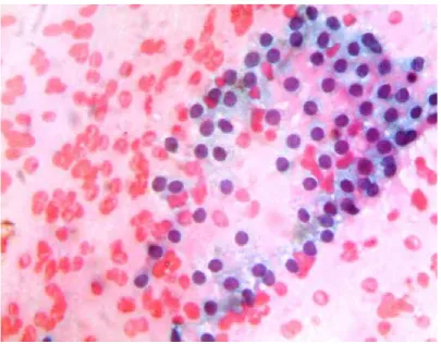
(HASHIMOTO’S THYROIDITIS/LYMPHOCYTIC THYROIDITIS)
Smears are hypercellular. A bloody background with lymphoid cells,
degenerative changes in follicular cells and infiltration of follicular cells by
lymphoid cells are characteristic features of Hashimoto’s thyroiditis (2) . Variable features include oxyphilic cells (Hurthle cells), plasma cells,
epithelioid cell granulomas and multinucleated giant cells
The smear background shows lymphocytes with a variable number of
plasma cells. There is prominent oxyphilic (Hurthle cell/Askanazy cell)
change with single and syncytial aggregates of cells showing abundant,
dense, finely granular, gray-blue cytoplasm (MGG), and well-defined cell
borders (2) . Nuclei are 2–4 times the size of normal follicular cell nuclei and may show atypia and prominent nucleoli. Normal-appearing follicular
cells may be present, showing features of hyperactivity. A characteristic
feature is that of lymphocytes (and occasionally plasma cells) seeming to
adhere to or infiltrate follicular cells, supporting the theory of direct
epithelial damage by lymphocytes. Multinucleated giant cells and
epithelioid cells can be seen in up to 40% of cases. Neutrophils and
eosinophils may be seen adhering to or infiltrating follicular cells in early
An interpretation of lymphocytic thyroiditis does not require a minimum
number of follicular/Hürthle cells for adequacy.(60) The lymphoid
population is polymorphic, including small mature lymphocytes, larger
reactive lymphoid cells, and occasional plasma cells.
The lymphoid cells may be present in the background or infiltrate
epithelial cell groups. Intact lymphoid follicles and lymphohistiocytic
aggregates may be seen. Hürthle cells (oncocytes), when present, are
arranged in sheets or as isolated cells. They have abundant granular
cytoplasm, large nuclei, and prominent nucleoli
Anisonucleosis of Hürthle cells may be prominent. Sometimes mild
nuclear atypia is encountered, including scattered nuclear clearing and
grooves
DE QUERVAIN’S THYROIDITIS (SUBACUTE THYROIDITIS;
GRANULOMATOUS THYROIDITIS)
In smears of granulomatous thyroiditis large multinucleate giant
cells with numerous nuclei, granulomatous aggregates of epithelioid cells,
degenerating follicular cells, neutrophils, macrophages and lymphocytes in
a dirty smear background consisting debris and colloid.
The cellularity is variable and depends on the stage of disease.
Clusters of epithelioid histiocytes, i.e., granulomas, are present, along with
many multinucleated giant cells. The early stage demonstrates many
the smears are hypocellular. They show giant cells surrounding and
engulfing colloid , epithelioid cells, lymphocytes, macrophages, and scant
degenerated follicular cells.(61)
RIEDEL´S THYROIDITIS/DISEASE
This is the rarest form of thyroiditis and results in progressive
fibrosis of the thyroid gland with extension into the soft tissues of the neck.
Criteria : The thyroid gland feels very firm on palpation. The preparations
are often acellular. Collagen strands and bland spindle cells may be
present. There are rare chronic inflammatory cells. Colloid and follicular
cells are usually absent (51) .
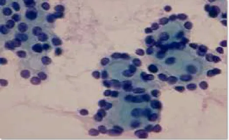
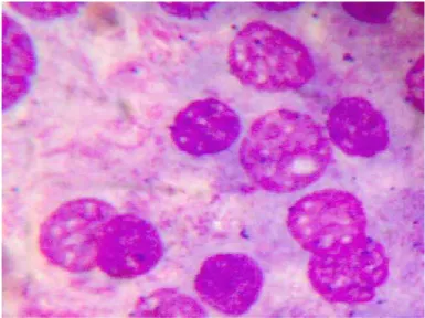
FOLLICULAR NEOPLASMS (FN):
Cytologically, follicular lesions include follicular adenoma
(FA),follicular carcinoma (FC) and cellular NG . Smears in FN are cellular
in a bloody background that is usually devoid of colloid. Many
uniform-sized follicular cell clusters, microfollicles and rosette formations are
present. Syncytial aggregates, nuclear crowding and overlapping are also
often seen. The repetitive smear pattern with uniform cell population is in
contrast to the variable pattern of different cell types seen in colloid and
hyperplastic nodules. Microacinar clusters with a central lumen represent