I
HYBRID SIMULTANEOUS INTEGRATED
BOOST TECHNIQUE IN LOCALLY ADVANCED
TONGUE CANCER-A PROSPECTIVE STDUY
A dissertation submitted to
the Tamil nadu Dr. M.G.R. Medical University, Chennai,
in partial fulfilment of the requirements for the award of the degree of
DOCTOR OF MEDICINE (M.D.) IN RADIOTHERAPY
II
CERTIFICATE
This is to certify that this dissertation titled,
“HYBRID
SIMULTANEOUS
INTEGRATED
BOOST
TECHNIQUE IN LOCALLY ADVANCED TONGUE
CANCER-A PROSPECTIVE STDUY”
is a bonafide record of
the work done by Dr. Vijayaveeran.P, in the Division of Radiation
Oncology, Cancer Institute (W. I. A.), Chennai, during the period of
his postgraduate study for the degree of M.D. (Branch X –
Radiotherapy) from 2014-2017 under my direct guidance and
supervision.
Date : Sep 30 , 2016
Place: Chennai
Dr. G.Selvaluxmy
Professor and H.O.D
Division of Radiation Oncology
Cancer Institute (W.I.A.)
III
ACKNOWLEDGEMENT
I am ever-grateful to Late. Dr. S.Krishnamurthi, Advisor, Dr. V.Shanta, Chairman, Cancer Institute (WIA), Adyar, for providing me all the facilities for this study.
I express my gratitude to Dr. G.Selvaluxmy, Professor and H.O.D, Division of Radiation Oncology, for her encouragement, constant support and guidance throughout my postgraduate career and during this study.
I also thank Dr. A.Vasanthan, Professor and chairman, Division of Radiation Oncology, for his support and advice throughout my post-graduate days and in this study.
I am also thankful to Dr. Alexander John, Dr. M.N.Arun Kumar, Dr. C. Vasanth Christopher, Dr. Aswin N, Dr. Ramaniah for their support.
Special thanks to my colleague and friend who was always there when I needed support or any help with this dissertation.
I thank all the other faculty members, my colleagues, physicists, radiotherapy technologists and tumour registry staff without whom this study would not have materialized.
I express my gratitude to all the patients who form the most important part of this study.
I thank my parents and all my family members all of whom have been the greatest sources of motivation and support for me.
V
Index pages
I) Introduction 1
1) Institute data 2
2) Risk factors for carcinoma of tongue 4
3) Histopathology 8
4) Pre-malignant lesion 10
5) Anatomy of oral tongue 11
6) Investigation 22
7) Treatment 23
II) Aim 45
III) Material and Methods 48
IV) Results 57
V) Discussion 68
1
I.INTRODUCTION:
Head and neck cancer is the sixth most common cancer worldwide.(1)
50% of the overall global head and neck cancer burden is from Asia, India in
specific. (2)Each year 2 lakhs new patients are diagnosed and 1 lakh patient die
with oral cavity cancers.(3) In India carcinoma buccal mucosa and carcinoma
tongue are more common among the oral cavity cancers. Carcinoma tongue is
second most common cancer of oral cavity. It is more common in men
compared to women. The ratio is 3:1 in sex distribution.(4) The incidence of
carcinoma tongue according to Madras Metropolitan Tumour Registry (MMTR)
is 10.3 /100000 population for the year 2012-13. (5)
Incidence rates for the year 2012-13.
Source Sex CIR ASR No of cases Total cases
World GLOBOCAN
(Oral cavity)
Male 5.6 5.5 198975 7410376
Female 2.9 2.5 101398 6657518
India IARC
(Oral cavity)
Male 8.3 10.1 53842 477482
Female 3.8 4.3 23161 537452
Chennai MMTR
(Tongue)
Male 8.1 7.4 380 5448
2
1).INSTITUTE DATA
609 patients presenting to the Institute from 2006 to 2011 are diagnosed
with carcinoma tongue. Most of the patients report to the institute in locally
advanced stages. Stage wise 5 years Disease free survival (DFS) and 5 year
Overall survival (OS) is given below.
Stage Total cases DFS 5 years OS
I 87 52% 74%
II 122 36% 50%
III 160 40% 46%
IV 237 19% 23%
5years Relative survival of Oral cancer (1988-2001) from SEER database (6).
Tongue (%) Oral Tongue (%)
Stage I 70.7 75.9
Stage II 58.6 57.5
Stage III 47.3 38.8
3
Distribution of treatment modalities used in our institute from 2006 -11
Treatment modality No of cases Percentage
Concurrent chemo-radiation 327 54%
Radiation alone 170 28%
Surgery followed adjuvant Radiation 58 9.5%
Surgery followed by concurrent
chemo-radiation
36 6%
Surgery alone 16 2.5%
Surgery followed by chemotherapy 1 <0.2%
Palliative chemotherapy 1 <0.2%
Total 609
5 Years DFS and 5 years OS with concurrent chemo-radiation and Radiation
alone are given below.
Treatment DFS 5 years OS
RT+CT 30% 38%
4
2).RISK FACTORS FOR TONGUE CANCER:
The common causes for tongue cancer are
Five S’s
Smoking
Spirit (alcohol)
Sharp (Septic) tooth
Spicy food
Syphilis
SMOKING AND ALCOHOL:
Use of tobacco and alcohol is associated with 80-90% of all case of
carcinoma tongue. A Case control study with two arms, smoking alone and
smoking along with alcohol showed that smoking increases the risk of
developing cancer by about 25 times than in non-smokers.(7) Tobacco in any
form increases the risk of development tongue cancer by three folds. Both
smoking and alcohol have synergistic effect and will increase incidence of
cancers. Stopping smoking for more than 9 years will decrease the incidence of
oral cancer by about 50 %(8). Second primary is more common in patients who
5
Carcinogen in Tobacco:
Aromatic hydrocarbons benzopyrene,
Tobacco specific nitrosamines (TSNs),
N-nitrosononicotine (NNN),
N-nitrosopyrrolidine (NPYR),
N-nitrosodimethylamine (NDMA),
4-methylnitrosamino-1-(3-pyridyl)-1-butanone (NNK).
Mechanisms of tobacco carcinogenesis:
These products will act on ketatinocyte stem cells. They produce DNA
adducts (O-6-methylguanine) which interfere and disrupt the accuracy of DNA
replication producing mutations, Leading to chromosomal instability. (9)
Mechanisms of alcohol synergism:
1) Alcohol increases the permeability of carcinogens in oral mucosa
2) It will produce poor oral hygiene.
6
NON SMOKING CAUSE OF ORAL CANCERS:
DIETARY
OCCUPATIONAL
ENVIRONMENTAL
LICHEN PLANUS:
It will progress to malignant transformation in around about 0.3%
patients. It is also associated with tobacco usage.
GRAFT VERUS HOST DISEASE
VIRUS:
1) Human papilloma virus (HPV):
Up to 20% of oral cavity cancer shows HPV 16 DNA.
2) HIV infection
3) Herpes simplex virus(HSV)
4) Epstein-barr virus(EBV)
5) Hepatitis C virus.(HCV)
Fungal infection:
7
CANCER SYNDROMES associated with carcinoma tongue
Xeroderma pigmentosum
Ataxia telangiectasia
Li fraumeni syndrome
Fanconi’s anemia
NATURAL HISTORY:
Patients most commonly present with chronic non healing ulcer with or
without pain, young women may present with white patch-leukoplakia. Also
difficulty in speech and swallowing. Tongue cancers grow rapidly either
infiltrative or exophytic. Infiltrative tumours may be very large at presentation.
Thick tumours have worse prognosis. Thickness >4 mm increased probability of
occult metastasis to neck nodes.(10)
Cytological Atypia
Hyperplasia Dysplasia
Carcinoma in situ
Premalignant lession
8
3).HISTOPATHOLOGY:
Squamous cell carcinoma:
It is most common. Seen in 90% cases. It has several variants, mostly
Basaloid and verrucous carcinoma.(11)
Basaloid squamous cell carcinoma:
It has worse prognosis than normal squamous cell carcinoma. It is
associated with advance disease, distant metastasis and poor overall survival.
Verrucous squamous cell carcinoma:
It is less common variant. It is a low grade malignancy with good local control
and good overall survival. It presents as a thickened white patchy mucosa in
9
Treatment is excision of the lesion but it is prone to recur after excision.
Non squamous cell carcinoma:
Adenocarcinoma,
Melanoma,
Lymphoma,
Leiomyosarcoma,
Kaposi’s sarcoma.-mostly in oral cavity (gingiva, palate and tongue). After
HAART treatment incidence decreased into 50%
Adenosquamous carcinoma,
Papillary squamous cell carcinoma,
10
Prognostic factors in histology:
1) Tumour margins
2) Perineural invasion
3) Histopathology of tumour
4) Grade of tumour
5) Lymphatic/vascular invasion
6) Angiogenesis.
4).PREMALIGNANT LESSIONS:
Leukoplakia:
It has two types homogenous and nonhomogenous.
Homogenous: uniform white lesion and low (<1%) malignant potential.
Non homogenous: nodular, sparkled lesion with central ulceration. It has more
malignant potential. It has 18 % progress to cancer.(12)
Leukoplakia will regress spontaneously no treatment needed. Need to do
biopsy for malignant transformation. If it shows dysplatic changes we have to
do excision of the lesion. Leukoplakia will occur 6 folds more in smokers
compared to non-smokers
11
It is due to virus infection of oral cavity. Malignant transformation is less
common.
Erythroplakia:
It is chronic red patch on the mucosa. If erythroplakia is present we have
find out other erythematous lesion in the patient. It has very high potential for
malignant transformation compare to other pre malignant lesions. Erythroplakia
progress to invasive carcinoma about 51%, carcinoma in situ about 40% and
mild to moderate dysplasia about 9%. We have to do surgical excision of all
erythroplakia lesions.
5).ANATOMY OF TONGUE:
Parts of oral cavity:
It has lips, oral tongue, floor of mouth, alveolar ridge, retromolar trigone,
hard palate and buccal mucosa.
Tongue:
It is a muscular structure in oral cavity, triangle in shape, root of tongue
attached to mandible and hyoid bone. It is divided in to two parts by a V shaped
terminal sulcus marked by circumvallate papillae, anterior two third (oral
tongue) - part of oral cavity, posterior one third (base of tongue) –part of
oropharynx.
12
Anterior two third of tongue:
It has five parts tip, dorsal surface, ventral surface, right and left lateral borders.
Dorsal surface of tongue:
Papillae: Superior surface of tongue covered by mucosal folds having taste buds
called as papillae for taste.
1) Filiform papillae: cone shaped distributed throughout dorsum of tongue
2) Fungiform papillae: round shaped predominantly in the border.
3) Circumvallate papillae: cylindrical shaped in 8-12 in number arranged in
signal V shaped line anterior to terminal sulcus.
4) Foliate papillae: Linear shaped border of tongue near terminal sulcus.
Ventral surface of tongue:
Frenulum: tongue is divided in to right and left half by sagittal septum.
Frenulum is median fold of mucosa overlying lower margin of sagittal septum.
On each side of frenulum lingual veins present.
Pharyngeal surface: base of tongue - sub mucosal lymphoid tissue which forms
the lingual tonsil.
Muscles:
Intrinsic muscle originate and inserted within the substance of tongue.
13
structures outside tongue and inserted in to the tongue. They are responsible for
[image:18.595.71.419.145.392.2]movement of tongue and tumour infiltrated causes ankyloglossia.
14
Figure shows Intrinsic muscles of the tongue
Muscles of tongue:
Muscles Origin insertion innervation Functions
Intrinsic muscles
Superior longitudinal Base of tongue
and median septum Hypoglossal nerve (XII) Shorten tongue, curl tip and sides
Inferior longitudinal Root of tongue Apex of
tongue Hypoglossal nerve (XII) Shorten tongue, uncurls tip and turns down
Transverse Median septum Lateral
border
Hypoglossal
nerve (XII)
Narrow and
elongates
Vertical Dorsum of
15
Muscles
Extrinsic muscle
Origin insertion innervation Functions
Genioglossus Mandible(Mental
tubercle) Entire tongue and hyoid bone Hypoglossal nerve (XII) Protrusion
Hyoglossus Hyoid bone(
greater horn) Lateral surface Hypoglossal nerve (XII) Depression
Styloglossus Styloid process Lateral
16
Arterial supply:
Lingual artery: it is a major artery of tongue, originate from external carotid
artery. Tonsilar and ascending pharyngeal arteries also supplies oral tongue.
Venous drainage:
Deep lingual vein: it is visible in ventral surface of tongue. It ends in to internal
jugular vein. Dorsal lingual vein also drains from tongue and ends in to internal
jugular vein.
Nerve supply:
Motor supply: All intrinsic and extrinsic muscles are innervated by hypoglossal
nerve expect patatoglossus is innervated by vagus nerve.
Lymphatic drainage:
It has five primary level.
Level I: IA- sub mental and IB- submandibular node.
Level II: Upper jugular group of lymph nodes.
Level III: Mid jugular group of lymph nodes.
Level IV: Inferior jugular group of lymph nodes (Supraclavicular lymph node).
Level V: Posterior triangle group of lymph nodes.
Lymphatic drainage of tongue:
Anterior tongue drains in to sub mental (level IA).
17
jugular group of lymph nodes (level II).
Posterior tongue drains in to upper jugular group of lymph nodes (level II).
Tongue is midline structure it cause bilateral lymph node metastasis.
Skip metastasis: some patient will get level IV lymph node metastasis without
involving level I, II and III.
Salivary glands: parotid gland, sub mandibular gland and sub lingual gland.
Mandible: It has ramus and body ramus has medial surface, lateral surface,
condylar process and coronoid process. Lateral surface of ramus provide
attachment to massticator muscle.
Posterior and inferior border intersect to form angle of mandible.
Coronoid process flat triangle process at the junction of superior and anterior
border provides attachment for temporalis muscle. Condylar process at the
junction of posterior and superior border, has two parts head and neck. Head
form tempromandibular joint, neck provides attachment for lateral pterygoid
muscle.
Medial surface from lateral wall of infrotemproal fossa provides
attachment to medial pterygoid muscle, it has mandibular foram, inferior
18
Temporo-mandibluar joint:
It mandible articulate with temporal bone. When tumour infiltrates TM
joint patients cannot open mouth. It will cause trismus. Its difficult assess the
oral cavity with trismus.
STAGING:
T
stage
Description
Tx Primary tumour cannot be assessed
T0 No evidence of primary tumour
Tis Carcinoma in situ
T1 Tumour 2 cm or less in greatest dimension
T2 Tumour more than 2 cm but less than 4 cm in greatest dimension
T3 Tumour more than 4 cm in greatest dimension
T4a Tumour invades adjacent structures. Eg. cortical bone,
deep(extrinsic)muscles of tongue ( genioglossus, hyoglossus,
palatoglossus and styloglossus), maxillary sinus and skin of face.
T4b Tumour invades masticator space, pterygoid plate, or skull base or
19
N
stage
Description
Nx Regional lymph node cannot be assessed
N0 No regional lymph node metastasis
N1 Metastasis to a single ipsilateral lymph node, 3 cm or less in
greatest dimension
N2a Metastasis to a single ipsilateral lymph node, > 3 cm but < 6
cm in greatest dimension
N2b Metastasis to multiple ipsilateral lymph node, not more than
6 cm in greatest dimension
N2c Metastasis to bilateral or contralateral lymph node, not more
than 6 cm in greatest dimension
N3 Metastasis to a lymph node more than 6 cm in greatest
dimension
M stage Description
Mx Distant metastasis cannot be assessed
M0 No distant metastasis
20
Figure shows T4a tongue lesion with ankyloglassia
Stage group TNM stage
0 Tis N0 M0
I T1 N0 M0
II T2 N0 M0
III T3 N0 M0, T1 N1 M0, T2 N1 M0 or T3 N1 M0
Iva T4a N0 M0, T4a N1 M0, T4a N2 M0, T1 N2 M0, T2 N2
M0, T3 N2 M0 or T4a N2 M0
IVb Any T N3 M0, T4b any N M0
21
ORAL CAVITY EXAMINATION:
Simple visual inspection:
We have to see tongue surface for description of the lesion (ulcerative,
proliferative, exophytic, endophytic, fungating tumour) and mobility for muscle
infiltration or hypoglossal nerve damage.
Palpation:
We can demonstrate the three dimension view of tumour, consistency and
tongue fixation. Tumour in tongue starts as small painless ulcer than progress
to invade the muscle of tongue. Squamous cell carcinoma of tongue commonly
present in lateral border of tongue. Bimanual examination of oral cavity will
assess the depth of invasion of tongue tumour.
Examination of head and neck cancer: it includes inspection and
palpation of skin over face, scalp, oral cavity, nasal cavity, nasopharynx, ear,
oropharynx, hypo pharynx and larynx. Than examination of cervical lymph
nodes and cranial nerves. Tumour around 30% to 40% will has cervical lymph
node metastasis. Patients die due to uncontrolled local disease with
haemorrhage and aspiration. Distant metastasis is rarely seen before death. 19%
patients may developed second malignancy in upper aero-digestive track which
22
6).INVESTIGATION:
Basic
1) Routine blood investigation,
2) Viral markers,
3) ECG
Diagnostic
1) Biopsy from tumour
Staging
1) Endoscopy (Direct laryngoscopy and pharyngoscopy)
2) X ray chest PA view
3) CT scan head and neck.
4) MR image head and neck.
23
7).TREATMENT:
Curative intent of treatment
Surgery is main modality of treatment for early stage carcinoma tongue.
Early stage disease treated with surgically with adequate margins followed by
adjuvant treatment depends upon histopathalogically.
Late stage disease is approached with multimodality treatments
(Surgery, radiation and chemotherapy).
Ultimate aim of treatment:
1) Cure the cancer,
2) Restore the function
3) Minimize the complication of treatment
4) Prevent metastasis.
Selection of single or combined treatment modality depends on stage,
size and site of the tumour. Multidisciplinary team assessment should be done
before starting treatment of tongue cancer. It includes surgical oncologist,
medical oncologist, radiation oncologist, nursing staff, dentist, speech therapist,
dietician and counsellor for family & social support.
NCCN (National Comprehensive Cancer Network) recommend early
stage lesion will treated with single modality of treatment (surgery or radiation).
24
Factors affecting treatment modality:
1) Location of the tumour,
2) Size of the tumour,
3) Cervical lymphadenopathy,
4) Histology
5) Previous treatment
6) Age and comorbidity
7) Socio-economic status.
Early stage disease (Stage I, Stage II):
Surgery alone: T1- Local excision with margin, T2- Partial glossectomy
Brachytherapy alone: Hairpin technique, Plastic tube loop technique
Surgery preferred over brachytherapy in
1) Bone involvement
2) Melanoma
3) Adenocarcinoma
4) Tip of the tongue lesion
5) Tumour surrounding by pre malignant lesion
25
ADVANCED STAGE DISEASE:
Surgery:
Subtotal or total glossectomy
Marginal/segmental mandibulectomy
With free tissue transfer or regional pedicled flaps
Approach:
Median or para-median mandibulotomy, Trans-cervical
Management of Neck:
Primary lesion <2 mm-neck is managed expectantly
Larger lesion (including N0)-Selective neck dissection of levels I to IV
Lesions close to, or involving the mid line- Bilateral neck dissection
Postoperative RT indications: it is decrease local recurrence
1. Close or positive margins,
2. Extra-capsular invasion,
3. Bone invasion,
4. Pathological node positive disease,
26
RADIATION:
External Beam Radiotherapy upto 40 Gy followed interstitial
brachytherapy. Decroix and Ghissein study showed combine external beam
radiation and brachytherapy to be beneficial (14)
Concurrent chemo-radiotherapy:
Concurrent chemo-radiation will increase the overall survival in locally
advanced head and neck cancer is about 5% and absolute benefit in survival 8%
in five years.(15)
Salvage surgery:
It is the second attempt at cure after definite treatment or final attempt for
cure in recurrent disease following surgery, radiation and chemo-radiation. It is
only cure option available and mainly final curative attempt.
Palliative intent of treatment:
Patients with more advanced disease not curable disease will be treated
with palliative to reduce the symptoms (like pain, bleeding and reduce the
burden of the disease). It may be surgery, radiation and chemotherapy.
Radiation used to relive the pain, bleeding as in palliative treatment.
Chemotherapy used in metastatic disease. Surgery is mainly used to debulk the
27
SURGERY:
Selecting the type, approach and extend of surgery depends on size, site
and depth of the infiltration and proximity of tumour from mandible or maxilla,
supine Position: 30 degree angle and anaesthesia.
Anaesthesia:
Small lesion are excised with local anaesthesia.
Larger lesion require General anaesthesia with Naso-tracheal tube intubation.
Surgical approach:
1. Trans-oral,
2. Lower cheek flap
3. Mandubulotomy
4. Trans cervical
With
1) Healing by secondary intension,
2) Primary closure,
3) Split thickness skin grafts
28
RADIATION THERAPY:
Radiation treatment can be given as teletherapy, brachytherapy and
internal therapy.
Mechanism of action of Radiation:
Radiation will have two types of action, Direct action and indirect action.
Direct action is fast moving electron interact direct with DNA molecule and
cause DNA damage. Indirect action is fast moving electron will interact with
other atoms or molecules in the cell. This will produce free radicals and free
radical will cause DNA damage. Radiation will produce cell DNA damage by
double-strand or single-strand breaks. Double strand breaks will leads to cell
death. Single strand breaks will cause sub lethal DNA damage, it may repair by
themselves. Before repair that cell will undergone one more single-strand break
than this may leads to cell death. But the repair mechanism is superior in normal
cell compared to cancer cells. Cell death means cells unable to divide
furthermore and cause growth and spread of disease. Chromosomal aberration
will leads to cell death while they attempt to divide after lethal dose of radiation
(mitotic cell death).
Aim of the radiation is to deliver precise measured dose to the target
29
BRACHYTHERAPY:
It is form of radiotherapy where the source of the radiation is kept very
close to the tumour or actually implanted in to the tumour. Iridium-192 is
commonly using radioactive source in most of the canters. It has low photon
energy makes protection easier than the Radium or Cesium-137. It gives high
dose to tumour by a short period of time and we can spear adjacent normal
tissue. Brachytherapy gives the benefit of function preservation, which is
achieved through tissue preservation. Local control rates are 85% which are on
par with that achieved by surgery. Local control rates decrease with increasing
tumour size. (16)
Brachytherapy techniques in tongue:
1) Hairpin technique:
Implant is in the shape of hairpin, the two limbs are 12 mm apart and 6
cm in length. This forms a double plan. Single plan implantation is not
preferred as it does not cover deeper parts. Stainless steel slotted hairpin
guides are inserted under general anaesthesia. First anterior most and
posterior most hairpin guides are implanted and later the required number
of guides are incorporated between them, with a 12 mm gap between
them. The limbs are cut depending upon the tumour depth. Top of the
30
cover bottom adequately limb of the hairpin should be longer than the
tumour.
2) Plastic tube loop technique:
It is used for larger volume tumour difficult to cover with hairpin
implants. Plastic tubes is formed in to a loop, this also a two plan
implant. Stainless steel guide needles are pushed into the tumour from the
skin below in jaw line.
Brachytherapy Vs External Beam Radiation Therapy (EBRT)
Local control rates are suboptimal in EBRT in contrast to brachytherapy
LDR Vs HDR brachytherapy:
HDR brachytherapy schedule- 59 Gy in 6 Gy twice daily compared to
LDR brachytherapy schedule- 61 Gy over 5 to 6 days showed inferior local
[image:35.595.105.420.529.714.2]control rates with HDR (T1: 85% Vs 64%, T2: 71% Vs 38%) (17)
31
EXTERNAL BEAM RADIATION THERAPY
It is a modality of radiation delivery in which the source of radiation is at
a distance from the human body. The radiation source may be a radioactive
nuclide, Cobalt 60 emitting Gamma rays, is used presently, or it may be X-rays
Produced by a Linear Accelerator, Electrons, protons, heavy charged particles
etc. Accuracy of treatment delivery is of utmost importance.
Positioning the patient:
Patient in supine position, straight and neck extended. We have to place
custom-made mouth bite to depress the tongue. Because we have to avoid
radiation dose to hard palate and parotid glands. Than patient should be
immobilized with thermoplastic mould or perspex. It is helped to fix the
well-defined position to treat the patient.
If a patient is having short neck, both shoulders should be depressed by
tensioning device to loop the feet. Because we have to treat whole neck region.
Effective immobilization will reduce setup error.
Radiation techniques:
1) 2D conventional technique
2) 3D conformal technique
3) IMRT technique
32
2D CONVENTIONAL TECHNIQUE:
Target volume includes primary tumour with 2 cm margin and ipsi lateral
submandibular and upper deep cervical nodes.
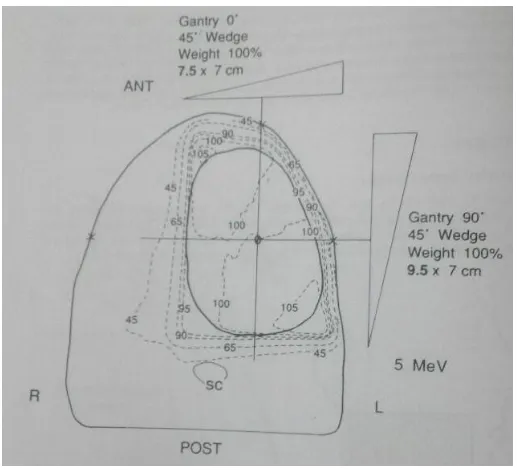
Early lesion in lateral tongue is treated using anterior and lateral wedged
fields, this gives homogenous dose distribution with sparing of contralateral oral
cavity and spinal cord. For large tumours near or crossing midline two opposing
lateral fields are used. Anterior and lateral wedged fields produce less mucositis
and xerostomia compared to two opposing lateral fields used in extensive
[image:37.595.71.330.423.658.2]tumour.
33
RT field borders in two opposing lateral fields:
Superior border: 2 cm above the primary lesion,
Inferior border: below hyoid bone,
Anterior border: 2 cm front of primary tumour (usually in front of mandible),
[image:38.595.72.375.270.480.2]Posterior border: back to the vertebral corpuses.
Figure shows 2D planning with MLC shielding of lateral field
Supraclavicular field border:
Superior border: inferior border of lateral fields,
Inferior border: bottom supraclavicular joint,
Lateral border: including medial two third of clavicle.
34
Figure shows 2D planning with MLC shielding of anterior field
After completing 45Gy of external beam radiation in two opposing lateral fields
the posterior border is shifted anterior to spinal cord to spare the spinal cord.
35
3D CONFORMAL TECHNIQUE:
In conformal technique target volume is defined with three dimensions
view for contouring purpose. It has multiple beams because to reduce the
normal tissue toxicity and confined dose to the target alone. But concavity not
archived in conformal technique. Setup errors also present in conformal
technique. Contouring is drawing in CT scan in slice by slice and exact
conformity we can achieve. Beam shaping done with multi-leaf collimators
(MLC). It usually has 4 – 6 beam angles. It is 3Dinemtional images will give
precise treatment planning. In this no need to change the blocks during the
treatment. Intensity of the beam is uniform.
Planning CT scan:
CT scan should be done from base of skull to arch of aorta with supine
position of the patient with immobilization thermoplastic mould. In the CT scan
will send to planning system, there is got registered in planning system, than we
have do contouring the structure of the patients. This CT scan will be in axial
Cuts depending upon the technique we can take the slices from 0.25 cm, 0.5 cm
36
ICRU Volumes for contouring:
Gross Tumour Volume (GTV):
It is grossly demonstrable tumour, macroscopic lesion. Tumour id visible,
palpable and demonstrable in imaging.
Clinical Target Volume (CTV):
It is demonstrable tumour and any other surrounding tissues with
presumed tumour. It delineate
Planning Target Volume (PTV):
In this volume that includes CTV, setup margin and setup uncertainties.
Organ at risk (OAR):
Spinal cord, brain stem, brain, contralateral parotid, larynx and inferior
constructor. Tolerance doses are given in Table 4
OAR Tolerance dose
Spinal cord Max - 45Gy
Brain stem Max – 54Gy
Brain 60Gy point dose
Contralateral parotid Mean dose 26Gy
Larynx Mean dose 40Gy
37
IMRT (INTENSITY-MODULATED RADIATION THERAPY):
Non uniform fluence of dose distributed to the patient with any given
position of treatment beam to optimize the composite dose distribution. It also
called as Inverse planning technique. It is to treat a patient with multiple number
of beam direction non uniform fluence, it optimize high dose to target volume
and low dose to surrounding normal tissue. In which we can achieve good dose
conformity and steep dose gradient outside the target. It has two MLC window
technique 1) dynamic MLC delivery, 2) step and shoot delivery.
Considerable factors in IMRT is increased cost, increased worked,
increased treatment time/delivery, high integral dose, increased risk of second
primary and low dose hypersensitivity. In head and neck cancers IMRT will
spear major salivary glands and reduce xerostomia. Geographic miss due to
improper positioning of the patient, organ motion and inadequate target
delineation.
Limitation of IMRT:
Most dose distribution not physically achieved, distortion of internal
38
ALTERED FRACTIONATION SCHEDULES:
During conventional fractionation using conformal and IMRT techniques
are reduce normal tissue toxicities. But improving loco-regional control will
achieve by increasing dose to tumour should be needed. In prolonged treatment
time the tumour cell allow for repopulation. For that few newer fractionation
schedule were tried. It is alerted fractionation.
Hyper-fractionation:
It is increase daily fraction with reduce the daily dose, which will leads to
decrease the overall treatment time. It will reduce late toxicities, increase
tumour control and slightly increase acute toxicities.
Accelerated fractionation:
It is increase daily fractions without alter the daily dose and decrease the
overall treatment time. It will increase the local control, slight increase acute
and late toxicities.
Hypo-fractionation:
It is decrease total fractionation along with increase the daily dose. It
decrease overall treatment time and increase local control. Acute toxicities are
39
Accelerated repopulation:
The cells are treated with cytotoxic agents (e.g Radiation) can trigger the
surviving cells to divide faster than before. Analysis suggested that accelerated
repopulation starts in human cancer cells about 4 weeks after starting radiation.
So dose increment about 60 cGy/fraction will compensate the accelerated
repopulation.
Pre-treatment doubling time for head and neck cancers is 30 to 60 days,
but after radiation starts the doubling time is reduced to 3 - 5 days. In head and
neck cancers each day delay during treatment will reduce the local control about
from 0.4 to 2.5%. It is better to delay initiation of treatment than introduce delay
during treatment. (18)
CONCOMITANT BOOST:
It is during the conventional fractionation boost dose is given in last two
weeks radiation. It will counteract the accelerated repopulation. In boost
treatment duration daily dose will decrease and gap between two fractions is 6
hour. It is increase loco-regional control and disease free survival. It will leads
to more severe acute toxicities and no effect on late toxicities.
40 Conventional fractionation Hyper fractionation Accelerated fractionation Concomitant Boost
Total no of fractionation
30
fractionation Increased
Same as
conventional Increased
Dose/ # 200cGy/ # Decreased
Same as
conventional Decreased
Overall time 6 weeks same/increased
Same as
conventional Decreased
No of #/day 1 > 1 fractionation Increased
> 1 # in last 2weeks
Total dose 60Gy Increased Decreased Decreased
SIMULTANEOUS INTEGRATED BOOST-INTENSITY MODULATED
RADIOTHERAPY (SIB-IMRT)
It is intuitive radiation technique. It will deliver extra boost
simultaneously along with normal fractionation. It will increase to dose to target
sub-volumes and reduce dose to critical structures. IMRT is using
computer-optimized intensity distribution are controlled by dynamic MLCs will delivered
multiple beam along with simultaneously irradiating nearby tissue in different
dose in single treatment session. This technique will allows for improved dose
conformity, excellent dose homogeneity within the target volumes, and
conformal avoidance of adjacent critical normal tissues throughout the
treatment course. Using this approach, the fractional dose delivered to gross
tumour will be increased, at the same time the radiation doses and dose
41
uninvolved lymph nodes are preserved. This approach is called as Simultaneous
Integrated Boost (SIB). (19, 20, 21, 22, 23, 24, 25, 26)
Acute toxicities:
Mucositis:
It is most common side effects of radiation and chemotherapy, But short
term side effect. It depends on type ionizing radiation, dose of RT, rate of RT
delivered. It cause pain which needs stop therapy to mucositis to heal. It is due
to mitotic death of cells.
RTOG grading of Mucositis:
1) Grade I- Injection or mild pain.
2) Grade II- Patchy mucositis and moderate pain.
3) Grade III- Confluent fibrious mucositis and severe pain.
4) Grade IV- Ulceration, haemorrhage and necrosis.
RTOG grading of Dermatitis:
1) Grade I- Faint or dull erythema, epilation,
2) Grade II- Bright erythema, patchy moist desquamation,
3) Grade III- Confluent moist desquamation other than skinfolds,
42
RTOG grading of Dysphagia:
1) Grade I- Mild dysphagia may require soft diet,
2) Grade II- Moderate dysphagia may require liquid diet,
3) Grade III- Severe dysphagia may require IV fluids, NG tube insertion.
4) Grade IV- complete obstruction, ulceration, perforation and fistula.
Late toxicities:
1) Xerostomia
2) Dysphagia
3) Taste alterations
43
CHEMOTHERAPY:
Cisplatin (cis-diamminedichloroplatinum) (CDDP)
It is used in wide variety of cancers, it is a prototype drug and alkylating
agent. It has high affinity to electrons so it increase sensitive of radiation.
Mechanism of action:
1) Inhibition of DNA synthesis,
2) Inhibition of transcription elongation by DNA cross links,
3) Inhibition of repair of radiation induced DNA damage.
Dose regimen:
1) 100 mg/m2 or 70 mg/m2 IV every 3 weekly for 3 cycles,
2) 40 mg/m2 IV every weekly for 6 cycles.
Administration:
1) Monitoring with serum creatinine, electrolyte, magnesium and
calcium.
2) Antiemetics: we should started with ondensetron, dexamethasone.
3) Hydration: we have hydrate the patient with adequate Intravenous
44
Toxicity:
Dose limiting toxicity are acute renal failure, peripheral neuropathy
(cumulative dose >400mg/m2) and ototoxicity (Dose > 100 mg/m2). Common
side effects are severe nausea, vomiting will happened all patients, it will leads
to electrolyte imbalance. Other common side effects are anorexia, metallic taste
of foods, myelosuppression and sterility. Other occasional side effects are
SIADH, cortical blindness, congestive cardiac failure and tetany.
Indication:
1) Unresectable squamous cell head and neck cancers,
2) Nasopharyngeal cancer,
3) Laryngeal cancer,
45
II.AIM:
To evaluate the feasibility of hybrid simultaneous integrated boost (SIB)
technique and its efficacy in the treatment of locally advanced carcinoma
46
Inclusion Criteria:
1) ECOG performance score: 0-2,
2) Age 30-60 years,
3) Both sexes
4) Locally advanced carcinoma anterior 2/3rd tongue
(T4a, N1, 2 & 3, M0),
5) Patient is planned for definitive concurrent chemo radiation
(3 weekly Cisplatin 70 mg/m2)
6) Histopathology: Squamous cell carcinoma.
7) Non metastatic disease.
8) Adequate blood function test,
47
Exclusion Criteria:
1) Patients with poor performance status (3 & 4),
2) Patients initially treated with Radiation therapy, Chemotherapy and
surgery,
3) Patients who had other histology: adenocarcinoma, melanoma and
lymphoma.
4) Patients with palliative intent of treatment,
5) Patients who has not willing to participate in this study,
6) Patients with previously diagnosed as parotid glands disorder ( e.g
Sjogren’s syndrome)
7) Patients with predisposing to enhancing radio sensitivity (e.g
ataxiatelangiectasia or past/active connective tissue disorders).
48
III.MATERIAL AND METHODS:
All patients who came to Cancer Institute were examined
thoroughly. Most of the patients were referred by non-oncology
physicians. Diagnostic and staging investigations are done on outpatient
basis. After conforming non-metastatic squamous cell carcinoma of oral
tongue. Patient with stage IVa and IVb were enrolled in the study. Patients
were counselled clearly about treatment modality, technique, toxicities and
disease outcome. After getting informed consent these patients were
enrolled into our study.
All patients were advised about the importance of proper nutritional,
encouraged to avoid spicy food. All patients were Counselled for strict cessation
of smoking and/or tobacco chewing habits, Clearly explained that smoking or
tobacco chewing during treatment will reduce the treatment outcome and wil
also increase the risk of occurrence of second primary in aero-digestive track. .
They were explained about the importance of oral hygiene All patients
underwent dental examination and if needed dental extraction done. Patients
were all admitted in ward.
Thermoplastic mould was done to immobilise patients in supine position,
49
Planning CT scan done for all patients in flat couch from base of skull to
arch of aorta with thermoplastic mould.
Treatment:
All patients were planned for concurrent chemo-radiation. Chemotherapy
schedule was 70 mg/m2 Cisplatin in day 1, 22 and 43.
Radiation was designed to be delivered in two phases. All patients were
treated with 6 MV photons in 600c unit at our institute.
Treatment was delivered in two phases as planned.
Phase I:
Patient were treated with 6 MV X-ray beam therapy to deliver a Total
Dose of 36Gy using conformal technique with 200cGy daily dose per fraction
for 5 days/week over 3.5 week
In this phase PTV includes primary tumour and bilateral whole neck
nodes along with setup error margins. This area received TD 36Gy in phase I.
After completion 36Gy all patients were underwent re-planning. A repeat
Mould and repeat CT scan.
Phase II:
It has two PTV’s, the high risk PTV (residual primary and residual neck
50
cord) were contoured. Patients were treated with 6 MV X-ray beam therapy
using IMRT technique.
A Total Dose of 24Gy as 240cGy/day to the high risk PTV and a Total
Dose of 20Gy as 200cGy/day to the low risk PTV was delivered simultaneously
in 10 days over 2 weeks.
Chemotherapy delivered was three weekly Cisplatin.
These patients were compared with 10 matched control patients treated in
2015 with conformal technique TD 60Gy (200cGy/day), 5 days per week, over
6 - 7 weeks concurrent with chemotherapy.
During the treatment all patients are advised to take a proper nutritional
support. They are advised to do sodium bicarbonate mouth wash 6-8 times per
day, to avoid tooth brushing in order avoid mucosal injury. All patients were
advised to have non-spicy soft bland easily digestible high protein diet with
added salt. They were all advised to take more vegetables, more water intake to
reduce renal toxicity by Cisplatin. They were also told to avoid hot or chilled
food as it may aggravate mucositis. Multivitamins and antioxidants were given
daily for all patient. Patients were started on chymerol forte at around the end of
third week of treatment. Amifostine is not usually administered at our institute.
During the treatment time good psychological support were given to all patient
51
52
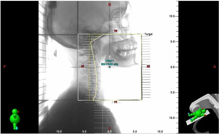
Figure shows dose distribution of 3DCRT plan (Sagittal plane)
Figure shows dose distribution of 3DCRT plan (Transverse plane)
53
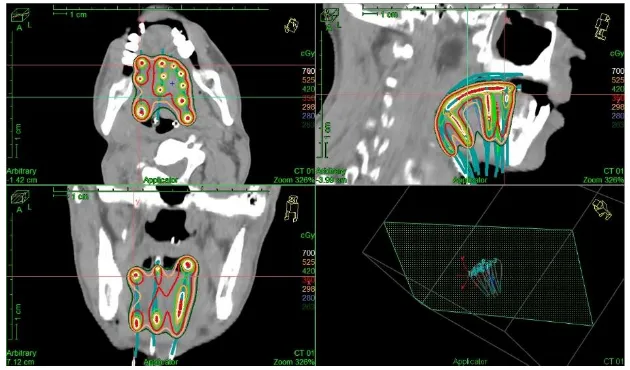
Figure shows 3D representation of PTV in SIB-IMRT plan
54
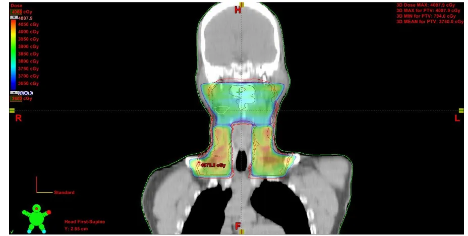
Figure shows dose distribution of SIB-IMRT plan (Coronal plane)
55
Toxicity Assessment and management:
All patients were assessed for tumour response for primary, nodal
region and acute toxicity during the treatment period on weekly basis.
During the radiation treatment patients developed acute toxicity like
mucositis, dermatitis and dysphagia. These patients advised for
conservative management. Patients with mucositis were advised for
frequent mouth wash, lignocaine viscous gel/Mucaine gel for pain relief.
Opioids and NSAIDS in the form of tramadol or ibuprofen along with
paracetmol were used for relief of mucositis/dermatitis induced pain when
ever needed. Patients who developed grade III acute toxicities advised for
Naso-gastric tube insertion for better nutritional support.
Before delivering chemotherapy all patient’s complete blood count,
renal function test, liver function test and serum electrolytes was checked.
Chemotherapy was delivered only if all blood investigation were normal,
patient vitals are stable and hydration was adequate. If investigation are
de-arranged patient was admitted to ward and conservative management was
given to that patients along with pending that schedule of chemotherapy or
56
During treatment weekly twice white blood cell count was checked
for all patients.
Response assessment:
Patient response to treatment is assessed 6 weeks after completion of
treatment according to World health organization criteria for solid tumour.
Local disease control is evaluated by clinical examination and imaging and
biopsy is done whenever necessary. For each follow up review complete
physical examination, response assessment and toxicities assessment were
done.
Response Evaluation Criteria in Solid Tumours (RECIST)
1. Complete response (CR): disappearance of all known disease by two
observations not less than four weeks apart.
2. Partial response (PR): 50 % or more decrease the tumour size by two
observations not less than four weeks apart.
3. No changes (NC): 50 % decrease in tumour size not assessed and
more than 25% decrease in tumour size assessed.
4. Progressive disease (PD): 25 % more increase the tumour size or
57
IV.RESULTS:
From September 2015 to august 2016, there were total number of
case diagnosed with carcinoma tongue, were stage IV, Twelve patients fit
in to the inclusion criteria. These twelve patients with locally advanced
carcinoma tongue stage IVA and IVB were enrolled in to study after taking
a written informed consent. But, one patient had tumour bleeding at 6Gy
itself so patient was treated with haemostatic radiation to tongue TD
500cGy in two fractionation. Hence, patient in not included in this study.
Another one patient had progressive disease in the form of nodal region
ulcerated and development of post auricular nodes was treated with
palliative radiation. Hence this patient also not included in this study. Total
ten patients were assessed in the study. All the 10 patient completed the
treatment.
Patient characteristics:
Median age of 43.7years (range of 39 to 65 years), all are male
patients diagnosed to have stage IVa carcinoma tongue. In this patients 4
patients were smoker and 5 were chewing, one patient having both the
58
cm to 8 x 7 cm. Seven patients had tumour in the ventral surface of the
tongue, two patients had extensive tumour occupying entire tongue, one
patients alone tumour was limited to lateral border. Clinically two patients
had no neck nodes palpable, two patients had ipsi lateral single node < 3
cm, one patient had ipsi lateral node 4cm, four patients had multiple ipsi
lateral nodes < 6 cm, and one patients had bilateral neck nodes < 6 cm.
Acute toxicity:
During course of treatment at 30Gy two patients developed Grade I
mucositis and four patients had grade II mucositis who progresses to have
Grade III mucositis at the end of first phase of treatment requiring
treatment break.
At the end of first phase four patients had grade I mucositis, two
patients had grade II mucositis and four patients had grade III mucositis.
At the end of treatment two patients each developed Grade I, II and III
mucositis. None had grade IV mucositis. Two patients developed oral
candidiasis were treated with local anti-fungal.
Five patients had Grade II-III dysphagia requiring nutrition support
in the form of Ryles tube and Intravenous fluids.
Six patients had dry desquamation of skin field area, two patients had
59
At some points of treatment only two patients developed grade III
neutropenia with nadir ANC-900/Cu mm. These patients are managed
with prophylactic IV antibiotics. None of the patient developed febrile
neutropenia. These two patients received Inj. GCSF (Granulocyte colony
stimulating factor) for 5 days. During this time patient was treated with
radiation.
Treatment break:
Out of ten patients three patients had a treatment break of more than
10 days owing to Grade III mucositis, 2 patients also had Grade III
neutropenia. All patient put together had a mean treatment break 7.2 days
ranging from 3 to 12 days.
Completion of chemotherapy:
Out of the ten patients five patients all the three cycles of
chemotherapy on scheduled time. Four patients had only two cycles
chemotherapy, one patient had only one cycle of chemotherapy. None of
the patient had other Cisplatin complications like ototoxicity,
nephrotoxicity and peripheral neuropathy other complication.
Treatment results:
Out of ten patients four patient had a satisfactory primary tumour
60
the end of treatment all four patients continue to have good loco-regional
control, one patient had static response and remaining five patients had
residual disease.
Follow up:
All patients were reviewed after 6 weeks for assessing the first follow
up. Then all patients were regular assessed by monthly once for one year.
After that patients will be follow up by three months once for two years,
than six months once for two more years after that annual follow up.
Retrospective Control group:
Retrospective data of patients diagnosed as carcinoma tongue and
treated with concurrent chemo-radiation using conformal technique in
conventional fractionation in our institute in the year 2015 was collected.
Of these, ten patients matched for age, stage, site of tumour were taken as
control group and analysed in detail and compared with the study group.
Comparison:
In age distribution analysis younger age (< 40 years) patients are well
response to treatment compared to old age (> 50 years) patients in both
61
age patients. Elderly age patients are not tolerate the chemotherapy and
more acute toxicities and treatment breaks are more in both groups.
Loco-regional response with respect to different age group:
AGE Years Study group (10) LR response Control group (10) LR response
< 40 4 2 3 2
40-50 4 2 4 1
50-60 1 0 2 0
>60 1 0 1 0
In study group all patients are having smoking /or tobacco chewing
habits. Tobacco chewing having more incidence of oral cavity cancer
compared smoking habit. Compare to both group smoker are less response
to treatment compared with tobacco chewer.
Loco-regional control with respect to habits:
HABITS Study group (10) LR response Control group (10) LR response
62
Chewing 5 3 5 2
No habits 0 0 1 1
Both habits 1 0 2 0
In study group seven patients are ventral surface of tongue, two
patients are entire tongue and one patient is lateral surface involvement. In
control group six patients are entire tongue and four patients are ventral
surface involvement present. Both entire tongue and ventral surface tongue
are poor prognosis patients. Compared in both ventral surface tongue
patients response to treatment is well on both group. Ventral surface
tongue we cannot assess the deeper invasion of the tumour. Hence it is
consider bad prognosis.
Loco-regional control with respect to site of the lesion
0 1 2 3 4 5 6 7
Study group (10) LR response Control group (10) LR response
SITE OF THE LESION
63
In our study nodal stage more than N2b not response well compared
to nodal stage N1 and N2a. The study group having two patients are N1
node and one patient is N2a node were responded well to the treatment.
Among the four patient were having N2b nodes three patient responded
well. In control group two patients having N1 node and two patients were
having N2c were responded well. Along five patients having N2b node
two patients only responded only.
NODE Study
group (10) Node regression
Control
group (10) Node regression
N0 2 2 0 0
N1 2 2 2 2
N2a 1 1 0 0
N2b 4 3 5 2
N2c 1 0 2 1
64
In nodal response is 80% vs 50% (p=0.16) in study group and
control group respectively
NODE
Study
group (10)
LR response
Control
group (10)
LR response
N0 2 0 0 0
N1 2 2 2 0
N2a 1 1 0 0
N2b 4 1 5 2
N2c 1 0 2 1
N3 0 0 1 0
0 1 2 3 4 5 6 7 8
study group (10) control group (10)
65
Treatment breaks:
Treatment breaks are comparable in both groups. There are four
patient having < 5 days treatment in both group for which response is one
and two patients respectively. In 5 – 10 days treatment break were three in
study group and four in control group, among them response well were two
patients in study group and one patient in control group. In more than 10
days treatment break three patients in study group and two patients in
control group, among them in study group one patient responded well
compared to control group none of the responded.
Loco-regional control with respect to treatment break:
0 1 2 3 4
Study group (10) LR response Control group (10) LR response
TREATMENT BREAKS
66
In comparing tumour size, small size tumours were responded well
in both group. In contrast study group on patient had tumour size of 8 x 7
cm tumour responded well with hybrid technique. In study group 5 cm
tumours responded well comparable with control group. Five patients were
having the tumour size of 6 x 5 cm in study group none the patients
responded. Four patients were having tumour size of 6 x 4 cm in control
group none of them responded. In study group four patients responded well
compared to control group three were responded well. Large volume
tumour response usually poor in both group.
Loco-regional control with respect to volume of tumour
SIZE
Study group
(10)
LR response
Control group
(10)
LR response
5 x 3 cm 2 2 2 1
5 x 5 cm 0 0 2 2
6 x 4 cm 1 1 4 0
6 x 5 cm 5 0 1 0
7 x 6 cm 1 0 1 0
67
Loco-regional response:
In study group among the ten patients four responded well with
hybrid simultaneous integrated boost technique. In control group among
the ten patients three patients responded well. The p value is 0.639.
The loco-regional response is slightly better in study group
compared to control group. But, the p value is not significant in this study
because of poor response of the disease.
Statistical Analysis:
The loco-regional response and toxicity profile was analysed by
Kaplan-Meier method. P value was also calculated in chi-square test.
0 1 2 3 4
study group (10) control group (10)
68
V.DISCUSSION:
Management of tongue cancer depends on extent of primary tumour and
nodal status. The options are radical RT with or without neck dissection and
primary surgery with or without postoperative RT. Definitive/ Initial
Radiotherapy has a benefit mainly due to functional preservation. Base of
tongue tumours are more radiosensitive than oral tongue. Overall treatment time
plays a major role in local control.
For locally advanced head and neck cancer treated with primary
radiation, It is believed that radiation alone is inferior therefore the current
standard of care is concurrent chemo-radiation. Concurrent chemotherapy along
with altered fractionation is superior in the form of loco-regional control and
comparable toxicity profile.
External Beam Radiotherapy up to 40Gy followed interstitial
brachytherapy. Decroix and Ghissein study showed combine external beam
radiation and brachytherapy to be beneficial.
Brachytherapy gives the benefit of function preservation, which is
achieved through tissue preservation. Local control rates are 85% which are on
par with that achieved by surgery. Local control rates decrease with increasing
69
Using Linear-quadratic modal we can compare the radiation dose of two
different technique by using Biologic Effectiveness Dose (BED).
BED = (total dose) x (relative effectiveness)
BED = (nd) x (1+ d/ α/β)
For 200cGy conventional fractionation schedule for TD 60Gy
BED = 30 x 2 (1 + 2/10) = 72Gy
In study group patient was treated with two phase of treatment.
In phase I, they were treated with 200cGy per fractionation for TD of 36Gy
over 18 fractions. The BED of this schedule is
BED 36 = 18 x 2 (1 + 2/10) = 43.2Gy
In phase II, they were treated with 240cGy per fractionation for TD of 24Gy
over 10 fractions. The BED of this schedule is
BED 24 =10 x 2.4 (1+ 2.4/10) = 29.76Gy.
Total BED of the study group is 43.2 + 29.76 = 73Gy.
70
HYBRID TECHNIQUE:
Locally advanced squamous cell carcinoma of tongue cause major burden
of disease. It needs aggressive treatment for satisfactory loco-regional control.
Radical surgery is not feasible and more surgical morbidity. Therefore
chemo-radiation is the main modality of treatment in locally advanced tongue cancer.
In standard fractionation schedule will achieve limited loco-regional control.
Therefore the newer approach to evaluate the altered fractionation for better
loco-regional control and overall survival. In altered fractionation concomitant
boost technique causes better loco-regional control achieved compared with
other techniques. In this study we were attempted hybrid simultaneous
integrated boost technique for treating locally advanced carcinoma tongue.
RTOG 90-03 study:
Improvement on disease free survival is 8 % in hyper-fractionation,
accelerated concomitant boost fractionation compared to standard fractionation.
(27) In this hyper-fractionation having high cost, more work compared to
concomitant boost technique. There will be an inconvenience for multiple
fractionation will leads to more acute toxicity. It will increase the workload of
radiation treatment units, technologist, physicist and radiation oncologist. In our
study we can escalate the dose in phase II will be like concomitant boost
71
Accelerated repopulation:
During end of treatment accelerated repopulation of tumour cell are more
it will leads to tumour proliferation. In this study designed for additional dose
given during end of treatment will compensate the accelerated repopulation.
Concomitant boost radiation is reduce the treatment duration. It leads to
counteract the accelerated repopulation of clonogenic cells on radiation.(28)
In contrast to concomitant boost technique in this study patients were
treated with single fractionation schedule not in concomitant boost technique
patient was treated two fraction for 2 weeks with 6 hours gap. This will reduce
the workload of machine and manpower especially in large volume patient
treating radiation centres (29). In this centres we cannot treat all patients with
advance radiation technique. Because this will consume more treatment time of
the machine and working staffs. For that we attempted hybrid simultaneous
integrated boost technique for locally advanced tongue cancers (30).
72
Ghoshal et al studied 216 patients with oropharyngeal cancers treated
with concomitant boost radiation and compared with concurrent
chemo-radiation. Median treatment break was 11 days but in our study 7.3 days.
Hospital admission needed was 54% but my study 20%. Grade III Mucositis
were 54% comparable with standard conventional fractionation. Loco-regional
response is 74% Vs 68% (p=0.3) and partial response were 25% and 22%
reported (31).
2 years disease free survival in this study is 61% (p=0.2).
Acute toxicities Grade III mucositis were 55% and 38%. Grade III dermatitis
were 61% and 27%. Grade III dysphagia were 34% and 42%.
Bhavana rai et al 60 patients of locally advanced head and neck cancer
were compared concomitant boost chemo-radiation vs standard concurrent
chemo-radiation. Local control for both arm had no significant difference (54%
vs 49%) but toxicity profile were comparable. In acute toxicities Grade III
mucositis were 43.5% and 48.2%. Grade II or more dermatitis were 78.3% and
81.7%. Grade II or more dysphagia were 87.2% and 96.4%. Grade II or more
weight loss were 57% and 75.2%. Compare to our study local control were 40%
and 30%. Acute toxicities were Grade III mucositis were 60% and 40%. Grade
III dermatitis were 40% and 40%. Grade III dysphagia were 20% and 30%.
73
Sabine Bieri et al 42 locally advanced oro-pahryngeal cancer was treated
with concomitant boost technique from May 1991 to October 1995. Local
control after 3 years 79% and 61% (p-0.005). Acute toxicities like Grade III
mucositis were 78%, grade III dysphagia were 68 and 25%, ryles tube insertion
were 68% and 14%.(33)
In the University of Texas M.D Anderson Cancer Centre, a study was
done with increasing fractionation after 12 days of initial radiation. It is based
on incremental dose needed to compensate the cancer cell proliferation at near
the end of radiation treatment (34).
SIB:
SIB-IMRT is more needed in busy radiation treatment centre with limited
treatment machine and manpower. Because it is single daily treatment with
escalating dose to target volume. This dose escalation radio biologically will
toxicity to late responding normal tissue near or inside the GTV receiving
higher dose. For that in this study dose escalation done towards end of therapy.
Treatment breaks is due to acute toxicity. To overcome the repopulation during
this treatment break. Increase dose per fraction and or number of fraction in a
week can be increased.
Davide et al there were 50 patients head neck cancer enrolled in this
study, they were treated with SIB 70 and SIB 66 schedules. In SIB 70 three