FUNCTIONAL AND RADIOLOGICAL OUTCOME OF
POSTERIOR PLATE OSTEOSYNTHESIS FOR
DISPLACED TIBIAL PLATEAU FRACTURES
Dissertation submitted in partial fulfillment of the regulation for the award of
M.S. DEGREE IN ORTHOPAEDIC SURGERY BRANCH II
MAY 2018
THE TAMILNADU
CERTIFICATE
This is to certify that the work “FUNCTIONAL AND RADIOLOGICAL OUTCOME OF POSTERIOR PLATE OSTEOSYNTHESIS FOR DISPLACED TIBIAL PLATEAU FRACTURES” which is being submitted for M.S.Othopaedics, is a bonafide work ofDr.SENTHILKUMAR.K, Post Graduate Student at Department of Orthopaedics, Madurai Medical College, Madurai.
CERTIFICATE
This is to certify that this dissertation entitled “FUNCTIONAL AND RADIOLOGICAL OUTCOME OF POSTERIOR PLATE OSTEOSYNTHESIS FOR DISPLACED TIBIAL PLATEAU FRACTURES” is the bonafide work done by
Dr.SENTHILKUMAR.K, under my direct guidance and supervision in the Department of Orthopaedic Surgery, Madurai Medical College, Madurai-20
Prof.Dr.P.V.PUGALENTHI, M.S.Ortho., D.Ortho, Professor and Head of the Department, Department of Orthopaedcis & Traumatology,
CERTIFICATE
This is to certify that this dissertation entitled “FUNCTIONAL AND RADIOLOGICAL OUTCOME OF POSTERIOR PLATE OSTEOSYNTHESIS FOR DISPLACED TIBIAL PLATEAU FRACTURES” is the bonafide work done by
Dr.SENTHILKUMAR.K, under my direct guidance and supervision in the Department of Orthopaedic Surgery, Madurai Medical College,Madurai-20.
Prof.Dr.R.SIVA KUMAR M.S.Ortho., D.Ortho, Professor,
Department of Orthopaedcis & Traumatology, Madurai Medical College,
ACKNOWLEDGEMENT
At the very outset I would like to thankDr.D.MARUTHU PANDIYAN M.S., FICS., FAIS., the Dean, Madurai Medical College and Govt. Rajaji Hospital, Madurai for permitting me to carry out this study in this hospital.
I am grateful to Prof.Dr.P.V.Pugalenthi M.S.Ortho., D.Ortho,
Professor and Head, Department of Orthopaedic Surgery and Traumatology, Madurai Medical College in guiding me to prepare this dissertation.
I am greatly indebted to my beloved chief, Prof.Dr.R.SivaKumar M.S.Ortho., D.Ortho, Ortho III unit, Department of Orthopaedic Surgery and Traumatology, Madurai Medical College for his invaluable help, encouragement and guidance rendered to me in preparing this dissertation.
I am most indebted and take immense pleasure in expressing my d eep sense of gratitude toProf.Dr.R.Arivasan M.S.Ortho., D.Ortho,
I also take this opportunity to thank my co-guide Dr.T.C.Premkumar M.S ortho., and my Assistant Professors, Department of Orthopaedics, Madurai Medical College, Dr.J.Maheswaran M.S ortho., Dr.K.P.Saravanakumar M.S ortho., Dr.S.Ramanathan M.S ortho., Dr.S.Madhu M.S ortho., Dr.M.N.Karthi M.S ortho., Dr.T.Saravanamuthu M.S ortho., Dr.V.A.Prabhu M.S ortho., Dr.R.Karthikraja M.S ortho., Dr.R.Ashokkumar M.S ortho., Dr.K.Senthil kumar M.S ortho., Dr.Gopimanohar D.Ortho., DNB Ortho., Dr.S.Karthikeyan M.S ortho., Dr.K.Singaravel M.S ortho., Dr.Gokulnath M.S ortho., Dr.Anbarasan M.S ortho., for their timely help and guidance given to me during all stages of the study.
DECLARATION
I, Dr.SENTHILKUMAR.K, solemnly declare that the dissertation titled
“FUNCTIONAL AND RADIOLOGICAL OUTCOME OF POSTERIOR PLATE OSTEOSYNTHESIS FOR DISPLACED TIBIAL PLATEAU FRACTURES” has been prepared by me. This is submitted to “The Tamil Nadu Dr.M.G.R.Medical University, Chennai”, in partial fulfillment of the regulations for the award of M.S. degree branch II Orthopaedics.
Place: Madurai Dr.SENTHILKUMAR.K
CONTENTS
S.No TITLE PAGE No
1. Introduction 1
2. Aim of the study 5
3. Review of literature 6
4. Materials and methods 48
5. Statistical analysis 61
6. Results 72
7. Discussion 91
8. Conclusion 98
ANNEXURES:
I. Bibliography II. Proforma III. Consent IV. Master chart
INTRODUCTION
Tibial plateau fractures are one of the commonest periarticular fractures. These fractures include 1% of all fractures and 8% of fractures in elderly. Motor vehicle accidents account for the majority of these fractures in younger individuals with good bone stock, but in elderly individuals these fractures may result from simple fall due to osteopenic bone.
These fractures are associated with high energy violence and extensive soft tissue injury. Each fracture type has its own morphology, treatment considerations and prognosis. Apart from bony injury, meniscal tear and ligament injuries should also be assessed.
approaches, implants and whether to use single plate, dual plate o r 3 c o l u m n f i x a t i o n for fractures of tibial plateau exits.
Conservative methods may result in malunion and articular congruity cannot be restored. Surgical treatment can regain articular congruity and restore mechanical alignment, and can allow early range of motion. Open reduction and internal fixation techniques had been associated with wound complications. This has led to the emergence of alternate methods such as Ilizarov ring fixation, external fixation with limited internal fixation and hybrid external fixation. Achieving good reduction and stable fixation sparing knee joint was not possible with
external fixation.1
Rigid fixation with good articular congruity is the goal of surgery to
get good knee function.2 Open reduction and internal fixation achieves this goal. Open reduction and internal fixation can be achieved by
Bicondylar tibial plateau fractures require good exposure which can be achieved either by single (Mercedes Benz, Single anterior midline) or double (anterolateral and posteromedial) incisions. Study conducted by
Raykov et alstated that Mercedes Benz incision had high rates of wound
necrosis3. Tibial plateau fractures are managed with single anterola teral or dual plating. In high energy tibial plateau fractures involving posteromedial fragment dual plating is better than the single plating.
To achieve satisfactory fixation of the tibial plateau fractures it is mandatory to know the fracture morphology, soft tissue and ligament status of the injury.
Luo CF et al stated Three column classification concept provides
excellent inter-observer reliability than conventional schatzker classification and AO/OTA classification and allows better understanding of the fracture morphology and detection of posterior column fractures which guides in pre
Isolated Posterior tibial fractures best fixed in prone position than
supine position7,8, and in multiplanar fractures involving the posterior
column and combination of posterior and anterior-lateral approaches is a safe and effective way to have direct reduction and satisfactory fixation in supine
or combined supine and prone positions9. Intraoperative repositioning of patient, skin preparation and draping is time consuming and prolongs surgical
AIM OF THE STUDY
REVIEW OF LITERATURE
The principles and techniques of treating tibial plateau fractures have evolved dramatically over the last 50 years. Initially various types of external fixators to avoid soft tissue damage which was already injured due to high energy violence. Due to inadequacy in reduction with external fixators open reduction and internal fixation methods were introduced.
Apley G controlled deformity using longituidinal traction, couraged
early knee motion, and reported satisfactory results12.
At University of Lowa authors began treating tibial plateau and bicondylar proximal tibial fractures with early application of a cast brace. They encouraged early motion, weight bearing to tolerance and unrestricted activities using crutches or other supports only when
necessary which lead to improved knee function13.
X-rays taking AP oblique inclined 10° caudally useful in doubtful fractures. It also gives information regarding associated tibial shaft fractures that can be treated by intramedullary nailing coupled with
buttress plate15.
In early 20th century palmer reported two studies that had
satisfactory good to excellent short and long term outcome with surgical
method of treatment16,17.
Duparc and Ficat reported a including 159 cases of all types of tibial plateau fracture , treated by conservative (46%) and surgery (54%),
evaluated by Hohl and Luck method, reported better results for
surgery (84%) than conservative (62%) methods18.
Porter reported a study of 68 cases, both non surgical and
surgical methods observed excellent-good results in 96% of cases by conservative methods with depression < 10mm, 47% in depression > 10 mm and 80% in surgical methods. They advocated good anatomical
reduction for best results19.
and 58% of conservatively treated patient. 88% acceptable results with
ORIF with plating and bone grafting20.
Lachiewicz PF and Funik, published a study on studied 43
tibial plateau fractures treated by open reduction and internal fixation (AO-ASIF principles) and followed for an average of 2.7 years. Excellent to good results in 93% cases. Poor results were due to technical faults or
absence of bone graft21.
The tibial plateau fractures are associated with soft tissue injuries in 10-30% of cases, need to be evaluated pre-operatively as well as after fixation .The ligament injuries to be treated immediately or after fracture union. The instability can be overcome by adequately treating such
injuries is shown by recent studies 22,23.
Tscherne H and Lobenhoffer P, in their study of ‘complex
Sushil H Mankar, Anil V Golhar,with studied external fixator in outcome of complex tibial plateau fractures treated to reduce soft tissue complications is a better treatment alternative if near anatomical reduction
can be achieved 25.
Thomas G, Padanilam and Nabil A, in their study 18 Tibial plateau fractures in which extensile meniscal detachment approach was a safe and effective method for excellent exposure and accurate reduction significant depression of lateral tibial plateau with 72% excellent and
38% good results.26.
Ballmer Hetel, Notzli evaluated the small fragment implants for proximal tibia fractures. 17 patients with AO classification Type B and Type C fractures of proximal tibia were included in their study. Postoperative radiographs showed 86.7% anatomic or near anatomic
reduction with respect to the articular surfaces. During follow-up the result was 53.3% excellent, 33.3% fair. They concluded that the use of small fragment implants combined with atraumatic soft tissue dissection offers
include anatomic reduction of articular surface, restoration of limb axis, spanning metaphyseal comminution if present. All these are met by ORIF. advantage of ORIF over other techniques are the ability to recognize and repair associated menisci and collateral ligament injuries, greater visualization of articular surface, avoidance of prolonged immobilization &
wires28.
Jong-keun O has made a difference in treating tibial plateau fractures. minimal invasive methods of fixation like MIPPO with key hole incision and using locking compression plate. The results of this study are encouraging because of less infection rate, minimal soft tissue damage, high rate of early
fracture union and above all it is a biological fixation29.
Kye Youl Cho, Hyun Sup Oh, Jae ho yoo, treatment of Schatzker type V, VI tibial plateau fractures using a midline longitudinal incision and dual plating
resulted in satisfactory clinical and radiological outcomes 30 .
Ebrahim Ghayem Hassankhani, treatment of complex proximal
tibia fractures by double plate fixation with single anterior incision is
both the condyles will prevent this varus collapse with acceptable soft tissue compromise. Also dual plating restores the mechanical alignment of the knee joint.
Horwitz et alstated double plate fixation with either double
buttress or lateral buttress and medial antiglide construct has significantly
higher stability than isolated lateral buttress plate33. The lateral
buttress/medial antiglide technique is favored by the
Osteosynthesefragen (AO) /Association for the Study of Internal
Fixation34.
Barei et al.35 found posteromedial fragment in nearly 33% of
bicondylar fractures with CT scan. And in his another study of 83 patients were treated for a complex bicondylar tibial plateau fracture with 2-incision
technique with successful results36. midline anterior approach or
Mercedes-Benz incision were associated with high wound complication rate. Reaching the posteromedial fragment through a single incision causes wide periosteal stripping and extensive muscle dissection and may hamper reduction as
The Lobenhoffer approach provides better access to the fixation of displaced posteromedial fragment and it is difficult to reduce and
stabilize through traditional approaches37.
Yoo et al studied 30 patients with posteromedial fragments
shows superiority of posteromedial approach and plating when
combined with anterolateral plating than single anterolateral plate alone38 Classifying the fractures for the purpose of selecting optimal treatment
became of increased importance.Luo CF et alevaluated 525 patients CT scan
and proposed Three column classification concept5 provides excellent inter-observer reliability than conventional schatzker classification and AO/OTA classification and allows better understanding of the fracture morphology and detection of posterior column fractures which guides in pre operative
planning4,5,6.
Isolated Posterior tibial fractures best fixed in prone position than
patient, skin preparation and draping is time consuming and prolongs surgical
time thus dual plate fixation in supine position can also be done20.
Lin et al, in recently studied 184 patients and prone position and direct posterior approach has great advantages in terms of reduction and stable
fixation, yielding good results in his study7.
Lin et al and De boek et al, studied individually about Posterior tibial
fractures best fixed in prone position than supine position7,8 and in multiplanar fractures involving the posterior column and combination of posterior and anterior-lateral approaches is a safe and effective way to have direct reduction and satisfactory fixation in supine or combined supine and
prone positions9.
Cuellar et al52on his Cadaveric study,posteromedial fragment initially
seem nondisplaced after injury and it is likely to displace during knee range of motion exercises in non-weight-bearing conditions and this unstable fragment is need to be fixed.
modes of fixation (anteroposterior lag-screws, an anteromedial limited contact dynamic compression plate (LC-DCP), a lateral locking plate).
Chang et alstudied Posterior coronal plating of bicondylar tibial plateau fractures through posteromedial and anterolateral approaches in a healthy
ANATOMY
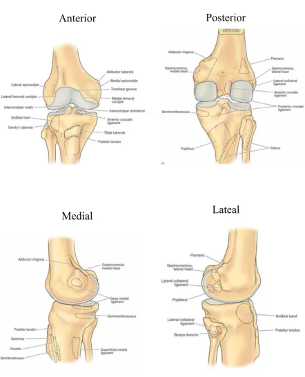
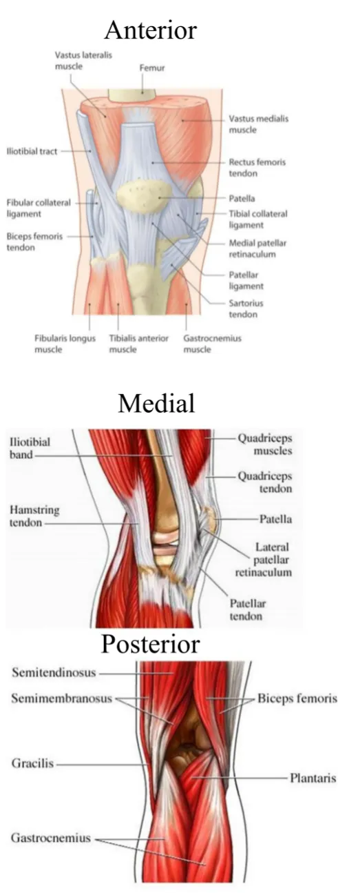
Thorough knowledge of anatomy of the knee joint and proximal tibia is necessary to plan the management and postoperative care in proximal tibia fractures. Tibial plateau fractures include fractures of the proximal tibia extending from articular surface upto the length corresponding to the maximum transepiphyseal width.
The proximal surface of the tibia contains the medial and the lateral tibial plateaus, which are separated by the intercondylar tibial eminences.
KNEE JOINT:
It is a large complex joint. It consists of 1) Patellofemoral - saddle joint
2) Tibiofemoral - condylar joint 3) Tibiofibular - condylar joint
Proximal tibia flares out from the shaft to form medial and lateral condyles. Tibial plateau is the articular surface of the tibial condyles. They articulate with the femoral condyles. The tibial plateau is sloped
anteroposteriorly from 7°-10°. The spinous processes present translation and protect the insertion of anterior cruciate ligament. The most posterior portion of the interspinous region is not covered by articular cartilage.
MEDIAL TIBIAL CONDYLE 44:
Figure 1: Right tibia and fibula: bony landmarks with attachments
Anterior
Posterior
LATERAL TIBIAL CONDYLE:
The lateral plateau is smaller and higher than the medial plateau and articular cartilage on the lateral plateau(4mm) is slightly thicker than that on the medial side(3mm). The lateral tibial plateau is convex in the sagittal plane and nearly flat to slightly convex in the coronal plane. Lateral meniscus covers the outer portion of the lateral plateau. Proximal tibiofibular joint is located posterolaterally on the lateral tibial condyle.
In the frontal plane, the tibial articular surface forms an angle of approximately 3 degrees of varus with the long axis of the tibia. This varus, as well as the slight difference in cartilaginous thickness between the medial and lateral plateaus, results in the lateral plateau being slightly higher than the medial plateau. This difference is further exacerbated by the convexity of the lateral side and the concavity of the medial side.
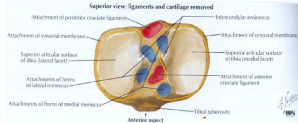
INTERCONDYLAR AREA:
intercondylar eminence to appropriately restore the anatomic width of the proximal end of the tibia as a whole.
Following structures are attached intercondylar area from before backwards
Anterior horn of the medial meniscus Anterior cruciate ligament
[image:28.595.67.497.470.685.2] Anterior horn of the lateral meniscus Posterior horn of the lateral meniscus Posterior horn of the medial meniscus Posterior cruciate ligament
Figure 4: Plateau – superior view : ligaments and cartilage removed
TIBIAL TUBEROSITY AND GERDY’S TUBERCLE:
Tibial tuberosity and Gerdy’s tubercle are bony prominences located in the subcondylar region for insertion of the patellar tendon and iliotibial tract respectively.
Figure 5: MUSCLES AROUND KNEE JOINT
Anterior
Medial
Knee joint is stabilised by the following structures:
FIBROUS CAPSULE:
The capsule is a fibrous membrane containing areas of thickening that may be referred to as discrete ligaments. The anterior capsule is thin, and directly anteriorly it is replaced by the patellar ligament. Proximally, to the femur approximately three to four fingerbreadths above the patella. Distally, it attaches circumferentially to the tibial margin except where the popliteal tendon enters the joint through the hiatus. Posteriorly, the capsule consists of vertical fibers that arise from the condyles and the walls of the intercondylar fossa of the femur.
MEDIAL COLLATERAL LIGAMENT:
It is embryologically the distal end of the adductor magn us muscle. It is divided into
1. Superficial MCL or tibial collateral ligament – it originates from the medial epicondyle of femur just distal to the adductor tubercle and inserts to the upper part of the medial surface of the tibia.
LATERAL COLLATERAL LIGAMENT or FIBULAR COLLATERAL LIGAMENT:
It stretches obliquely downward and forward from lateral femoral condyle below the attachment of lateral head of gastrocnemius and above the tendon of the popliteus proximally to the head of fibula distally and overlapped by the tendon of biceps femoris, a bursa intervening between them. The infero-lateral genicular vessels and nerves separate it from the capsule.
In contrast to medial collateral ligament it neither fuses with the capsular ligament or the lateral meniscus thus making it more susceptible to be torn in the knee injuries and it resists varus forces.
OBLIQUE POPLITEAL LIGAMENT:
ARCUATE POPLITEAL LIGAMENT:
It is a ‘Y’ shaped thickening of the posterior capsular fibres. It extends from the head of the fibula, arches over the tendon of the popliteus and attached to the posterior border of the intercondylar area of tibia.
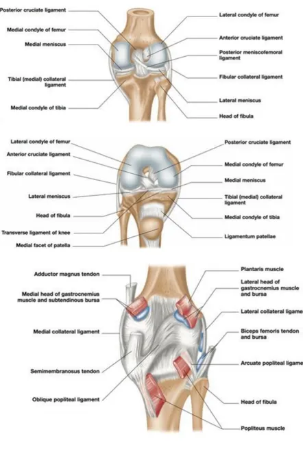
CRUCIATE LIGAMENTS:
These are a pair of very strong ligaments connecting the tibia to the femur. They are extrasynovial and intracapsular. It is covered by synovial membrane on their front and sides but not posteriorly.
The anterior cruciate ligament is attached to the anterior part of the intercondylar area between the attachments of anterior horns of medial and lateral menisci. It ascends posterolaterally and attached to the posteromedial aspect of the lateral femoral condyle. Consists of 2 parts-anterimedial (taut in flexion) and posterolateal (taut in extension)
posteromedial (taut in extension). Both are supplied by middle genicular artery and posterior articular nerve a branch of posterior tibial nerve.
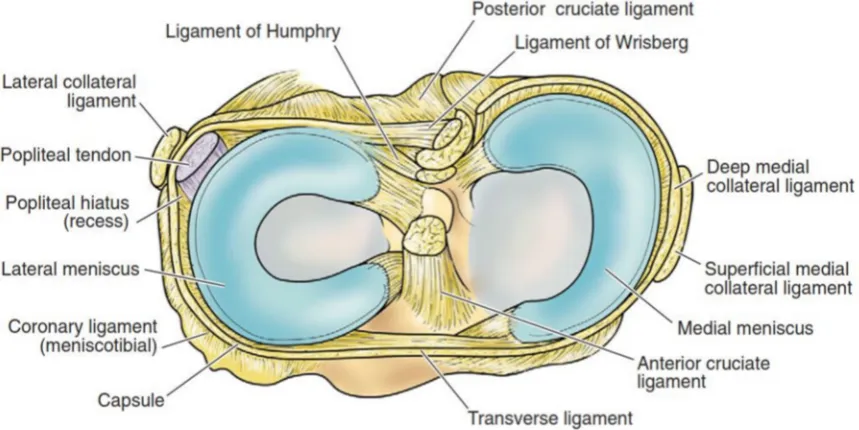
MENISCI (SEMILUNAR CARTILAGE):
The medial and lateral menisci are both semilunar, triangular-shaped fibrocartilage that rest between the femoral condyles and the tibial plateaus. They srve an important function in load sharing by protecting the articular cartilage from up to 60% of the load encountered by the knee. They are composed of collagen fibers which are primarily circumferential, they also contain radial and perforating fibers. They are largely avascular except at their peripheral attachment to the coronary ligaments which attach to their respective tibial plateaus.
The medial meniscus is almost a semicircle. It is broad posteriorly. Its anterior horn is attached to the intercondylar area in front of the anterior cruciate ligament, while the posterior horn is attached in front of the posterior cruciate ligament.
horn is attached behind the intercondylar eminence in front of the posterior horn of the medial meniscus. It is attached to both cruciate ligaments and posteriorly to the medial femoral condyle by Ligament of Humphry (Anterior meniscofemoral lig.) and Ligament of Wrisberg (Posterior
Meniscofemoral lig.) The lateral meniscus covers larger surface of the articular surface, smaller in diameter, thicker in periphery and more mobile than the medial meniscus. Meniscotibial ligaments attach the menisci to the periphery of the tibial plateau.
TRANSVERSE LIGAMENT:
It is a variable band that connects the anterior convexity of the lateral meniscus and anterior horn of the medial meniscus.
SYNOVIAL MEMBRANE:
It is attached to the articular margins of the femur, tibia, patella and lines the deep aspect of the capsule, but it is separated from the capsule by popliteus muscle and the cruciate ligaments. Infrapatellar fat pad separates synovial membrane from the patellar ligament anteriorly.
LIGAMENTUM PATELLAE:
of the apex of patella and below to the tibial tuberosity. The superficial fibers pass in front of the patella. It is related to the infrapatellar bursa and to the infrapatellar pad of fat.
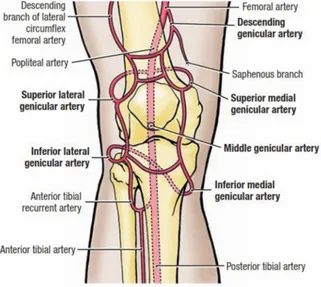
BLOOD SUPPLY OF THE KNEE JOINT 45 :
Knee joint is supplied by genicular anastomosis formed by 1. Superior genicular artery
2. Medial genicular artery
3. Lateral inferior genicular artery 4. Middle genicular artery
5. Anterior and posterior tibial recurrent arteries This supplies bone, capsule and synovial membrane.
Medially by
1. Descending genicular artery 2. Superior medial genicular artery 3. Inferior medial genicular artery
Laterally by
5. Posterior tibial recurrent artery 6. Circumflex fibular artery
Medial and lateral arteries are connected by long anastomosis which are interconnected by horizontal anastomosis just above and below the patella.
NERVE SUPPLY:
[image:37.595.67.388.422.709.2]1. Femoral nerve through its branches to vastii especially vastus medialis. 2. Sciatic nerve through genicular branches of common peroneal nerve 3. Obturator nerve through its posterior division.
MECHANICS OF KNEE JOINT:
Because of the disparity between the lengths of the articular surfaces of the femoral condyles and the tibial condyles, two types of motion during flexion and extension are produced.
1) Ginglymus (hinge).
2)Trochoid (pivot joint) articulation.
Knee joint permits flexion and extension in the sagittal plane and also some degrees of internal and external rotation when the knee is flexed. The complex flexion and extension motion is a combination of rocking and gliding. The rocking motion occurs in the first twenty degrees of flexion, after which the motion becomes predominantly of gliding type.
The natural outward deflection of the tibia on the femur at the knee joint produces greater weight bearing stresses on the lateral condyle of femur than the medial femoral condyle. But because the medial femoral condyle is prolonged forwards than the lateral femoral condyle, the vertical axis of
SCREW-HOME MOVEMENT:
The articular surface of the medial femoral condyle is prolonged anteriorly than the lateral condyle. When the knee comes into full extension, the femur rotates internally until the remaining articular surface on the medial condyle comes in contact with the articular surface of the tibia. The posterior portion of the lateral condyle rotates forward, thus providing a screwing home movement, and locks the knee in fully extended position.
TIBIAL PLATEAU FRACTURES:
INCIDENCE:
Tibial plateau fracture constitute 1% of all fractures and 8%
in the elderly. These fractures include many and varied fracture configurations that involve the medial condyle (10-23%), lateral condyle (55-70%) or both condyles (11-30%) with varying degrees of articular depression and
displacement47.
NATURE OF VIOLENCE:
DIRECT:
Automobile accident, which is one of most frequently encountered. 1. Road traffic accidents/ automobile accidents.
2. Fall from a height 3. Industrial accidents
4. Valgus stress/varus stress 5. Athletics
6. Assault
INDIRECT:
MECHANISM:
Fractures of the upper tibia occur due to strong valgus or varus forces along with axial loading. When a patient sustains varus or valgus force with an axial load, the respective femoral condyle exerts shearing and compressive forces on the articular surface of the tibia.
This frequently results in a split fracture, a depressed fracture or both. Isolated split fractures are virtually confined to adults with dense cancellous bone that is capable of withstanding the compressive forces on the joint surface. With advancing age, strong cancellous bone of the proximal tibia gradually becomes more sparse and is no longer able to withstand the compressive forces. With impact loading, a depressed or split depressed fracture results.
fracture, conversely, the tears of the lateral collateral ligament or cruciate ligaments may be associated with fractures of the medial tibial plateau.
The location of the fracture depends on the degree of flexion/extension of the knee. However when axial loads exceeds 8000 pounds, explosive severely comminuted fractures are produced. This mechanism is thought to occur clinically in a fall from height on the extended knee.
o Fracture line and degree of flexion of knee combined with valgus/varus strain and axial loading contribute to determine the fracture line and the site of depression whether it is anterior, middle or posterior.
o Collateral ligament integrity and forces determine the type of fracture.
o Pure axial loading or axial loading combined with valgus/varus stress determines the type of bicondylar fractures.
o Very high velocity injuries are associated with ligament and neurovascular injuries.
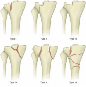
CLASSIFICATIONS:
A) SCHATZKER CLASSIFICATION48
TYPE I - PURE CLEAVAGE:
A wedge shaped uncomminuted fragment is split off and displaced laterally and downwards. This type is common in younger patients with good bone stock.
TYPE II - CLEAVAGE COMBINED WITH DEPRESSION:
A lateral wedge of fragment is split off and the articular surface is depressed down into the metaphysis. This tends to occur in older people with osteoporotic bone.
TYPE III - PURE CENTRAL DEPRESSION:
The articular surface is driven into the metaphysis. The lateral cortex is intact. These tend to occur in older patients with osteoporotic bone.
TYPE IV - FRACTURES OF MEDIAL CONDYLE:
TYPE V - BICONDYLAR FRACTURES:
Both tibial plateaus are split off. The distinguishing feature is that the metaphysis and diaphysis retain the continuity. Intercondylar eminence may or may not be fractured. Sometimes only the anterior or posterior portions of both the condyles will be fractured and remaining portion will be intact. One third of the bicondylar fractures will have a postermedial fracture. These patterns are important in planning the treatment of these type of fractures.
TYPE VI - PLATEAU FRACTURE WITH DISSOCIATION OF METAPHYSIS AND
DIAPHYSIS:
[image:44.595.136.420.494.783.2]A transverse or oblique fracture of the proximal tibia is present in addition to fracture of one or both tibial condyles and articular surfaces.
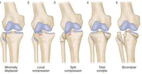
B) HOHL AND MOORES CLASSIFICATION:
B1)FRACTURE PATTERN:
TYPE 1:Split fractures of the lateral condyle.
TYPE 2:Lateral compression.
TYPE 3:Split with compression fracture.
TYPE 4:Total condylar fractures.
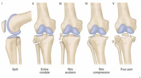
[image:45.595.69.553.441.694.2]B2) FRACTURE - DISLOCATION PATTERN:
TYPE 1:Coronal split fracture dislocation.
TYPE 2:Entire condylar fracture dislocation.
TYPE 3:Rim avulsion fracture dislocation.
TYPE 4:Rim compression fracture dislocation.
[image:46.595.64.534.373.633.2]TYPE 5:Four part fracture dislocation.
C) AO CLASSIFICATION OF TIBIAL PLATEAU FRACTURES:
TYPE 1:Wedge fractures.
TYPE 2:Depression fractures.
TYPE 3:Wedge and depression fractures.
TYPE 4:‘Y’ and ‘T’ fractures/comminuted fractures of both the condyles
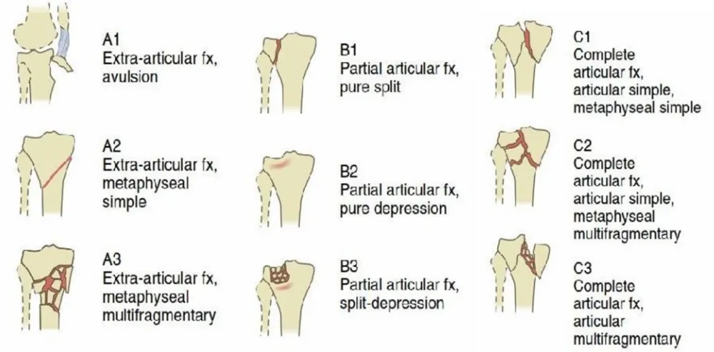
D) THE AO CLASSIFICATION OF PROXIMAL TIBIAL FRACTURES:
TYPE A - Metaphyseal fractures that do not involve the joint surface.
TYPE B - Partial articular fractures.
B1 - Pure split fracture.
B2 - Pure depression fracture.
B3 - Split depression fracture.
TYPE C - Complete articular fractures.
C1 - Articular simple, metaphyseal simple.
C2 - Articular simple, metaphyseal multifragmentary.
Figure 10: AO/OTA CLASSIFICATION
E) 3 COLUMN CONCEPT (Luo et al – 2010)4,5,6:
whole proximal tibia anatomically as a triangle trunk and defined it on transec- tion as 3 columns. A column fracture is defined as an articular depression in combination with a fracture of the column wall (cortex).
The column borders are demarcated by four points on the transversal CT- image:
point B - posteromedial ridge of the tibial plateau; point C - most anterior border of the fibular head
zero-column fracture - Pure articular depression with intact cortex (Schatzker III) is not a column fracture
[image:49.595.60.503.357.531.2]3 Columns : medial, lateral and posterior depending on the columns involved fracture described as single, double or three column fractures.
F) REVISED 3 COLUMN CONCEPT
According to the revised three-column classification approach, column fractures that include the Posterolateral column, are not classified exclusively on the basis of the column borders but rather by clinical reasoning.
[image:50.595.185.413.386.572.2]The Posterolateral column is defined by connecting both the anterior and posterior border of the fibular head, respectively, point C and D, with point O. Hence, a lateral column fracture extending posteriorly into the Posterolateral column is classified as a single, extended lateral column fracture
Figure 12: revised 3 column concept
INVESTIGATIONS:
Plain Radiograph:
o Traction films and stress x-rays. To assess the efficacy of an applied ligamentotaxis force.
CT scan with 3D reconstruction:
Especially useful in determining the extent of injury, amount of articular depression but gives limited information about the soft tissue status.
Magnetic Resonance Imaging:
Advantage of MRI over CT is that it gives a better status of the associated soft tissue injuries.
Angiography
Useful in High velocity injuries associated with vascular compromise.
Role of arthroscopy:
a) Diagnostic :in assessing the degree of injury to menisci, collateral and articular surface.
MODALITIES OF TREATMENT OF TIBIAL PLATEAU FRACTURES:
A) CONSERVATIVE:
o Closed reduction and POP application o Skeletal traction and mobilisation o Functional brace
B)SURGICAL:
o Percutaneous screw fixation
o ORIF with cancellous screws and bone grafting o ORIF with plate osteosynthesis
o ORIF with plate osteosynthesis and bone grafting
o External fixator/Hybrid external fixator/Illizarov ring fixator o Arthroscopic assisted internal fixation
o MIPPO (Minimally Invasive Percutaneous Plate Osteosynthesis) o Total knee replacement
COMPLICATIONS:
A) Early Complications:
o Bleeding
o Wound infection /Dehiscence - Superficial or Deep
o Compartment syndrome
o Pain
o Swelling
o Knee stiffness
o Nerve Injury ( Lateral politeal. N)
o Vascular Injury (Anterior tibial A)
o Loss of fracture reduction
o Limb length discrepancy
o Deep vein thrombosis
B) Late Complications:
o Knee stiffness
o Malunion
o Knee instability – varus/valgus/anterior/posterior
o Angular deformities
SURGICAL APPROACHES 49:
Figure 13: surgical approaches
There are about 7 surgical approaches in tibial plateau fracture (Anteromedial , Anterolateral, Posteromedial, Posterolateral, Medial, Anterior midline, Direct
posterior) of which, most commonly used ones are the anterolateral and
Double incision for fracture reduction has more chance of wound dehiscence than single incision but it canbe minimized with minimum distance between the incision should be 5-7cm. Single anterior midline incision as in TKR can also be employed in any type of tibial plateau fracture.
Direct posterior, posteromedial approach, posterolateral approaches are needed to reduce the fractures involving the posterior column. It can be combined with anterolateral approach with plate osteosynthesis without much soft tissue injury if needed.
MATERIALS AND METHODS
In our institution 10 patients with displaced tibial plateau fractures with posterior column involvement were selected for the study. This is a prospective study done from September 2015 to August 2017.
Mode of injury was road traffic accident in all 10 patients. Patients were evaluated with X rays (AP and lateral views) and Computed Tomography (axial, coronal, sagittal with 3D reconstruction views). Fractures classified based on 3 column concept classification. Mean follow up period was 13.7 months (6 – 21 months). Functional and radiological outcome was assessed using Modified Rasmussen’s clinical and radiological criteria.
INCLUSION CRITERIA:
• Fractures with posterior column involvement • Closed injury
• Age more than 18 years
EXCLUSION CRITERIA:
• Fractures with zero column(pure depression type), pure lateral or medial column involvement without posterior column involvement
PRE-OPERATIVE PLANNING:
•clinical examination • knee aspiration
• x ray knee AP view, lateral view •CT knee with 3D reconstruction
•HB, Blood sugar, urea, creatinine, viral markers, electrocardiogram for anaesthetic Assessment
PRE OPERATIVE MANAGEMENT:
• Stabilisation of fracture with AK slab
• Control of co morbid conditions – diabetes and hypertension
TIME INTERVAL BETWEEN INJURY AND SURGERY:
Three patients were operated between 4 to 7 days. Five patients were operated between 7 to 14 days. Two patients were operated between 14 to 21 days. The mean time interval between injury and surgery was 11.7 days.
o 6.5,5mm cancellous screws (locking, non-locking)
o 4.5,5mm cortical screws (locking, non-locking)
Proximal tibial ‘T’ Buttress plate ‘L’ Buttress plate
locking plate
Posterior plate posteromedial plate 3.5mm screws
SURGERY:
• Postero Medial Plate Osteosynthesis • Posterior plate osteosynthesis
ANAESTHESIA:
Regional / Spinal anaesthesia was used in all patients.
POSITION:
Supine position or prone position depend on the approach used to fix the fracture.
SURGICAL APPROACHES 8,9,10:
POSTEROMEDIAL APPROACH TO THE MEDIAL TIBIAL PLATEAU: SUPINE POSITION
Externaly rotate and slightly flex the knee. Make a longitudinal incision along the posteomedial aspect of the tibia, beginning 3cm above the joint line and
extend it as far distally as needed. Avoid the great saphenous vein and saphenous nerve anterior to the incision.
Mobilize and retract the pes anserinus tendons proximally and anteriorly or distally and posteriorly. Retract the medial gastrocnemius and soleus muscles posteriorly, exposing the junction of popliteal fascia, the semimembranosus
PRONE POSITION: GALLA AND LOBENHOFFER AS DESCRIBED BY FAKLER ET AL.
Make a straight 6 to 8 cm long longitudinal skin incision along the medial border of the medial head of the gastrocnemius, beginning at the level of the joint line. Incise the subcutaneous tissue and popliteal fascia sharply. Free up the medial head of the gastrocnemius muscle without detaching it and retract it laterally. Bluntly dissect the semimembranosus complex and retract it medially. Identify the upper edge of the popliteus muscle and detach it subperiosteally, exposing the posteromedial tibial plateau. If more exposure is needed, incise the tibial insertion of the semimembranosus in a subperiosteal fashion.
POSTEROLATERAL APPROACH: minkoff et al
Patient in prone position
Begin the skin incision 1 to 2 cm below the popliteal crease slightly medial to the midline of the midline of the knee, carrying it transversely and curving it distally just medial and parallel to the head of the fibula, ending 5 to 6 cm distal to it
gastrocnemius medially along with popliteal artery and vein and tibial nerve.
Dissect free the fibular origin of the soleus muscle and retract it distally. Retract the underlying popliteus muscle medially to expose the posterior aspect of the lateral tibial plateau and proximal tibiofibular joint.
ANTEROLATERAL APPROACH: (KANDEMIR AND MACLEAN)
Place the patient supine. Begin the incision 2 to 3 cm proximal to the joint line and extend it 3 cm below the inferior margin of the tibial tubercle crossing Gerdy’s tubercle at the midpoint of the incision. Detach the iliotibial band and
develop the interval between it and joint capsule.reflect the origin of tibialis anterior muscle from the anterolateral surface of the tibial plateau.
SURGICAL TECHNIQUE:
SURGICAL STEPS:
After inflation of high thigh tourniquet
In supine posion: Open interval between posterior pes anserinus and medial gastrocnemius and avoid saphenous vein and nerve
In prone position: Open interval between semitendinosus complex and gastrocnemius medial head and protect saphenous nerve, great saphenous vein, popliteal vessels and tibial nerves
Expose the posteromedial tibia subperiosteally
Place retractor along posterior tibia to protect popliteal fossa structures Apply reduction clamp with manual traction and manipulation
Reduce fracture and fix provisionally with K wire
Confirm reduction of fracture and overall joint alignment fluoroscopically
Fix with antiglide plate for buttressing the fracture and interfragmentary screws through upper hole of the plate if necessary.
Posterior column fixed first with antiglide plate using T/L buttress or 3.5mm locking posterior or posteromedial plates with minimal screws in posterior column fracture alone. Posteromedial plate osteosynthesis combined with
POSITION - PRONE
INCISION SUPERFICIAL DISSECTION
DEEP DISSECTION C ARM PICTURE WITH PROVISIONAL K WIRE
C ARM PICTURE AFTER ANTIGLIDE PLATE AND SCREW FIXATION
INTRAOPERATIVE PICTURE IN SUPINE POSITION IN POSTEROMEDIAL AND ANTEROLATERAL PLATE OSTEOSYNTHESIS
AFTER SKIN CLOSURE
fixation or anterolateral plate fixation in supine or prone position as per the fracture morphology and surgeon’s preference. One patient with with anterior tibial spine fracture mayer and Mc Keevers type I with single column fracture fixed with posterior plating and tibial spine fracture treated conservatively.
POSTOPERATIVE FOLLOW UP:
Patients were treated with i.v.antibiotics for 5 days postoperatively. EOT done on POD 2, 5, 8 and POD 10. Sutures removal done on POD 12.
Patients were mobilised as early as possible. Ankle pump and quadriceps exercises were performed from second POD. Knee bending exercises were started after the pain subsides and as tolerated by the patient.
GRADING CRITERIA:
Modified Rasmussen Criteria for Clinical Assessment50,51
Pain
None 6
Occasional 5
Stabbing pain in certain position 3
Constant pain after activity 1
Significant rest pain -3
Walking capacity
Normal walking capacity for age 6 Walking outdoor more than one hour 5 Walking outdoor 15 mins to 1 hour 3
Walking outdoor < 15 mins 1
Walking indoor only 0
Wheel chair or bed ridden -3
Knee extension
Normal 4
Total Range of Motion
Full 6
Atleast 120° 5
Atleast 90° 3
Atleast 60° 1
< 60° -3
Stability
Normal stability in extension and 20° flexion 6 Abnormal instability in 20° flexion 4 Instability in extension < 10° 2
Instability in extension > 10° 0
Instability in extension > 10°
Power of Quadriceps
Grade 5 2
Grade 3-5 1
Grade <3 -2
Maximum score 30
Modified Rasmussen Criteria for Radiological Assessment 50,51 Articular depression None 3 <5mm 2 6-10mm 1 >10mm 0 Condylar widening None 3 <5mm 2 6-10mm 1 >10mm 0
Valgus varus angulation
None 3
<10° 2
10°-20° 1
>20° 0
Osteoarthrosis
None/ No progress 1
Progression by 1 grade 0
Progression by >1 grade -1
Maximum score 10
Excellent 9-10
Good 7-8
STATISTICAL ANALYLSIS
1. AGE INCIDENCE:
[image:69.595.51.506.205.744.2]In this study 70% of patients were in the fourth and fifth decades of life.
TABLE 1
AGE (YEARS) FREQUENCY PERCENTAGE
20-30 1 10
31-40 2 20
41-50 5 50
51-60 1 10
61-70 1 10
TOTAL 10 100
AGE INCIDENCE 10 20 50 10 10 20 30 40 50 60
2. SEX INCIDENCE:
[image:70.595.56.503.269.702.2]In this study 90% of the cases were male patients and only 10% were female patients. This shows high incidence of tibial plateau fractures in male patients.
TABLE 2
SEX FREQUENCY PERCENTAGE
MALE 9 90
FEMALE 1 10
TOTAL 10 100
SEX INCIDENCE
FREQUENCY
3. SIDE OF INJURY:
[image:71.595.52.498.340.695.2]In this study both lower limbs are equally involved and there is no significant difference in side of injury.
TABLE 3
SIDE FREQUENCY PERCENTAGE
RIGHT 5 50
LEFT 5 50
TOTAL 10 100
SIDE OF INJURY
0 10 20 30 40 50 60 70 80 90 100
5 5 10
50 50
100
4. MODE OF INJURY:
In this study all the patients sustained fracture due to traffic accident. This clearly shows high velocity direct impact force sustained during road traffic accidents are the commonest cause for tibial plateau fractures.
[image:72.595.58.496.430.702.2]TABLE – 4
MODE OF INJURY FREQUENCY PERCENTAGE
Road traffic accident 10 100
Total 10 100
mode of injury
5. FRACTUES DISTRIBUTION ACCORDING TO TYPE:
50% of schatzker type V cases, 30% with type IV and 20% Hohl and moore type I fracture with coronal splitting was included in this study.
[image:73.595.53.493.464.741.2]TABLE – 5
RACTURE TYPE FREQUENCY PERCENTAGE
SCHATZKER TYPE IV 3 30
SCHATZKER TYPE V 5 50
MOORE TYPE I 2 20
TOTAL 10 100
FRACTUES DISTRIBUTION ACCORDING TO TYPE:
30
50
20 20
30 40 50 60
6. THREE COLUMN CLASSIFICATION:
In our study 3 column involvement were more than single or 2 column fracrers (40%)
[image:74.595.53.496.439.739.2]TABLE – 6
Three column classification
FREQUENCY PERCENTAGE
1 3 30
2 3 30
3 4 40
TOTAL 10 100
THREE COLUMN CLASSIFICATION
7. SCHATZKER VS THREE COLUMN FRACTURE CLASSIFICATION:
Moore type I fractures 20% in our study are not classified under schatzker classification also included with 3 column fracture classification which provides better pre op planning and future comparative study using with three classification system.
TABLE – 7
SINGLE COLUMN TWO COLUMN THREE COLUMN TOTAL
SCHATZKER TYPE IV 1 2 0 3
SCHATZKER TYPE V 0 1 4 5
MOORE TYPE I 2 0 0 2
TOTAL 3 3 4 10
SCHATZKER VS THREE COLUMN FRACTURE CLASSIFICATION
[image:75.595.51.508.252.779.2]8. INTERVAL BETWEEN INJURY AND SURGERY
In our study 30 % of cases operated within one week and 50% of cases operated within 2 weeks. 20% of patients operated after 2 weeks of injury.
[image:76.595.53.495.386.733.2]TABLE – 8
TIME INTERVAL (DAYS) NO OF CASES PERCENTAGE
0-7 3 30
8-14 5 50
15-21 2 20
TOTAL 10 100
INTERVAL BETWEEN INJURY AND SURGERY
30
50
20 20
30 40 50 60
9. SURGICAL POSITION AND PROCEDURE
In our study all dual plating are done in supine position and posterior plating done in prone position. For fixation of posteromedial fragment selected supine or prone depending on the surgeon’s preference.
[image:77.595.50.520.238.743.2]TABLE - 9
POSITION POSTERIOR PLATING POSTEROMEDIAL PLATING POSTEROMEDIAL PLATING WITH SCREW FIXATION DUAL (PM/AL) PLATING
SUPINE 0 1 1 4
PRONE 2 1 1 0
TOTAL 2 2 2 4
SURGICAL POSITION AND PROCEDURE
2 3 4 5 6 2 1 1 1 4 0
DUAL (PM/AL) PLATING
POSTEROMEDIAL PLATING WITH SCREW FIXATION POSTEROMEDIAL PLATING
10.COMPLICATION:
One patient with 3 column fracture developed wound necrosis for which flap cover done and eventually patient developed deep infection for whom implant removal was done.
[image:78.595.49.517.215.780.2]TABLE - 10
COMPLICATIONS FREQUENCY PERCENTAGE
Deep Infection 1 10
Infection and wound necrosis 0 0
Valgus/ Varus deformity 0 0
Peroneal nerve injury 0 0
Vascular injury 0 0
Normal 9 90
total 10 100
Functional and radiological outcomes were assessed by Modified Rasmussen’s clinical and radiological criteria.
FUNCTIONAL OUTCOME:
The functional outcome was assessed with Modified Rasmussen’s clinical criteria which includes pain (maximum points-6), working capacity (maximum points-6), knee extension (maximum points-4), range of motion (maximum points-6), stability (maximum points-6) and power of quadriceps (maximum points-2) with maximum total points-30. Results were graded as excellent (28-30), good (24-27), fair (20-23) and poor (<20).
RADIOLOGICAL ASSESSMENT:
RESULTS
10 patients with tibial plateau fractures with displaced posterior column fractures admitted at Government Rajaji Hospital, Madurai were included in this study. All the patients were treated with posterior plating with or without additional anterolateral plating in supine or prone position was done after assessing fracture morphology with CT scan and three column classification and surgeon’s preference on positioning the patients. The longest follow up period was 21 months and shortest follow up period was 6 months. Mean follow up period was 13.7 months. Follow up analysis was made using Modified Rasmussen’s Clinical and Radiological Criteria.
20%, type V 10%, 40% of three column fractures(LUO) - schatzker type V 40% included.
In this study, 40% patients had excellent outcome, 50% patients had good outcome, 10% patients had poor clinical and 10% fair radiological outcome.
In this study, One patient 68 years old male with 3 column fracture
developed superficial blisters over the tibial plateau region and patient taken into
surgery after soft tissue healing and after normalisation of ESR and CRP on 3rd week after the injury. Patient developed wound necrosis for which flap coverdone and eventually patient developed deep infection for whom implant removal was done after 6 months.
FUNCTIONAL AND RADIOLOGICAL OUTCOME:
MODIFIED RASMUSSEN
FUNCTIONAL – no of patients
FUNCTIONAL – percentage
RADIOLOGICAL – no of patients
RADIOLOGICAL – PERCENTAGE
EXCELLENT 4 40 4 40
GOOD 5 50 5 50
FUNCTIONAL AND RADIOLOGICAL OUTCOME: 4 5 0 1 40 50 0 10 4 5 1 0 40 50 10 0 0 10 20 30 40 50 60
EXCELLENT GOOD FAIR POOR
CASE 1
S.NO.1SUBRAMANI 51 Years/male 3 COLUMN TIBIAL PLATEAU # LEFT
CT SCAN WITH 3D RECONSTRUCTION PRE OP XRAY
Modified Rasmussen criteria -outcome
6 MONTH FOLLOWUP 1 YEAR FOLLOWUP
KNEE EXTENSION
SCAR
CASE 2
S.NO:2KUMAR 40 Years/male 3 COLUMN TIBIAL PLATEAU # RIGHT
PRE OP XRAY CT SCAN WITH 3D RECONSTRUCTION
1 YEAR FOLLOWUP 6 MONTH FOLLOWUP
CLINICAL PICTURE
CASE 3
S.NO:3SUBRAMANI 44 Years/male 1 COLUMN TIBIAL PLATEAU # RIGHT
PRE OP XRAY CT SCAN WITH 3D RECONSTRUCTION
Modified Rasmussen criteria -outcome
1 YEAR FOLLOWUPKNEE FLEXION
CASE 4
S.NO:4MAHENDRAN 35 Years/male 2 COLUMN TIBIAL PLATEAU # RIGHT
PRE OP XRAY
CT SCAN WITH 3D RECONSTRUCTION
1 YEAR FOLLOWUP 6 MONTH FOLLOWUP
CLINICAL PICTURE
SCAR
CASE 5
S.NO.6
ROHINI 35 Years/Female 3 COLUMN TIBIAL PLATEAU # LEFT
PRE OP XRAY PRE OP CT SCAN WITH 3D RECONSTRUCTION
CLINICAL
SCAR
KNEE FLEXION
CASE 6
S.NO.7
PANDI 35 Years/male 1 COLUMN TIBIAL PLATEAU # LEFT
PRE OP XRAY
POST OP XRAY
CT SCAN WITH 3D RECONSTRUCTION
CASE 7
S.NO.8
MUTHUVELRAJA 23 Years/male 1 COLUMN TIBIAL PLATEAU # RIGHT
PRE OP XRAY
POST OP XRAY
CT SCAN WITH 3D RECONSTRUCTION
CASE 8
S.NO.9
VENKATESH 47 Years/male 3 COLUMN TIBIAL PLATEAU # LEFT
Modified Rasmussen criteria -outcome
PRE OP XRAYPOST OP XRAY
CT SCAN WITH 3D RECONSTRUCTION
CASE 9
S.NO.10
CHANDRASEKAR 37 Years/male 2 COLUMN TIBIAL PLATEAU # RIGHT
POST OP XRAY
PRE OP XRAY
CT SCAN WITH 3D RECONSTRUCTION
CASE 10
S.NO:5
COMPLICATION
JEGAN MOHAN 68 Years/male 3 COLUMN TIBIAL PLATEAU # LEFT
PRE OP XRAY CT SCAN WITH 3D RECONSTRUCTION
6 MONTH FOLLOWUP AFTER IMPLANT EXIT
POST OP – INFECTED WOUND
DISCUSSION
Tibial plateau fractures, one of the commonest intra articular fractures, occurring as a result of RTA, fall from height, violence etc. The management of these fractures has always been in debate because of their variety of fracture pattern and soft tissue complications. High energy tibial plateau fractures have associated with more severe fracture pattern, ligament injury and severe soft tissue injuries. Bicondylar fractures are best treated with dual plating than single lateral plating with better anatomic reduction and rigid fixation and it also has soft tissue complications as well. There are many approaches for fixation of tibial plateau fractures each one has its own merits and demerits. Selection of approach and fixation for tibial plateau fractures is still a debate for better outcomes.
In high energy tibial plateau fractures, posteromedial and posterior
fractures are often not able to fix with anterolateral plate alone. Fractures of the
decreased range of motion and clinical outcome in displaced tibial plateau fractures and posterior tibial fractures are best studied and planned for fixation
using the three column fixation proposed by Luo CF et al.
Posteromedial or posterior approaches either in prone or supine provides better visualization of the fractures and aid in better reduction and fixation and it also has the advantages of less soft tissue injury even when combined with
anterolateral incision and it can also be used to fix the posterior cruciate ligament injury if present. Posterior Column fixation through these approaches with
antiglide plate and medial and lateral column fixation with screws or lateral locking plates provides the accurate reduction of articular surfaces and rigid fracture
fixation thereby has advantages of early mobilization, reduced soft tissue complications, better range of movements, early mobilization than other modes of fixations.
In this study functional and radiological outcome in displaced tibial plateau fractures with posterior column involvement of 10 patients treated by posterior plate osteosynthesis with antiglide plating with interfragmentary screws through the upper hole of the antiglide plate if needed and it was combined with or without lateral support with 6.5 mm cancellous screws or plate osteosynthesis. Plateau fractures were studied with CT scan and classified and planned for
approach and plating using three column classification. Post operative functional and radiological outcome was assessed by using Modified Rasmussen Criteria.
In this study, tibial plateau fractures were more commonly seen in the active productive age group (31-50 years) due to high-energy trauma. Conservative management, extrernal fixators and routine anterolateral plate osteosynthesis are difficult to reduce and fix the posterior column fractures especially in posteromedial fragment and coronal splitting moore type I fractures. It is extremely important to adequately visualize the fragments, reduce the fracture, regain articular congruity and obtain stable rigid fixation.
In our study three column classification utilized to evaluate the fracture morphology and to plan for fracture fixation. Fracture distribution in our study was 30% of single column fractures (schatzker type IV 10%, Hohl and moore type I coronal split fracture 20%), 30% of two column fracture (schatzker type IV 20%, type V 10%), 40% of three column fractures (schatzker type V 40%) included.
In this study, One patient 68 years old male with 3 column fracture
developed superficial blisters over the tibial plateau region and patient taken into surgery after soft tissue healing and after normalisation of ESR and CRP on
3rd week after the injury. Patient developed wound necrosis for which flap cover done and eventually patient developed deep infection for whom implant removal was done after 6 months.
Soft tissue complications are a major concern in the treatment of bicondylar tibial plateau fractures with plates. Papers reporting the results of Dual Plate osteosynthesis through a single extensile incision have shown the incidence of
(10%) for which flap cover and eventually implant exit done after 6 months comparing with other studies infection rate was little higher.
In a study conducted by Waddell et al42, patients treated with single lateral
plating, developed varus malunion at the fracture site. Zheng43 et al, stated that patients treated with dual plating provided better stability and none of the patient developed malunion. When comparing to the above studies, none of the patient developed malunion in our study. This clearly shows dual plating offers better stability than single lateral plating.
In a study conducted by Zhi et al53, west et al54, Luo et al55on tibia bone
model, Posteromedial T plate can improve the strength and stiffness of
posteromedial fragment fixation and had a buttress effect preventing descent of the fragment under load than other modes of fixation (anteroposterior lag-screws, an anteromedial limited contact dynamic compression plate (LC-DCP), a lateral locking plate). Hence, reduce the varus collapse and increase in range of
movements by fixing the unstable posterior fragments. No patient developed varus collapse in our study.
Limitations of the study:
CONCLUSION
1) Posterior plate osteosynthesis improve the strength and stiffness of posteromedial fragment fixation and had a buttress effect preventing descent of the fragment under load than other modes of fixation Hence, reduce the varus collapse and increase in range of movements by fixing the unstable posterior fragments. Rigid anatomical fixation without much soft tissue complication allow the patient to mobilize early and increase range of movement and functional outcome.
2) In high energy displaced tibial plateau fractures of 3 column fractures can be treated with dual plating with conventional lateral locking plate or screw fixation when combined with posterior antiglide plating effectively.
3) Posterior plate osteosynthesis of posterior column fracture fixation
through posteromedial or posterior approach in either supine or prone position provides better visualization and rigid fixation of posterior tibial fractures without major soft tissue complications if the surgery is done once the soft tissue condition improves.
ANNEXURE – I
BIBLIOGRAPHY
1. Mallik AR, Covall DJ, Whitelaw GP. Internal versus external fixation of
bicondylar tibial plateau fractures. Orthop Rev 1992;21:1433-6.
2. Su EP, Westrich GH, Rana AJ, Kapoor K, Helfet DL. Operative treatment of tibial
plateau fractures in patients older than 55 years. Clin Orthop Relat
Res2004;421:240-8.
3.Dimitar Raykov, Stoyan Ivanov, Pavlin Apostolov. Standard and specific surgical
approaches. Scripta Scientifica Medica, Vol 45, No.3, 2013, pp 74-81.
4.Yang G, Zhai Q, Zhu Y, Sun H, Putnis S, Luo C.Arch Orthop Trauma Surg. 2013
Jul;133(7):929-34. doi: 10.1007/s00402-013-1735-4. Epub 2013 Apr 16. PMID:23589062
5.Zhu Y, Yang G, Luo CF, Smith WR, Hu CF, Gao H, Zhong B, Zeng BF. J Trauma Acute Care
Surg. 2012 Sep;73(3):731-7. doi: 10.1097/TA. 0b013e31825c17e7. PMID: 22929503
6. Zhu Y, Hu CF, Yang G, Cheng D, Luo CF.J Trauma Manag Outcomes. 2013 Sep
11;7(1):7. doi: 10.1186/1752-2897-7-7. PMID: 24025650
7. Lin KC, Tarng YW, Lin GY, Yang SW, Hsu CJ, Renn JH.Orthop Traumatol Surg Res. 2015
Jun;101(4):477-82. doi: 10.1016/j.otsr.2014.12.021. Epub 2015 Apr 20. PMID:25907515
8. Galla M, Lobenhoffer P. Unfallchirurg.2003 Mar;106(3):241-7. German. PMID:2658343
9.Luo CF, Sun H, Zhang B, Zeng BF. J Orthop Trauma. 2010 Nov;24(11):683-92.
doi:10.1097/BOT.0b013e3181d436f3. PMID: 20881634
11. Hoekstra H, Rosseels W, Luo CF, Nijs S.A combined posterior reversed L- shaped and
anterolateral approach for two column tibial lateaufractures in Caucasians: A technical
note. Injury. 2015 Dec;46(12):2516-9. doi:10.1016/j.injury.2015.10.014. Epub 2015 Oct 19.
PMID: 26520364
12. Apley A.G. Fractures of lateral tibial condyle treated by skeletal traction and early
mobilization. J Bone & Joint Surg 1956; 383: 699.
13.George A, Brown and Sprague. Cast brace treatment for plateau and
bicondylar fractures of tibia. Clin Orthop 1976; 119: 184.
14.Sirkin MS, Bono CM, Reilly MC and Behrens FF. Percutaneous methods of tibial
plateau fixation. Clin Orthop June 2000; 375:60-68.
15. Waddell JP.Fracture of the tibial plateau in tibia and fibula. Court Brown
C & Penning D. ed. Oxford, Butterworth Heinemann, 2000; 38-54.
16.Palmer I. Compression fracture of lateral tibial condyle and their treatment. J
Bone & Joint Surg 1939; 2 (Am): 674.
17.Palmer I. Fracture of the upper end of tibia. J Bone & Joint Surg 1951;33(Br): 160.
18.Duparc and Ficat. Fracture of the tibial plateau in Insall et al Surgery of the knee 2nd
ed. New York, Churchill Livingstone, 1995; Vol 2. : 1074.
19.Porter B.B. Crush fractures of lateral tibial table, factors influencing the prognosis. J
Bone & Joint Surg 1970; 52(Br): 676.
20.Schatzkar J, Mc Broom R and Bruce D. The tibial plateau fractures – Toronto
22.Dalamarter R, Hohl M and Hopp E Jr.Ligament injuries associated with tibial plateau
fractures. Clin Orthop 1990; 250: 226.
23.Bennett WF and Browner B. Tibial plateau fractures- a study of associated soft
tissue injury. J.Ortho.Trauma 1992; 6: 78.
24.Tscherne H and Lobenhoffer P. Tibial plateau fractures – management and expected
results. Clin Orthop 1993; 292: 87.
25.Sushil H Mankar, Anil V Golhar.Outcome of complex tibial plateau fractures treated
with external fixator, Indian J Orthop. 2012. 570-574.
26.Thomas G, Padanilam and Nabil A. Meniscal detachment to approach lateral tibial
plateau fractures. Clin Orthop 1995; 314: 192-198.
27.BallmerFT, Hertel R, NOtzil HP.Treatment of tibial plateau fractures with small
fragment internal fixation; a preliminary report.J Orthop Trauma 2000;14(7):467-74.
28.Mills WJ and Nork SE. Open reduction and internal fixation of High energy tibial plateau
fractures. Orthop Clin North Am. 2002;33: 177-194.
29.Jong-keun O, Chang-wug O, In-Ho J, Sung-Jung K, Hee-Soo K, Il-Hyung P et al.
Percutaneous plate stabilisation of proximal tibial fractures. J Trauma Aug 2005; 5: 431-437.
30.Kye Youl Cho, Hyun Sup Oh, Young Joo Cho. Treatment of Schatzker type V, VI
fractures using a midline longitudinal incision and dual plating. Knee Surg Relat Res
2013;25(2):77-83.
31.Ebrahim Ghayem Hassankhani. Treatment of complex proximal tibia fractures by double
33.Horwitz DS, Bachus KN, Craig MA, Peters CL. A biomechanical analysis of internal
fixation of complex tibial plateau fractures. J Orthop Trauma1999;13:545-9.
34. Watson JT. Tibia: Proximal. In: Rüedi TP, Murphy WM, editors. AO Principles of
Fracture Management. Stuttgart: Thieme; 2000. p. 499-517.
35. Barei DP, O′Mara TJ, Taitsman LA, Dunbar RP, Nork SE. Frequency and fracture
morphology of the posteromedial fragment in bicondylar tibial plateau fracture patterns. J
Orthop Trauma 2008;22:176-82.
36. Barei DP, Nork SE, Mills WJ, Henley MB, Benirschke SK. Complications associated with
internal fixation of high-energy bicondylar tibial plateau fractures utilizing a two-incision
technique. J Orthop Trauma 2004;18:649-57
37. Moore TM, Patzakis MJ, Harvey JP. Tibial plateau fractures: Definition, demographics,
treatment rationale, and long term results of closed traction management or operative
reduction. J Orthop Trauma 1987;1:97-119
38. Young MJ, Barrack RL. Complications of internal fixation of tibial plateau fractures.
Orthop Rev1994;23:149-54.
39. Stevens DG, Beharry R, McKee MD, Waddell JP, Schemitsch EH. The long term
functional outcome of operatively treated tibial plateau fractures. J Orthop Trauma
2001;15:312-20
40. Egol KA, Su E, Tejwani NC, Sims SH, Kummer FJ, Koval KJ. Treatment of complex
tibial plateau fractures using the less invasive stabilization system plate: Clinical experience