Copyright © 2006, American Society for Microbiology. All Rights Reserved.
Alpha and Lambda Interferon Together Mediate Suppression
of CD4 T Cells Induced by Respiratory Syncytial Virus
Bo Chi,
1Harold L. Dickensheets,
2Kirsten M. Spann,
3Marc A. Alston,
1Cindy Luongo,
3Laure Dumoutier,
4Jiaying Huang,
5Jean-Christophe Renauld,
4Sergei V. Kotenko,
5Mario Roederer,
3Judy A. Beeler,
1Raymond P. Donnelly,
2Peter L. Collins,
3and Ronald L. Rabin
1*
Center for Biologics Evaluation and Research,1and Center for Drug Evaluation and Research,2U.S. Food and Drug Administration,
Bethesda, Maryland; National Institute of Allergy and Infectious Diseases, Bethesda, Maryland3; Ludwig Institute for
Cancer Research, Brussels Branch, and Experimental Medicine Unit, Universite´ de Louvain, Brussels, Belgium4; and
Department of Biochemistry and Molecular Biology, University of Medicine and Dentistry of New Jersey,
New Jersey Medical School, Newark, New Jersey5
Received 4 November 2005/Accepted 21 February 2006
The mechanism by which respiratory syncytial virus (RSV) suppresses T-cell proliferation to itself and other antigens is poorly understood. We used monocyte-derived dendritic cells (MDDC) and CD4 T cells and
measured [3
H]thymidine incorporation to determine the factors responsible for RSV-induced T-cell suppres-sion. These two cell types were sufficient for RSV-induced suppression of T-cell proliferation in response to
cytomegalovirus orStaphylococcusenterotoxin B. Suppressive activity was transferable with supernatants from
RSV-infected MDDC and was not due to transfer of live virus or RSV F (fusion) protein. Supernatants from RSV-infected MDDC, but not MDDC exposed to UV-killed RSV or mock conditions, contained alpha interferon
(IFN-␣; median, 43 pg/ml) and IFN-(approximately 1 to 20 ng/ml). Neutralization of IFN-␣with monoclonal
antibody (MAb) against one of its receptor chains, IFNAR2, or of IFN-with MAb against either of its receptor
chains, IFN-R1 (interleukin 28R [IL-28R]) or IL-10R2, had a modest effect. In contrast, blocking the two
receptors together markedly reduced or completely blocked the RSV-induced suppression of CD4 T-cell proliferation. Defining the mechanism of RSV-induced suppression may guide vaccine design and provide insight into previously uncharacterized human T-cell responses and activities of interferons.
Respiratory syncytial virus (RSV) is a member of the
Paramyxoviridae family of negative-strand RNA viruses that
includes parainfluenza virus 1 (PIV1), PIV3, mumps, and mea-sles, as well as newly recognized pathogens such as metapneu-movirus, Nipa virus, and Hendra virus. RSV is often the first pathogen encountered by infants and accounts for about 24% of hospitalizations for pediatric respiratory tract disease. RSV is the primary cause of infantile pneumonia and bronchiolitis, the latter being an obstructive disease characterized by wheez-ing and hypoxia due to necrosis of the respiratory epithelium and airway plugging (9, 14, 22, 33). Severe infantile RSV-induced lower respiratory infections (LRI) may herald the onset of allergic diseases and/or asthma later in life (57). While RSV-induced LRI beyond age 3 are uncommon, RSV is a known trigger for wheezing in older asthmatics, and frequent upper respiratory infections in children and adults are com-mon. Finally, RSV is a significant cause of morbidity in the elderly and mortality in bone marrow transplant recipients (22). AmongParamyxoviridae family members, RSV, PIV3, and measles are known to suppress T-cell responses to secondary antigenic stimuli (43, 45, 50, 55). Clinical immunosuppression is a common complication of measles, and secondary bacterial and viral infections contribute to its high morbidity and
mor-tality (6). The mechanism by which measles suppresses T-cell responses is not clear, but altered cytokine expression and impaired antigen presentation have been implicated (for re-views, see references 35 and 50). PIV3 is also immunosuppres-sive, perhaps a consequence of increased expression of inter-leulin-10 (IL-10) (54, 55).
Suppression of in vitro T-cell responses by human RSV (43, 46) and bovine RSV has been described (26, 51, 52). Unlike measles, however, it is not immediately obvious that suppres-sion of T-cell responses in vitro is clinically relevant. It is quite possible that this immunosuppressive activity contributes to the phenomena of repeat infections with RSV, despite a lim-ited number of subtypes and a genome that is not prone to mutation, as is, for example, influenza. In addition, secondary infection with Pasteurella haemolytica is not rare in bovine RSV-infected sheep and goats (10), andStreptococcus
pneu-moniae may complicate human RSV LRI more commonly
than has been appreciated (30). Furthermore, immunosup-pression may be mechanistically linked to a bias toward re-sponses dominated by Th2 cytokines such as IL-4 and IL-13 induced by early infection with RSV in animal models (15). This effect may in turn contribute to subsequent episodes of wheezing in childhood with repeat exposure to this virus and may also cause predisposition to the development of asthma and allergic diseases.
The mechanism by which RSV suppresses T-cell responses is not well understood. Contact-dependent mechanisms (49), IL-1 receptor antagonist (IL-1RA) (32, 46), and alpha
inter-* Corresponding author. Mailing address: Center for Biologics Eval-uation and Research, 29 Lincoln Drive, Room B1, Bethesda, MD 20892. Phone: (301) 496-8806. Fax: (301) 402-5177. E-mail: rrabin @helix.nih.gov.
5032
on November 8, 2019 by guest
http://jvi.asm.org/
feron (IFN-␣) (42) have each been implicated but inconclu-sively so. The implication that IFN-␣is immunosuppressive is somewhat paradoxical, since it is well documented that the nonstructural genes of RSV, NS1 and NS2, also decrease ex-pression of the cellular responses to type I IFN (IFN-␣and -) (47, 48, 58).
To date, T-cell responses to RSV have been studied using peripheral blood mononuclear cells, which is a heterogeneous population. This may have impeded efforts to define the mech-anisms of RSV-induced immunosuppression. Therefore, we first designed a simplified experimental system using only monocyte-derived dendritic cells (MDDC) and autologous CD4 T cells and then focused on identifying soluble factors expressed by MDDC in response to RSV that suppress T-cell proliferation to the superantigenStaphylococcusenterotoxin B (SEB).
MATERIALS AND METHODS
Cell culture.Human MDDC were generated from elutriated monocytes ob-tained from healthy adult donors (⬍65 years of age) at the National Institutes of Health (NIH) Clinical Center blood bank. The monocytes were cultured at 3⫻ 105/ml in RPMI medium with 1% nonessential amino acids and 1% sodium pyruvate (Biosource, Rockville, MD), 10% fetal bovine serum (HyClone, Logan, UT), 0.05 mM-mercaptoethanol (Sigma, St. Louis, MO), 20g/ml gentamicin (Invitrogen, Grand Island, NY), 800 U/ml recombinant human granulocyte-macrophage colony-stimulating factor (Leukine; Immunex, Seattle, WA), and 500 U/ml recombinant human IL-4 (rhIL-4; BD Pharmingen, San Diego, CA) for 7 days.
Elutriated lymphocytes were purified with Ficoll-Hypaque density centrifuga-tion (Sigma) and frozen in liquid nitrogen. After thawing and overnight incuba-tion, CD4 T cells were purified from the lymphocytes by positive selection with a magnetic column and beads (AutoMACS; Miltenyi Biotec, Auburn, CA) ac-cording to the manufacturer’s protocol, and purity was verified by flow cytometry. The Institutional Review Boards of the NIH and at the U.S. Food and Drug Administration approved the study.
Monoclonal antibodies and recombinant cytokines.Mouse anti-IFN-␣/ re-ceptor chain 2 (IFNAR2, CD118) monoclonal antibody (MAb) was purchased from PBL laboratory (Piscataway, NJ). Anti-IL-10R2 MAb and mouse immu-noglobulin G2a (IgG2a) and IgG1 isotype controls were obtained from R & D systems (Minneapolis, MN). The humanized MAbs palivizumab and omali-zumab were obtained from Medimmune (Gaithersburg, MD) and Genentech (San Francisco, CA), respectively. Anti-IL28R was generated as previously de-scribed (19).
Viral infection of MDDC.PIV3 was propagated at 32°C in LLC-MK2 cells or Vero cells, as previously described (23). The Udorn strain of influenza A virus (A/Udorn/72) was propagated in HEp-2 cells in the presence of 7.5g of trypsin/ml as previously described (62). The wild-type A2 strain of RSV and recombinant RSV expressing green fluorescent protein (rgRSV) (24) were grown in HEp-2 cells (14). Wild-type RSV was further purified by centrifugation through a sucrose gradient to eliminate cytokine contaminants (58). Virus con-centrations were determined by plaque assay (28). RSV was inactivated with UV light (UV-RSV) in a Stratalinker UV cross-linker for 10 min (0.12 J/cm2; Strat-agene, La Jolla, CA). Complete inactivation of RSV was confirmed by plaque assay. Cytomegalovirus (CMV) grade 2 antigen was purchased from Microbix Biosystems (Toronto, Ontario).
MDDC (1⫻106/ml) were exposed to virus for 4 h at 37°C and, when stated, washed extensively in culture medium to remove any nonadherent virus prior to coculture with lymphocytes or CD4 T cells. MDDC supernatants were collected after overnight culture of infected and washed MDDC. Mock-infected MDDC were exposed to sucrose solutions of a concentration equal to those in the viral stocks as measured by the invertase-hexokinase-G6PD assay (Sigma). Infection of MDDC with rgRSV was verified with an Olympus BX41 (Tokyo, Japan) fluorescent microscope and digitally photographed with the Spot RT Color system (Sterling Heights, MI).
Cell stimulation and in vitro proliferation assay.Virus-exposed MDDC were cultured with lymphocytes at a ratio of 1:10 or with CD4 T cells at a ratio adjusted according to the percentage of CD4 T cells in lymphocytes of that donor, which was usually between 1:3 and 1:5. CD4 T cells were stimulated with
Staphylococcusenterotoxin B (1g/ml; Sigma). Unless otherwise stated, the cells were pulsed with [3
H]thymidine (0.5Ci/well; MP Biomedicals, Inc., Irvine, CA) on day 4 and harvested on day 5 for measurement of cell lysate-associated tritium by-scintillation counting (Perkin-Elmer, Boston, MA). All proliferation exper-iments were performed in triplicate and reported as counts per minute.
To test MDDC supernatants for antiproliferative activity, CD4 T cells (0.6⫻ 105to 1⫻105/well) were incubated in 96-well plates with SEB for 3 days in the presence of 100l tissue culture medium/well, of which a maximum of 84l was supernatant from an overnight culture of virus or mock-infected MDDC. The cells were then pulsed with [3
H]thymidine and harvested for-scintillation the next day. To test dependence on IFN-␣and IFN-, MAb to IFNAR2, IL-10R2, and IL-28R were used at concentrations of 15, 10, and 20g/ml, respectively. CD4 T cells were incubated with MAb in half the final volume of medium for 1 h at 37°C before addition to the MDDC supernatant and SEB.
Transwell cultures.Virus-exposed MDDC were placed in the top chamber of 24-well Transwell culture plates (1.4⫻105
cells/well in 100l; Costar, Cam-bridge, MA). SEB (1g/ml) and CD4 T cells (4⫻105to 7⫻105) treated with MAb were incubated in 600l of media in the lower chamber. After 4 days, the CD4 T cells were transferred to 96-well plates (0.6⫻105to 1⫻105/well) for measurement of proliferation using [3
H]thymidine.
Enzyme-linked immunosorbent assay (ELISA) for RSV proteins.Rabbit anti-RSV polyclonal Abs were generated by immunizing a rabbit with UV-inactivated sucrose gradient-purified virus at a dose equivalent to 6.0 log10 50% tissue culture infective doses given subcutaneously four times at 3-week intervals and then terminally bled 1 month after the last dose. Rabbit anti-RSV antisera had a 50% plaque reduction neutralization titer of 1:11,000 when tested against the A2 strain of RSV (not shown).
For detection of RSV proteins, 96-well plates were coated at 4°C overnight with either MDDC supernatants or RSV standards (RPMI medium containing serial twofold dilutions of live RSV). The sample, standard dilutions, and control wells were washed three times with phosphate-buffered saline–0.05% Tween 20 solution and blocked with 1% bovine serum albumin in phosphate-buffered saline–0.05% Tween 20 solution prior to incubation with rabbit polyclonal anti-RSV. Horseradish peroxidase-conjugated goat anti-rabbit IgG and 3,3⬘,5,5⬘-tetramethylbenzidin (KPL, Inc., Gaithersburg, MD) were used to reveal RSV-derived proteins. Concentrations of RSV proteins in MDDC supernatants are expressed in equivalents relative to the amount of protein in the standard con-taining 1⫻106
/ml of live RSV.
Measurements of cytokines.ELISA kits for IFN-␣and IFN-were purchased from PBL Laboratories (Piscataway, NJ). All other cytokines were detected with Luminex multiplex bead assay (Linco Research, St. Charles, MO), except IL-1␣, for which the bead assay was purchased from Biosource (Camarillo, CA). The data were analyzed using the MasterPlex QT quantitation software (MiraiBio, Alameda, CA).
Quantitative reverse transcription-PCR (RT-PCR) for IFN-.Total cellular RNA of virus-infected or mock-infected MDDC was isolated using RNeasy (QIAGEN, Valencia, CA) and reverse transcribed to cDNA with Superscript II reverse transcriptase (Invitrogen, Carlsbad, CA). Primers from SuperArray (Fre-derick, MD) specific for IFN-1 and-actin were used for PCR amplification. The cDNA was mixed with Brilliant SYBR green quantitative PCR (qPCR) master mix (Stratagene, La Jolla, CA), and qPCR was performed using the Mx3005P system (Stratagene). IFN-1 mRNA quantification was normalized using-actin.
Biological assays for IFN-activity.Signal transduction by electrophoretic mobility shift assay (EMSA) was used to detect activation of STAT1 by IFN-in the HT-29 cell line transfected with the IL-28 receptor complex. The effect of IFN-on proliferation of BWLICR2 cells (stable transfectants with hIL-28R) was measured with the hexominidase-based chromogenic viability assay as pre-viously described (19, 27, 29).
Statistical analysis.Differences were analyzed for statistical significance with the Wilcoxon signed rank test using JMP software (version 5; SAS Institute, Inc., Cary, NC).
RESULTS
RSV infects MDDC.To determine whether RSV actively
in-fects MDDC, we exposed MDDC overnight to rgRSV. Figure 1 shows that approximately 10 to 15% of the MDDC express green fluorescent protein, suggesting that RSV is actively tran-scribing and probably replicating in those cells. By contrast, CD4 T cells exposed to rgRSV did not fluoresce (data not
VOL. 80, 2006 IFN-␣ AND IFN-MEDIATE SUPPRESSION OF CD4 T CELLS 5033
on November 8, 2019 by guest
http://jvi.asm.org/
shown). To determine the amounts of virus in the supernatant after overnight incubation with MDDC, we performed plaque assays using supernatants generated from unwashed RSV-ex-posed MDDC. To our surprise, only 1.9% of infectious PFU were in the supernatants after overnight incubation. In con-trast, 25% of infectious PFU was recovered when RSV was incubated overnight in medium alone (not shown). Further-more, analysis of MDDC supernatants by ELISA demon-strates that the concentration of RSV proteins after overnight incubation (whether from live or dead virus) was 38%⫾10% (mean⫾ standard deviation [SD]) of the dose to which the MDDC were exposed. The RSV infectious PFU and proteins found in the MDDC supernatants may be either newly repli-cated or due to virus that had been added to the MDDC but had not adsorbed onto the cells. Either way, it is clear that RSV replicates poorly in MDDC for the first 12 to 18 h of exposure.
RSV inhibits proliferation in response to either SEB or
CMV antigens. To determine factors responsible for
RSV-induced immunosuppression, we first sought to design a sim-plified, reproducible experimental system. We used MDDC as antigen-presenting cells and to measure proliferation of
autol-ogous T cells in response to SEB as the primary readout for responses and suppression. Figure 2A shows that RSV and not influenza virus inhibits proliferation of T cells in response to SEB. Exposure of MDDC to PIV3, another paramyxovirus, modestly inhibited T-cell proliferation as previously reported (54, 55). To simplify the experimental system, we mixed puri-fied CD4 T cells (97 to 99% pure, not shown) with RSV-exposed MDDC and found suppression to be essentially the same (Fig. 2A). The addition of RSV directly to CD4 T cells (in the absence of MDDC), however, did not suppress SEB-induced proliferation (Fig. 2B). Thus, RSV-SEB-induced immuno-suppression could readily be demonstrated in a system in which MDDC were treated with RSV and mixed with superantigen and T cells (either CD4 or CD8 T cells).
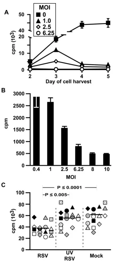
[image:3.585.317.519.67.337.2]Since lymphocytes were harvested on day 5 after exposure to MDCC, we sought to determine whether the immunosuppres-sive effects could be observed earlier and to verify the appro-priate experimental time course. Figure 3A demonstrates that T-cell proliferation in response to CMV antigens is suppressed as early as day 3, in a dose-dependent manner. However, max-imal suppression was observed on day 5 (Fig. 3A and B).
FIG. 1. RSV infects MDDC. MDDC (1⫻106/ml) were exposed
overnight to rgRSV (multiplicity of infection of 3) and washed twice before being examined by fluorescent microscopy. The fluorescent (A) and bright field (B) images of the same microscopic field were photo-graphed. The figure is representative of the results of three experi-ments.
FIG. 2. RSV induces MDDC-mediated suppression of T-cell pro-liferation. (A) CD4 T cells together with MDDC are sufficient to demonstrate RSV-induced immunosuppression. MDDC were exposed to RSV (multiplicity of infection of 2.5), influenza, or PIV3 (multiplic-ity of infection of 3) or mock infected for 4 h before the addition of either SEB-stimulated autologous lymphocytes or purified CD4 T cells. The cells were pulsed with [3H]thymidine on day 4 and harvested
on day 5. Each symbol shape is one of three donors. Open symbols indicate proliferation of T cells in a population of total lymphocytes, and filled symbols represent purified CD4 T cells. (B) RSV does not directly inhibit CD4 T-cell proliferation. RSV (final density, 1⫻106
PFU/ml) or the same volume of sucrose buffer was added to SEB-stimulated CD4 T cells and SEB for 3 days prior to pulsing with [3H]thymidine. The bar indicates the median of data from five donors,
each represented by a different symbol.
on November 8, 2019 by guest
http://jvi.asm.org/
[image:3.585.54.274.69.401.2]Figure 3C demonstrates reproducibility of experiments in which only CD4 T cells and MDDC were used and the neces-sity of live RSV for maximal immunosuppression. For these, and subsequent experiments, the MDDC were washed 4 h
after incubation with RSV to minimize any effect of RSV carryover. Consistent with the dose response shown in Fig. 3A, immunosuppression was diminished when exposure of MDDC to live RSV was limited to 4 h.
Supernatants from RSV-exposed MDDC transfers
immuno-suppression. To determine whether soluble factors might be
responsible for RSV-induced immunosuppression, we exposed MDDC to RSV, UV-RSV, or sucrose buffer (mock) for 4 h, after which time the cells were washed and incubated overnight before harvesting supernatant. Figures 4A and B show that only supernatants from MDDC exposed to live RSV suppress proliferation of CD4 T cells in response to SEB and that the suppressive effect is dose dependent. To confirm that RSV carryover is not responsible for this immunosuppression, we first incubated virus with humanized MAb (1 mg/ml) against either RSV F (fusion) protein (palivizumab), or human IgE (omalizumab) as a control to demonstrate the former’s neu-tralization capacity. As expected, supernatants from MDDC exposed to RSV that was neutralized by palivizumab did not suppress CD4 T-cell proliferation (Fig. 4C). In contrast, when added to supernatant that was harvested from RSV-exposed MDDC, palivizumab did not reverse immunosuppression (Fig. 4C), just as live RSV added to supernatants from mock-in-fected MDDC did not immunosuppress (Fig. 2B). Thus, the immunosuppressive activity transferred in supernatants from RSV-exposed MDDC is not due to RSV itself and is more likely due to one or more soluble factors that are MDDC derived.
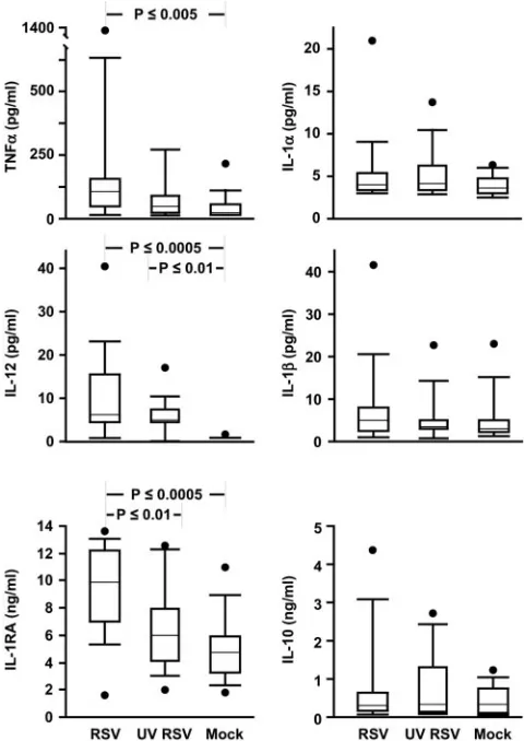
Analysis of cytokines in MDDC supernatants.To determine
which soluble factors might be responsible for immunosup-pression, we measured levels of a number of cytokines using multiplex bead assays. Figure 5 shows that there were no dif-ferences in supernatant derived from RSV-, UV-RSV-, or mock-exposed MDDC in concentrations of IL-1␣ or IL-1. Consistent with previous reports (32, 46), IL-1RA levels were high in RSV supernatants and low in UV-RSV and mock-infected supernatants. Since we found very little IL-1␣or -1, with no differences between the conditions and since the only known activity of IL-1RA is to block binding of IL-1␣/ to IL-1R, we did not explore further an immunosuppressive role for IL-1RA.
There were no differences in IL-10, and in all three condi-tions,transforming growth factor1 (TGF-1) was either very low or undetectable (not shown). Because IL-10 and TGF-1 are known to suppress T-cell proliferation, we tested whether addition of rIL-10 (25 ng/ml) or TGF-1 (10 ng/ml) would suppress proliferation in our system and found that they did not (not shown). Similarly, when we removed suppressor CD4 T cells, which secrete IL-10 and TGF-1, by magnetic bead selection (i.e., CD25⫹, CD4⫹lymphocytes), we saw no differ-ences in SEB-induced proliferation (not shown).
Tumor necrosis factor alpha levels were highest in the su-pernatants from RSV-infected MDDC, a difference that was significant only when compared to mock-infected MDDC su-pernatants. IL-12 was found at high levels in both RSV and UV-RSV supernatant but not in supernatant from the mock-infected MDDC. Exogenous IL-12 (2 ng/ml) or IL-2 (20 U/ml) and anti-IL-12 (2g/ml) also had no effect in our system (not shown).
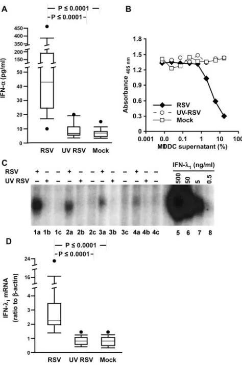
[image:4.585.59.258.65.522.2]IFN-␣has been previously reported to be solely responsible
FIG. 3. RSV-induced immunosuppression is dependent on time, dose, and live virus. (A and B) Time and dose dependency of RSV-induced immunosuppression of CMV antigen-stimulated proliferation. MDDC were exposed to CMV antigen (vol/vol⫽1:100) and RSV (mul-tiplicity of infection [MOI] of 0, 0.4, 1, 2.5, 6.25, 8, and 10) for 4 h before incubation with autologous lymphocytes. Cells were pulsed with [3
H]thy-midine at different time points for proliferation assay. The results, ex-pressed as mean cpm⫾SD, are from one representative experiment of three. (A) Only MOI 0, 1, 2.5, and 6.25 are shown for clarity. (B) Prolif-eration on day 5 of each experimental condition showing dose depen-dency at that time point. (C) Only live RSV induces immunosuppression. MDDC were incubated with live RSV or UV-RSV, each at an MOI of 1, or an equal volume of sucrose buffer for 4 h (mock), washed, and incu-bated with SEB-stimulated autologous purified CD4 T cells for 4 days before harvest on day 5 for [3H]thymidine uptake assay. The bar indicates
the median of data from 15 donors.
VOL. 80, 2006 IFN-␣ AND IFN-MEDIATE SUPPRESSION OF CD4 T CELLS 5035
on November 8, 2019 by guest
http://jvi.asm.org/
for RSV-induced immunosuppression (42). We therefore mea-sured IFN-␣using an ELISA that detects 12 of the 14 isoforms and found high concentrations uniquely in the RSV-exposed MDDC supernatants (Fig. 6A). By contrast, IFN-was unde-tectable in any of the MDDC supernatants (data not shown). Type III interferons, which include IFN-1, 2, and 3 (also known as IL-29, -28A, and -28B, respectively), are expressed by macrophages at low levels in response to RSV (58). Currently, there are no commercially available MAbs for measurement of IFN-by ELISA, so we used two biological assays. The first assay system measures activation of signal transducer and ac-tivator of transcription 1 (STAT1) induced by IFN- in a hamster cell line that is stably transfected with a cDNA encod-ing a chimeric receptor composed of the extracellular ligand-binding domain of IFN-R1 (IL-28R) and the intracellular domain of the IFN-␥R1 chain (27). The second assay system measures IFN-activity by quantifying the suppression of cell division using a chromogenic viability assay of a human lym-phoma cell line that was engineered to express the full-length IFN-R1 chain (19). Figures 6B and C demonstrate that only
FIG. 4. Supernatants from RSV-exposed MDDC transfer immuno-suppressive activity. (A) Supernatants from RSV, but not UV-RSV or mock-exposed MDDC, inhibit SEB-stimulated CD4 T-cell proliferation. MDDC were exposed to RSV or UV-RSV or mock exposed for 4 h and washed. Supernatants were harvested after overnight incubation and in-cubated with SEB-stimulated CD4 T cells for 3 days prior to pulsing with [3H]thymidine for 24 h. The bar indicates the median of data from 15
donors. (B) Inhibitory effect of RSV-exposed MDDC supernatants decreases with dilution. Means and SD of results from one representative experiment of three are shown. (C) RSV or RSV F proteins in the supernatants are not responsible for immunosuppression of CD4 T cells. RSV or superna-tants from RSV- or mock-exposed MDDC were incubated with palivi-zumab or omalipalivi-zumab (1-mg/ml final concentration) for 1 h at 37°C before addition to MDDC or SEB-stimulated CD4 T cells, respectively. The data, expressed as mean counts per minute⫾SD, are cumulative from three donors. Filled bars, MAb added directly to RSV prior to addition of virus to MDDC; open bars, MAb added to MDDC supernatants prior to mixing with CD4 T cells; a-RSV F, anti-RSV F protein; a-hIgE, anti-hIgE.
FIG. 5. RSV induces secretion of tumor necrosis factor alpha, IL-12, and IL-1RA, but not IL-1␣, IL-1, or IL-10, from MDDC. Data from 14 donors are shown. The upper and lower error bars indicate the 90th and 10th percentiles, respectively; the borders of each box indi-cate the 75th and 25th percentiles; the bar within the box indiindi-cates the median, and the filled circles show the highest (and in the case of IL-1RA, the lowest) data points of each set.
on November 8, 2019 by guest
http://jvi.asm.org/
supernatants from RSV-exposed MDDC contained apprecia-ble levels of IFN-, The concentration of type I IFN in the supernatants is estimated to be between 1 to 5 ng/ml and 5 to 20 ng/ml, based on the specific activity of rIFN-in the EMSA and proliferation assays, respectively. Expression of IFN-by RSV-infected MDDC was confirmed by qRT-PCR (Fig. 6D).
IFN-␣and IFN-contribute to the inhibitory effect of
RSV-exposed MDDC supernatants.We then asked whether IFN-␣
and IFN- mediate RSV-induced immunosuppression by treating the CD4 T cells with neutralizing MAb to IFNAR2 (CD118), one of the two chains of IFN-␣/ receptor, and
[image:6.585.44.281.67.425.2]MAbs to both of the members of the IFN-receptor complex, IL-10R2 and IL-28R. Two experimental systems were used: transfer of supernatants and Transwell cultures by placing MDDC in the top chamber and the CD4 T cells in the bottom chamber for the first 3 days of incubation. RSV-induced im-munosuppression in the Transwell system is shown in Fig. 7A. While each of the MAbs alone may have a modest effect, the combination of either anti-IFNAR2 with anti-IL10R2 or with anti-IL28R substantially abrogated suppression after transfer
[image:6.585.315.524.77.454.2]FIG. 6. RSV induces secretion of IFN-␣and IFN-from MDDC supernatants. (A) IFN-␣protein is expressed in response to live RSV. Data from 15 donors are shown. (B and C) IFN-activity is expressed in response to live RSV. (B) Supernatants from RSV-infected MDDC inhibit proliferation of BWLICR2 cells. The parent line BW5147 was unaffected by any of the supernatants (not shown). The figure shown is representative of data from five donors. (C) HT-29 cells transfected with the IL-28 receptor complex were exposed to MDDC superna-tants, harvested, and analyzed by EMSA for activation of STAT1 in response to supernatants from MDDC that were exposed to RSV (lanes a), UV RSV (lanes b), or mock infection (lanes c). Lanes 5 to 8 are nuclear extracts from transfected HT-29 cells treated with rhIFN-1 at 500, 50, 5, and 0.5 ng/ml, respectively. ⫹, present;⫺, absent. (D) IFN-1 mRNA is expressed in response to live RSV. RNA harvested from MDDC was analyzed for expression of the gene for IFN-1 by qRT-PCR and shown as a ratio to expression of the gene for -actin. Data from 16 donors are shown.
FIG. 7. IFN-␣ and IFN- contribute to the inhibitory effect of RSV-exposed MDDC supernatants. (A) RSV-induced immunosup-pression can be demonstrated with the Transwell system. MDDC were exposed to mock, live, or UV-inactivated RSV for 4 h, washed, and placed in the top chamber of Transwell plates (pore size, 0.4m). The bottom chamber contained autologous purified CD4 T cells stimulated with SEB (1 g/ml). Each symbol represents a different donor. (B) Neutralizing MAbs to IFN-␣and IFN-receptors together sub-stantially reverse the inhibitory effect of RSV-exposed MDDC super-natant (top) or in the Transwell system (5-m pore size, bottom). Each symbol represents a different donor. Since annof 6 is required for the Wilcoxon signed-rank test, the data from the top and bottom plots were pooled for additional statistical analysis. -IFNAR2, anti-IFNAR2; -IL10R2, anti-IL10R2; -IL28R, anti-IL28R; ⫹, present;⫺, absent.
VOL. 80, 2006 IFN-␣ AND IFN-MEDIATE SUPPRESSION OF CD4 T CELLS 5037
on November 8, 2019 by guest
http://jvi.asm.org/
of supernatants and in the Transwell system (Fig. 7B, top and bottom, respectively).
DISCUSSION
We explored the mechanism of RSV-induced immunosup-pression by first simplifying the experimental system to MDDC and autologous CD4 T cells, thus eliminating interactions with CD8 T cells, B cells, and NK cells. Our approach was similar to that of Bartz and colleagues (5), who used cord blood CD4 T cells, which are all naive, and cord blood CD34⫹cells to derive dendritic cells (DC). In that report, treatment of DC with RSV suppressed IFN-␥secretion but not proliferation in response to superantigen. Preliminarily, we have seen no difference in suppression by RSV-infected MDDC of sorted naive versus memory CD4 T cells (B. Chi and R. L. Rabin, unpublished data), but the difference in the source of DC, age of donor, or both may easily account for the difference in functional sup-pression between the two reports.
Since RSV had no direct effect on CD4 T cells (Fig. 2B), we could concentrate on MDDC as a source of suppressive activ-ity, and since suppressive activity could be transferred with supernatants from MDDC (Fig. 4A), we turned our attention toward soluble factors secreted by them. And since suppres-sion of T-cell proliferation is induced only with infection of MDDC by live RSV (Fig. 3C and 4A), we ruled out the pos-sibility that live RSV directly suppresses CD4 T-cell prolifer-ation by demonstrating, first, that CD4 T-cell suppression was unaffected by addition of palivizumab to supernatants from RSV-infected MDDC(Fig. 4C)and, second, that direct addi-tion of live (Fig. 2B) or UV-killed RSV (not shown) to CD4 T cells did not suppress proliferation in response to SEB. Fur-thermore, consistent with a previous report using a similar experimental system (5), live RSV, and RSV-derived protein levels in harvested supernatants actually diminished. However, there are some obvious limitations to these experiments that must be acknowledged. First, it is not known if palivizumab blocks virus or F binding to cells or if palivizumab neutraliza-tion is mediated at a postbinding step. Because palivizumab specifically interacts with a single epitope within the F1 region, it would not be expected to block virus attachment mediated by the F2 portion of the fusion glycoprotein or inhibit interactions with the G glycoprotein. Second, these experiments do not rule out the possibility that RSV-secreted G glycoprotein may play a role in T-cell suppression. It is apparent, however, that type I and III IFN-mediated suppression is distinct from inhibition of NF-B mediated by RSV-soluble G glycoprotein, the latter of which is not dependent on live virus (1, 41). Taken together, it appeared unlikely that RSV virions or proteins directly sup-press T cells in our experiments; thus, we focused on MDDC-derived soluble factors.
One such factor that has been held responsible for RSV-induced immunosuppression is IL-1RA (32, 46). We found high levels of IL-1RA in the MDDC supernatants but very little IL-1␣and IL-1, suggesting that IL-1RA probably has no role in this context. Similarly, TGF-1 and IL-10 are immuno-suppressive cytokines that were ruled out because TGF-1 was undetectable in the MDDC supernatants (not shown) and IL-10 levels were similar in all experimental conditions. Fur-thermore, neither addition of rhTGF-1 or rhIL-10 to control
MDDC supernatants nor exclusion of suppressor CD4 T cells (CD4⫹, CD25⫹lymphocytes), an important source of TGF-1 and IL-10 blocked RSV-induced suppression (not shown). IL-12 enhances T-cell proliferation (61) and was induced by RSV and UV-RSV. However, neither neutralization with anti-IL-12 MAb nor supplementation with rhanti-IL-12 blocked RSV-induced immunosuppression (not shown). Similarly, IFN-␥, classically a product of T and NK cells, can be also expressed by DC (60) and may be immunosuppressive (12, 37). Consis-tent with a previous report (39), neutralizing MAb to IFN-␥ enhanced, rather than diminished, suppression of proliferation (data not shown).
IFN-␣levels were elevated in supernatants of MDDC ex-posed to live RSV (Fig. 6A). Similarly, RSV induces expres-sion of IFN- in monocyte-derived macrophages (58) and MDDC (Fig. 6B, C, and D). IFN-1, IFN-2, and IFN-3 comprise the newly described type III IFN, which may be expressed by a variety of cells, including plasmacytoid DC, MDDC, and macrophages in response to a variety of stimuli, including viruses (13). The receptor for the type III IFN con-sists of the ligand-binding chain, IL-28R, and the accessory receptor chain, IL-10R2 (27, 53). IL-28R is unique to the type III IFN, but IL-10R2 also dimerizes with IL-10R1, IL-22R, and IL-26R to signal in response to their respective cytokines (for a review, see reference 18). In contrast to these three IL-10 family members, STAT1 and STAT2 primarily transduce sig-nals through the IFN-R complex, as they do for the IFN-␣/ and -␥receptors (16, 38).
To determine whether IFN-␣and IFN-contribute to RSV-induced immunosuppression, we used MAb to one of the two chains of IFN-␣/receptor, IFNAR2 (CD118), and to the two chains of the IFN-R complex, IL-10R2 and IL-28R, to block receptor binding. In contrast to a previous report that IFN-␣ is solely responsible for RSV immunosuppression (42), anti-IFNAR2 only modestly diminished immunosuppression (Fig. 7). Similarly, anti-IL10R2 or anti-IL-28R was relatively inef-fective. But when IFNAR2 was combined with either anti-IL-10R2 or anti-IL-28R, the level of suppression was substan-tially decreased, thus demonstrating synergism between the two IFN. Since type I and III IFN both signal through STAT1 and STAT2, the two IFN might synergize by surpassing a threshold of signaling that neither could achieve alone. On the other hand, the two IFN may qualitatively signal such that only when present together are unique pathways activated or blocked. To our knowledge, this is the first report that type III IFN affect T-cell function.
The dependence of RSV-induced immunosuppression on type I and III IFN is paradoxical considering that (i) IFN are expressed in response to a variety of stimuli, including many other viruses that are not associated with T-cell suppression; (ii) plasmacytoid DC may express 100-fold as much IFN-␣as MDDC and yet are critical to the ultimate antiviral adaptive response (56); and (iii) type I IFN prevent activation-induced death of murine T cells in vitro after stimulation with super-antigen in vivo (31). The emerging story, however, is that the effects of type I IFN on CD4 T cells are dependent on the context in which they are exposed. For example, Dondi and colleagues showed that, in the context of TcR stimulation, exogenous IFN-␣is antiapoptotic early and proapoptotic later (17). Most relevant to our report, entry into the cell cycle was
on November 8, 2019 by guest
http://jvi.asm.org/
delayed when TcR stimulation followed IFN-␣exposure (17), apparently due to sequestration by the type I IFN receptor complex of CD45, Lck, and ZAP-70 (40), all of which are necessary to initiate, but not to sustain, cellular responses to stimulation through the TcR (11). Similarly, high concentra-tions of IFN-␣ cause STAT2 to associate with STAT6 and enhance signaling through the IL-4 receptor for the first 6 h of exposure; thereafter, IFN-␣ inhibits IL-4 responses (20). Fi-nally, IFN-␣may activate STAT4 (36), which while insufficient to induce Th1 differentiation (7), may enhance it when induced by IL-12. It is likely that the contextual complexity of T-cell responses to IFN-␣is only increased by the addition of type III IFN into the equation. The combination of type I and type III IFN could favor signaling through pathways that preferentially activate antiproliferative effects of IFN rather than those path-ways that mediate the antiviral effects.
RSV has a number of strategies to minimize its vulnerability to IFN-␣. The products of the NS1 and NS2 genes inhibit the upstream expression of IFN-␣(8, 25, 47, 58) by suppressing activation and nuclear translocation of interferon regulatory factor 3 (8, 59) and block downstream responses by decreasing STAT2 levels (2, 44). Since cellular responses to IFN-␣ are complex and dependent upon context, it is reasonable to sus-pect that RSV may have achieved the fine balance of impairing antiviral properties of IFN-␣while its suppressive activity may persist. Proliferation is not likely the only impaired T-cell re-sponse. For example, since IL-4 attenuates CD8 T-cell cyto-lytic function (3, 4), cytolysis may be further impaired when IFN-␣enhances signaling in response to IL-4 (20). This is but one potential mechanism by which RSV may deviate T-cell responses in ways that are highly relevant to infection with RSV and its sequelae, including childhood wheezing and asthma.
Conversely, suppression of T-cell responses during RSV in-fection may actually offer some benefit to the host. Our model system uses MDDC and autologous CD4 T cells derived from healthy, young adult donors, a population in which RSV lower respiratory tract disease is rare (22). Since the host inflamma-tory response contributes significantly to lower respirainflamma-tory dis-ease (34), diminished or absent suppressive activity in infants 2 to 6 months of age (22) and the infirm elderly (21) may account for their susceptibility to RSV pneumonia.
ACKNOWLEDGMENTS
We thank Yeqiang Li for excellent technical support.
This work was supported by CBER intramural funds and a collab-orative grant from the Vaccine Research Center, NIAID. S.V.K. is supported by Public Health Services grant RO1 AI51139 from the National Institute of Allergy and Infectious Diseases and by American Heart Association grant AHA 0245131N.
The views expressed in this report are the personal opinions of the authors and are not the official opinion of the U.S. Food and Drug Administration, the National Institutes of Health, or the Department of Health and Human Services.
REFERENCES
1.Arnold, R., B. Konig, H. Werchau, and W. Konig.2004. Respiratory syncytial virus deficient in soluble G protein induced an increased proinflammatory response in human lung epithelial cells. Virology330:384–397.
2.Atreya, P. L., and S. Kulkarni.1999. Respiratory syncytial virus strain A2 is resistant to the antiviral effects of type I interferons and human MxA. Virology261:227–241.
3.Aung, S., and B. S. Graham.2000. IL-4 diminishes perforin-mediated and
increases Fas ligand-mediated cytotoxicity In vivo. J. Immunol.164:3487– 3493.
4.Aung, S., J. A. Rutigliano, and B. S. Graham.2001. Alternative mechanisms of respiratory syncytial virus clearance in perforin knockout mice lead to enhanced disease. J. Virol.75:9918–9924.
5.Bartz, H., O. Turkel, S. Hoffjan, T. Rothoeft, A. Gonschorek, and U. Schauer.2003. Respiratory syncytial virus decreases the capacity of myeloid dendritic cells to induce interferon-gamma in naive T cells. Immunology 109:49–57.
6.Beckford, A. P., R. O. Kaschula, and C. Stephen.1985. Factors associated with fatal cases of measles. A retrospective autopsy study. S. Afr. Med. J. 68:858–863.
7.Berenson, L. S., N. Ota, and K. M. Murphy.2004. Issues in T-helper 1 development–resolved and unresolved. Immunol. Rev.202:157–174. 8.Bossert, B., S. Marozin, and K. K. Conzelmann.2003. Nonstructural
pro-teins NS1 and NS2 of bovine respiratory syncytial virus block activation of interferon regulatory factor 3. J. Virol.77:8661–8668.
9.Breese Hall, C.1998. Respiratory syncytial virus, p. 2084–3011.InR. D. Feigin and J. D. Cherry (ed.), Textbook of pediatric infectious diseases, 4th ed. Saunders, Philadelphia, Pa.
10.Brogden, K. A., H. D. Lehmkuhl, and R. C. Cutlip.1998. Pasteurella hae-molytica complicated respiratory infections in sheep and goats. Vet. Res. 29:233–254.
11.Chan, A. C., D. M. Desai, and A. Weiss.1994. The role of protein tyrosine kinases and protein tyrosine phosphatases in T cell antigen receptor signal transduction. Annu. Rev. Immunol.12:555–592.
12.Chin, Y. E., M. Kitagawa, W. C. Su, Z. H. You, Y. Iwamoto, and X. Y. Fu. 1996. Cell growth arrest and induction of cyclin-dependent kinase inhibitor p21 WAF1/CIP1 mediated by STAT1. Science272:719–722.
13.Coccia, E. M., M. Severa, E. Giacomini, D. Monneron, M. E. Remoli, I. Julkunen, M. Cella, R. Lande, and G. Uze.2004. Viral infection and Toll-like receptor agonists induce a differential expression of type I and lambda interferons in human plasmacytoid and monocyte-derived dendritic cells. Eur. J. Immunol.34:796–805.
14.Collins, P., R. M. Chanock, and B. R. Murphy.2001. Respiratory syncytial virus, p. 1443–1485.InD. M. Knipe, P. M. Howley, D. E. Griffin, R. A. Lamb, M. A. Martin, B. Roizman, and S. E. Straus (ed.), Fields virology, 4th ed. Lippincott Williams and Wilkins, Philadelphia, Pa.
15.Culley, F. J., J. Pollott, and P. J. Openshaw.2002. Age at first viral infection determines the pattern of T cell-mediated disease during reinfection in adulthood. J. Exp. Med.196:1381–1386.
16.Decker, T., S. Stockinger, M. Karaghiosoff, M. Muller, and P. Kovarik.2002. IFNs and STATs in innate immunity to microorganisms. J. Clin. Investig. 109:1271–1277.
17.Dondi, E., G. Roue, V. J. Yuste, S. A. Susin, and S. Pellegrini.2004. A dual role of IFN-alpha in the balance between proliferation and death of human CD4⫹T lymphocytes during primary response. J. Immunol.173:3740–3747. 18.Donnelly, R. P., F. Sheikh, S. V. Kotenko, and H. Dickensheets.2004. The expanded family of class II cytokines that share the IL-10 receptor-2 (IL-10R2) chain. J. Leukoc. Biol.76:314–321.
19.Dumoutier, L., A. Tounsi, T. Michiels, C. Sommereyns, S. V. Kotenko, and J. C. Renauld.2004. Role of the interleukin (IL)-28 receptor tyrosine resi-dues for antiviral and antiproliferative activity of IL-29/interferon-lambda 1: similarities with type I interferon signaling. J. Biol. Chem.279:32269–32274. 20.Eriksen, K. W., V. H. Sommer, A. Woetmann, A. B. Rasmussen, C. Brender, A. Svejgaard, S. Skov, C. Geisler, and N. Odum.2004. Bi-phasic effect of interferon (IFN)-alpha: IFN-alpha up- and down-regulates interleukin-4 sig-naling in human T cells. J. Biol. Chem.279:169–176.
21.Falsey, A. R., P. A. Hennessey, M. A. Formica, C. Cox, and E. E. Walsh.2005. Respiratory syncytial virus infection in elderly and high-risk adults. N. Engl. J. Med.352:1749–1759.
22.Hall, C. B.2001. Respiratory syncytial virus and parainfluenza virus. N. Engl. J. Med.344:1917–1928.
23.Hall, S. L., A. Stokes, E. L. Tierney, W. T. London, R. B. Belshe, F. C. Newman, and B. R. Murphy.1992. Cold-passaged human parainfluenza type 3 viruses contain ts and non-ts mutations leading to attenuation in rhesus monkeys. Virus Res.22:173–184.
24.Hallak, L. K., D. Spillmann, P. L. Collins, and M. E. Peeples.2000. Glycos-aminoglycan sulfation requirements for respiratory syncytial virus infection. J. Virol.74:10508–10513.
25.Hengel, H., U. H. Koszinowski, and K. K. Conzelmann.2005. Viruses know it all: new insights into IFN networks. Trends Immunol.26:396–401. 26.Keles, I., A. K. Sharma, Z. Woldehiwet, and R. D. Murray.1999. The effects of
bovine respiratory syncytial on normal ovine lymphocyte responses to mitogens or antigens in vitro. Comp. Immunol. Microbiol. Infect. Dis.22:1–13. 27.Kotenko, S. V., G. Gallagher, V. V. Baurin, A. Lewis-Antes, M. Shen, N. K.
Shah, J. A. Langer, F. Sheikh, H. Dickensheets, and R. P. Donnelly.2003. IFN-lambdas mediate antiviral protection through a distinct class II cytokine receptor complex. Nat. Immunol.4:69–77.
28.Krempl, C., B. R. Murphy, and P. L. Collins.2002. Recombinant respiratory syncytial virus with the G and F genes shifted to the promoter-proximal positions. J. Virol.76:11931–11942.
VOL. 80, 2006 IFN-␣ AND IFN-MEDIATE SUPPRESSION OF CD4 T CELLS 5039
on November 8, 2019 by guest
http://jvi.asm.org/
29.Landegren, U.1984. Measurement of cell numbers by means of the endog-enous enzyme hexosaminidase. Applications to detection of lymphokines and cell surface antigens. J. Immunol. Methods67:379–388.
30.Madhi, S. A., and K. P. Klugman.2004. A role for Streptococcus pneu-moniae in virus-associated pneumonia. Nat. Med.10:811–813.
31.Marrack, P., J. Kappler, and T. Mitchell.1999. Type I interferons keep activated T cells alive. J. Exp. Med.189:521–530.
32.McCarthy, D. O., F. M. Domurat, J. E. Nichols, and N. J. Roberts, Jr.1989. Interleukin-1 inhibitor production by human mononuclear leukocytes and leukocyte subpopulations exposed to respiratory syncytial virus: analysis and comparison with the response to influenza virus. J. Leukoc. Biol.46:189–198. 33.McIntosh, K.2004. Respiratory syncytial virus, p. 1076–1079.InR. E. Be-hrman, R. M. Kleigman, and H. B. Jenson (ed.), Nelson textbook of pedi-atrics, 17th ed. Saunders, Philadelphia, Pa.
34.McNamara, P. S., and R. L. Smyth.2002. The pathogenesis of respiratory syncytial virus disease in childhood. Br. Med. Bull.61:13–28.
35.Moss, W. J., M. O. Ota, and D. E. Griffin.2004. Measles: immune suppres-sion and immune responses. Int. J. Biochem. Cell. Biol.36:1380–1385. 36.Nguyen, K. B., W. T. Watford, R. Salomon, S. R. Hofmann, G. C. Pien, A.
Morinobu, M. Gadina, J. J. O’Shea, and C. A. Biron.2002. Critical role for STAT4 activation by type 1 interferons in the interferon-gamma response to viral infection. Science297:2063–2066.
37.Novelli, F., M. M. D’Elios, P. Bernabei, L. Ozmen, L. Rigamonti, F. Alm-erigogna, G. Forni, and G. Del Prete.1997. Expression and role in apoptosis of the alpha- and beta-chains of the IFN-gamma receptor on human Th1 and Th2 clones. J. Immunol.159:206–213.
38.O’Shea, J. J., M. Gadina, and R. D. Schreiber.2002. Cytokine signaling in 2002: new surprises in the Jak/Stat pathway. Cell109(Suppl.):S121–S131. 39.Paton, A. W., and P. N. Goldwater.1990. Respiratory syncytial virus
modu-lation of adult and neonatal lymphocyte mitogenic responses and the role of interferon-gamma. Microb. Pathog.9:235–241.
40.Petricoin, E. F., III, S. Ito, B. L. Williams, S. Audet, L. F. Stancato, A. Gamero, K. Clouse, P. Grimley, A. Weiss, J. Beeler, D. S. Finbloom, E. W. Shores, R. Abraham, and A. C. Larner.1997. Antiproliferative action of interferon-alpha requires components of T-cell-receptor signalling. Nature 390:629–632.
41.Polack, F. P., P. M. Irusta, S. J. Hoffman, M. P. Schiatti, G. A. Melendi, M. F. Delgado, F. R. Laham, B. Thumar, R. M. Hendry, J. A. Melero, R. A. Karron, P. L. Collins, and S. R. Kleeberger.2005. The cysteine-rich region of respiratory syncytial virus attachment protein inhibits innate immunity elicited by the virus and endotoxin. Proc. Natl. Acad. Sci. USA102:8996– 9001.
42.Preston, F. M., P. L. Beier, and J. H. Pope.1995. Identification of the respiratory syncytial virus-induced immunosuppressive factor produced by human peripheral blood mononuclear cells in vitro as interferon-alpha. J. In-fect. Dis.172:919–926.
43.Preston, F. M., P. L. Beier, and J. H. Pope.1992. Infectious respiratory syncytial virus (RSV) effectively inhibits the proliferative T cell response to inactivated RSV in vitro. J. Infect. Dis.165:819–825.
44.Ramaswamy, M., L. Shi, M. M. Monick, G. W. Hunninghake, and D. C. Look.2004. Specific inhibition of type I interferon signal transduction by respiratory syncytial virus. Am. J. Respir. Cell Mol. Biol.30:893–900. 45.Roberts, N. J., Jr.1982. Different effects of influenza virus, respiratory
syncytial virus, and Sendai virus on human lymphocytes and macrophages. Infect. Immun.35:1142–1146.
46.Roberts, N. J., Jr., A. H. Prill, and T. N. Mann.1986. Interleukin 1 and interleukin 1 inhibitor production by human macrophages exposed to influ-enza virus or respiratory syncytial virus. Respiratory syncytial virus is a potent inducer of inhibitor activity. J. Exp. Med.163:511–519.
47.Schlender, J., B. Bossert, U. Buchholz, and K. K. Conzelmann.2000. Bovine respiratory syncytial virus nonstructural proteins NS1 and NS2 cooperatively antagonize alpha/beta interferon-induced antiviral response. J. Virol. 74: 8234–8242.
48.Schlender, J., V. Hornung, S. Finke, M. Gunthner-Biller, S. Marozin, K. Brzozka, S. Moghim, S. Endres, G. Hartmann, and K. K. Conzelmann.2005. Inhibition of toll-like receptor 7- and 9-mediated alpha/beta interferon pro-duction in human plasmacytoid dendritic cells by respiratory syncytial virus and measles virus. J. Virol.79:5507–5515.
49.Schlender, J., G. Walliser, J. Fricke, and K. K. Conzelmann.2002. Respi-ratory syncytial virus fusion protein mediates inhibition of mitogen-induced T-cell proliferation by contact. J. Virol.76:1163–1170.
50.Schneider-Schaulies, S., K. Bieback, E. Avota, I. Klagge, and V. ter Meulen. 2002. Regulation of gene expression in lymphocytes and antigen-presenting cells by measles virus: consequences for immunomodulation. J. Mol. Med. 80:73–85.
51.Sharma, R., and Z. Woldehiwet.1991. Depression of lymphocyte responses to phytohaemagglutinin in lambs experimentally infected with bovine respi-ratory syncytial virus. Res. Vet. Sci.50:152–156.
52.Sharma, R., and Z. Woldehiwet.1990. Increased susceptibility to Pasteurella haemolytica in lambs infected with bovine respiratory syncytial virus. J. Comp. Pathol.103:411–420.
53.Sheppard, P., W. Kindsvogel, W. Xu, K. Henderson, S. Schlutsmeyer, T. E. Whitmore, R. Kuestner, U. Garrigues, C. Birks, J. Roraback, C. Ostrander, D. Dong, J. Shin, S. Presnell, B. Fox, B. Haldeman, E. Cooper, D. Taft, T. Gilbert, F. J. Grant, M. Tackett, W. Krivan, G. McKnight, C. Clegg, D. Foster, and K. M. Klucher.2003. IL-28, IL-29 and their class II cytokine receptor IL-28R. Nat. Immunol.4:63–68.
54.Sieg, S., C. King, Y. Huang, and D. Kaplan.1996. The role of interleukin-10 in the inhibition of T-cell proliferation and apoptosis mediated by parain-fluenza virus type 3. J. Virol.70:4845–4848.
55.Sieg, S., C. Muro-Cacho, S. Robertson, Y. Huang, and D. Kaplan.1994. Infection and immunoregulation of T lymphocytes by parainfluenza virus type 3. Proc. Natl. Acad. Sci. USA91:6293–6297.
56.Siegal, F. P., N. Kadowaki, M. Shodell, P. A. Fitzgerald-Bocarsly, K. Shah, S. Ho, S. Antonenko, and Y. J. Liu.1999. The nature of the principal type 1 interferon-producing cells in human blood. Science284:1835–1837. 57.Sigurs, N., R. Bjarnason, F. Sigurbergsson, and B. Kjellman.2000.
Respi-ratory syncytial virus bronchiolitis in infancy is an important risk factor for asthma and allergy at age 7. Am. J. Respir. Crit. Care Med.161:1501–1507. 58.Spann, K. M., K. C. Tran, B. Chi, R. L. Rabin, and P. L. Collins.2004. Suppression of the induction of alpha, beta, and lambda interferons by the NS1 and NS2 proteins of human respiratory syncytial virus in human epi-thelial cells and macrophages. J. Virol.78:4363–4369.
59.Spann, K. M., K. C. Tran, and P. L. Collins.2005. Effects of nonstructural proteins NS1 and NS2 of human respiratory syncytial virus on interferon regulatory factor 3, NF-kappaB, and proinflammatory cytokines. J. Virol. 79:5353–5362.
60.Yamaguchi, N., Y. Fujimori, Y. Fujibayashi, I. Kasumoto, H. Okamura, K. Nakanishi, and H. Hara.2005. Interferon-gamma production by human cord blood monocyte-derived dendritic cells. Ann. Hematol.84:423–428. 61.Zeh, H. J., III, S. Hurd, W. J. Storkus, and M. T. Lotze.1993. Interleukin-12
promotes the proliferation and cytolytic maturation of immune effectors: implications for the immunotherapy of cancer. J. Immunother.14:155–161. 62.Zhang, L., M. E. Peeples, R. C. Boucher, P. L. Collins, and R. J. Pickles. 2002. Respiratory syncytial virus infection of human airway epithelial cells is polarized, specific to ciliated cells, and without obvious cytopathology. J. Vi-rol.76:5654–5666.