Evolution of Developmental Control Mechanisms
Coral
emx-Am
can substitute for
Drosophila empty spiracles
function in head, but not
brain development
Beate Hartmann
a, Martin Müller
a, Nikki R. Hislop
b, Bettina Roth
a, Lucija Tomljenovic
b,
David J. Miller
b, Heinrich Reichert
a,⁎
a
Biozentrum, University of Basel, Klingelbergstrasse 50, CH-4056 Basel, Switzerland
b
ARC Centre of Excellence for Coral Reef Studies and Comparative Genomics Centre, James Cook University, Townsville, Qld. 4811, Australia
a b s t r a c t
a r t i c l e i n f o
Article history:
Received for publication 13 May 2008 Revised 22 December 2009 Accepted 24 December 2009 Available online 4 January 2010
Keywords:
Brain development Evolution
ems Emx
Cephalic gap gene Hexapeptide
Drosophila
Acropora
The ems/Emxgenes encode homeodomain transcription factors that have conserved actions in anterior embryonic patterning in bilaterian animals ranging from insects to mammals. Recently, genes of theems/ Emx family have been identified in cnidarians raising the possibility that some of their developmental functions might be conserved throughout the Eumetazoa. To determine to what extent functions of a cnidarianems/Emxprotein have been retained across phyla, we carried out cross-phylum rescue expression experiments in which the coralAcropora emx-Amgene was misexpressed inDrosophila emsmutants. Our findings demonstrate that coralemx-Amcan substitute forflyemsin embryonic head development and rescue the open head defect and the loss of segmentalengrailedexpression domains inDrosophila ems mutants. In contrast, the coralemx-Amgene can not substitute forflyemsin embryonic brain development. Even when a hexapeptide motif of the type present in theDrosophila emsgene is inserted into the coral emx-Amgene, rescue of the developmental brain defects inflyemsmutants fails. Thesefindings have implications for understanding the evolutionary origins of head versus brain patterning mechanisms.
© 2009 Elsevier Inc. All rights reserved.
Introduction
Comparative molecular analyses are providing increasing evidence for the evolutionary conservation of key developmental control genes involved in embryonic regionalization and patterning. This is exemplified by the genes of the empty spiracles (ems/Emx) family which play key roles in anterior embryonic patterning in animals ranging from insects to mammals. The ems gene was originally discovered in aDrosophilascreen for zygotic patterning mutations and is a member of the cephalic gap gene group (Cohen and Jürgens, 1991; Cohen and Jürgens, 1990; Jürgens et al., 1984). It encodes a homeodomain containing transcription factor and was grouped into the“dispersed superclass”of homeobox genes (Gehring et al., 1994). It shares with the homeobox genes of the“complex superclass,”which includes the Hox genes, a hexapeptide motif of the core consensus sequence YPWL, located N-terminal of the homeodomain. The hexapeptide plays a role in the cooperative DNA binding of Hox proteins with the cofactor extradenticle (Exd) (Mann and Chan, 1996). However it is not known if the hexapeptide of theEmsprotein serves a similar function.
During early embryogenesis, the Drosophila ems gene is first expressed at the early cellular blastoderm stage in a single circumferential stripe at the anterior end of the embryo. Later in embryogenesis,ems expression is detected in discrete ectodermal patches of the labral, antennal and intercalary segment of the anterior head as well as in lateral regions of ectodermal and neural cell patches in all trunk segments (Dalton et al., 1989; Walldorf and Gehring, 1992). The large antennal expression domain of the ectoderm gives rise to the ems expressing neuroblasts of the deutocerebral brain anlage and the smaller intercalary expression domain gives rise to the
emsexpressing neuroblast of the tritocerebral brain anlage (Urbach and Technau, 2003; Younossi-Hartenstein et al., 1996). Mutation of
emsleads to a gap-like phenotype in the embryonic head, which includes deletions of cuticular and cephalic sensory structures in the ocular, antennal and intercalary segments, and results in an open-head phenotype (Cohen and Jürgens, 1990; Dalton et al., 1989; Jürgens et al., 1984; Walldorf and Gehring, 1992). Mutation ofems
also results in deletion of deutocerebral and tritocerebral brain anlagen, which in turn leads to massive structural defects in the embryonic brain (Hirth et al., 1995). This brain phenotype is due to defective specification of the neuroectoderm in the antennal and intercalary domains ofemsmutants and correlates with the absense of the proneural genelethal of scute, which is thought to be required for neuroectodermal cells to adopt the competence to become neuroblasts (Younossi-Hartenstein et al., 1997).
⁎Corresponding author. Fax: +41612671613.
E-mail address:[email protected](H. Reichert). 0012-1606/$–see front matter © 2009 Elsevier Inc. All rights reserved. doi:10.1016/j.ydbio.2009.12.038
Contents lists available atScienceDirect
Developmental Biology
Comparative studies suggest that the expression pattern and the function ofems/Emxgenes in anterior patterning are evolutionarily conserved throughout the Bilateria (reviewed byLichtneckert and Reichert, 2005). Thus, the murine homologues of theDrosophila ems
gene,Emx1andEmx2, are both expressed in the anterior brain and genetic loss-of-function analyses suggests that they are involved in anterior brain development (Pellegrini et al., 1996; Qiu et al., 1996; Simeone et al., 1992a,b; Yoshida et al., 1997). Further evidence for the functional equivalence of the ems and Emx2 gene products comes from cross-phylum rescue experiments in which ubiquitous overexpression of a mouseEmx2transgene in an emsnull mutant background rescues the brain phenotype of the mutantfly embryos (Hartmann et al., 2000). The notion of an evolutionarily conserved role ofems/Emx genes in anterior patterning of the brain and/or head during embryonic development is supported by expression data from a number of different vertebrate and invertebrate bilaterian species including mouse (Simeone et al., 1992a), chick (Bell et al., 2001), frog (Pannese et al., 1998), zebrafish (Morita et al., 1995), shark (Derobert et al., 2002), lamprey (Myojin et al., 2001), hemichordate (Lowe et al., 2003) and nematode (Aspöck et al., 2003). Indeed,ems/Emxgenes appear to be such universal anterior markers that (together with the homeotic genes) they were included in the original description of the“zootype,”the universal genetic toolkit of animals (Slack et al., 1993).
ems/Emx genes have also been identified in the sister group of bilaterians, the Cnidaria (de Jong et al., 2006; Mokady et al., 1998). The Cnidaria are thought to represent one of the most ancient metazoan phyla and thus provide a useful outgroup for comparative studies of the molecular control of development in the more complex bilaterians. Among cnidarians, the Anthozoa, which includes the sea anemoneNematostellaand the coralAcropora,is viewed as the most basal class, and these animals appear to reflect most faithfully ancestral characteristics (Bridge et al., 1992, 1995; Kortschak et al., 2003; Technau et al., 2005). Recently it has been demonstrated that in
Acropora millepora, anemxgene is expressed in putative neurons in the aboral half of the planula larva (de Jong et al., 2006), which is the anterior region with respect to swimming direction. This apparent neuron-specific and axially restricted expression of theAcropora emx
gene (emx-Am) in a developing cnidarian larva is reminiscent of the expression of ems/Emx genes in the anterior CNS and/or head systems of developing bilaterian embryos. This raises the question of whether theems/Emxgenes are part of an ancient axis- and/or CNS-specification system that predates the cnidarian/bilaterian split. If this were the case, then the protein encoded by the coralemx-Amgene might still retain conserved functions in development that could be uncovered in cross-phylum rescue experiments performed on a bilaterian such asDrosophila. Thus, a challenge now is to determine which functional aspects of the proteins encoded by theems/Emx
genes have been conserved and which aspects have diverged after the Cnidaria/Bilateria split.
To address this question, we carried out targeted gene expression experiments in which the coral emx-Am gene was expressed in appropriate embryonic domains of Drosophila ems mutants. Our findings demonstrate thatemx-Amcan indeed substitute foremsin epidermal head development and, thus, rescue the open head defect as well as restore the intercalary and antennal head segments of
Drosophila emsmutants. The relative rescue efficiency of the emx-Am gene in these targeted gene expression experiments was comparable to that of the Drosophila ems gene. In contrast, the coralemx-Amgene was not able to rescue the brain defect of Dro-sophilamutants. Even when a hexapeptide motif of the type present in theDrosophila emsgene was inserted into the coralemx-Amgene, rescue of the developmental brain defects inflyemsmutants failed. The implications of thesefindings for understanding the evolution-ary origins of head vs. brain patterning mechanisms in Eumetazoa are discussed.
Material and methods
Transgene constructs
Modified coral andflyemsconstructs were generated from cDNA templates (in Bluescript) using the Stratagene QuikChange site-directed mutagenesis system under the manufacturers recommended conditions. In the case of theDrosophila emscDNA, the YPWL amino acid motif that forms the core of the hexapeptide was mutated to AAAL using the primer 5′gatagctatcagctggccgccgcgctgctcagccgcc3′ and its reverse complement. The coral Emx protein lacks a definitive hexapeptide (de Jong et al., 2006); in this case, the amino acid sequence YPCA which occurs 28-31 residues N-terminal of the homeodomain was mutated to YPWL using the primer 5′ ctttttcattc-tatccgtggctgtcaagtcatcgatatgtac3′and its reverse complement. In both cases the introduction of modifications was confirmed by DNA sequencing prior to cloning the cDNAs into pUAST. Both mutant and wild-typeAcropora emxcDNAs were cloned in via the NotI and XhoI sites, and the correspondingDrosophila emsconstructs were gener-ated via the EcoRI site.
Fly husbandry
Flies were reared on standard corn meal medium at room temperature. Transgenicflies for the 3 P{w+mC, UAS-cDNA} constructs were generated according to standard procedures (Rubin and Spradling, 1982). For UAS-ems and UAS-emx-Am, additional lines were generated by mobilization of an existing transgene withΔ2-3 transposase (Robertson et al., 1988). Linkage of each insert was determined by standard genetic crossing procedures.
For the rescue experiment,first, three double balanced stocks were established: (1) nocSco/CyO, LacZ;ems9H83e/TM3, Sb e LacZ, (2) L/
CyO, LacZ;ems9H83e/TM3, Sb e LacZ and (3) L/CyO, LacZ;ems9Q64e/
TM3, Sb e LacZ. These stocks were used to establish all UAS-(cDNA)/ CyO, LacZ;ems9H83 e/TM3, Sb e LacZ and UAS-(cDNA)/CyO, LacZ;
ems9Q64e/TM3, Sb e LacZ stocks used for this study. Thesca-Gal4
driver line (Klaes et al., 1994) of the genotypesca-Gal4;ems9H83e/
TM3, Sb e LacZ and the nos-Gal4-3GCN4-Bcd3′UTR driver line (Janody et al., 2000) of the genotype nos-Gal4-3GCN4-Bcd3′UTR;ems9H83e/
TM3, Sb e LacZ were obtained accordingly. The sca-Gal4 driver is active during neuroectoderm specification and neuroblast formation (Sprecher et al., 2004). The nos-Gal4-3GCN4-Bcd3′UTR transgene allows expression of the Gal4-GCN4 protein as a maternal anterior gradient in the earlyDrosophilaembryo (Janody et al., 2000). For rescue experiments, Gal4 driver virgins were collected and crossed to UAS-(cDNA)/CyO, LacZ;ems9H83e/TM3, Sb e LacZ or UAS-(cDNA)/
CyO, LacZ;ems9Q64e/TM3, Sb e LacZ males. As negative control, Gal4
driver virgins were mated withems9H83e/TM3, Sb e LacZ or ems9Q64
e/TM3, Sb e LacZ males. Embryo collections of these crosses were done at 25 °C.
For targeted ectopic expression oforthodenticle, the responder line
UAS-otd(Blanco et al., 2009) was used.
Immunocytochemistry and cuticular preparations
Cuticular preparations were made according to standard proce-dures (Nüsslein-Volhard et al., 1984).
Genetic rescue analysis
In genetic rescue experiments,sca-Gal4 ornos-Gal4 driven P{UAS-cDNA} activity in homozygousemsnull mutants (ems9H83/ems9H8or
ems9H83/ems9Q64) was confirmed by the absence of balancer-specific (CyO,LacZ; TM3,LacZ)β-gal immunoreactivity.
The brain phenotype in ems−/− embryos was scored as fully
rescued, when there was no gap in the neuronal tissue between the protocerebral brain hemispheres and the subesophageal neuromeres. The open head phenotype inems−/−embryos was scored as fully
rescued, when the brain lobe was not protruding out of the embryo and the dorsal ectodermal tissue appeared normal. Embryos of different genetic background were rated in a double blind experi-mental setup. A total of 50 hemispheres were judged for each experimental group as well as for the negative control group (ems9H83/ems9H83 without rescue construct). Thus, we obtained a rescue score for each experimental group (RS) and a rescue score for the negative control group (RSems−/−), the latter reflecting the fact
that there is no complete penetrance of the mutant phenotype. To correct for this incomplete phenotypic penetrance, the rescue scores were transformed into relative rescue efficiencies (RRE in %) according to the formula: RRE = RS−RSems−/−/50− RSems−/−⁎
100. For statistical analysis we used the Fisher test. Values with p≤0.01 are referred to as significant.
A rescue of the head patterning defect inems−/−embryos was
judged by scoring each embryonic hemisphere for the presence of the engrailed intercalary stripe, the antennal stripe or the engrailed head spot. A total of 50 hemispheres were judged for each experimental group as well as for the negative control group (ems9H83/ems9Q64 without rescue construct). Thus, for each group the total number of observed head segments could vary between 0 and 150. For statistical analysis we used the independentt test. Values with p≤0.01 are referred to as significant.
Laser confocal microscopy
For laser confocal microscopy a Leica TCS SP microscope was used. Optical sections were taken approximately every 1μm, recorded in line average mode with picture size of 512 × 512 pixels. Captured images from optical sections were arranged and processed using IMARIS (Bitplane). Figures were arranged and labeled using Adobe Photoshop.
Results
The coral emx gene does not rescue embryonic brain defects in Drosophila ems mutants
TheDrosophila emsgene is a cephalic gap gene that is important for the development of the embryonic brain (Hartmann et al., 2000; Hirth et al., 1995; Urbach and Technau, 2003; Younossi-Hartenstein et al., 1996). The brain inemsnull mutant embryos has a deletion in the deutocerebral and tritocerebral brain anlagen, which leads to a prominent gap devoid of neuronal cells between the remaining protocerebral brain hemispheres and the subesophageal neuromeres (Figs. 1A and B). We analysed the penetrance of this embryonic brain phenotype and found it present in 56% of theemsmutant embryos. For quantification of the following rescue experiments, a penetrance of 56% of the mutant brain phenotype is defined as a relative rescue efficiency of 0% (see Methods).
To determine if the brain phenotype can be rescued by targeted expression of theflyemsgene, we made use of the GAL4-UAS system (Brand and Perrimon, 1993). We generated two UAS responder lines
with independent inserts of an ems transgene in an otherwise homozygousemsmutant background (seeMaterial and Methods). In order to target theemstransgenes to the early ectodermal tissue of the head, we used asca-GAL4 driver (Klaes et al., 1994). In a wild-type background this results in expression of ems throughout the neuroectoderm starting at stage 9 and includes the anterior cephalic domain in which ems is normally expressed (data not shown). Targeted expression of the fly ems transgenes in an ems mutant background resulted in a significant rescue of the brain phenotype in both transgenicfly lines (seeFig. 2). The relative rescue efficiency was 57% for the UAS-ems6insertion line and 50% for UAS-ems9. In rescued embryos, contiguous neuronal tissue extended from the protocerebral hemispheres to the mandibular neuromere and prominent cellular gaps in this tissue were not observed (Fig. 1C, arrow).
To determine whether the brain phenotype inDrosophilaembryos can be rescued by targeted expression of the coralemx-Amgene, we generated two UAS responder lines with independent insertions of an
emx-Am transgene (see Material and Methods). The targeted expression of theemx-Amgene inemsmutantDrosophilaembryos did not result in a significant rescue of the brain phenotype in either of the two insertion lines (seeFig. 2). The relative rescue efficiency was 4% for the UAS-emx-Am5insertion line and−11% for UAS-emx-Am6. In the majority of the transgenic embryos, large cellular gaps were observed between the remaining brain hemispheres and the subesophageal neuromeres (Fig. 1D, arrow).
The Drosophila ems gene without a normal hexapeptide domain rescues embryonic brain defects in ems mutants
The failure of the coral Emx protein to rescue the brain defects in
ems mutants might be due to the fact that it lacks a definitive hexapeptide motif (de Jong et al., 2006); this motif is assumed to have a role in cooperative DNA binding of some Antp-superclass home-odomain proteins with the cofactor extradenticle (Mann and Chan, 1996). It is conceivable that this motif is essential foremsaction in embryonic brain development. To test this, we generated UAS responder lines carrying anemx-Am+hptransgene, which contained the coral emx-Am gene with an added hexapeptide motif (see Material and Methods).
However, even with the addition of a hexapeptide the coralemx
transgene remained unable to restore the brain phenotype in ems
mutant embryos (seeFig. 2). The relative rescue efficiency for
UAS-emx-Am+hpwas 7%. Most mutant embryos still showed a prominent gap in the brain between the brain hemispheres and the subesopha-geal neuromeres (Fig. 1F, arrow).
Given that the presence or absence of the hexapeptide motif did not alter the rescue efficiency of the coralemx-Amgene, we wondered if the hexapeptide motif in the fly ems gene might be similarly dispensable for a rescue of the embryonic brain mutant phenotype. To investigate this, we generated UAS responder lines with independent inserts of anems−hptransgene in which the hexapeptide motif had been mutated (seeMaterial and Methods).
Targeted expression of theflyemsgene without a normal hexapep-tide motif inemsmutant embryos resulted in a significant rescue of the brain phenotype (seeFigs. 2 and 1E). One insertion line (UAS-ems−hp3) showed a relative rescue efficiency of 71% and a second insertion line (UAS-ems−hp2) showed a relative rescue efficiency of 36% for the mutant phenotype. Thesefindings indicate that the hexapeptide motif of theems
gene is not necessary for embryonic brain development.
expression in the head is found in the antennal and preantennal neuroectoderm as well as in many neuroblasts delaminating from these regions (Cohen and Jürgens, 1990; Finkelstein and Perrimon, 1990; Urbach and Technau, 2003). Targeted misexpression of otd
under the control of thesca-GAL4 driver inemsmutants did not result in a rescue of the brain phenotype (seeFig. 2). The relative rescue efficiency for this line was 11%.
The coral emx gene rescues the embryonic open-head defect in Drosophila ems mutants
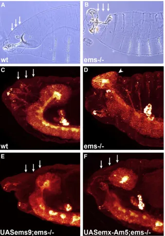
[image:4.595.123.464.51.583.2]In addition to its role in embryonic brain development,emsis also involved in proper embryonic development of the head ectoderm, and in ems mutants embryonic head development is severely perturbed (Cohen and Jürgens, 1990; Dalton et al., 1989; Fig. 1.The coralemxgene does not rescue theflyemsmutant brain defect. (A–F) Reconstructions of lateral confocal microscopy sections of stage 15 embryos; anti-HRP immunolabeling; anterior of body axis to the left. (A) Wild type; (B)emsnull mutant; a gap devoid of neuronal cells appears between the remaining protocerebral brain anlage and the subesophageal neuromeres (arrow, compare to A); (C)emsnull mutant carrying asca-GAL4/UAS-ems6 rescue construct; (D)emsnull mutant carrying asca-GAL4/UAS- emx-Am5 rescue construct; (E)emsnull mutant carrying asca-GAL4/UAS-ems−hp3 rescue construct; and (F)emsnull mutant carrying asca-GAL4/UAS-emx-Am+hprescue construct.
(C and E) Both, targeted expression of theflyemsgene or theflyemsgene with a mutated hexapeptide are able to rescue theemsmutant brain phenotype. No gap is observed in the brain (arrow) and the neuronal tissue extends from the protocerebral neuromere to the mandibular neuromere. (D and F) Neither targeted expression of the coralemx-Am
Jürgens et al., 1984; Walldorf and Gehring, 1992). Accordingly, analysis of cuticular preparations fromemsmutants reveals a gap-like, open-head phenotype in which the anterodorsal head ectoderm is defective (Figs. 3A and B). Associated with the defective ectodermal tissue in the dorsal head are perturbed head involution and protrusion of the brain hemispheres out of the embryo proper (Figs. 3C and D). We analysed the penetrance of this open-head phenotype and found it present in 66% of the homozygous ems
mutant embryos. For quantification of the following genetic rescue experiments, we define a penetrance of 66% of the mutant head phenotype as a relative rescue efficiency of 0% (see Methods).
To ensure that the open-head phenotype could be rescued by targeted expression of theflyemsgene we again used thesca-GAL4 driver line together with the two UAS responder lines that had independent inserts of anemstransgene. Targeted expression of the fly ems transgene in an ems mutant background resulted in a significant rescue of the open-head phenotype in both transgenicfly lines (seeFig. 4); the relative rescue efficiencies were 97% (UAS-ems9) and 85% (UAS-ems6). In rescued embryos the anterodorsal head ectoderm was completely intact and the brain hemispheres remained contained within the embryo proper (Fig. 3E).
To examine whether the open-head phenotype in Drosophila
embryos can be rescued by targeted expression of the coral emx-Am gene, we used the sca-GAL4 line to drive expression of the two UAS responder lines that had independent inserts of an emx-Am transgene. Remarkably, targeted expression of the emx-Am
gene under the control of the sca-GAL4 driver in ems mutant
Drosophilaembryos resulted in rescue of the open-head phenotype with near equivalent efficiency to that of the Drosophilagene (see Fig. 4). Thus relative rescue efficiencies of 82% (UAS-emx-Am5) and 70% (UAS-emx-Am6) were determined for the two insertion lines. In general, ems mutant embryos rescued by the coral emx genes were indistinguishable from those rescued by the fly ems gene (compare Figs. 3E and F) with respect to the head ectodermal phenotype. Thus, in contrast to its inability to replace the Droso-phila emsgene in embryonic brain development, the coralemx-Am
gene might be able to replace the ems gene in embryonic head development.
The Drosophila ems gene without a normal hexapeptide domain rescues the embryonic open-head defect in ems mutants
Since the coral emx-Am gene lacks the hexapeptide motif the rescue result reported above suggests that this motif might also be dispensable for proper ems protein function during head develop-ment. To test this, we made use of the two UAS responder lines with independent inserts of anems−hp transgene with a mutated hexapeptide motif. Targeted expression of the mutated fly ems
transgene under the control of a sca-GAL4 driver in ems mutant
Drosophila embryos rescued the open-head phenotype with the same efficiency as the wild-type gene (seeFig. 4). One insertion line (UAS-ems−hp3) showed complete rescue (100% relative rescue efficiency), and the second insertion line (UAS-ems−hp2) showed a relative rescue efficiency of 97%. These findings indicate that the hexapeptide motif is not necessary for the action of theemsgene in embryonic development of the head ectoderm.
To control for the specificity of this rescue effect we again analysed the ability oforthodenticle(otd) to rescue the open-head defect ofems
mutant embryos. Targeted misexpression of anotdtransgene under the control of the sca-GAL4 driver in ems mutants resulted in a relative rescue efficiency of 33% which was not statistically significant (at the pb0.01 level) (seeFig. 4).
The coral emx gene rescues embryonic head patterning defects in Drosophila ems mutants
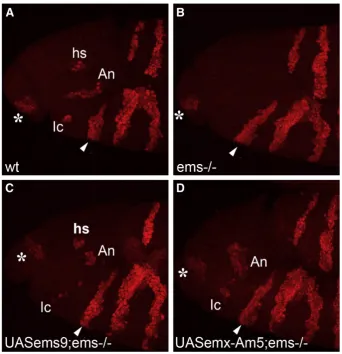
The ability of the coralemx-Amgene to rescue the open head defect suggests that the coral protein can replace thefly protein in embryonic head development. Ems is necessary for the establishment and correct development of the ocular, antennal and intercalary segments of the developing head (Cohen and Jürgens, 1990; Dalton et al., 1989; Jürgens et al., 1984; Walldorf and Gehring, 1992). We next wanted to test whether the coralemx-Amgene is able to rescue the development of these three head segments. For this, we focused on the pattern ofengrailed(en) expression in the head of early embryos, which in the wild-type characteristically delimit the different cephalic segments (Cohen and Jürgens, 1990; Schmidt-Ott and Technau, 1992; Schock et al., 2000). As expected, in the wild-typeen expression domains corresponding to each cephalic segment can be clearly identified, while inemsmutant embryos expression ofenis lacking in the intercalary and antennal segments and in a portion of the preantennal (ocular) region (Figs. 5A and B). In order to target the
emsor emx-Am transgenes at an early embryonic time point, we used anos-Gal4 driver (Janody et al., 2000, see Methods). In a wild-type background this resulted in expression ofemsexclusively and throughout the cephalic region starting from early blastoderm until stage 11 of embryogenesis (data not shown). Targeted expression of theflyemstransgenes with and without a hexapeptide motif in an
emsmutant background resulted in a significant rescue of the head segmentation defect as indicated by wild-type-like en expression patterns in the transgenic lines UAS-ems9, UAS-ems6, UAS-ems−hp3 and UAS-ems−hp2 (seeFig. 6). In rescued embryos,enexpression was restored in the intercalary and antennal segments as well as in the preantennal domain which is marked by theenhead spot (Fig. 5C). Remarkably, targeted expression of the emx-Am gene under the control of thenos-GAL4 driver inemsmutantDrosophilaembryos also resulted in a significant rescue of the head segment defect as monitored byenexpression domains (Figs. 5D and 6). In embryos of both transgenic lines (UAS-emx-Am5 and UAS-emx-Am6) the en
[image:5.595.53.288.54.192.2]intercalary spot and theenantennal stripe were restored. The domain of theenantennal stripe appeared broader than in thefly rescue and was arranged in a more vertical orientation compared to the horizontal orientation of the antennal stripe in the wild type. This might reflect a slight disarrangement of the ectodermal tissue, but could also indicate a rescue of the preantennal head spot which is
Fig. 2.Relative rescue efficiency of embryonic brain defect inemsnull mutants for different rescue constructs. The relative efficiency of rescue is shown for theflyems
gene (two independent insertion lines;ems6 andems9), the coralemx-Amgene (two independent insertion lines;emx-Am5 andemx-Am6), the coralemx-Amgene with an added hexapeptide motif (emx-Am+hp), theflyemsgene with a mutated hexapeptide
motif (two independent insertion lines;ems−hp3 andems−hp2) and, as a negative
control, for theotdgene. All genes were expressed in theemsmutant under the control of the samesca-GAL4 driver line. See Methods for explanation of relative rescue efficiency. A significant rescue resulted from targeted expression of theflyemsgene. A rescue of the brain defect is also achieved by targeted expression of theflyemsgene with a mutated hexapeptide motif. Neither targeted expression of the coralemx-Am
fused to the antennal stripe. Targeted expression of theotdgene in
emsmutantDrosophila embryos did not rescue the head segment defect (Fig. 6). We conclude that in contrast to its inability to replace theDrosophila emsgene in embryonic brain development, the coral
emx gene is able to replace the ems gene in embryonic head patterning.
Discussion
Theems/Emxgenes are key elements of the conserved molecular genetic mechanisms that control the patterning and specification of the anteroposterior body axis and neuraxis in bilaterians. Although
the roles of these patterning genes are well understood inDrosophila
and mouse, their evolutionary origins are equivocal (Holland, 2000; Martindale, 2005). Given that the Cnidaria are the sister group to the Bilateria (Medina et al., 2001), a comparative analysis of cnidarian vs. bilaterianems/Emxgene homolog function is likely to be informative as to the origin of bilaterian patterning mechanisms. In this report we have used targeted gene expression experiments inDrosophila to demonstrate the cross-phylum functional conservation of the cnidar-ianemx-Amgene. Ourfindings indicate thatemx-Amcan substitute foremsin embryonic head development and rescue the open head defect as well as cephalic segmental patterning as indicated byen
[image:6.595.125.465.52.538.2]expression domains in Drosophila ems mutants. In contrast, the
Fig. 3.The coralemxgene rescues theflyemsmutant head defect. (A and B) Cuticular preparations of late embryos; lateral views of anterior part of animal. (A) Wild type; (B)ems
Fig. 4.Relative rescue efficiency of embryonic head defect inemsnull mutants for different rescue constructs. The relative efficiency of rescue is shown for theflyems
gene (two independent insertion lines;ems9 andems6), the coralemx-Amgene (two independent insertion lines;emx-Am5 andemx-Am6), theflyemsgene with a mutated hexapeptide motif (two independent insertion lines;ems−hp3 andems−hp2) and, as a
negative control, for theotdgene. All genes were expressed in theemsmutant under the control of the samesca-GAL4 driver line. See Methods for explanation of relative rescue efficiency. A significant rescue (with pb0.01; bars with two stars) resulted from targeted expression of theflyemsgene, the coralemx-Amgene and theflyemsgene with a mutated hexapeptide motif. Targeted expression ofotddid not result in a significant rescue of theemsmutant phenotype (p = 0.02).
Fig. 5.The coralemxgene rescues the loss of specific head segments inemsmutantflies. (A–D) Reconstructions of lateral confocal microscopy sections of stage 11 embryos; anti-en immunolabeling; anterior of body axis to the left, arrowhead indicates mandibular segment, star indicates labral segment. (A) Wild type; in the headenis expressed in the labial, maxillary, mandibular, intercalary (Ic) and antennal segment (An), a preantennal region as indicated by theenhead spot (hs) and the labral segment. (B)emsnull mutant; loss ofen
expression in three head segments; the preantennal head spot, the intercalary and antennal segment. En expression in the labral and the three gnathal segments is unaffected. (C)
emsnull mutant carrying anos-GAL4/UAS-ems9 rescue construct;enexpression is restored in theenhead spot, the antennal and intercalary segment. (D)emsnull mutant carrying a
nos-GAL4/UAS-emx-Am5 rescue construct. Targeted expression of the coralemx-Amgene results in a rescue of theemsmutant head defect in the intercalary and antennal segment.
[image:7.595.55.286.54.173.2]enexpression in the antennal segment is broader than in wild type which might be due to a fusion of the preantennal head spot with the antennal stripe.
Fig. 6.Total number of rescued head segments as judged byenexpression pattern in
emsnull mutants for different rescue constructs. The number of rescued head segments is shown for theflyemsgene (two independent insertion lines;ems9 andems6), the coralemx-Amgene (two independent insertion lines;emx-Am5 andemx-Am6), thefly
emsgene with a mutated hexapeptide motif (two independent insertion lines;ems−hp3
andems−hp
2) and, as a control, for theotdgene. All genes were expressed in theems
[image:7.595.131.474.323.678.2]cnidarianemx-Amgene cannot rescue the embryonic brain defect of
Drosophila emsmutants, even when a hexapeptide motif of the type present in theDrosophila emsgene was inserted into the cnidarian gene. Although there appears to be no simple relationship between the major longitudinal axis of the cnidarians (oral-aboral) and the anteroposterior axis in bilaterians, the ectodermal expression pat-terns of the coralemx-Amgene and theflyemsgene are comparable in that both are axially regionalized. Theemsgene is expressed in a regionally restricted manner in the anterior cephalic ectoderm of the fly embryo (Dalton et al., 1989; Walldorf and Gehring, 1992). The
emx-Amgene is regionally expressed in the ectoderm in the aboral half to two thirds of the planula larva (de Jong et al., 2006). The cross-phylum rescue experiments reported here imply that in addition to their comparable expression patterns, these genes are functionally related. Implied conservation of function of Ems/Emx proteins has previously been shown between insects and vertebrates and between insects and nematodes (Aspöck et al., 2003; Hartmann et al., 2000). Thesefindings suggest that functional conservation of the Ems/Emx proteins might predate the origins of the Bilateria. TheAcroporato
Drosophila cross-phylum rescue experiments presented here now suggest that the functional aspects of the proteins utilized in Droso-philato mediate anterior cephalic development has been conserved by Ems/Emx proteins from the very beginning of eumetazoan evolution to the present. This is remarkable not only because of the large evolutionary distance between cnidarians and insects but also because the former do not display cephalization. Furthermore, the fact that these proteins act as transcription factors implies that a subset of their downstream targets, be these genes or protein cofactors, may also have been evolutionarily conserved. Thus, the simplest explana-tion of the cross-phylum rescue data presented here is that at least some components of the anteroposterior specification systems known from Bilateria coexisted in the common ancestor of modern cnidarians and bilaterians but reached their present sophistication in bilaterian cephalization only after evolutionary separation and divergence (Hobmayer et al., 2000).
The nervous system-specific expression of both cnidarian emx-Am(de Jong et al., 2006) andDrosophila emsgenes (Hartmann et al., 2000; Hirth et al., 1995; Urbach and Technau, 2003; Younossi-Hartenstein et al., 1996) could indicate that the two genes might also have comparable and evolutionarily conserved roles in nervous system development. However, our cross-phylum rescue experi-ments provide no support for this. Thus, while a general ectodermal function may be conserved between the Acropora and Drosophila ems/Emx genes, the lack of rescue in these experiments suggests that the roles of these genes in central nervous system development may not be conserved. It is highly likely that the emergence of the first nervous system predated the evolutionary divergence of Bilateria and Radiata, which is likely to have been in late pre-Cambrian time, given the fact that neurons and nervous systems are present in both. However, in terms of complexity there are marked differences between the two animal groups. Cnidarian nervous systems are relatively simple, indeed, the nerve nets of sessile anthozoan such as corals are the simplest known nervous systems of extant animals (Bullok and Horridge, 1965; Mackie, 2004). In contrast, the central nervous system of an insect likeDrosophilais a highly complex structure composed of hundreds of thousands of neurons that are interconnected in exquisitely organized ganglionic neuropil structures (Burrows, 1996; Strausfeld, 1976). Failure of the
Acroporaprotein to rescue thefly brain phenotype may therefore reflect the greater number of regulatory interactions that must be mediated by the Drosophila protein in constructing a highly complex centralized nervous system. Indeed it seems reasonable to assume that many of these interactions, protein–DNA or protein– protein, evolved in the lines leading to animals with complex brains after their separation from the lines leading to animals with simple nerve nets.
Surprisingly, in our experiments the hexapeptide motif was not required for rescue of either the cephalic ectoderm or the embryonic nervous system phenotypes, suggesting that it might be redundant for normal Ems function. There are precedents for this. For example, mutation of the AbdA hexapeptide does not appear to alter its binding site specificity and this motif is dispensable with respect to AbdA epidermal functions (Merabet et al., 2003). Whilst the classical function of the hexapeptide is thought to be in modification of the binding site specificity of Hox proteins by recruiting the Exd/Pbx proteins (Chan et al., 1994, 1995; Johnson et al., 1995), otherfindings demonstrate this hexapeptide-mediated interaction with cofactors has additional complexity. For example, in the Hox protein Lab, the hexapeptide appears to inhibit Lab function by inhibiting DNA binding. This is because Lab proteins, in which this motif was deleted or mutated, bind DNA with higher affinity than do proteins with an intact hexapeptide. Moreover, this hexapeptide mutant of Lab had an increased ability to activate transcription in vivo (Chan et al., 1996). In this case, Exd appears to induce a conformational change in Lab, thereby overcoming the negative function of the hexapeptide. If the hexapeptide of the Ems protein functions similarly, then mutating the hexapeptide should increase the ability of Ems to activate its transcriptional targets. This would explain why rescue experiments withemstransgenes with or without a hexapeptide lead to similar results. Alternatively, the fact that the Ems hexapeptide is dispensable in our rescue experiments might be an artefact of driving high-level pan-neural expression via thesca-GAL4 line. In the case of Ubx, the hexapeptide appears to provide an interface for the assembly of transcription repression complexes; Ubx lacking the hexapeptide can form repression complexes, but much higher concentrations are required (Tour et al., 2005).
Acknowledgments
We thank Nathalie Dostatni (Curie Institute, Paris) for the nos-Gal4-3GCN4-Bcd3′UTR transgenic stock, Uwe Walldorf for the Ems antibody, Tanja Radimerski and Simon Sprecher for help at the initial stages of this work, Susanne Flister for technical assistance and Frank Hirth for helpful discussions. Supported by the Swiss NSF (to H.R.) and from the Australian Research Council (both directly and via the Centre of Excellence for Coral Reef Studies) and the Australian Academy of Science (to DJM).
References
Aspöck, G., Ruvkun, G., Bürglin, T.R., 2003. TheCaenorhabditis elegansems class homeobox gene ceh-2 is required for M3 pharynx motoneuron function. Development 130, 3369–3378.
Bell, E., Ensini, M., Gulisano, M., Lumsden, A., 2001. Dynamic domains of gene expression in the early avian forebrain. Dev. Biol. 236, 76–88.
Blanco, J., Seimiya, M., Pauli, T., Reichert, H., Gehring, W.J., 2009. Wingless and Hedgehog signaling pathways regulate orthodenticle and eyes absent during ocelli development inDrosophila. Dev. Biol. 329, 104–115.
Brand, A.H., Perrimon, N., 1993. Targeted gene expression as a means of altering cell fates and generating dominant phenotypes. Development 118, 401–415. Bridge, D., Cunningham, C.W., Schierwater, B., DeSalle, R., Buss, L.W., 1992. Class-level
relationships in the phylum Cnidaria: evidence from mitochondrial genome structure. Proc. Natl. Acad. Sci. U. S. A. 89, 8750–8753.
Bridge, D., Cunningham, C.W., DeSalle, R., Buss, L.W., 1995. Class-level relationships in the phylum Cnidaria: molecular and morphological evidence. Mol. Biol. Evol. 12, 679–689.
Bullok, T.H., Horridge, G.A., 1965. Structure and function in the nervous systems of invertebrates. Freeman, San Francisco.
Burrows, M., 1996. The neurobiology of an insect brain. Oxford Univ. Press, Oxford. Chan, S.K., Jaffe, L., Capovilla, M., Botas, J., Mann, R.S., 1994. The DNA binding specificity
of Ultrabithorax is modulated by cooperative interactions with extradenticle, another homeoprotein. Cell 78, 603–615.
Chan, S.K., Popperl, H., Krumlauf, R., Mann, R.S., 1996. An extradenticle-induced conformational change in a HOX protein overcomes an inhibitory function of the conserved hexapeptide motif. EMBO J. 15, 2476–2487.
Chang, C.P., Shen, W.F., Rozenfeld, S., Lawrence, H.J., Largman, C., Cleary, M.L., 1995. Pbx proteins display hexapeptide-dependent cooperative DNA binding with a subset of Hox proteins. Genes Dev. 9, 663–674.
Cohen, S.M., Jürgens, G., 1990. Mediation ofDrosophilahead development by gap-like segmentation genes. Nature 346, 482–485.
Dalton, D., Chadwick, R., McGinnis, W., 1989. Expression and embryonic function of
empty spiracles: aDrosophilahomeobox gene with two patterning functions on the anterior-posterior axis of the embryo. Genes Dev. 3, 1940–1956.
de Jong, D.M., Hislop, N.R., Hayward, D.C., Reece-Hoyes, J.S., Pontynen, P.C., Ball, E.E., Miller, D.J., 2006. Components of both major axial patterning systems of the Bilateria are differentially expressed along the primary axis of a “radiate”
animal, the anthozoan cnidarianAcropora millepora. Dev. Biol. 298, 632–643. Derobert, Y., Plouhinec, J.L., Sauka-Spengler, T., Le Mentec, C., Baratte, B., Jaillard, D., Mazan,
S., 2002. Structure and expression of three Emx genes in the dogfishScyliorhinus canicula: functional and evolutionary implications. Dev. Biol. 247, 390–404. Finkelstein, R., Perrimon, N., 1990. Theorthodenticlegene is regulated bybicoidand
torsoand specifiesDrosophilahead development. Nature 346, 485–488. Gehring, W.J., Affolter, M., Burglin, T., 1994. Homeodomain proteins. Annu. Rev.
Biochem. 63, 487–526.
Hartmann, B., Hirth, F., Walldorf, U., Reichert, H., 2000. Expression, regulation and function of the homeobox gene empty spiracles in brain and ventral nerve cord development ofDrosophila. Mech. Dev. 90, 143–153.
Hirth, F., Therianos, S., Loop, T., Gehring, W.J., Reichert, H., Furukubo-Tokunaga, K., 1995. Developmental defects in brain segmentation caused by mutations of the homeobox genesorthodenticleandempty spiraclesinDrosophila. Neuron 15, 769–778. Hobmayer, B., Rentzsch, F., Kuhn, K., Happel, C.M., von Laue, C.C., Snyder, P., Rothbacher,
U., Holstein, T.W., 2000. WNT signalling molecules act in axis formation in the diploblastic metazoan Hydra. Nature 407, 186–189.
Holland, L.Z., 2000. Body-plan evolution in the Bilateria: early antero-posterior patterning and the deuterostome-protostome dichotomy. Curr. Opin. Genet. Dev. 10, 434–442.
Janody, F., Reischl, J., Dostatni, N., 2000. Persistence of Hunchback in the terminal region of theDrosophilablastoderm embryo impairs anterior development. Development 127, 1573–1582.
Johnson, F.B., Parker, E., Krasnow, M.A., 1995. Extradenticle protein is a selective cofactor for theDrosophilahomeotics: role of the homeodomain and YPWM amino acid motif in the interaction. Proc. Natl. Acad. Sci. U. S. A. 92, 739–743. Jürgens, G., Wieschaus, E., Nüsslein-Volhard, C., Kluding, H., 1984. Mutations affecting
the pattern of the larval cuticle in Drosophila melanogaster. Roux's Arch. Development. Biol. 193, 283–295.
Klaes, A., Menne, T., Stollewerk, A., Scholz, H., Klämbt, C., 1994. The ETS transcription factors encoded by theDrosophilagene pointed direct glial cell differentiation in the embryonic CNS. Cell 78, 149–160.
Kortschak, R.D., Samuel, G., Saint, R., Miller, D.J., 2003. EST analysis of the cnidarian
Acropora milleporareveals extensive gene loss and rapid sequence divergence in the model invertebrates. Curr. Biol. 13, 2190–2195.
Lichtneckert, R., Reichert, H., 2005. Insights into the urbilaterian brain: conserved genetic patterning mechanisms in insect and vertebrate brain development. Heredity 94, 465–477.
Lowe, C.J., Wu, M., Salic, A., Evans, L., Lander, E., Stange-Thomann, N., Gruber, C.E., Gerhart, J., Kirschner, M., 2003. Anteroposterior patterning in hemichordates and the origins of the chordate nervous system. Cell 113, 853–865.
Mackie, G.O., 2004. Central neural circuitry in the jellyfish Aglantha: a model“simple nervous system”. NeuroSignals 13, 5–19.
Mann, R.S., Chan, S.K., 1996. Extra specificity from extradenticle: the partnership between HOX and PBX/EXD homeodomain proteins. Trends Genet. 12, 258–262.
Martindale, M.Q., 2005. The evolution of metazoan axial properties. Nat. Rev., Genet. 6, 917–927.
Medina, M., Collins, A.G., Silberman, J.D., Sogin, M.L., 2001. Evaluating hypotheses of basal animal phylogeny using complete sequences of large and small subunit rRNA. Proc. Natl. Acad. Sci. U. S. A. 98, 9707–9712.
Merabet, S., Kambris, Z., Capovilla, M., Berenger, H., Pradel, J., Graba, Y., 2003. The hexapeptide and linker regions of the AbdA Hox protein regulate its activating and repressive functions. Dev. Cell. 4, 761–768.
Mokady, O., Dick, M.H., Lackschewitz, D., Schierwater, B., Buss, L.W., 1998. Over one-half billion years of head conservation? Expression of an ems class gene inHydractinia symbiolongicarpus (Cnidaria: hydrozoa). Proc. Natl. Acad. Sci. U. S. A. 95, 3673–3678.
Morita, T., Nitta, H., Kiyama, Y., Mori, H., Mishina, M., 1995. Differential expression of two zebrafish emx homeoprotein mRNAs in the developing brain. Neurosci. Lett. 198, 131–134.
Myojin, M., Ueki, T., Sugahara, F., Murakami, Y., Shigetani, Y., Aizawa, S., Hirano, S., Kuratani, S., 2001. Isolation of Dlx and Emx gene cognates in an agnathan species,
Lampetra japonica, and their expression patterns during embryonic and larval development: conserved and diversified regulatory patterns of homeobox genes in vertebrate head evolution. J. Exp. Zool. 291, 68–84.
Nüsslein-Volhard, C., Wieschaus, E., Kluding, H., 1984. Mutations affecting the pattern of the larval cuticle inDrosophila melanogaster. I Zygotic loci on the second chromosome. Wilhelm Roux's Arch. Dev. Biol. 193, 267–282.
Pannese, M., Lupo, G., Kablar, B., Boncinelli, E., Barsacchi, G., Vignali, R., 1998. The
XenopusEmx genes identify presumptive dorsal telencephalon and are induced by head organizer signals. Mech. Dev. 73, 73–83.
Patel, N., 1994. Imaging neuronal subsets and other cell types in whole mount Droso-philaembryos and larvae using antibody probes. In: Goldstein, L., E, F. (Eds.),
Drosophila melanogaster: practical uses in cell biology. Academic Press, New York. Pellegrini, M., Mansouri, A., Simeone, A., Boncinelli, E., Gruss, P., 1996. Dentate gyrus
formation requiresEmx2. Development 122, 3893–3898.
Qiu, M., Anderson, S., Chen, S., Meneses, J.J., Hevner, R., Kuwana, E., Pedersen, R.A., Rubenstein, J.L., 1996. Mutation of the Emx-1 homeobox gene disrupts the corpus callosum. Dev. Biol. 178, 174–178.
Robertson, H.M., Preston, C.R., Phillis, R.W., Johnson-Schlitz, D.M., Benz, W.K., Engels, W.R., 1988. A stable genomic source of P element transposase inDrosophila melanogaster. Genetics 118, 461–470.
Rubin, G.M., Spradling, A.C., 1982. Genetic transformation of Drosophila with transposable element vectors. Science 218, 348–353.
Schmidt-Ott, U., Technau, G.M., 1992. Expression ofenandwgin the embryonic head and brain ofDrosophilaindicates a refolded band of seven segment remnants. Development 116, 111–125.
Schock, F., Reischl, J., Wimmer, E., Taubert, H., Purnell, B.A., Jackle, H., 2000. Phenotypic suppression of empty spiracles is prevented by buttonhead. Nature 405, 351–354. Simeone, A., Acampora, D., Gulisano, M., Stornaiuolo, A., Boncinelli, E., 1992a. Nested
expression domains of four homeobox genes in developing rostral brain. Nature 358, 687–690.
Simeone, A., Gulisano, M., Acampora, D., Stornaiuolo, A., Rambaldi, M., Boncinelli, E., 1992b. Two vertebrate homeobox genes related to theDrosophila empty spiracles
gene are expressed in the embryonic cerebral cortex. EMBO J. 11, 2541–2550. Slack, J.M.W., Holland, P.W.H., Graham, C.F., 1993. The zootype and the phylotypic
stage. Nature 361, 490–492.
Sprecher, S.G., Müller, M., Kammermeier, L., Miller, D.F., Kaufman, T.C., Reichert, H., Hirth, F., 2004. Hox gene cross-regulatory interactions in the embryonic brain of
Drosophila. Mech. Dev. 121, 527–536.
Strausfeld, N.J., 1976. Atlas of an insect brain. Springer Verlag, Berlin.
Technau, U., Rudd, S., Maxwell, P., Gordon, P.M., Saina, M., Grasso, L.C., Hayward, D.C., Sensen, C.W., Saint, R., Holstein, T.W., Ball, E.E., Miller, D.J., 2005. Maintenance of ancestral complexity and non-metazoan genes in two basal cnidarians. Trends Genet. 21, 633–639.
Therianos, S., Leuzinger, S., Hirth, F., Goodman, C.S., Reichert, H., 1995. Embryonic development of theDrosophilabrain: formation of commissural and descending pathways. Development 121, 3849–3860.
Tour, E., Hittinger, C.T., McGinnis, W., 2005. Evolutionarily conserved domains required for activation and repression functions of theDrosophilaHox protein Ultrabithorax. Development 132, 5271–5281.
Urbach, R., Technau, G.M., 2003. Molecular markers for identified neuroblasts in the developing brain ofDrosophila. Development 130, 3621–3637.
Walldorf, U., Gehring, W.J., 1992.Empty spiracles, a gap gene containing a homeobox involved inDrosophilahead development. EMBO J. 11, 2247–2259.
Yoshida, M., Suda, Y., Matsuo, I., Miyamoto, N., Takeda, N., Kuratani, S., Aizawa, S., 1997.
Emx1andEmx2functions in development of dorsal telencephalon. Development 124, 101–111.
Younossi-Hartenstein, A., Nassif, C., Green, P., Hartenstein, V., 1996. Early neurogenesis of theDrosophilabrain. J. Comp. Neurol. 370, 313–329.