0022-538X/05/$08.00⫹0 doi:10.1128/JVI.79.24.15388–15397.2005
Copyright © 2005, American Society for Microbiology. All Rights Reserved.
Production of Infectious Human Cytomegalovirus Virions Is Inhibited
by Drugs That Disrupt Calcium Homeostasis in the
Endoplasmic Reticulum
Jennifer A. Isler, Tobi Goldberg Maguire, and James C. Alwine*
Department of Cancer Biology, Abramson Family Cancer Research Institute, School of Medicine, University of Pennsylvania, Philadelphia, Pennsylvania 19104-6142
Received 25 July 2005/Accepted 28 September 2005
We previously reported that human cytomegalovirus (HCMV) infection induces endoplasmic reticulum (ER) stress, resulting in activation of the unfolded protein response (UPR). Although some normal consequences of UPR activation (e.g., translation attenuation) are detrimental to viral infection, we have previously shown that HCMV infection adapts the UPR to benefit the viral infection (14). For example, UPR-induced translation attenuation is inhibited by viral infection, while potentially beneficial aspects of the UPR are maintained. In the present work, we tested the ability of HCMV to overcome a robust induction of the UPR by the drugs thapsigargin and clotrimazole (CLT), which disrupt ER calcium homeostasis. A 24-h treatment with these drugs beginning at 48, 72, or 96 h postinfection (hpi) completely inhibited further production of infectious virions. HCMV could not overcome the inhibition of global translation by CLT; however, between 48 and 72 hpi, HCMV overcame translational inhibition caused by thapsigargin. Despite the restoration of translation in thapsigargin, the accumulation of immediate-early and early gene products was modestly retarded (50% or less), whereas the accumulation of an early-late and late gene product was significantly retarded. Electron microscopic analysis shows that the drugs severely disrupt the maturation of HCMV virions. This can be accounted for by both the retarded accumulation of late gene products and the drug-induced depletion of ER calcium, which disrupts critical cellular functions needed for maturation.
The unfolded protein response (UPR), which results from the accumulation of misfolded proteins in the endoplasmic reticulum (ER), triggers a complex signal transduction path-way mediated by three ER membrane resident protein sensors: PKR-like ER kinase (PERK), activating transcription factor 6 (ATF6), and inositol-requiring enzyme 1 (IRE-1). The net effect of the UPR is to provide the cell with a means of eliminating unfolded proteins from the ER by (i) inhibiting translation to reduce the load of proteins in the ER; (ii) in-ducing the expression of genes encoding chaperones and deg-radation factors to properly fold and degrade, respectively, the misfolded proteins; and (iii) inducing the expression of genes whose products will increase metabolism to help the cell re-cover. PERK phosphorylates eucaryotic initiation factor 2␣ (eIF2␣), leading to global translation inhibition, and, in a seemingly contradictory fashion, stimulates the translation of specific mRNAs, such as the mRNA encoding ATF4 (8, 9). Expression of ATF4 leads to transcriptional activation of genes whose products are involved in metabolism. UPR-induced ac-tivation of ATF6 occurs at the level of protein cleavage (36). The N-terminal product of ATF6 functions as a transcription factor which activates transcription from many ER chaperone genes. In addition, ATF6 induces the transcription of the Xbp-1 gene (20, 34, 37). The Xbp-1 transcript becomes fully processed through splicing mediated by the third UPR sensor, IRE-1. The resulting mRNA encodes XBP-1, a transcription
factor which activates expression of genes encoding chaper-ones and, in particular, protein degradation factors (19, 20).
Several viruses, including human cytomegalovirus (HCMV), have been shown to induce ER stress and activate UPR sig-naling (14, 15, 30, 31). Viruses which induce ER stress and activate the UPR are faced with the consequences of UPR activation, some of which may be beneficial to the viral infec-tion (e.g., expression of chaperones) and some of which may be detrimental (e.g., translation attenuation). Hepatitis C virus has been shown to manipulate the UPR by inhibiting PERK phosphorylation of eIF2␣ and inhibiting the transcriptional activity of XBP-1, thereby maintaining protein translation and stability, respectively (25, 32). Similarly, we recently demon-strated that infection by HCMV induces the UPR but also modulates its effects (14). In HCMV-infected cells, phosphor-ylation of eIF2␣occurs but is regulated in such a manner that global translation is not inhibited and yet phospho-eIF2␣ -de-pendent translation of ATF4 is maintained. HCMV infection inhibits cleavage of ATF6, yet activates chaperone genes by an alternative mechanism. In addition, HCMV induction of the UPR results in the splicing of Xbp-1 mRNA, yet transcrip-tional activation of the XBP-1 target genes (e.g., those encod-ing protein degradation factors) is inhibited, presumably to keep viral proteins in the ER from being degraded. However, it has also been proposed that HCMV activation of the UPR aids the viral infection by facilitating the degradation of class I major histocompatibility complex proteins (33).
Thapsigargin and clotrimazole (CLT) are two drugs known to induce the UPR through their disruption of calcium ho-meostasis in the ER. Both drugs lead to attenuation of protein synthesis. Thapsigargin causes calcium release from the ER * Corresponding author. Mailing address: 314 Biomedical Research
Building II/III, 421 Curie Blvd., University of Pennsylvania, Philadel-phia, PA 19104. Phone: (215) 898-3256. Fax: (215) 573-3888. E-mail: alwine@mail.med.upenn.edu.
15388
on November 8, 2019 by guest
http://jvi.asm.org/
and activates calcium store-regulated calcium influx, thereby allowing calcium stores to be refilled (28). CLT also causes calcium release from intracellular stores but inhibits calcium-store regulated influx; thus, calcium calcium-stores cannot be refilled (3). The release of ER calcium by both drugs (i) decreases the level of ER calcium, resulting in decreased activity of calcium-dependent chaperones and, consequently, the accumulation of misfolded proteins and activation of the UPR, and (ii) in-creases the concentration of cytosolic calcium, resulting in stimulation of calcium-dependent enzymes (e.g., protein ki-nases) that regulate the function of cytosolic proteins. How-ever, disruption of calcium homeostasis is not the only phar-macological means to induce the UPR; tunicamycin induces ER stress by inhibiting N-linked glycosylation of proteins.
Given our previous finding that HCMV infection induces mechanisms which specifically regulate the three branches of UPR signaling (PERK, ATF6, and IRE-1) to favor viral rep-lication (14), we asked whether these virally induced mecha-nisms could circumvent a deliberate and severe activation of the UPR by thapsigargin, CLT, and tunicamycin. We find that thapsigargin and CLT have a profound inhibitory effect on infectious virion production. However, in the case of thapsi-gargin this is not the result of UPR-induced inhibition of global translation, since this is largely overcome by 48 to 72 hours postinfection (hpi). Despite the restoration of translation in thapsigargin, the accumulation of immediate-early and early gene products was somewhat retarded (50% or less), whereas the accumulation of early-late and late gene products was significantly retarded. Electron microscopic (EM) analysis showed that the drugs severely disrupt the maturation of HCMV virions. This can be accounted for by both the retarded accumulation of late gene products and the drug-induced de-pletion of ER calcium, which disrupts critical cellular functions needed for maturation.
MATERIALS AND METHODS
Cells and viruses.Life-extended human foreskin fibroblasts (HFFs) (4) were propagated and maintained at 37°C in 5% CO2in Dulbecco’s modified Eagle’s
medium supplemented with 10% fetal calf serum, Glutamax, and antibiotics. Cells were infected with the Towne strain of HCMV at a multiplicity of infection (MOI) of 2.5.
Drugs.All drugs were dissolved in dimethyl sulfoxide (DMSO), and DMSO was used as a control in all experiments. All drugs were obtained from Sigma (St. Louis, MO), and final drug concentrations were as follows: 2M thapsigargin, 10
M CLT, and 2g/ml tunicamycin.
Virus growth curves.HFFs were grown in six-well plates and infected at an MOI of 2.5. Twenty-four hours prior to each harvest time point, drug or DMSO was added to the medium. At designated time points, all but 1 ml of medium was removed from each plate and placed in a 15-ml conical tube. The cells in the well were scraped into the remaining 1 ml of medium, sonicated to dislodge cell-associated viruses, and centrifuged for 10 min in a microcentrifuge at 4°C. The supernatants were combined with previously collected medium, aliquoted, and stored at⫺80°C. Viral titration was carried out using a 50% tissue culture infective dose method described previously (11).
Metabolic labeling.HFF cells were infected at an MOI of 2.5 with HCMV under normal conditions. The cells were treated with DMSO (solvent control) or drugs during three 24-h time periods: 6 to 30 hpi, 24 to 48 hpi, and 48 to 72 hpi. Fifteen minutes prior to the end of the drug treatment, the cells were washed and incubated in Dulbecco’s modified Eagle’s medium lacking methionine and cys-teine (met/cys-free medium; Gibco, Carlsbad, CA), which had been prewarmed to 37°C. The cells were then pulse labeled for 30 min by the addition of pre-warmed met/cys-free medium containing [35S]methionine and [35S]cysteine at
125Ci/ml. The appropriate drugs or DMSO were included in the medium during washes and pulse labeling. At the end of the 30-min labeling, the
radio-active medium was removed, cells were washed twice with cold phosphate-buffered saline (PBS), and extracts were prepared using radioimmunoprecipita-tion assay buffer. Incorporaradioimmunoprecipita-tion of35S into proteins was determined by standard
trichloroacetic acid (TCA) precipitation.
The HCMV major immediate-early protein IE1/IEP72 was immunoprecipi-tated from the labeled extracts using rabbit polyclonal antibody specific to the first 85 amino acids common to all HCMV major immediate-early proteins (MIEPs) (10); protein A agarose was then added, and immunoprecipitates were collected by centrifugation, washed with PBS, and analyzed by sodium dodecyl sulfate-polyacrylamide gel electrophoresis (SDS-PAGE) (11% polyacrylamide gels). Gels were dried and analyzed with a Molecular Dynamics PhosphorIm-ager.
Western analysis.Cell lysates were prepared in radioimmunoprecipitation assay buffer. Proteins were quantitated using a Bio-Rad protein assay (Bio-Rad, Hercules, CA), and equal amounts of protein were separated by SDS-PAGE (11% polyacrylamide gels). Proteins were transferred to nitrocellulose mem-branes, which were blocked by incubation for 30 min at room temperature in 5% nonfat milk diluted in PBS containing 0.5% Tween 20 (PBS-T). Specific anti-bodies were diluted in PBS-T containing 5% bovine serum albumin (BSA).
Immunofluorescence.HFF cells were grown on coverslips and infected with HCMV at an MOI of 0.5. At 24 hpi, cells were treated with thapsigargin for 1 h, prepared for microscopy by being washed three times with standard PBS, and then fixed with 4% formaldehyde in PBS at room temperature for 30 min. Cells were again washed three times with PBS and then permeabilized by incubation in 0.1% Triton X-100 in PBS-0.5% Tween with 10% BSA for 30 min at room temperature. Immunostaining was performed by adding mouse anti-eIF4G an-tibody (sc-11373; Santa Cruz Biotechnologies, Santa Cruz, CA) diluted 1:300 in 10% BSA in PBS-0.5% Tween for 90 min. Cells were washed three times with PBS and then incubated with a rhodamine-conjugated anti-mouse secondary antibody (Santa Cruz Biotechnologies, Santa Cruz, CA) diluted 1:50 in PBS-0.5% Tween with 10% BSA for 30 min. Cells were again washed three times with PBS. Finally, cell nuclei were stained with 4⬘-6-diamidino-2-phenylindole (DAPI) by removing PBS, washing them one time with water, and adding a 1:4,000 dilution of DAPI in water for 2 min. Coverslips were washed in water prior to mounting on slides.
Electron microscopy.HFF cells were mock infected or infected with HCMV at an MOI of 2.5. At 72 hpi, cells were treated with DMSO, thapsigargin, or CLT; cells were harvested at 96 hpi. For harvest, medium was removed and cells were washed one time with PBS and fixed with prewarmed 2.5% glutaraldehyde, 2 mM CaCl2, and 0.1 M sodium cacodylate (pH 7.4) for 2 h. The cells were then rinsed
in 0.1 M sodium cacodylate (pH 7.4), collected from the culture plate in 1 ml of 0.1 M sodium cacodylate (pH 7.4), and centrifuged into a compact pellet. Cells were then treated with 1% osmium tetroxide, dehydrated with ascending con-centrations of ethanol, embedded in LX-112 medium, and polymerized at 68°C for 72 h. Ultrathin sections (⬃80 nm) were cut with a diamond knife, mounted on 200-mesh thin-bar copper grids, stained with uranyl acetate and lead citrate, and examined with a Phillips CM-100 electron microscope operated at 60 kV accelerating voltage.
RESULTS
Thapsigargin and CLT inhibit the production of infectious
HCMV late in infection, but tunicamycin does not. Viral
growth curves were generated to determine the effect of UPR-inducing drugs on the production of HCMV in order to deter-mine whether the virally induced mechanisms known to regu-late UPR signaling (14) could circumvent a deliberate and severe activation of the UPR by thapsigargin, CLT, and tuni-camycin. HFF cells were infected with HCMV at an MOI of 2.5 and harvested at various time points up to 144 h. Twenty-four h prior to each harvest time point, drugs or DMSO (sol-vent control) was added to the cultures. In this manner we could determine whether the drugs altered virus production at different time points during infection and whether sensitivity to the drugs changed during the course of infection. Cells treated with these drugs prior to HCMV infection, or at the time of infection, produced no virions, apparently because the viral infection never becomes established to counteract the effects
VOL. 79, 2005 THAPSIGARGIN AND CLT DISRUPT HCMV FORMATION 15389
on November 8, 2019 by guest
http://jvi.asm.org/
of the drug-induced UPR; in the case of thapsigargin, cells die before infectious virions are produced.
Fig. 1 shows the viral growth curve produced from control cells treated with the solvent control DMSO and the 24-h periods during which drugs were added, i.e., from the time drug was added to the time at which virus was harvested. No matter when thapsigargin or CLT was added, both drugs in-hibited any increase in viral titer over each 24-h treatment period; that is, at the end of each 24-h treatment with thapsi-gargin or CLT, the viral titer was equal to or slightly less than the titer at the point of drug addition. These results suggest that preexisting virions were not destabilized by the drugs, but few, if any, new infectious virions were produced after the addition of thapsigargin or CLT. The use of tunicamycin dur-ing the 48- to 72-hpi period inhibited the production of infec-tious virions, an effect similar to those of thapsigargin and CLT. However, tunicamycin treatment beginning at 72 hpi and later had little to no effect on virion production, suggesting that by 72 hpi, enough viral structural proteins and genomic DNA had been synthesized to allow continued virion production despite the induction of the UPR by tunicamycin.
Translation attenuation by thapsigargin can be partially
overcome during HCMV infection.The above data suggest that
drugs which disrupt ER calcium homeostasis dramatically in-hibit virion production at all times during infection. Transla-tion attenuaTransla-tion is a well-established consequence of UPR induction by thapsigargin and CLT (1, 27). To determine whether the inhibition of virus production by thapsigargin and CLT was due to inhibition of translation, cells were mock infected or infected with HCMV for various times (6, 24, and 48 h) and then treated with DMSO, thapsigargin, or CLT for
an additional 24 h, at which time the cells were pulse labeled (30 min) with [35S]methionine/[35S]cysteine to measure protein
synthesis rates. Thus, as shown in Fig. 2A, mock-infected cells were treated with drug for 24 h, pulsed, and harvested, and HCMV-infected cells were treated with drugs for the 24-h periods from 6 to 30 hpi, 24 to 48 hpi, and 48 to 72 hpi and then pulsed and harvested. Total protein synthesis was quantitated using TCA precipitation (Fig. 2A). The 24-h treatment of mock-infected cells with thapsigargin and CLT caused a 35 to 45% decrease in total protein synthesis. In HCMV-infected cells, the 24-h treatment with CLT caused a 45% or greater decrease in total translation during each time period tested. However, the translational inhibition caused by thapsigargin decreased as the infection proceeded, and there was no effect of the drug on total translation, as indicated by the pulse labeling at the end of the 24-h treatment between 48 and 72 hpi.
As shown in Fig. 2B, specific viral protein synthesis was examined using the same infected cell samples. Total cell ly-sates were immunoprecipitated using an antibody which rec-ognizes the HCMV major immediate-early protein IE1/IEP72. Immunoprecipitates were separated by SDS-PAGE, and the IE1/IEP72 band intensities were quantitated using a Molecular Dynamics PhosphorImager. The data (Fig. 2B) agree with the total protein synthesis data. CLT inhibited pulse labeling of IE1/IEP72 by 40 to 60% at the end of each of the 24-h treat-ment periods. While thapsigargin treattreat-ment significantly re-duced pulse labeling of IE1/IEP72 synthesis during the 6- to 30- and 24- to 48-hpi periods, normal levels of pulse labeling were observed at the end of the 48- to 72-hpi period.
[image:3.585.52.530.69.310.2]We previously described the ability of HCMV infection to FIG. 1. Thapsigargin and CLT inhibit production of infectious HCMV virions. Viral growth curves were generated in the presence of thapsigargin, CLT, tunicamycin, or DMSO (drug solvent control; No Drug). HFF cells were infected with HCMV at an MOI of 2.5. At 48, 72, 96, and 120 hpi, the drugs were added for 24 hours, and then viruses were harvested and titers were determined. The solid black lines with open circles indicate the normal growth curves in which DMSO was added during the 24-h treatment periods. The dashed lines culminating in solid circles indicate the effect of the UPR-inducing drugs over the 24-h treatment periods.
on November 8, 2019 by guest
http://jvi.asm.org/
reverse the inhibition of translation induced by ER stress and the UPR during a normal infection (14). The data in Fig. 2 suggest that between 48 and 72 hpi, these virally induced mech-anisms can also reverse the inhibition of translation caused by the robust induction of the UPR by thapsigargin but not by CLT.
HCMV infection suppresses stress granule formation by
thapsigargin.Intracellular structures known as stress granules
form in response to the phosphorylation of eIF2␣ (16, 17). These granules are believed to be sites where stalled transla-tion initiatransla-tion complexes accumulate or sites where mRNA is either stabilized or destabilized (16, 17) under conditions of cellular stress. Stress granules contain a number of translation initiation proteins, including the components of the eIF4F cap-binding complex. Treatment with thapsigargin has been shown to induce the formation of intracellular stress granules due to its initiation of the UPR and phosphorylation of eIF2␣ (16, 17). We have previously shown that, although HCMV infection induces many features of the UPR, the phosphory-lation of eIF2␣ is modulated and translation is maintained (14). Since the above data suggest that HCMV infection in-duces mechanisms that allow translation to be maintained when ER stress is induced by thapsigargin, we asked whether stress granule formation was suppressed by HCMV infection in thapsigargin-treated cells.
As shown in Fig. 3, immunofluorescence microscopy was used to determine the intracellular status of the translation factor eIF4G (the scaffolding protein of the eIF4F cap-binding complex), which enters stress granules upon treatment with thapsigargin (17). Cells were fixed and treated with anti-eIF4G and a secondary antibody conjugated to rhodamine (shown in red). The HCMV used for infection expressed green fluores-cent protein, which indicated the infected cells (green), and nuclei were labeled with DAPI (blue).
In untreated, uninfected cells (Fig. 3A), the eIF4G was dif-fuse in the cytoplasm. However, after 1 h of thapsigargin treat-ment (Fig. 3B), stress granules were readily detected in the cytoplasm. In cells infected with HCMV for 24 h (Fig. 3C), the diffuse cytoplasmic staining of eIF4G was maintained. Figure 3D shows cells infected for 24 h and then treated with thapsi-gargin for 1 h; a low MOI was used so that not all the cells would be infected. Two fields are shown, each containing un-infected and un-infected (green) cells. While stress granules were present in the uninfected cells, their formation was significantly suppressed in the infected cells. The data show that after only 24 h of HCMV infection, mechanisms which reverse the inhib-itory effects of thapsigargin have been induced, as indicated by the suppression of stress granule formation. This agrees with the above data showing that HCMV infection can overcome the translational inhibition mediated by thapsigargin.
Thapsigargin differentially affects the accumulation of HCMV immediate-early, early, early-late, and late gene
ex-pression.The data in Fig. 2 and 3 suggest that inhibition of
translation induced by thapsigargin can be overcome during HCMV infection. Comparison of the effects of the 48- to 72-hpi thapsigargin treatment on virion production (Fig. 1) and translation (Fig. 2) suggest that the inhibition of virion pro-duction during this period may not be due to inhibition of translation. However, it is possible that the accumulation of particular viral proteins is specifically inhibited by thapsigargin.
To test this possibility, we infected cells for 48, 72, and 96 h in the absence of thapsigargin (Fig. 4A) and treated a parallel set of infected cultures with thapsigargin for the 24-h periods between 48 and 72 hpi and between 72 and 96 hpi (Fig. 4A). The infected cells were extracted, and the steady-state levels of the major immediate-early proteins (IE1 and IE2), the pp52 early protein (ppUL44 or ICP36), the pp65 early-late protein (ppUL83), and the pp28 late protein (UL99) were determined by Western analysis. In addition, to assure that thapsigargin had induced the UPR, we measured the levels of the chaper-FIG. 2. Protein synthesis in HCMV-infected cells following thapsi-gargin and CLT treatment. Thapsithapsi-gargin (black bars), CLT (gray bars), and DMSO (open bars) were added to HCMV-infected HFF cultures during the indicated 24-h periods after infection (6 to 30, 24 to 48, and 48 to 72 hpi). Mock-infected cells were similarly treated for 24 h. At the end of the 24-h treatment periods, the cells were pulse labeled with [35S]methionine-[35S]cysteine for 30 min, and total protein extracts were prepared to measure protein synthetic levels. (A) Total protein was TCA precipitated from each sample; the bars indicate the levels of 35S incorporation relative to that of the DMSO-treated control (set at 100%). (B) The level of [35S]methionine-[35S]cysteine incorporation into HCMV IE1/IEP72 in each sample was evaluated by immunopre-cipitation from lysates, which was followed by SDS-PAGE and quan-titation of the IE1/IEP72 bands by PhosphorImager analysis.
VOL. 79, 2005 THAPSIGARGIN AND CLT DISRUPT HCMV FORMATION 15391
on November 8, 2019 by guest
http://jvi.asm.org/
[image:4.585.314.522.66.463.2]one Bip (GRP78), which is induced when the UPR is activated by thapsigargin (18, 21). In the absence of thapsigargin, viral proteins increased during the 24-h periods between 48 and 72 hpi and between 72 and 96 hpi. These increased levels were compared to the levels detected when thapsigargin was present during these 24-h periods. The data in Fig. 4A show that between 48 and 72 hpi, the levels of MIEPs and pp52 increased in the presence of thapsigargin, but the levels attained were about 50% of those seen by 72 hpi in a normal infection. Making the same comparisons during the period from 72 to 96 hpi, we again noted 50% or less inhibition of the accumulation of the MIEPs. The level of pp52 in the normal infection did not increase substantially between 72 and 96 h, and a similar level of pp52 was detected in the 72- to 96-hpi thapsigargin-treated sample.
Examination of the synthesis of the early-late protein, pp65 (Fig. 4A), and that of the late protein, pp28 (Fig. 4B), showed additional significant effects of thapsigargin treatment. In the presence of thapsigargin, pp65 accumulation was significantly retarded during both the 48-to-72- and the 72-to-96-hpi peri-ods. Under the normal conditions, pp28 was not yet detectable at 72 hpi but appeared by 96 hpi and had accumulated more by 120 hpi (Fig. 4B). Thapsigargin treatment during the 72-
to-96-and 96- to-120-hpi periods halted further accumulation of pp28.
Although the pulse labeling data (Fig. 2) suggest that gen-eral translational inhibition induced by thapsigargin can be overcome during infection, the data in Fig. 4 suggest that thapsigargin treatment moderately retards accumulation of im-mediate-early and early gene products and significantly retards post-early accumulation of viral gene products (early-late and late).
Thapsigargin and CLT lead to alterations in virion
matu-ration.To determine whether addition of thapsigargin or CLT
to HCMV-infected cells affected the formation of intracellular virions, we performed electron microscopy. At 72 hpi, mock- or HCMV-infected cells were treated with DMSO (control), thapsigargin, or CLT, and cells were prepared for EM analysis at 96 hpi. Thapsigargin and CLT treatment showed slight changes in the cellular morphology in mock-infected cells (Fig. 5A to C). In HCMV-infected cells (Fig. 5D to F), far fewer extracellular virions were observed in thapsigargin- and CLT-treated cells than in the DMSO-CLT-treated control cells. This suggests that treatment with thapsigargin and CLT leads to a decrease in the number of virions released from the infected cell. In agreement with this suggestion, intracellular virions FIG. 3. HCMV-infected cells do not form stress granules in response to thapsigargin treatment. HFF cells were (A) mock infected for 24 h; (B) mock infected for 24 h and then treated with thapsigargin for 1 h; (C) HCMV infected (MOI⫽0.5) for 24 h; or (D) HCMV infected (MOI ⫽0.5) for 24 h and then treated with thapsigargin for 1 h. The cells were then prepared for immunofluorescence microscopy (see Materials and Methods) and examined for eIF4G (4G; red), HCMV infection (GFP; green), and nuclei (blue).
on November 8, 2019 by guest
http://jvi.asm.org/
(shown at higher magnification in Fig. 6) appeared abnormal and clustered within the cytoplasm of thapsigargin- and CLT-treated cells. In the control (DMSO)-infected cells (Fig. 6A and at a higher magnification in Fig. 6D), small dark dense bodies, or tegument bodies, can be seen with individual virions dispersed around them. However, in both the thapsigargin-treated (Fig. 6B and E) and CLT-thapsigargin-treated (Fig. 6C and F) cells, the tegument bodies are significantly enlarged, and virions appear to be clustered and clumped with each other around the tegument bodies. In Fig. 7, areas indicated in Fig. 6 D, E, and F are enlarged to examine virion formation. In the thap-sigargin- and CLT-treated cells, the electron-dense nucleocap-sids appear malformed and improperly or incompletely tegu-mented and are not undergoing envelopment. In sum, the data suggest that the drugs disrupt virion maturation. The definitive cause of this disruption cannot be gleaned from EM data; however, interference with tegumentation and envelopment is suggested. This interference with maturation and release agrees with the virus growth data, which indicate that infec-tious virus production is inhibited by these drugs (Fig. 1).
DISCUSSION
We have previously shown that during HCMV infection, ER stress and the unfolded protein response are induced; however, HCMV specifically regulates the three branches of UPR
sig-naling (PERK, ATF6, and IRE-1) to promote viral replication (14). For example, HCMV induces activation of the eIF2␣ kinase, PERK; however, the amount of phosphorylated eIF2␣ in the infected cells is limited, and total eIF2␣ levels are in-creased. Consequently, translational attenuation is minimized, an outcome which benefits viral replication (14). Given the existence of viral mechanisms that modulate the UPR during normal infection, we asked whether these mechanisms could circumvent a deliberate and severe activation of the UPR by the UPR-inducing drugs thapsigargin, CLT, and tunicamycin. Using pulse labeling to measure protein synthesis rates, we found that translational inhibition, a major effect of UPR in-duction by thapsigargin, can be largely overcome during the period between 48 and 72 hpi. That HCMV infection can overcome thapsigargin-induced translational inhibition is sup-ported by the observation that thapsigargin-induced stress granule formation was inhibited in infected cells as early as 24 hpi. In contrast, CLT inhibited translation of total cellular proteins and viral proteins by 45% or more at every time point tested. Although each of these drugs depletes ER calcium, thapsigargin activates calcium store-regulated calcium influx, thereby allowing calcium stores to be refilled (28), whereas CLT inhibits calcium-store regulated influx, thus preventing calcium stores from being refilled (3). This significant differ-ence may be critical for the functioning of the virally induced mechanisms which maintain translation when the UPR is in-duced.
Regardless of the ability of the viral infection to overcome translation inhibition induced by thapsigargin, virus production was quickly and efficiently inhibited by the addition of either thapsigargin or CLT to infected cultures at any point in the infection time course (Fig. 1). This contrasts with the result seen with tunicamycin, which induces ER stress by inhibiting N-linked glycosylation. Although tunicamycin inhibited virion production when added at 48 hpi, it did not do so when added at 72 hpi or later (Fig. 1). The tunicamycin inhibition at earlier times most likely results both from translational inhibition and from the inability to properly glycosylate critical viral glyco-proteins. However, the data suggest that by 72 hpi, adequate levels of properly processed viral structural proteins and ge-nomes are present; thus, virion formation can proceed even when the UPR was induced by the addition of tunicamycin at 72 hpi.
If the levels of structural proteins and genomes are sufficient to support virion formation by 72 hpi, then why does the addition of thapsigargin or CLT at 72 hpi completely block further virion formation? Our data suggest that several mech-anisms may be functioning. The retardation of the accumula-tion of viral late proteins (Fig. 4A and B) in the presence of thapsigargin may have an effect. At this point, we feel that the retarded accumulation is not the result of inhibited synthesis of late proteins but is more likely due to decreased stability re-sulting from the induction of the UPR. It is well established that degradation enzymes are induced during the UPR to rid the cell of excess and misfolded proteins (19). During normal HCMV infection, the pathway leading to induction of degra-dation enzymes is blocked (14); however, this may not be the case during the robust induction of the UPR caused by thap-sigargin and CLT.
The decreased accumulation of pp28 (Fig. 4B) could have a FIG. 4. Thapsigargin retards early-late and late protein
accumula-tion. (A) HFF cells were infected (MOI⫽2.5) for 48, 72, and 96 h under normal conditions, and a parallel set of infected cultures was treated with thapsigargin for the 24-h periods between 48 and 72 hpi (48-72 Th.) and between 72 and 96 hpi (72-96 Th.). Total protein was extracted from the infected cells, and the steady-state levels of the MIEPs, the pp52 early protein, and the pp65 early-late protein were determined by Western analysis. To assure that thapsigargin had in-duced the UPR, we measured the levels of the chaperone Bip, which is induced when the UPR is activated by thapsigargin (18, 21). (B) Us-ing an identical approach but extendUs-ing the infection time to 120 h, the accumulation level of the pp28 late protein was determined.
VOL. 79, 2005 THAPSIGARGIN AND CLT DISRUPT HCMV FORMATION 15393
on November 8, 2019 by guest
http://jvi.asm.org/
FIG. 5. Thapsigargin and CLT inhibit the release of infectious virions. Mock-infected (A to C) or HCMV-infected (D to F) cells were treated with DMSO (A and D), thapsigargin (B and E), or CLT (C and F) at 72 hpi and harvested at 96 hpi for analysis by electron microscopy. Arrows at the cell surface indicate extracellular virions released in DMSO-treated cells (D) and the lack thereof in thapsigargin- or CLT-treated cells (E and F). Scale is shown below the figure.
on November 8, 2019 by guest
http://jvi.asm.org/
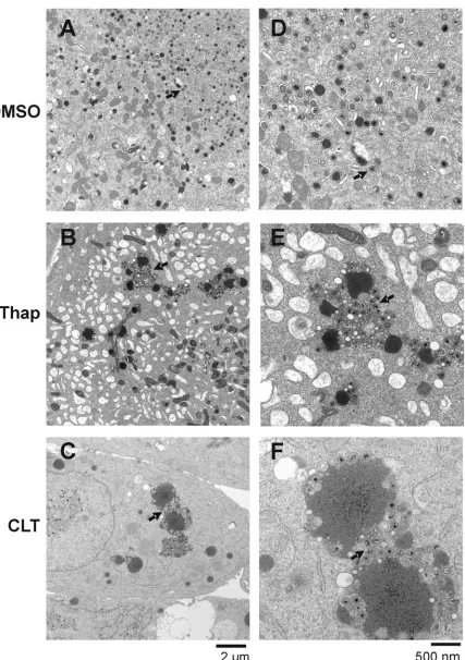
FIG. 6. Thapsigargin and CLT inhibit virion maturation. HCMV-infected HFF cells were treated with DMSO (A and D), thapsigargin (Thap) (B and E), or CLT (C and F) at 72 hpi and harvested at 96 hpi for analysis by electron microscopy. Panels D, E, and F show higher-magnification images of panels A, B, and C, respectively. Arrows indicate properly formed virions in the control samples (panels A and D) and abnormal clustering and formation of virions and tegument bodies in thapsigargin- and CLT-treated cells (panels B, C, E, and F). Scales are shown below the left and right sets of panels (panels A to C and D to F, respectively).
VOL. 79, 2005 THAPSIGARGIN AND CLT DISRUPT HCMV FORMATION 15395
on November 8, 2019 by guest
http://jvi.asm.org/
significant effect on virion formation, since pp28 has been reported to have a role in cytoplasmic envelopment of tegu-ment-associated capsids (29). In this regard, our electron mi-croscopic analysis confirmed that thapsigargin and CLT inhibit the formation of infectious virions. Drug-treated cells showed significantly decreased levels of extracellular virions compared with control (DMSO)-treated cells. In addition, tegument bod-ies were significantly enlarged in the drug treated cells, with nascent virions clustered and clumped around the tegument bodies. Further, many nucleocapsids appeared malformed and improperly tegumented and were not undergoing envelop-ment.
We think it is unlikely that the decreased accumulation of pp28 (or other late proteins) is the sole cause of all of these problems in maturation. Additional effects of thapsigargin and CLT may come directly from ER calcium depletion (or the concomitant increase in cytosolic calcium). A role for calcium in various aspects of virus maturation has been established, and mechanisms by which calcium may influence specific viral processes are beginning to emerge. Thapsigargin has been shown to inhibit the maturation of both African swine fever virus (7) and rotavirus (24). An intact ER calcium store is necessary for African swine fever virus capsid assembly as well as for enwrapment of the capsid by the ER membrane (7). In the case of rotavirus, calcium depletion in the ER alters the oligomerization of virus-encoded proteins (26). In contrast, thapsigargin was shown to enhance virion core assembly of hepatitis B virus, suggesting that cytosolic calcium ions mote assembly (5). Similarly, calcium ions mediate capsid pro-tein interactions and virion assembly for polyomavirus (6, 13) and are contained within SV40 virions (22).
A role for calcium-dependent chaperones in HCMV assem-bly also should be considered. It has been established that ER calcium can regulate the formation of chaperone complexes in the ER. As a result, the folding and maturation of many pro-teins can be affected by the depletion of ER calcium (2). In this regard, it has been suggested that the calcium-dependent ER chaperone protein calnexin is involved in the proper folding of the essential HCMV glycoprotein gB (35). Thus, decreased ER calcium concentration may affect the proper folding and/or processing of essential HCMV tegument proteins and glyco-proteins as well as that of cellular glyco-proteins involved in virus maturation.
[image:9.585.54.275.88.651.2]It should also be considered that the ER is a dynamic struc-ture continually exchanging lipid and protein components with the Golgi. ER-Golgi transport is also a calcium-dependent process (2); thus, changes in calcium homeostasis may disrupt the formation of ER-Golgi-derived vesicles which provide the membranous shell of dense bodies necessary for tegumenta-tion and virion maturategumenta-tion during late stage envelopment (12). This may contribute to the abnormal tegument bodies we have noted in drug-treated cells. In addition, it must be remembered that thapsigargin- and CLT-induced depletion of ER calcium is accompanied by a concomitant increase in cytosolic calcium. Increased cytosolic calcium influences the activity of many regulatory proteins, such as kinases, which may play a role in regulating the function of proteins involved in HCMV virion maturation, specifically tegument proteins (23). Given the complexity of virion maturation, many details of which remain FIG. 7. Thapsigargin and CLT inhibit virion maturation. The areas
indicated by the arrows in Fig. 6D, E, and F are enlarged to show normal virion maturation in the DMSO-treated control cells (A) and abnormal virion formation, tegumentation, and envelopment in thap-sigargin-treated cells (B) and CLT-treated cells (C).
on November 8, 2019 by guest
http://jvi.asm.org/
unclear, the disruption of calcium homeostasis during this pro-cess may have far-reaching effects.
ACKNOWLEDGMENTS
We thank the members of the Alwine laboratory for helpful sion and critical evaluation of the data, Alan Diehl for helpful discus-sions and critical evaluation, Sherri Adams for critical reading of the manuscript, and Q.-C. Yu of the Penn Biomedical Imaging Facility for expert electron microscopy and interpretation of micrographs. Cheers to all.
J.A.I. was supported by an NIH postdoctoral training grant (F32 AI062055-01). This work was supported by NIH grant CA28379-25 awarded to J.C.A. by the National Cancer Institute.
REFERENCES
1.Aktas, H., R. Fluckiger, J. A. Acosta, J. M. Savage, S. S. Palakurthi, and J. A. Halperin.1998. Depletion of intracellular Ca2⫹stores, phosphorylation of elF2a, and sustained inhibition of translation initiation mediate the antican-cer effects of clotrimazole. Proc. Natl. Acad. Sci. USA95:8280–8285. 2.Ashby, M. C., and A. V. Tepikin.2001. ER calcium and the function of
intracellular organelles. Semin. Cell Dev. Biol.12:11–17.
3.Benzaquen, L. R., C. Brugnara, H. R. Byers, S. Gatton-Celli, and J. A. Halperin.1995. Clotrimazole inhibits cell proliferation in vitro and in vivo. Nat. Med.1:534–540.
4.Bresnahan, W. A., G. E. Hultman, and T. Shenk. 2000. Replication of wild-type and mutant human cytomegalovirus in life-extended human dip-loid fibroblasts. J. Virol.74:10816–10818.
5.Choi, Y., S. Park, J. Yoo, and G. Jung.1993. Calcium ions affect the hepatitis B virus core assembly. Virology332:454–463.
6.Chromy, L., J. Pipas, and R. Garcea.2003. Chaperone-mediated in vitro assembly of polyomavirus capsids. Proc. Natl. Acad. Sci. USA100:10477– 10482.
7.Cobbold, C., S. M. Brookes, and T. Weilman.2000. Biochemical require-ments of virus wrapping by the endoplasmic reticulum: involvement of ATP and endoplasmic reticulum calcium store during envelopment of African swine fever virus. J. Virol.74:2151–2160.
8.Harding, H. P., Y. Zhang, A. Bertolotti, H. Zeng, and D. Ron.2000. PERK is essential for translation regulation and cell survival during unfolded pro-tein response. Mol. Cell5:897–904.
9.Harding, H. P., Y. Zhang, and D. Ron.1999. Protein translation and folding are coupled by an endoplasmic-reticulum-resident kinase. Nature397:271– 274.
10.Harel, N. Y., and J. C. Alwine.1998. Phosphorylation of the human cyto-megalovirus 86-kilodalton immediate-early protein IE2. J. Virol.72:5481– 5492.
11.Heider, J. A., Y. Yu, T. Shenk, and J. C. Alwine.2002. Characterization of a human cytomegalovirus with phosphorylation site mutations in the immedi-ate-early 2 protein. J. Virol.76:928–932.
12.Homman-Loudiyi, M., K. Hultenby, W. Britt, and C. Soderberg-Naucler. 2003. Envelopment of human cytomegalovirus occurs by budding into Golgi-derived vacuole compartments positive for gB, Rab 3, trans-Golgi network 46, and mannosidase II. J. Virol.77:3191–3203.
13.Ishizu, K., S. Watanbe, S. Han, S. Kanesashi, M. Hoque, H. Yajima, K. Kataoka, and H. Handa. 2001. Roles of disulfide linkages and calcium ion-mediated interactions in assembly and disassembly of virus-like particles composed of simian virus 40 VP1 capsid protein. J. Virol.75:61–72. 14.Isler, J. A., A. H. Skalet, and J. C. Alwine.2005. Human cytomegalovirus
infection activates and regulates the unfolded protein response. J. Virol. 79:6890–6899.
15.Jordan, R., L. Wang, T. M. Graczyk, T. M. Block, and P. R. Romano.2002. Replication of a cytopathic strain of bovine viral diarrhea virus activates PERK and induces endoplasmic reticulum stress-mediated apoptosis of MDBK cells. J. Virol.76:9588–9599.
16.Kedersha, N., and P. Anderson.2002. Stress granules: sites of mRNA triage
that regulate mRNA stability and translatability. Biochem. Soc. Trans.30: 963–969.
17.Kimball, S. R., R. L. Horetsky, D. Ron, L. S. Jefferson, and H. P. Harding. 2003. Mammalian stress granules represent sites of accumulation of stalled translation initiation complexes. Am. J. Physiol. Cell Physiol. 284:C273– C284.
18.Lee, A. S.1992. Mammalian stress response: induction of the glucose-regu-lated protein family. Curr. Opin. Cell Biol.4:267–273.
19.Lee, A. S., N. N. Iwakoshi, and L. H. Glimcher.2003. XBP-1 regulates a subset of endoplasmic reticulum resident chaperone genes in the unfolded protein response. Mol. Cell. Biol.23:7448–7459.
20.Lee, K., W. Tirasophon, X. Shen, M. Michalak, R. Prywes, T. Okada, H. Yoshida, K. Mori, and R. J. Kaufman.2002. IRE1-mediated unconventional mRNA splicing and S2P-mediated ATF6 cleavage merge to regulate XBP1 in signaling the unfolded protein response. Genes Dev.16:452–466. 21.Li, M., P. Baumeister, B. Roy, T. Phan, D. Foti, S. Luo, and A. S. Lee.2000.
ATF6 as a transcription activator of the endoplasmic reticulum stress ele-ment: thapsigargin stress-induced changes and synergistic interactions with NF-Y and YY1. Mol. Cell. Biol.20:5096–5106.
22.Liddington, R., Y. Yan, J. Moulai, R. Sahli, T. Benjamin, and S. Harrison. 1991. Structure of simian virus 40 at 3.8-A resolution. Nature354:278–284. 23.Mettenleiter, T. C.2002. Herpesvirus assembly and egress. J. Virol.76:1537–
1547.
24.Michelangeli, F., F. Liprandi, M. E. Chemello, M. Ciarlet, and M. Ruiz. 1995. Selective depletion of stored calcium by thapsigargin blocks rotavirus maturation but not the cytopathic effect. J. Virol.69:3838–3847. 25.Pavio, N., P. R. Romano, T. M. Graczyk, S. M. Feinstone, and D. R. Taylor.
2003. Protein synthesis and endoplasmic reticulum stress can be modulated by the Hepatitis C virus envelope protein E2 through the eukaryotic initia-tion factor 2a kinase PERK. J. Virol.77:3578–3585.
26.Poruchynsky, M., D. Maass, and P. Atkinson.1991. Calcium depletion blocks the maturation of rotavirus by altering the oligomerization of virus-encoded proteins in the ER. J. Cell Biol.114:651–661.
27.Prostko, C. R., J. N. Dholakia, M. A. Brostrom, and C. O. Brostrom.1995. Activation of the double-stranded RNA-regulated protein kinase by deple-tion of endoplasmic reticular calcium stores. J. Biol. Chem.270:6211–6215. 28.Sagara, Y., F. Fernandez-Belda, L. deMeis, and G. Inesi.1992. Character-ization of the inhibition of intracellular Ca2⫹transport ATPases by
thapsi-gargin. J. Biol. Chem.267:12606–12613.
29.Silva, M. C., Q. C. Yu, L. Enquist, and T. Shenk.2003. Human cytomega-lovirus UL99-encoded pp28 is required for the cytoplasmic envelopment of tegument-associated capsids. J. Virol.77:10594–10605.
30.Su, H. L., C. L. Liao, and Y. L. Lin. 2002. Japanese encephalitis virus infection initiates endoplasmic reticulum stress and an unfolded protein response. J. Virol.76:4162–4171.
31.Tardif, K. D., K. Mori, and A. Siddiqui.2002. Hepatitis C virus subgenomic replicons induce endoplasmic reticulum stress activating an intracellular signaling pathway. J. Virol.76:7453–7459.
32.Tardif, K. D., K. I. Mori, R. J. Kaufman, and A. Siddiqui.2004. Hepatitis C virus suppresses the IRE1-Xbp1 pathway of the unfolded protein response. J. Biol. Chem.279:17158–17164.
33.Tirosh, B., N. N. Iwakoshi, B. N. Lilley, A. Lee, L. H. Glimcher, and H. L. Ploegh.2005. Human cytomegalovirus protein US11 provokes an unfolded protein response that may facilitate the degradation of class 1 major histo-compatibility complex products. J. Virol.79:2768–2779.
34.Wang, Y., J. Shen, N. Arenzanna, W. Tirasaphon, R. Kaufman, and R. Prywes.2000. Activation of ATF6 and an ATF6 DNA binding site by the endoplasmic reticulum stress response. J. Biol. Chem.275:27013–27020. 35.Yamashita, Y., K. Shimokata, S. Mizuno, T. Daikoku, T. Tsurumi, and Y.
Nishiyama.1996. Calnexin acts as a molecular chaperone during the folding of glycoprotein B of human cytomegalovirus. J. Virol.70:2237–2246. 36.Ye, J., R. B. Rawson, R. Komuro, X. Chen, U. P. Dave, R. Prywes, M. S.
Brown, and J. L. Goldstein.2000. ER stress induces cleavage of membrane-bound ATF6 by the same proteases that process SREBPs. Mol. Cell6:1355– 1364.
37.Yoshida, H., T. Matsui, A. Yamamoto, T. Okada, and K. Mori.2001. Xbp1 mRNA is induced by ATF6 and spliced by IRE1 in response to ER stress to produce a highly active transcription factor. Cell107:881–891.
VOL. 79, 2005 THAPSIGARGIN AND CLT DISRUPT HCMV FORMATION 15397