JOURNAL OFVIROLOGY, Dec. 2005, p. 14570–14585 Vol. 79, No. 23 0022-538X/05/$08.00⫹0 doi:10.1128/JVI.79.23.14570–14585.2005
Copyright © 2005, American Society for Microbiology. All Rights Reserved.
Effect of Mutations in the Mouse Hepatitis Virus 3
⬘
(
⫹
)42 Protein
Binding Element on RNA Replication
Reed F. Johnson,
1†‡ Min Feng,
1† Pinghua Liu,
1Jason J. Millership,
1§ Boyd Yount,
2Ralph S. Baric,
2and Julian L. Leibowitz
1*
Department of Pathology and Laboratory Medicine, Texas A&M University System College of Medicine, College Station, Texas 77843-1114,1and Department of Epidemiology, University of North Carolina
at Chapel Hill, Chapel Hill, North Carolina 27599-74352
Received 18 October 2004/Accepted 13 July 2005
The mouse hepatitis virus (MHV) genome’s 3ⴕuntranslated region containscis-acting sequences necessary for replication. Studies of MHV and other coronaviruses have indicated a role for RNA secondary and tertiary elements in replication. Previous work in our laboratory has identified four proteins which form ribonucleo-protein complexes with the 3ⴕ-terminal 42 nucleotides [3ⴕ(ⴙ)42] of the MHV genome. Defective interfering (DI) RNA replication assays have demonstrated a role for the 3ⴕ(ⴙ)42 host protein binding element in the MHV life cycle. Using gel mobility shift RNase T1protection assays and secondary structure modeling, we have
characterized a possible role for RNA secondary structure in host protein binding to the 3ⴕ-terminal 42-nucleotide element. Additionally we have identified a role for the 3ⴕ-terminal 42-nucleotide host protein binding element in RNA replication and transcription using DI RNA replication assays and targeted recombination and by directly constructing mutants in this protein binding element using a recently described MHV reverse genetic system. DI RNA replication assays demonstrated that mutations in the 3ⴕ(ⴙ)42 host protein binding element had a deleterious effect on the accumulation of DI RNA. When the identical mutations were directly inserted into the MHV genome, most mutant genomes were viable but formed smaller plaques than the wild-type parent virus. One mutant was not viable. This mutant directed the synthesis of genome-sized negative-sense RNA approximately as efficiently as the wild type did but had a defect in subgenomic mRNA synthesis. These results point to a potential role for sequences at the extreme 3ⴕend of the MHV genome in subgenomic RNA synthesis.
Mouse hepatitis virus(MHV) is a positive-sense single-strand RNA virus of the orderNidovirales. MHV is a prototypical coronavirus; its genome is 32 kilobases in length and contains
cis-acting elements responsible for viral mRNA transcription and genome replication (1, 6, 7, 11, 16, 19, 34). The genome also serves as an mRNA which contains two overlapping read-ing frames translated by an RNA frameshiftread-ing mechanism to produce a large polypeptide, which is autocatalytically proteo-lyzed into 16 different polypeptides, many of which contain or are predicted to contain a variety of enzymatic activities (31). These include RNA-dependent RNA polymerase, helicase, pa-pain-like protease, picornavirus 3C-like protease, methyltrans-ferase, and two different nuclease activities, all of which are likely required for viral replication. Coronavirus-infected cells contain five to eight subgenomic mRNAs, each of which is made up of two noncontiguous segments, an approximately 70-nucleotide (nt) leader RNA identical to the corresponding 5⬘end of the genome, which is joined to the downstream body of the mRNA (20, 28). Current thinking is that discontinuous
transcription during negative-strand synthesis results in the production of a nested set of subgenomic negative-strand RNAs containing antileader RNAs that function as the sole templates for subgenomic mRNA synthesis (2, 26). Genome replication is thought to occur through the production of a negative-sense full-length RNA followed by synthesis of genomic RNA which is packaged and released as fully infec-tious virions.
Most studies of coronavirus cis-acting replication signals have utilized defective interfering (DI) RNAs as model repli-cons due to the large size of coronavirus genomes. DIssE is a 2.2-kb, naturally occurring DI RNA which was isolated by serial passage of MHV-JHM (19). DIssE is composed of three regions from the MHV-JHM genome. Region I is from nt 1 to 864, region II is derived from a 784-nt internal region 3.3 to 4 kb from the 5⬘end of the genome, and region III is derived from the 601 3⬘-terminal nucleotides (10). Studies of DIssE deletion mutants demonstrated that both the 5⬘-terminal 474 nt and the 3⬘-terminal 436 nt are necessary for replication, as is a 58-nt element derived from region II (1, 11, 19). Studies of various MHV-A59 DI RNAs have indicated that the require-ment for the region II elerequire-ment is specific for MHV-JHM DI RNAs (11, 25). Spagnolo and Hogue demonstrated that at least five A residues in the poly(A) tail are required for repli-cation and poly(A) tail formation (29). Relatively few studies have attempted to dissect particular functions in genome rep-lication for the variouscis-acting sequences. Lin et al. demon-strated that DI RNAs in which the last 55 nt had been deleted were unable to serve as templates for negative-strand RNA
* Corresponding author. Mailing address: Department of Pathology and Laboratory Medicine, Texas A&M University System College of Medicine, 208 Reynolds Medical Building, 1114 TAMU, College Station, TX 77843-1114. Phone: (979) 845-7288. Fax: (979) 862-1299. E-mail: [email protected].
† These two authors contributed equally to this work.
‡ Present address: Department of Pathobiology, University of Penn-sylvania School of Veterinary Medicine, Philadelphia, PA 19104-6049. § Present address: Fort Dodge Animal Health, 800 5th St. NW, P.O. Box 518, Fort Dodge, IA 50501.
14570
on November 8, 2019 by guest
http://jvi.asm.org/
synthesis when transfected into MHV-infected cells (16), thus implicating these sequences in negative-strand RNA synthesis. RNA secondary structures withincis-acting sequences often function in viral replication. Computer-aided predictions of secondary structure combined with biochemical probing to detect such structures demonstrated the existence of a 68-nucleotide bulged stem-loop near the 5⬘ end of the MHV 3⬘ untranslated region (UTR) (6, 7, 21). Replication studies with DI RNAs containing mutations within this structure demon-strated that this stem-loop was essential for DI RNA replication (6). These studies used targeted recombination to generate viruses containing mutations within this 68-nt bulged stem-loop and demonstrated that both primary and secondary struc-tures were important for viral replication. Immediately down-stream of this stem-loop structure is a second stem-loop, first identified in the 3⬘UTR of bovine coronavirus, which can form a pseudoknot with the 3⬘-most segment of the upstream bulged stem-loop (32). The interaction of these two RNA secondary structures is proposed to form a molecular switch that may regulate viral RNA synthesis (4). Mutagenesis of these two structures in the MHV genome supports their existence and function in viral replication. Computer-assisted modeling and enzymatic probing of the secondary structure of the MHV 3⬘ UTR downstream of the pseudoknot indicated the formation of a complex multiple stem-loop structure in this region of the
3⬘ UTR (18). Mutagenesis of this structure in the DI RNA
DIssE, combined with studies of the ability of these mutant DI RNAs to replicate, supported a role for this RNA secondary structure in MHV replication.
Studies of protein binding to the 3⬘UTR identified two host protein binding elements within the 3⬘-terminal 166 nucleo-tides (18, 34, 35). Mutation of these protein binding elements decreased host protein binding and decreased the ability of DIssE RNAs carrying these mutations to replicate, suggesting that these protein binding elements were functionally impor-tant during MHV replication (17, 34, 35). Recently we identi-fied mitochondrial aconitase and mitochondrial HSP70, as well as HSP60 and HSP40, as the four proteins which bound to the 3⬘-terminal 42-nucleotide [3⬘(⫹)42] element within the 3⬘ UTR (23, 24). The role of secondary structure of the 3⬘(⫹)42 element in binding to mitochondrial aconitase and
mitochon-drial HSP70, HSP60, and HSP40 and the effect of 3⬘-end
RNA-protein interactions on viral replication have not been studied. Computer-based secondary structure modeling of the 3⬘(⫹)42 element suggests the formation of a stem-loop struc-ture. Nuclease probing of RNA secondary structure did not provide any information on the terminal 12 nucleotides but suggested the formation of a mostly single-stranded, bulged stem-loop structure which differs from the predominantly dou-ble-stranded model proposed by Yu and Leibowitz (34, 35).
In this report we demonstrate that mutations within the
3⬘-terminal 42-nt genomic RNA lead to increased and
de-creased RNP complex formation. Introduction of these same mutations into DI replicons lead to decreases in positive-strand DI RNA replication. Viable viruses with altered plaque morphology were recovered when these mutations were intro-duced into the MHV genome, with one exception. For one mutation, viable virus could not be recovered due to a defect in subgenomic RNA synthesis.
MATERIALS AND METHODS
Cells, cell lysates, and viruses.17Cl-1 cells were maintained at 37°C and 5%
CO2in Dulbecco modified Eagle medium (DMEM) supplemented with 10% calf
serum (DMEM10). L2 cells were maintained at 37°C and 3% CO2in DMEM
supplemented with 10% calf serum. L2 cells for targeted recombination
exper-iments were adapted to grow in suspension and maintained at 37°C with 3% CO2
in spinner DMEM supplemented with 10% calf serum. Cytoplasmic lysates for RNA binding assays were prepared from 17Cl-1 cells as previously described (35).
The origin and growth of MHV-A59 have been described previously (17). A temperature-sensitive mutant of MHV-A59, Alb4, was generously provided by Paul S. Masters (12) and used in targeted recombination experiments.
Template preparation and transcription of gel mobility shift RNase T1
pro-tection assay probes.PCR was carried out on DE25, a cDNA of a naturally occurring DI RNA, DIssE, using primers listed in Table 1. PCR products were
then purified by electrophoresis in a 1⫻Tris-acetate-EDTA, 4% NuSieve GTG
agarose gel; the amplified fragment was excised from the gel and recovered using the Qiaquick gel extraction kit. Transcription of radiolabeled RNAs was con-ducted as described previously (35). Probes were quantified based upon
incor-poration of [␣-32
P]UTP, and 1.4 picomoles of labeled RNA was used for each binding reaction. Unlabeled wild-type probe was prepared in the same manner as labeled probe, quantitated by spectrophotometry, and resuspended in diethyl pyrocarbonate-treated water to appropriate concentrations.
Gel mobility shift RNase T1protection assays.The gel mobility shift RNase T1
protection assays were carried out as described by Yu and Leibowitz (34). Radiolabeled probe was incubated with 17Cl-1 cell lysate for 20 minutes followed
by digestion with 0.006125 units of RNase T1per 1.4 pmol of RNA for 20
minutes at 22°C. All reactions were carried out with unlabeled wild-type
com-petitor, and reactions with nonspecific tRNA competitor at 10, 25, 50, and 100⫻
molar concentrations were carried out in parallel. The complete reaction mixture
was loaded onto a 1⫻Tris-buffered EDTA, 6% nondenaturing polyacrylamide
gel and electrophoresed for 4.5 h. Gels were transferred to Whatman 3CHR paper and dried at 80°C under vacuum for 3 h and then exposed to Kodak Biomax MS film and quantitated with a phosphoimager. Six gel shift assays of each mutant were used to generate an average binding efficiency, with wild type always set to 100%.
Computer models.The Mfold RNA version 3.0 folding algorithm was used to predict RNA secondary structure (36). The following parameters were used for all secondary structure modeling and are in keeping with previous work in our
lab (34): no folding constraints, RNA sequence⫽linear, temperature fixed by
the program at 37°C, ionic conditions fixed by the program at 1 M NaCl with no
divalent cations, percent suboptimality⫽5, upper band⫽50, default⫽window,
maximum distance between paired bases set, output at high resolution, format⫽
bases, base number frequency⫽default, structure rotation angle ⫽0, and
structure annotation by p-num.
UV cross-linking assays.UV cross-linking of RNA binding proteins to radio-labeled probes was performed as described previously (35). Samples were sub-jected to sodium dodecyl sulfate-polyacrylamide gel electrophoresis, and the labeled proteins were visualized by autoradiography.
Oligonucleotide primers.The oligonucleotide primers used in this work are shown in Table 1. For many of the primers, the position of the primer relative to the MHV-A59 genome, GenBank accession no. NC001846, is given in paren-theses as part of the primer name.
Site-directed mutagenesis and pB36 mutant construction.Paul S. Masters kindly provided the plasmid pB36, which contains a cDNA representing the B36
synthetic DI RNA (7). To ensure that only mutations in the 3⬘(⫹)42 host protein
binding element were introduced into pB36, a subcloning strategy to create a mutagenesis template was adapted. The B36 HindIII-SacI 178-nucleotide frag-ment was generated as a mutagenesis template by PCR and TA cloning using primers B36SacI and B36HindIII and Invitrogen’s 2.1 TA cloning kit to produce plasmid 2-1.
The MT3C, M19C, ATW, ATW5⬘, and ATW3⬘mutations were introduced
into plasmid 2-1 using Stratagene’s QuickChange site-directed mutagenesis kit with primers listed in Table 1. After the procedure for mutagenesis was carried out, potential mutant 2-1 plasmids were screened for their intended mutations. Plasmids containing the desired mutations were used to introduce these muta-tions into pB36 by restriction fragment exchange using the SacI and HindIII restriction sites. The presence of the mutations and the absence of other changes were verified by sequencing the mutant pB36 plasmids using primer 5666
(5⬘-GTAGTGCCAGATGGGTTA).
In vitro transcription of DI RNAs.DI RNAs for targeted recombination were generated by linearizing pB36 wild type and mutants with NsiI followed by gel
on November 8, 2019 by guest
http://jvi.asm.org/
purification. Transcription reactions were carried out using Ambion’s mMessage mMachine kit following the manufacturer’s protocol. Each transcription reaction was phenol:chloroform:isoamyl alcohol (25:24:1) extracted twice, chloroform ex-tracted twice, ethanol precipitated, redissolved in diethylpyrocarbonate-treated wa-ter, and quantitated by spectrophotometry.
Targeted recombination.Targeted recombination was carried out as described by Koetzner et al. (12). L2 cells grown in suspension were electroporated with
5g of DI RNA and then infected with Alb4. Virus stocks were prepared and
heat inactivated at 40°C for 24 h, and representative heat-resistant viruses were plaque purified. Isolated virus was incubated until syncytia involved 90 to 95% of a 17Cl-1 monolayer. The medium was drawn off the monolayer and stored
at⫺70°C, the monolayer was washed once with Dulbecco’s phosphate-buffered
saline, and total cellular RNA was isolated using the RNeasy Mini kit (QIA-GEN).
Reverse transcription PCR (RT-PCR) and sequencing of isolated virus.RT reactions were carried out using the primer 17TG following Invitrogen’s protocol
for SuperScript II RNase H⫺reverse transcriptase. PCR was carried out on
1 microliter of RT product using the primers B36For and B36Rev, which flank the 87-nucleotide deletion present in Alb4 (21). PCR products were resolved on
2% agarose gels run in parallel toX174 HaeIII markers. Viruses in which the
87-nucleotide deletion was repaired by a recombination event were identified by the presence of a 542-bp fragment compared to the 455-bp fragment observed when Alb4 was used as template. Recombinant viruses were subjected to further analysis to determine if they carried the desired mutations. A second PCR was carried out using the B36For and 17TG primers and 17TG-primed cDNA under
the conditions used for screening. The amplified fragment was purified by gel electrophoresis and subjected to automated sequencing using the primer 5666.
B36 DI replication assays.The method established by Masters’ lab was used with modifications (21). 17Cl-1 and L2 cells were grown as monolayers until confluent. 17Cl-1 cells were washed, trypsinized, centrifuged, resuspended in DMEM10, and seeded onto six-well cluster plates 2 h prior to electroporation of L2 cells. Just prior to electroporation of the L2 cells the medium was changed to DMEM supplemented with 2% serum. To prepare L2 cells for electroporation, a confluent flask of cells was trypsinized, resuspended in DMEM containing 10% fetal bovine serum, and after centrifugation resuspended in DMEM containing
2% fetal bovine serum (DMEM2) at a concentration of 1.0⫻106cells/ml. For
each DI RNA 1.0⫻106
L2 cells were infected in suspension with MHV-A59 at a multiplicity of infection (MOI) of 3 for 2 h at 37°C. After 2 h of infection L2
cells were centrifuged at 1,000⫻g, resuspended in 10 ml Ca2⫹- and Mg2⫹-free
phosphate-buffered saline, and centrifuged again at 1,000⫻g. This wash
proce-dure was repeated two times. For each DI RNA 1.0⫻ 106
L2 cells were
electroporated with 5g of DI RNA, or without RNA for the mock-transfected
control, at maximum resistance, 0.8 kV and 25F. Two pulses were delivered 5
seconds apart. The electroporated cells were resuspended in 1 ml of DMEM2 and overlaid onto the 17Cl-1 cells.
The overlaid-infected-electroporated cells were incubated until syncytia con-sumed roughly 65% of the monolayer, usually 8 to 10 h postinfection. At this point the DMEM2 was removed and replaced with 2 ml phosphate-free DMEM
with 2.5g/ml actinomycin D and incubated at 37°C for 20 min. At the end of 20
[image:3.585.44.544.78.469.2]min the medium was replaced with phosphate-free medium containing 2.5g/ml
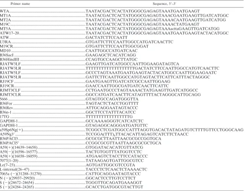
TABLE 1. Primers used for gel mobility shift RNase T1protection assays, site-directed mutagenesis, and RT-PCR
Primer name Sequence, 5⬘–3⬘
WTA...TAATACGACTCACTATGGGCGAGAGTAAATGAATGAAGT
MT1A...TAATACGACTCACTATGGGCGAGAGTAAACGAATGAAGTTGATCATGGC MT2A...TAATACGACTCACTATGGGCGAGAGTAAAACAATGAAGTTGATCATGG M19C...TAATACGACTCACTATGGGCGAGAGTAAAACTATGAAGT
MT5A...TAATACGACTCACTATGGGCGAGAGTAAAacttaGAAGTTGATCATGG ATW17–20...TAATACGACTCACTATGGGCGAGAGTAAATGAATGAAGTACTACATGGC ATW...GACTATCTTCCAATT
U3RA ...GTGATTCTTCCAATTGGCCATGATCAACTTC M19CR...GTGATTCTTCCAATTGGCGGAT
MD10 ...CAATTGGCCATGATCAAC B36SacI ...GAAGAGCTCACATCAGG B36HindIII ...CCAGTGCCAAGCTTATGC
RJATW3LF...GAAGTTGATCATGGCCAATTGGAAGATAGTCA
RJATW3LR ...TTTTTTTTTTTTTTTTTTTGACTATCTTCCAATTGGCCATGTCAACTTC RJATW5LF...GCCCTAGTAAATGAATGAAGTACTACATGGCCAATTGGAAGAATC RJATW5LR ...GATTCTTCAATTGGCCATGTAGTACTTCATTCATTTACTAGGGC RJ19CF ...GAATGAAGTTGATCATCGCCAATTGGAAG
RJ19CR...GAACCAATTGGCGATGATCAACTTCATTC
RJMT3CLF ...CCTGAATGCCCTAGTAAAACTATGAAGTTGATCATGGCC RJMT3CLR...GGCCATGATCAACTTCATAGTTTTACTAGGGCATTGCAGG 5666 ...GTAGTGCCAGATGGGTTA
B36For ...TAGTACTCTACCTGGTTTT B36Rev...ATTGCAGGAATAGTACCC B36n-1 ...GGCTTCCTATTTACATCC 17TG ...TTTTTTTTTTTTTTTTTG GAPDH-1...GCCAAAAGGGTCATCATCTC GAPDH-1...GTAGAGGCAGGGATGATGTTC
a59Sp6Ng(⫹)...TCGGCCTCGATGGCCATTTAGGTGACACTATAGATGTCTTTTGTTCCTGGGCAAG A59Ng3⬘...TCCGGA(TTT)8TTACACATTAGAGTCATCTTCTAACC
B36PACI3...GCGCGCTTAATTAACGCGCCGGTGCA B36PACI5⬘...CCGGCGCGTTAATTAAGCGCGCTGCA A59(⫹)(14639–14658)...GTGGATACACATCGTTATCG
A59(⫺)(16596–16577)...TACTGTGGTTTATGGTCCTC A59(⫹)(16038–16059)...ATGAAGTCTACCTTCCATACCC 7077(1–20) ...TATAAGAGTGATTGGCGTCC Le(7–23)...AGTGATTGGCGTCCGTA L-internal(26–47) ...TACCCTCTCAACTCTAAAACTC 7065a (⫺)(31288–31270)...CATTGCAGGAATAGTACCC N (⫺)(29937–29920) ...GGCACTCCTTGTCCTTCT S (⫺)(24672–24654) ...TGGGTTGCAGATGAAAGGT S (⫺)(24284–24265) ...GCACCTGATGGCGTACTTGT
14572 JOHNSON ET AL. J. VIROL.
on November 8, 2019 by guest
http://jvi.asm.org/
actinomycin D, 2% dialyzed serum, and 0.5 mCi of32PO
4and incubated at 37°C
until syncytia enveloped 90 to 95% of the monolayer, usually 14 to 16 h
post-infection. RNA was extracted with QIAGEN⬘s RNeasy kit. Three to sixg of
RNA was denatured in RNA loading buffer and resolved by electrophoresis in 1% agarose gels containing formaldehyde as described previously (35). The 18S rRNA bands in ethidium bromide-stained gels were quantitated by densitometry to normalize the phosphorimager signal to total RNA loaded per sample. B36 DI RNA was set at 100%, and replicating DI RNAs were also adjusted to account for small differences in quantification and loading based on rRNA staining. Gels were photographed under UV light, fixed in 80% methanol, dried, and exposed to film. Bands located between RNAs 6 and 7, which represent replicating DI RNAs, were quantified with Molecular Dynamics Phosphorimager and Storm 8.2 software; three experiments for each set of mutants were used to generate an average.
RT-PCR and sequencing of replicating DI RNAs.For each DI RNA evaluated,
1g of total RNA cell extracted from electroporated cells underwent reverse
transcription with 2 pmol of primer B36n-1 using Invitrogen’s Superscript II RT following the manufacturer’s protocol. B36n-1 is specific to the negative-strand B36 DI RNA. This ensured that only replicated DI B36 would be detected. No-RT, no-RNA, and A59 controls were included in the RT step. The no-RT control used RNA extracted from the B36 DI RNA-transfected culture to ensure that residual digested transcription template DNA was eliminated and not am-plified by PCR. RNA extracted from A59 infected-untransfected cells was used as a control to show specificity of the primers for B36. PCRs for each DI RNA were carried out for 31 cycles of 95°C for 30 seconds, 35°C for 60 seconds, and 72°C for 90 seconds, followed by a 15-minute extension at 72°C using 200 nM
primers B36n-1 and 17TG and 1l of RT product as template in a 50-l volume.
PCR products were electrophoresed on a 1.0% agarose gel, stained with ethidium bromide, photographed, excised, and sequenced using primer 5666.
Introduction of mutations into MHV-A59.The reverse genetic system de-scribed by Yount et al. (33) was used to recover wild-type MHV-A59 (known as
MHV-A59 1000) and mutant viruses carrying the MT3C, M19C, ATW3⬘, ATW5⬘,
and ATW mutations described above. Restriction fragment exchange was used to
move the 3⬘(⫹)42 mutations from pB36 into the reverse genetic system plasmid
G. To generate plasmids that contained compatible restriction sites, 5
micro-grams of DI B36 mutants MT3C, M19C, ATW, ATW5⬘, and ATW3⬘was
di-gested with NsiI, and linearized plasmids were gel purified. An adaptor contain-ing a PacI site was prepared by annealcontain-ing 100 nmol of oligonucleotides
B36PacI3⬘and B36PacI5⬘. The adaptor was ligated into NsiI-cut pB36, and
transformants containing the PacI site were identified by restriction digestion and verified by sequencing. These modified pB36 plasmids were digested with NruI and PacI, and the178-nucleotide NruI-PacI fragments were isolated. Plas-mid G was similarly digested with NruI and PacI and ligated with the pB36-derived NruI-PacI fragment. The identities of G plasmids carrying the MT3C,
M19C, ATW, ATW5⬘, and ATW3⬘mutations were verified by sequencing with
primer 5666. The MHV cDNAs were excised from mutant and wild-type G plasmids and ligated to MHV-A59 cDNAs A to F to generate cDNAs corre-sponding to the entire MHV genome under the control of a T7 promoter. Genome-sized RNAs were transcribed in vitro. Transcripts corresponding to the N gene coding sequence were also transcribed in vitro as described by Yount et al. (33). A mixture of genome and N gene transcripts was electroporated as described elsewhere to recover infectious virus (33).
After electroporation of the transcription reaction mixtures into MHV recep-tor-expressing BHK cells, the cells were overlaid onto a T-75 flask freshly seeded
with 2⫻106
DBT cells. Cultures were incubated for 2 to 3 days. The cells were
then frozen at⫺80°C, sonicated, and clarified by low-speed centrifugation.
Vi-ruses were plaque cloned on DBT or L2 cells. Individual plaques were isolated and inoculated onto a 70% confluent monolayer of 17Cl-1 cells until cytopathic effect enveloped 70 to 85% of the monolayer. At this point the medium was
removed and stored at⫺70°C.
RNA was extracted and purified from the monolayer of infected 17Cl-1 cells using the RNeasy RNA extraction kit (QIAGEN). Reverse transcription was
carried out using the primer 17TG. RT reactions were carried out using 1g of
total RNA and 500 ng of primer 17TG; following incubation the RT was heat inactivated at 70°C for 15 min. A fragment containing the last 590 nucleotides of the genome was amplified by PCR using the B36For and 17TG primers under the conditions described above. Amplified fragments were sequenced with the primer 5666.
One-step growth curves.17Cl-1 cells were grown in 96-well plates, and repli-cate wells were infected with MHV-A59 1000 or mutant virus at an MOI of three. Samples were frozen at 2, 4, 6, 8, 10, 12, 16, and 24 h postinfection, and virus production was quantitated by plaque assay. Triplicate samples were obtained for all time points.
Detection of negative-strand RNA and subgenomic RNAs by RT-PCR. RT-PCR was employed to detect negative-strand genomic RNA and subgenomic mRNAs that might be expressed after electroporation of mutant viral genomes which failed to give rise to viable virus in multiple independent experiments. To enhance the sensitivity of the assays and to be certain that electroporated DNA was not detected, the standard procedure used to recover virus described above was slightly modified. After transcription of MHV genome RNA and prior to electroporation, transcription reaction mixtures were digested with 2 U of RNase-free DNase I (Ambion) for 20 min at 37°C. After electroporation, trans-fected BHK-R cells were plated directly in T-25 tissue culture flasks in the absence of DBT cells. After 4 h and 8 h of incubation at 37°C or 34°C, cells were harvested and total RNA was isolated using the RNeasy kit (QIAGEN). For one experiment total RNA was extracted at 8, 12, and 24 h after
electropo-ration. One microgram of total RNA in a 10-l volume was treated with 2 U of
RNase-free DNase I for 30 min at 37°C; the DNase I was then inactivated at 65°C for 20 min. RT-PCRs to detect negative-strand genome-sized RNA and
sub-genomic RNAs were performed using SuperScript II RNase H⫺reverse
tran-scriptase according to the manufacturer’s protocol. To detect genome-sized
negative-strand RNA, oligonucleotide A59(⫹)(14639–14658) was used as the
RT primer for cDNA synthesis. This was followed by nested PCR. In the first
PCR oligonucleotide A59(⫹)(14639–14658) was the forward primer, while
oli-gonucleotide A59(⫺)(16596–16577) was used as the reverse primer. The primer
pair for the second PCR was oligonucleotide A59(⫹)(16038–16059) and
oligo-nucleotide A59(⫺)(16596–16577). A nested RT-PCR protocol was also used to
detect negative-strand RNA complementary to subgenomic mRNA7. One mi-crogram of total RNA was treated with DNase I, and cDNA synthesis was primed with oligonucleotide 7077(1-20) as described earlier. In the first PCR,
oligonucleotide Le(7–23) and oligonucleotide A59(⫺)(31288–31270) were used
as the forward and reverse primers, respectively. After 30 PCR cycles, 1 micro-liter of the first PCR product was used as template for the second nested PCR of 40 cycles. The forward and reverse primers were oligonucleotide
L-inter-nal(26–47) and oligonucleotide N(⫺)(29937–29925), respectively. Nested
RT-PCR was also used to detect subgenomic mRNA7. Oligonucleotide
7065a(⫺)(31288–31270) was used for cDNA synthesis during the RT reaction,
and then oligonucleotides Le(7–23) and 7065a(⫺)(31288–31270) were used for
the first 30 cycles of PCR. For the subsequent nested PCR, 1 microliter of this
PCR product was mixed with oligonucleotides L-internal(26–47) and
N(⫺)(29937–29925). A similar nested RT-PCR protocol was also used to detect
negative-strand RNA complementary to subgenomic mRNA3 (S gene). One microgram of total RNA was treated with DNase I, and cDNA synthesis was primed with oligonucleotide 7077(1-20) as described above. In the first PCR
oligonucleotide Le(7–23) and oligonucleotide S(⫺)(24672–24654) were used as
forward and reverse primers, respectively, for 30 cycles of PCR. One microliter of the first PCR product was mixed with oligonucleotide L-internal(26–47) and
oligonucleotide S(⫺)(24284–24265) and subjected to another 40 cycles of
am-plification. PCR mixtures were electrophoresed in 1.5% agarose gels and ethidium bromide stained. Parallel reactions in which reverse transcriptase was omitted from the cDNA step were always performed to ensure that the PCRs did not detect residual DNA transcription templates which entered the cells during electroporation. Nested RT-PCR was also used to detect subgenomic
mRNA3. The RT primer was oligonucleotide S(⫺)(24672–24654). Nested
PCR was performed with the same primers and conditions used for detecting negative-sense RNA3.
RESULTS
Gel mobility shift RNase T1protection assays and RNA
sec-ondary structure predictions.Previous work in our laboratory defined two host protein binding elements located at positions 154 to 129 upstream of the poly(A) tail and the 3⬘-terminal 42 nucleotides of the 3⬘UTR (18, 34, 35). A sequence comparison of the two elements revealed a shared 11-nt motif, UGARNG AAGUU. Secondary structure analysis using Mfold 2.0 pre-dicted that the 3⬘-terminal 42-nt host protein binding element formed a bulged stem-loop structure with the 11-nt motif form-ing part of the right portion of the bulged stem (35). Reexam-ination of the secondary structure with a more recent version of Mfold (version 3.0) predicted a different secondary struc-ture. Mfold 3.0 predicted that nucleotides 27 to 42 [all
on November 8, 2019 by guest
http://jvi.asm.org/
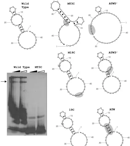
otides herein are referred to by their position relative to the 3⬘ terminus of the MHV genome, with the first nucleotide up-stream of the poly(A) tail designated as nucleotide 1] were single stranded with the remaining nucleotides forming a bulged stem-loop structure (Fig. 1). Nuclease structural prob-ing was more consistent with the 42-nt structure foldprob-ing into the Mfold-predicted structure shown here, rather than the published model based on a longer 166-nt RNA (17). Liu et al. (17) were able to obtain nuclease digestion data on nucleotides
13 to 42. Comparison of the predicted secondary structure of the wild-type 3⬘(⫹)42 model (Fig. 1) with the nuclease diges-tion of the 166-nt RNA indicated that the 42-nucleotide model matches the digestion data in 28 of 30 positions. When the Mfold 3.0-predicted secondary structure of the 166-nt RNA was compared to the same digestion data, only 18 of 30 nucle-otides matched (17).
To investigate the role of RNA secondary structure as a determinant of protein binding activity, the wild-type
second-FIG. 1. Gel mobility shift RNase T1protection assays for the mutant MT3C and secondary structure models of the 3⬘(⫹)42 host protein binding element mutants. Mutant nucleotides are shaded. The dark triangle represents increasing concentration of wild-type competitor RNA, and the open triangle indicates an increasing concentration of nonspecific tRNA competitor. The arrow indicates the RNP complexes which were quantitated (Table 2).
14574 JOHNSON ET AL. J. VIROL.
on November 8, 2019 by guest
http://jvi.asm.org/
[image:5.585.79.503.68.545.2]ary structure predicted by Yu and Leibowitz was used as a basis for designing mutations within the 42-nucleotide protein bind-ing element. Initially five mutations were created within this genome fragment (MT1A, MT2A, MT3C, MT5A, and MD10; Table 2). All of these mutations were located within the 11-nucleotide motif previously implicated in protein binding ac-tivity, with the exception of MD10, which is a 10-nt deletion
from the 3⬘ end. Two of these mutations were predicted by
Mfold to have no effect on RNA secondary structure, and three were predicted to produce major changes in secondary structure. The effect of these mutations on the RNA binding activity of the 42-nt protein binding element was then exam-ined in a series of gel mobility shift RNase T1 protection
assays. In our gel mobility shift RNase T1protection assays, the
complex of interest designated by the arrow in Fig. 1 repre-sented the largest RNP complex formed that could be resolved within the gel and was quantified by phosphoimager and com-pared with the wild type. The bands at the top of the gel are in the well and are most likely nonspecific complexes that could not enter the gel, while the faster-migrating complexes prob-ably represent RNP complexes which lack one or more of the four proteins composing the complete complex, as described by Nanda et al. (23).
RNAs which contained mutations predicted to maintain RNA secondary structure, MT1A and MT2A, had protein binding activity decreased to approximately 65% of that ob-served with wild-type RNA. RNAs that contained mutations predicted to disrupt secondary structure, like MT3C, MT5A, and MD10, disrupted protein binding activity to 6 to 40% of the wild-type value. The results are summarized in Table 2. Mutant MT3C is identical to mutant mB4 described by Yu and Leibowitz (34). The decrease in binding was expected based on previous studies but does not determine the contribution of secondary structure in RNP complex formation.
To further explore the role of RNA secondary structure in host protein binding, Mfold was used to identify a series of compensatory mutations that are predicted to restore the wild-type secondary structure. Compensatory mutation effects on host protein binding were determined by gel mobility shift RNase T1 protection assays. A representative mobility shift
RNase T1 protection assay for mutant MT3C is shown in
Fig. 1. MT3C had the greatest predicted change in secondary structure and the least amount of binding activity compared to wild-type virus (Fig. 1). The predicted compensatory G-to-C change at position 19 in the MT3C mutant not only restored wild-type RNA secondary structure (Fig. 1, mutant M19C) but dramatically restored protein binding activity of the substrate to about 74% of the wild-type value (Table 2). RNA carrying only the G-to-C mutation at position 19 (mutant 19C, Fig. 1 and Table 2) is predicted to maintain the wild-type secondary structure and has a protein binding activity which is approxi-mately 74% of that achieved by wild-type RNA. These data suggest that wild-type secondary structure, and to a lesser extent sequence context, may play a role in the ability of the 3⬘(⫹)42 host protein binding element to bind to host proteins. Because the updated version of Mfold predicted a different secondary structure for the wild-type sequence reported by Yu and Leibowitz (35), another panel of mutations were designed to disrupt interactions between nucleotides 23 to 26 and nu-cleotides 2 to 5. Nucleotide 26U is the last nucleotide
compos-ing the 11-nt motif. The ATW5⬘ mutation (Table 2) is
pre-dicted to disrupt RNA secondary structure (Fig. 1) with the only base pairing occurring between nucleotides 10 to 11 and 16 to 17 with a small loop formed by nucleotides 12 to 15. Gel mobility shift RNase T1 protection assays demonstrated that
ATW5⬘ exhibited 7.2% as much protein binding activity as
wild-type sequence. The ATW3⬘mutation (Table 2) was
[image:6.585.38.546.82.309.2]pre-dicted to produce an RNA secondary structure similar, but not
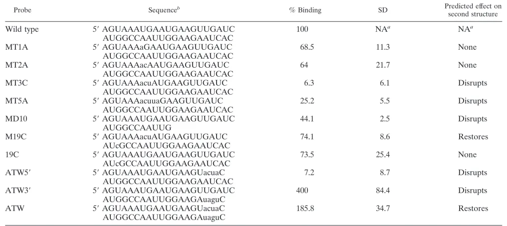
TABLE 2. Summary of RNA protein binding data
Probe Sequenceb % Binding SD Predicted effect on
second structure
Wild type 5⬘AGUAAAUGAAUGAAGUUGAUC 100 NAa NAa
AUGGCCAAUUGGAAGAAUCAC
MT1A 5⬘AGUAAAaGAAUGAAGUUGAUC 68.5 11.3 None
AUGGCCAAUUGGAAGAAUCAC
MT2A 5⬘AGUAAAacAAUGAAGUUGAUC 64 21.7 None
AUGGCCAAUUGGAAGAAUCAC
MT3C 5⬘AGUAAAacuAUGAAGUUGAUC 6.3 6.1 Disrupts
AUGGCCAAUUGGAAGAAUCAC
MT5A 5⬘AGUAAAacuuaGAAGUUGAUC 25.2 5.5 Disrupts
AUGGCCAAUUGGAAGAAUCAC
MD10 5⬘AGUAAAUGAAUGAAGUUGAUC 44.1 2.5 Disrupts
AUGGCCAAUUG
M19C 5⬘AGUAAAacuAUGAAGUUGAUC 74.1 8.6 Restores
AUcGCCAAUUGGAAGAAUCAC
19C 5⬘AGUAAAUGAAUGAAGUUGAUC 73.5 25.4 None
AUcGCCAAUUGGAAGAAUCAC
ATW5⬘ 5⬘AGUAAAUGAAUGAAGUacuaC 7.2 8.7 Disrupts
AUGGCCAAUUGGAAGAAUCAC
ATW3⬘ 5⬘AGUAAAUGAAUGAAGUUGAUC 400 84.4 Disrupts
AUGGCCAAUUGGAAGAuaguC
ATW 5⬘AGUAAAUGAAUGAAGUacuaC 185.8 34.7 Restores
AUGGCCAAUUGGAAGAuaguC
aNA, not applicable.
bMutations are indicated by lowercase type.
on November 8, 2019 by guest
http://jvi.asm.org/
identical, to that predicted for the wild-type sequence. Gel mobility shift RNase protection assays demonstrated that the ATW3⬘probe has a fourfold increase in protein binding activ-ity relative to the wild-type RNA probe. The double mutant
ATW contains both the ATW5⬘and ATW3⬘mutations; ATW
is predicted to restore the wild-type secondary structure and binds 1.85 times as well as wild type. Taken together, the results support the hypothesis that both primary and secondary structure play a role in host protein binding to the 3⬘(⫹)42 host protein binding element.
Since two of the mutant probes had increased protein bind-ing activity relative to wild-type RNA, we performed a UV cross-linking assay to determine if proteins of identical
molec-ular weights were bound by wild-type, ATW3⬘, and ATW
RNAs. After RNA complexes were allowed to form in solu-tion, the RNAs were cross-linked to their protein binding part-ners and the labeled proteins were resolved by sodium dodecyl sulfate-polyacrylamide gel electrophoresis as described in Ma-terials and Methods. The proteins that formed complexes with the mutant and wild-type probes were identical in molecular weight. Moreover, the relative amount of the four labeled proteins was similar in wild-type and mutant complexes except that the overall signal intensity of each sample paralleled that observed in the gel mobility shift RNase T1protection assays
for the wild-type, ATW3⬘, and ATW RNAs (data not shown).
DI RNA replication assays.To investigate the effect of these mutations on MHV replication, we first employed a series of DI
RNA replication assays. The M19C, MT3C, ATW5⬘, ATW3⬘,
and ATW mutations were introduced into a plasmid containing a cDNA corresponding to the B36 DI RNA (12). We chose the B36 DI RNA as our model replicon because it also would allow us to attempt to introduce these mutations into MHV-A59 by targeted recombination (12, 21). For simplicity, replication assays were performed in two groups, the first with the mutants involving the 11-nt host protein binding motif, MT3C and
M19C. The MT3C mutation in B36 is identical to the mB4 mutation in DIssE described by Yu and Leibowitz, which rep-licated poorly and readily recombined with wild-type genome sequences (34). We chose to explore this mutation in the con-text of the B36 DI RNA because it had the greatest effect on RNP formation and was predicted to cause a drastic change in RNA secondary structure. M19C was derived from MT3C and encoded a second compensatory change that largely restored protein binding activity as well as the predicted secondary structure of the RNA, establishing a possible connection be-tween secondary structure and RNP complex formation. Cells were infected with MHV-A59; wild-type B36, MT3C, M19C, or DIssE (an MHV-JHM-derived DI RNA which replicates to higher levels than B36) was electroporated into the infected cells; and the cultures were metabolically labeled with [32P]orthophosphate in the presence of actinomycin D as
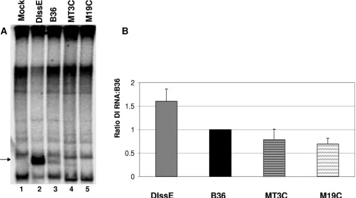
de-scribed in Materials and Methods. After the labeling period RNA was extracted and the MHV-specific RNAs were re-solved by electrophoresis (Fig. 2). Three complete experiments were used to determine an average DI RNA replication effi-ciency. A complete experiment was defined as one in which DI B36 RNA and DIssE RNA both replicated to detectable levels regardless of how well the mutant DI RNAs replicated and time constants for all electroporations were 0.8, indicating that electroporation conditions were optimal. MHV-A59-infected cells transfected with mutants MT3C and M19C synthesized positive-strand DI RNA with varying efficiency compared to wild-type DI B36, as detected by metabolic labeling. Quanti-tation by phosphorimager analysis revealed that MT3C repli-cated 78% as well as B36 and M19C replirepli-cated 69% as effi-ciently as B36. In order to determine if replicated DI RNAs contained the intended mutations, RT-PCR for negative-strand RNA was carried out for DI RNAs MT3C and M19C. The negative strand was amplified to avoid interference by transfected positive-strand DI RNAs. Sequence analysis
re-FIG. 2. Replication assays for DI RNAs B36, MT3C, and M19C. (A) Representative electropherogram of MHV-infected cells which were mock, DIssE, B36, MT3C, and M19C electroporated as indicated. Brightness and contrast have been adjusted to enhance the visibility of the B36, MT3C, and M19C bands using Adobe Photoshop 6.0. The arrow indicates the position of bands which were used to quantitate DI RNA replication. (B) Average replication efficiency for three experiments using mutants MT3C and M19C. DIssE replicated an average of 1.6-fold more efficiently than wild-type B36 in these experiments.
14576 JOHNSON ET AL. J. VIROL.
on November 8, 2019 by guest
http://jvi.asm.org/
[image:7.585.112.473.70.271.2]vealed that the mutant DI RNA had undergone recombination events that restored the wild-type sequence and that the re-paired, replication-competent DI RNAs became the dominant genotype detected in the culture, making it impossible to mea-sure levels of mutant DI RNA replication; any mutant DI RNA that could replicate did so well below our detection level. Recombination was not entirely unexpected, since recombina-tion to restore wild-type sequence is a common occurrence in DI RNA replication experiments (6, 17, 34, 35).
The second group consisted of the mutants based upon the
more recent Mfold data, two of which, ATW and ATW3⬘,
formed RNP complexes at higher levels than wild-type probe did in gel mobility shift RNase T1protection assays. The
rep-lication efficiency of ATW, ATW5⬘, and ATW3⬘ was
deter-mined in a similar set of experiments. In contrast to the first
set of mutants, ATW, ATW5⬘, and ATW3⬘ did not replicate
nearly as efficiently as DI B36 and were generally undetectable by metabolic labeling (Fig. 3). Sequencing was carried out by RT-PCR for negative-strand DI RNA followed by sequencing of the PCR products to determine if the intended mutations had been maintained. For two of the three complete experi-ments obtained, the mutations were maintained in ATW, ATW5⬘, and ATW3⬘. In one of those two experiments ATW3⬘ replicated at 4% of B36 levels and was undetectable in the
other, while ATW and ATW5⬘ were undetectable by
phos-phor-imager analysis in both experiments.
Sequencing data from the third experiment indicated a mixed population of DI RNAs. Some of the mutant nucleotides re-verted to wild type within the DI RNA population, while part of the population had either a mutant or a wild-type base in a particular position. Sequencing was repeated for these DI RNAs with identical results. The data are summarized in Table 3. The results from this experiment may point to particular nu-cleotides within and outside the 11-nt motif that play a role in replication and present an opportunity for further study of the 3⬘-terminal 42-nucleotide host protein binding element. DI
RNA replication assays for mutants ATW, ATW3⬘, and ATW5⬘ are consistent with the formation of an RNP complex at the 3⬘-terminal 42 nt of the MHV genome playing an important role in replication.
Isolation and phenotypic properties of MHV-A59 mutants.
Targeted recombination using B36 RNAs carrying the M19C,
MT3C, ATW5⬘, ATW3⬘, and ATW mutations as the donor
and Alb4 as the MHV-A59 recipient was carried out as de-scribed in Materials and Methods. A total of 87 individual plaques were screened, and 28 of these plaques were demon-strated to be recombinants (they repaired the deletion present in the Alb4 N gene) by RT-PCR. All 28 recombinant plaques were sequenced and found to be wild type; this indicated that an even number of recombination events had occurred and that under these conditions the introduced mutations were not recovered.
To evaluate the role of the 3⬘(⫹)42 host protein binding element in MHV replication in the context of a whole genome, we utilized the reverse genetic system recently developed in the
Baric lab (33). Mutants MT3C, M19C, ATW5⬘, ATW3⬘, and
[image:8.585.112.472.74.264.2]ATW were introduced into the G fragment which encompasses the 3⬘8.7 kb of the MHV genome by introducing a PacI site
FIG. 3. Replication assays for DI RNAs B36, ATW, ATW5⬘, and ATW3⬘. (A) Representative electropherogram of MHV-infected cells which were mock electroporated or DIssE, ATW, B36, ATW5⬘, and ATW3⬘electroporated as indicated. The arrow indicates the position of bands which were used to quantitate DI RNA replication. (B) Summation of the DI RNA replication data for the two experiments in which mutant DI RNAs did not recombine.
TABLE 3. Sequence variation during DI RNA replication assays with ATW5⬘, ATW3⬘, and ATW DI RNAs
Assay
Nucleotide at position:
26 25 24 23 5 4 3 2
Wild type U G A U A U C A
ATW A C U A U A G U
ATW recovered A/U C U A/U U/A U G/C U
ATW5⬘ A C U A A U C A
ATW5⬘recovered A/U C U A/U A U C A
ATW3⬘ U G A U U A G U
ATW3⬘recovered U G A/U U U/A U C U
on November 8, 2019 by guest
http://jvi.asm.org/
[image:8.585.301.542.625.725.2]downstream of the poly(A) tail in the pB36 constructs and then performing restriction fragment exchanges. All constructs were confirmed by sequencing. cDNAs representing the entire MHV-A59 genome with either the wild-type sequence or the MT3C,
M19C, ATW5⬘, ATW3⬘, and ATW mutations were
con-structed by ligation of the G fragments to fragments A to F as described previously (33). Mutant and wild-type MHV ge-nomes were transcribed and electroporated into cells as de-scribed by Yount et al. (33). After incubation for 72 h, or until cytopathic effect (syncytia) encompassed roughly 70% of the cell monolayer, whichever was first, potential mutant virus was harvested and plaque purified. For all mutants generated ex-cept ATW, viable virus was isolated and sequenced and found to carry the intended mutation. Syncytium formation became apparent for all viruses except the ATW mutant by 24 h after electroporation. Multiple independent experiments failed to recover virus containing the ATW mutation. Interestingly,
ATW encodes both the ATW3⬘ and ATW5⬘ mutation sets.
These included experiments in which the electroporated cells were incubated at 34°C and 40°C.
The plaque phenotypes of the MT3C, M19C, ATW5⬘, and
ATW3⬘viruses were different (Fig. 4) and are summarized in Table 4. In L2 cells wild-type MHV-A59 has a relatively large (2.4- to 3.38-mm-diameter) plaque size which varied from ex-periment to exex-periment. All of the mutants had somewhat reduced plaque sizes relative to wild-type MHV-A59 assayed in the same experiment. Virus carrying the MT3C mutation
produced plaques 46.4% (1.57 mm) of the diameter of those formed by wild-type virus, while virus carrying the M19C mu-tation had a plaque size only slightly smaller than that of
wild-type MHV-A59. Viruses carrying the ATW5⬘or ATW3⬘
mutation produced plaques approximately one-third the diam-eter of wild-type plaques. One-step growth curves were per-formed with all of these mutants to further characterize their ability to replicate (Fig. 5). At an MOI of 3, all viruses had growth kinetics similar to those of wild-type virus. Metabolic labeling experiments with [32P]orthophosphate in the presence
[image:9.585.79.500.70.376.2]of actinomycin D analyzed by denaturing gel electrophoresis
[image:9.585.302.541.588.715.2]FIG. 4. Average plaque sizes for viable 3⬘42-nucleotide mutant viruses generated with the MHV infectious clone. Plaque sizes were measured after monolayers were stained with crystal violet. The monolayer and a millimeter ruler were digitally photographed and projected. The projected plaque sizes and the degree of enlargement were determined with a millimeter ruler, and the actual plaque sizes were calculated.
TABLE 4. Summary of relative plaque sizes of mutants
Expt
no. Virus Viable
Plaque size (mm)
Relative plaque
size
1 A59 1000 Yes 3.38⫾0.50 1.0
MT3C Yes 1.57⫾0.20 0.46
2 A59 1000 Yes 2.40⫾0.05 1.0
M19C Yes 2.15⫾0.05 0.90
ATW5⬘ Yes 0.80⫾0.04 0.33
3 A59 1000 Yes 3.40⫾0.05 1.0
ATW3⬘ Yes 1.14⫾0.04 0.37
ATW No NAa NAa
aNA, not applicable.
14578 JOHNSON ET AL. J. VIROL.
on November 8, 2019 by guest
http://jvi.asm.org/
demonstrated that cells infected with these mutant viruses synthesized similar amounts of genomic and subgenomic MHV-specific RNAs (data not shown).
Since we could not recover viable ATW mutant virus and the DI RNA replication assays with this mutant indicated a defect
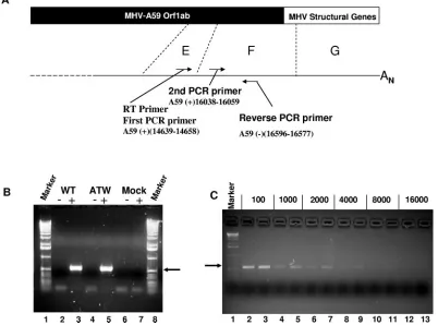
[image:10.585.124.457.68.236.2]in viral RNA synthesis, we employed a nested RT-PCR assay (shown schematically in Fig. 6A) to determine if genome-size negative-strand RNA was synthesized in cells electroporated with MHV-A59 genomic RNA containing this mutation. As shown in Fig. 6B, cells electroporated with ATW and wild-type
FIG. 5. Growth curves for MHV mutants. Replicate cultures in 96-well plates were infected at an MOI of 3 and harvested at the times shown, and titers were determined by plaque assays. Triplicate wells were assayed for each time point. Error bars represent the standard errors of the means.
FIG. 6. The defect in mutant ATW is not at the level of genome-length negative-strand RNA synthesis. (A) Schematic drawing of the assay used to detect negative-strand genome-sized RNA and the relative locations of the primers within the genome. (B) RT-PCR of RNA extracted from wild-type (WT)- and ATW mutant-electroporated cells at 8 hours after electroporation. A 1-kbp ladder was used as a molecular size marker. (C) RT-PCR assay done with the indicated dilutions of the RT product. For each dilution the wild-type sample is on the left and the ATW mutant sample is on the right.
on November 8, 2019 by guest
http://jvi.asm.org/
[image:10.585.92.494.381.679.2]MHV RNA yielded similar amounts of a 559-base-pair frag-ment in this assay (lanes 3 and 5). When reverse transcriptase was omitted from the RT step (lanes 2 and 4), no PCR product of the expected size was obtained from electroporated cells, demonstrating that any template DNA was sufficiently de-graded by DNase digestion so that it could not be amplified. To more accurately define the relative amounts of genome-sized negative-strand RNA present in the electroporated cells, we performed a second series of nested RT-PCRs using various dilutions of the initial RT product. Over this series of dilutions of cDNA, there is no noticeable difference between the amount of ATW and that of wild-type PCR products. This result, although not quantitative, indicates that the amounts of full-length negative-strand RNA in cells electroporated with these two RNA genomes are roughly similar and that the replication defect lies in another step of the viral repli-cation cycle.
To determine if negative-strand RNA complementary to subgenomic mRNAs was synthesized, we employed a second nested RT-PCR assay utilizing an RT primer, oligonucleotide
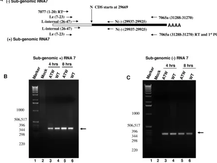
[image:11.585.65.511.76.411.2]7077(1–20), that corresponded to the first 20 nucleotides of the leader sequence present at the 5⬘end of all the MHV mRNAs and the genome (Fig. 7A and 8A). To detect negative-sense RNA complementary to mRNA7, encoding the N gene, nested PCR was carried out using primer sets that correspond to leader-specific primers [Le(7–23) and L-internal(26–47)] and primers within the N coding sequence. As shown in Fig. 7C, RNA extracted from cells electroporated with ATW or wild-type MHV RNA at 4 and 8 h after electroporation (prior to cell fusion in cultures electroporated with wild-type RNA) yielded roughly similar amounts of a 330-base-pair fragment in this assay. Sequencing of the amplicon showed that it corre-sponded to the targeted region and that the amplicon carried the leader sequence fused to the body of the N gene. When reverse transcriptase was omitted from the RT step, no PCR product of the expected size was obtained from electroporated cells (data not shown). A similar result was obtained when RNA was extracted from cells at later times (12, 16, and 24 h) after electroporation (data not shown). Real-time RT-PCR assays were performed using the same nested PCR strategy
FIG. 7. Nested RT-PCR analyses for RNA7. (A) Schematic representation of the RT-nested PCR strategy and primers used to detect negative-and positive-sense subgenomic RNA7. The relative sizes of the leader sequence in RNA7 negative-and the body of RNA7 are not to scale. The position of each primer in the MHV-A59 genome, GenBank accession no. NC0001846, is given in parentheses as part of the primer name. Primer sequences are listed in Table 1. (B) Representative RT-nested PCR for positive-strand subgenomic RNA7. The arrow indicates the specifically amplified RNA7 fragment. The sizes of the marker DNAs in base pairs are shown to the left of the electropherogram. (C) Representative RT-nested PCR for negative-strand subgenomic RNA7. The arrow indicates the specifically amplified RNA7 fragment. The sizes of the marker DNAs in base pairs are shown to the left of the electropherogram.
14580 JOHNSON ET AL. J. VIROL.
on November 8, 2019 by guest
http://jvi.asm.org/
to obtain a more quantitative comparison in the amount of negative-sense RNA7 present. These assays failed to demon-strate consistent differences between cultures electroporated with wild-type and those electroporated with ATW RNA, even at late times when cultures electroporated with the wild type had undergone considerable cell fusion. To investigate if mRNA transcription was impaired for the ATW mutant, a nested RT-PCR assay specific for subgenomic mRNA7 was used (Fig. 7A). As shown in Fig. 7B, RNA extracted from cells electroporated with ATW or wild-type MHV at 4 and 8 h after electroporation yielded roughly equal amounts of the expected 330-base-pair amplicon in this assay. Samples extracted at later times after electroporation also failed to demonstrate an ob-vious difference in the quantities of mRNA7 present (not shown). When reverse transcriptase was omitted from the RT step, no PCR product of the expected size was obtained from electroporated cells (data not shown). Somewhat surprisingly, real-time RT-PCR assays (data not shown) failed to demon-strate consistent differences in mRNA7 levels at either early times (4 and 8 h) or late times (12, 16, and 24 h) after elec-troporation, indicating that similar amounts of mRNA7 were present in cells electroporated with ATW and those electro-porated with wild-type genomes.
The data for mRNA7 are inconsistent with the ATW phe-notype; no syncytia are observed in cells electroporated with ATW genomes, leading us to believe that there must be a defect in subgenomic mRNA synthesis. One possible explana-tion for the nested RT-PCR result is recombinaexplana-tion between the in vitro-transcribed N coding sequence RNA lacking leader
and 3⬘UTR sequences with genomic sequences yielding mRNA7 and its negative-strand complement.
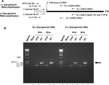
RNA3, which encodes the S protein responsible for cell fusion, was chosen as a target for RT-PCR to further examine the effect of the ATW mutation on subgenomic RNA synthesis (Fig. 8). A nested-set RT-PCR approach similar to that de-scribed for mRNA7 was employed (Fig. 8A). With RNA3 as a target it became obvious that negative- and positive-strand subgenomic RNA synthesis was impaired (Fig. 8B). Nested RT-PCR analysis of RNA extracted from cultures electropo-rated with wild-type genome (lanes 4 and 6) at 4 and 8 h postelectroporation amplified a DNA fragment of the ex-pected 401-bp size, indicated by the arrow in Fig. 8B, whereas cultures electroporated with ATW genomes (lanes 3 and 5) did not yield a fragment of the expected size. The approximately 220-bp fragment observed in the ATW lanes was also present in the mock-electroporated sample (lane 2), indicating that this likely represents nonspecific amplification of a host RNA. These results indicate that no mRNA3 was produced in ATW-electroporated samples, while mRNA3 is abundant in wild-type-electroporated samples. A similar nested RT-PCR experiment was performed to determine if negative-strand subgenomic RNA3 was present. At 4 and 8 h postelectroporation no neg-ative-strand ATW RNA3 could be detected (lanes 9 and 11), although negative-sense RNA3 was easily detected in cells electroporated with wild-type genome (lanes 10 and 12). The origin of the smaller (approximately 200- to 220-bp) fragments amplified in mock, ATW, and wild-type samples was not in-vestigated further. As subgenomic RNA abundance is
regu-FIG. 8. Cells electroporated with ATW genomes do not synthesize RNA3. (A) Schematic representation of the primers used to detect both positive- and negative-strand subgenomic RNA3. (B) Representative experiment for positive- and negative-strand subgenomic RNA3 synthesis at 4 and 8 hours postelectroporation. The sizes of the marker DNAs in base pairs are shown to the left of the electropherogram.
on November 8, 2019 by guest
http://jvi.asm.org/
[image:12.585.112.466.70.346.2]lated by 3⬘-end proximity and transcription regulatory se-quence regulatory domains, a similar experimental approach was used to investigate mRNA6, encoding the M gene, and identical results were obtained. Neither positive- nor negative-sense RNA corresponding to RNA6 was detected in cultures electroporated with ATW genomes (data not shown).
DISCUSSION
Using gel mobility shift RNase T1protection assays, we have
demonstrated a possible role for RNA secondary structure for
host protein binding to the MHV 3⬘ 42-nucleotide element.
Previous work in our lab has demonstrated that an 11-nt motif was important for RNA binding and DI RNA replication (17, 18, 34, 35). We have also identified the host proteins that bind specifically to this region as mitochondrial aconitase and mi-tochondrial HSP70, HSP60, and HSP40 (23, 24). Gel mobility shift RNase T1protection assays with probes containing
mu-tations within the 11-nt motif of the 3⬘42-nucleotide element demonstrated an overall decrease in host protein binding com-pared to wild-type probe. Two mutants, MT3C and MT5A, showed dramatic changes in computer-predicted RNA second-ary structure compared to wild-type sequence and had much lower protein binding activities, 6.3 and 25.2% of wild-type values, respectively. Probe M19C, which contained compensa-tory changes that restored the wild-type predicted secondary structure, also restored host protein binding activity to 74.1% of the wild-type level. The 19G-to-C mutation alone modestly decreased host protein binding to 73.5% of the wild-type level but did not alter computer-predicted RNA secondary struc-ture. These probes provide support for the hypothesis that proper RNA folding is essential for host protein binding to the 3⬘ 42-nucleotide element, although additional biochemical studies are needed to prove the existence of the predicted structures.
A second set of mutant probes was developed based on the Mfold 3.0 predictions of the wild-type 3⬘ 42-nucleotide
ele-ment. Probes ATW, ATW3⬘, and ATW5⬘carried mutations in
a predicted double-stranded region. Computer predictions
suggest that probes ATW3⬘ and ATW5⬘ would have altered
RNA secondary structures while the compensatory mutant ATW should have restored the wild-type structure. ATW5⬘, which is predicted to be mostly single stranded, had little pro-tein binding activity (7.2%), while probe ATW3⬘, which is predicted to have only a slight alteration in RNA secondary structure, had a protein binding activity fourfold greater than that of the wild type. The double mutant, ATW, is predicted to maintain wild-type secondary structure and formed RNP com-plexes 1.85 times more efficiently than wild-type probes did. The results with the ATW mutant suggest the possibility that this RNA may fold into two equally stable structures, with one structure binding RNA as efficiently as the ATW3⬘probe and the second conformation unable to efficiently form RNP com-plexes, producing the observed result. The failure to recover ATW mutant virus using the infectious clone underlines the importance of primary structure at the 3⬘end of the genome in the MHV life cycle. Other studies have established the impor-tance of host proteins interacting with the MHV genome. Polypyrimidine tract binding protein (PTB) binds to the leader RNA as well as the 5⬘end of the genome complement (9, 14).
It has recently been demonstrated that overexpression of PTB decreases viral mRNA transcription but not translation (3). PTB also alters RNA secondary structure and may help
medi-ate 5⬘-to-3⬘ interactions of the MHV genome through PTB
interactions with hnRNP-A1 (8). hnRNP-A1 interacts with
MHV transcription regulatory sequences and 3⬘ UTR; DI
mu-tants with decreased hnRNP-A1 binding also had a decrease in transcription and replication (8, 15) The importance of hnRNP-A1 in MHV transcription and replication is controver-sial, as cell lines deficient in hnRNP-A1 produce viral RNAs as efficiently as cell lines expressing hnRNP-A1 (27).
Spagnolo and Hogue characterized an interaction between the poly(A) tail of DI RNAs and poly(A) binding protein (PABP). PABP is a host cellular protein that is required for translation of eukaryotic mRNAs and is reported to play a vital role in poliovirus replication and transcription by circulariza-tion of the poliovirus genome via its interaccirculariza-tion with eIF4G (5, 22, 30). It is likely that PABP binding to the MHV 3⬘UTR has a similar effect on virus replication as what Spagnolo and Hogue hypothesize (29). Recently Zu´n˜iga et al. developed a model predicated upon a role for 5⬘and 3⬘ genomic interac-tions in the regulation of subgenomic RNA synthesis (37). They demonstrated that base pairing of transcription-regulat-ing sequences with the leader sequence of the genome directly correlated with the amount of subgenomic RNA produced and that discontinuous transcription occurs during negative-strand synthesis. Their data raise the possibility that specific regions of the 3⬘UTR may be required for interactions with the 5⬘end of the genome, either directly or through proteprotein in-teractions, to regulate additional aspects of MHV replication, such as negative-strand synthesis.
Nanda and Leibowitz established the identity of four pro-teins that specifically interact with the MHV 3⬘(⫹)42 host protein binding element as mitochondrial aconitase and mito-chondrial HSP70, HSP60, and HSP40 (23, 24). They also es-tablished that metabolic regulation of the RNA binding activity of mitochondrial aconitase did influence viral growth, although it’s less clear whether these proteins have high or low penetrant effects on virus replication. Mitochondrial aconitase has a cy-tosolic counterpart that is a conditional RNA binding protein, iron regulatory protein. Increasing iron concentrations in MHV-infected cell cultures increased viral titer at early times postinfection compared to infected cells that did not receive supplemental iron (23, 24), emphasizing the importance of RNP complex formation involving mitochondrial aconitase and the MHV genome.
Our functional studies to determine the role for RNP com-plexes of the 3⬘42-nucleotide element in viral replication dem-onstrated that mutations within the 3⬘42-nucleotide host protein element resulted in defects in replication. Targeted recombina-tion experiments to generate viruses containing the MT3C and
M19C and ATW, ATW3⬘, and ATW5⬘ mutations resulted in
the recovery of even-numbered recombinants which restored the wild-type sequence. The data suggest that the mutant vi-ruses which should have been generated by a single or an odd number of crossover events were at a replicative disadvantage compared to the wild-type genomes which were generated by a less-likely even number of crossovers. An alternative strategy that may have yielded a more favorable result would have been to use the feline coronavirus spike protein targeted
recombi-14582 JOHNSON ET AL. J. VIROL.
on November 8, 2019 by guest
http://jvi.asm.org/
nation system (13). Selection by spike protein would have been more stringent and probably reduced the background and even-number recombination events that likely occurred, giving our negative results.
DI RNA replication assays using DI B36 and DI B36
mu-tants MT3C, M19C, ATW, ATW3⬘, and ATW5⬘ were
per-formed. Mutant DI RNAs MT3C and M19C carry mutations within the 11-nt motif previously implicated in protein binding (18, 34, 35). DI mutants MT3C and M19C recombined to restore wild-type sequence in all three experiments evaluated, demonstrating that the mutants could not successfully compete against wild-type recombinant DI RNAs, suggesting the impor-tance of this element in replication. DI RNAs ATW, ATW3⬘, and ATW5⬘replicated poorly. In two experiments in which the mu-tant sequence was maintained in the DI RNAs (as shown by RT-PCR sequencing of the negative strand), ATW and ATW5⬘positive-strand replication was undetectable by meta-bolic labeling, and ATW3⬘replication was barely detectable in one of those experiments.
Comparison of the gel mobility shift RNase T1 protection
assays with the DI RNA replication data indicates the impor-tance of proper primary and secondary structures. Mutant MT3C was dramatically reduced in protein binding activity, while mutants M19C and 19C had more modest decreases in host protein binding. When introduced into DI B36 the DI RNAs MT3C and M19C were not able to replicate and only recombined wild-type DI RNA was detected. Although the gel shift data suggest that probe 19C and M19C, a mutation con-taining the MT3C and 19C mutations, have wild-type second-ary structures and near-wild-type binding activity, the M19C mutation did not restore the replicative capacity of the DI RNA, suggesting the importance of wild-type primary struc-ture in the 11-nt host protein binding motif. In contrast the ATW set of mutants suggest the importance of proper second-ary structure in host protein binding and replication. The mu-tant ATW3⬘could be detected only in one of two recombina-tion-free experiments and at 4% of the wild-type DI B36 level. The ATW3⬘mutants do not lie within the 11-nt element, and secondary structure is disrupted compared to wild type, sug-gesting that secondary structure is important for replication.
When the MT3C, M19C, ATW, ATW5⬘, and ATW3⬘
mu-tations were introduced into a full-length virus genome, results unfettered by recombination repair of DI RNAs were ob-tained. Kinetic analysis of the mutant viruses indicates that there is no impairment in the ability of MT3C, M19C, ATW5⬘, and ATW3⬘to replicate in one-step growth curve experiments performed at moderate multiplicities of infection, although more dramatic changes might be noted at lower MOIs. This result is somewhat difficult to reconcile with the negative ef-fects that these mutations had in DI RNA replication assays and underscores some of the difficulties in using DI RNA models for investigating genome replication. The inherent competition of mutant DI RNAs with both helper virus and any wild-type DI RNAs that arise by recombination compli-cates the interpretation of DI RNA replication assays. Recom-bination is a stochastic event; thus, DI RNA recomRecom-bination varies from experiment to experiment and recombination lead-ing to repair of introduced mutations is unlikely to be equiv-alent among constructs. Recombination probably varies de-pending upon the fitness of the mutation introduced and
transfection efficiency. Recombination is associated with rep-lication, and a slightly more robust mutant DI RNA may repair more quickly, resulting in wild-type DI RNAs. In spite of these limitations, which make interpretation difficult and quantita-tive comparisons of assays where recombination has repaired a mutation impossible, the inherent competitive nature of the DI RNA assay makes them similar in some respects to assays of competition between wild-type and mutant viruses, which may bring out deficits in replication fitness which are not apparent in one-step growth curve experiments.
Each of the mutant viruses produced plaque sizes smaller than wild-type virus. The cause of the small-plaque phenotype is not clear. Metabolic labeling experiments in cells infected
with the wild-type MHV-A59 and MT3C, M19C, ATW5⬘, and
ATW3⬘mutant viruses indicated that approximately the same amount of each species of virus-specific RNA is present regardless of the virus examined (J. L. Leibowitz and J. J. Millership, unpublished data). Since plaques are largely cre-ated by lysis of syncytial giant cells and syncytium formation is mediated by the amount of S protein on the cell surface, it is possible that the differences in plaque size reflect modest dif-ferences in translational efficiency of virus-specific mRNAs carrying these mutations in their 3⬘ UTRs. This question re-quires further investigation to be resolved.
Virus carrying the ATW mutation could not be recovered. A previous study by Lin et al. demonstrated that the 3⬘-terminal 55 nucleotides were necessary for negative-strand synthesis and subsequent DI RNA replication (16), and the ATW mu-tation falls within this region. Our results, using nested RT-PCR, demonstrate that negative-strand RNAs complementary to genome were produced by the ATW virus. However, it was impossible to determine if positive-strand genomes were pro-duced at early times (before the onset of cell fusion in cultures electroporated with wild-type genomes) after electroporation due to the presence of large amounts of the input genomes which persist up to 24 h postelectroporation even in the ab-sence of viral replication (H. Kang and J. L. Leibowitz, unpub-lished data). Subgenomic positive- and negative-strand mRNA3 could not be detected in cultures electroporated with ATW genomes (Fig. 8), and similar results were obtained for RNA6 (data not shown). Negative- and positive-strand sub-genomic mRNA7 was detected using nested RT-PCR, seem-ingly contradicting the data obtained for mRNA3 and mRNA6. The PCR product generated by nested RT-PCR of mRNA7 was cloned and sequenced and found to contain the
expected leader sequence. The 3⬘ UTR of RNA7 could not
be recovered and sequenced since RT-PCRs extending from the leader to the poly(A) tail failed to yield specific products. The RNA7 products are most likely the result of a recombi-nation event with N coding sequences electroporated with the ATW genome to increase the efficiency of generating recom-binant virus. However, we cannot rule out the possibility that ATW genomes support the synthesis of subgenomic RNA7 but not other subgenomic RNAs.
Our data support a role for the 3⬘42-nucleotide host protein binding element in mRNA production. It is possible that the 3⬘ 42-nucleotide host protein binding element is part of a larger complex of viral RNA viral proteins and host proteins which are involved in transcription through mediation of 5⬘-to-3⬘ interactions as recently proposed by Zu´n˜iga et al. (37), who