EFFECT OF VARIOUS IRRIGANTS ON WETTABILITY
AND PUSH-OUT BOND STRENGTH OF AN EPOXY
RESIN-BASED ROOT CANAL SEALER
–
AN IN VITRO
STUDY
Dissertation submitted to
THE TAMILNADU Dr. M.G.R. MEDICAL UNIVERSITY
In partial fulfillment for the Degree of
MASTER OF DENTAL SURGERY
BRANCH IV
CERTIFICATE
This is to certify that this dissertation titled “Effect of various irrigants on
wettability and push-out bond strength of an epoxy resin-based root canal sealer -an in vitro study” is a bonafide record of work done by Dr.KEERTHANA C under
my guidance and to my satisfaction during her postgraduate study period, 2014 – 2017. This dissertation is submitted to THE TAMILNADU Dr. M.G.R.
MEDICAL UNIVERSITY, in partial fulfillment for the award of the degree of
Master of Dental Surgery in Conservative Dentistry and Endodontics, Branch IV. It has not been submitted (partially or fully) for the award of any other degree or diploma.
________________________ ________________________
Dr. Prabhakar V, MDS, Dr. Subha Anirudhan, MDS,
__________________________
Date:
Place: Coimbatore
Dr. Prabhakar V, MDS, Principal, Professor and HOD
Sri Ramakrishna Dental College and Hospital
Coimbatore
Guide, Professor and HOD
Department of Conservative Dentistry and Endodontics,
Sri Ramakrishna Dental College and Hospital. Coimbatore.
Co-Guide, Reader,
Department of ConservativeDentistry and Endodontics,
ACKNOWLEDGEMENT
This thesis is the result of work done with immense support from many people and it is with immense pleasure that I express my heartfelt gratitude to all of them.
I devote my heartfelt thanks to Dr. V. Prabhakar, MDS, Principal & Head of Department and my guide whose care, matchless clinical and theoritical skills,
coupled with support, guidance and encouragement enabled me to successfully complete my dissertation.
I am indebted to my Co-Guide Dr. Subha Anirudhan, MDS, Reader, for her valuable guidance that enabled me to comprehend this dissertation and reach its successful culmination. I am grateful to her for sparing her valuable time in guiding me through this thesis.
I would like to thank and acknowledge Dr. Minu Koshy, MDS, Professor, for her innovative ideas, constructive suggestions, valuable criticism and constant encouragement.
I take this opportunity to express my sincere gratitude to Dr. M. Prabhu, MDS, Reader, Dr. S. Sudhakar, MDS, Reader, Dr. Sriman Narayanan, MDS, Senior Lecturer, Dr. V. Gayathri, MDS, Senior Lecturer and Dr. Mohan Kumar, MDS, Senior Lecturer, who supported me at every juncture throughout my
postgraduate curriculum.
I express my sincere thanks to Dr. Pijush Ghosh, Assistant Professor, Department of Applied Mechanics, IIT Chennai for his guidance in contact angle analysis.
I would like to express my heartfelt gratitude to Mr.V.Muthukumar, M.Tech, MBA, Project engineer and Mr. Selvakumar, M.Tech, MBA, Assistant professor at Department of Textile technology, PSG College of technology, Coimbatore for their sincere efforts that helped me carry out my study at ease.
I also wish to acknowledge the staff and friends at Ramachandra University, who helped me on this little journey.
I am thankful to Dr. Juniad Mohammed, for his guidance in the statistical works of this study.
I am thankful to my seniors, my colleagues and my juniors,who have been together as friends and of great support throughout my period of study here. I am thankful to all other department staff members, my fellow colleagues in other departments, all UG staff members and non-clinical staffs of my department for their great support and encouragement.
I express my dearest gratitude to parents and my husband, and the special people in my life who contributed in various ways towards my study and this dissertation.
Last but not the least, I am greatly indebted to God the Almighty, for blessing me with all the good things in my life and guiding me throughout.
ABSTRACT
AIM OF THE STUDY:
The aim of this in vitro study was to evaluate the effect of various final irrigation regimen on the wettability and push out bond strength of an epoxy resin root canal sealer AH plus.
METHODOLOGY:
To assess wettability, 20 premolars were cross-sectioned to prepare 40 dentin discs. Dentin discs were then divided randomly into 4 groups (n = 10) depending on the irrigation regimen: Group I: 5 ml of 3% NaOCl; Group II: 5 ml of 3% NaOCl + 5 ml of 17% EDTA; Group III: 5 ml of 3% NaOCl + 5 ml of 10%citric acid; Group IV: 5 ml of 3% NaOCl + 5 ml of Q mix. Irrigation regimens were performed for 1 min. Each specimen was placed inside a Dynamic Contact Angle Analyzer. A controlled-volume droplet of sealer was placed on each specimen and the static contact angle was analyzed.
RESULTS:
The contact angles of group 4 (3% NaOCl + Q mix) was significantly less compared to group 1(3% NaOCl), group2 (3% NaOCl + 17% EDTA) and group 3(3% NaOCl + 10% Citric acid). The contact angles of group 2 (3% NaOCl + 17% EDTA) and group 3 (3% NaOCl + 10% Citric acid) were significantly larger than group 1 (3% NaOCl). However, there was no significant difference between the contact angles produced by group 2 (3% NaOCl + 17% EDTA) and group 3 (3% NaOCl + 10% Citric acid).
The push out bond strength of group 2 (3% NaOCl + 17% EDTA), group 3 (3% NaOCl + 10% Citric acid) and group 4 (3% NaOCl + Q mix) were significantly higher than that of group 1 (3% NaOCl) . However there was no significant difference between group 2 (3% NaOCl + 17% EDTA), group 3 (3% NaOCl + 10% Citric acid) and group 4 (3% NaOCl + Q mix).
CONCLUSION:
CONTENTS
TITLE PAGE NO
1. Introduction 1
2. Aim and Objective 3
3. Review of Literature 4
4. Materials and Methods 18
5. Results 36
6. Discussion 45
7. Summary and Conclusion 52
Introduction
1
INTRODUCTION
Adhesion is a clinically favorable property of root canal sealers. Adhesion of an endodontic sealer is defined as its capacity to adhere to the root canal walls and promote the union of gutta-percha cones to each other and to the dentin. 1,2
Contemporary endodontics has seen an unequalled advance in technology and materials. These advances aim at improving the standard of care in endodontics. Adhesive root filling materials which were introduced to adhere to coronal dentine, could minimize leakage by increasing the seal between the core root filling material and the root canal walls.3 AH Plus, is an epoxy based root canal sealer. It is characterized by very good mechanical properties, high radiopacity, reduced polymerization shrinkage, low solubility and not the least, a high degree of stability on storage. Studies have shown that AH plus has higher bond strength to root dentine than methacrylate sealers.4,5
Introduction
2
infiltration.12 Also, chlorhexidine inhibits matrix metalloproteinases and has a positive effect on dentine bonding.13
Recently, an EDTA-based formulation was developed as final rinse solution. QMix (Dentsply Tulsa Dental, Tulsa, OK, USA) contains EDTA, chlorhexidine and a surfactant agent. This solution exhibits a lower level of toxicity than 17% EDTA,14 which also has low toxicity and antimicrobial activity associated with the ability to remove the smear layer.15–18
Aim and Objective
3
AIM AND OBJECTIVE
The aim of the study was:
To evaluate the effect of various final irrigation regimes on the wettability
of AH Plus root canal sealer on intraradicular dentine
To evaluate the effect of various final irrigation regimes on the push out
Review of Literature
4
REVIEW OF LITERATURE
Delivanis et al (1983)19 examined the survivability of F43 strain of Streptococcus
sanguis inside root canals filled with gutta-percha and Procosol Cement. They proved that a fluid tight obturation is essential for prevention of survival of microorganisms in the root canals by using the F43 strain of Streptococcus sanguis. Their results confirmed Grossman’s hypothesis that if a canal is completely filled laterally and
apically any microorganisms remaining inside will not be capable of surviving for a long time.
Drummond et al (1996)20 evaluated the effect of the following variables on shear dentin-bonding test results: mode of testing (cyclic fatigue versus static loading), surface treatments (32% phosphoric acid, 10% phosphoric acid, and no treatment [unetched], and type of shear test (traditional planar versus push-out). They stated that the bond strength resulting from cyclic fatigue of the etched specimens was approximately 51% of the static loading value. Ten percent phosphoric acid was as effective as 32% phosphoric acid for dentin bonding. They also stated that the push-out test provides a better evaluation of the bonding strength than the conventional shear test because with the push-out test fracture occurs parallel to the dentine-bonding interface, which makes it a true shear test for parallel-sided samples.
Review of Literature
5 statistically similar with respect to efficacy.
Goracci et al (2004)22 compared the trimming and non-trimming variants of the microtensile technique with the micro push-out test in the ability to measure accurately the bond strength of fiber posts luted inside root canals. They concluded that the push-out test appears to be more efficient and dependable than both the trimming and non-trimming versions of the micro- tensile technique.
Zehnder et al (2005)23 evaluated the effect of reducing surface tension in
endodontic chelator solutions on their ability to remove calcium from instrumented root canals. Surface tension in these solutions was measured using the Wilhelmy method. Calcium concentration in eluates was measured using atomic absorption spectrometry. They concluded that incorporation of wetting agents resulted in a reduction of surface tension values by approximately 50% in all tested solutions. They also stated that none of the solutions with reduced surface tension chelated more calcium from canals than their pure counterparts
Review of Literature
6
Jainaen et al (2007)25 evaluated the push-out bond strength of the dentine– sealer interface with and without main cone for three resin sealers universal testing machine . They stated that the epoxy resin-based sealer (AH Plus) had the highest push-out bond strength compared with UDMA-based sealers (EndoREZ) and methacrylate resin-based sealer (Resilon ) when used with a main cone and sealer. The bond strengths after filling with sealer alone were higher than those with main cone and sealer, and may reflect different patterns of behavior when the sealer is present as a thin layer.
Teixeira et al (2009)26 compared the shear bond strength (SBS) test and push-out test for evaluation of the adhesion of an epoxy-based endodontic sealer (AH Plus) to dentin and gutta- percha, and to assess the failure modes on the debonded surfaces by means of scanning electron microscopy (SEM). They stated that SEM analysis showed a predominance of adhesive and mixed failures of AH Plus sealer. They also stated that the comparison of the employed methodologies showed that the SBS test produced significantly lower bond strength values than the push-out test, was skillful in determining the adhesion of AH Plus sealer to dentin and gutta-percha, and required specimens that could be easily prepared for SEM, presenting as a viable alternative for further experiments.
Balguerie et al(2011)27 assessed the tubular adaptation and penetration depth
Review of Literature
7
Epoxy resin (AH Plus), Silicon (RSA). They stated that AH Plus showed the most optimal tubular penetration and adaptation to the root canal wall of the sealers tested.
De-Deus et al (2012)28 determined the correlation between leakage and sealer
penetration into dentinal tubule using glucose leakage measurements and Confocal Laser Scanning Microscope respectively. They concluded that there was no significant correlation between leakage and sealer penetration into dentinal tubules. The lack of correlation reported is of relevance as sealer penetration into dentinal tubules has been used as an advantageous property during the launch of new root filling materials and techniques.
Silva et al (2012)29
evaluated the efficacy of smear layer removal of chitosan compared with different chelating agents, and quantified, by atomic absorption spectrophotometry with flame (AASF), the concentration of calcium ions in these solutions after irrigation. They concluded that 15% EDTA, 0.2% chitosan and 10% citric acid effectively removed smear layer from the middle and apical thirds of the root canal. They also stated that 15% EDTA and 0.2% chitosan were associated with the greatest effect on root dentine demineralization, followed by 10% citric acid and 1% acetic acid.
Review of Literature
8 was comparable to EDTA.
Tummala et al (2012)30 compared the wetting behavior of three different root canal sealers namely zinc oxide (ZnOE), AH plus and GuttaFlow on the root canal dentin surface treated with irrigants and their combination using a dynamic contact angle analyzer. The root dentin surfaces were treated with 17% Ethylenediaminetetraaceticacid (EDTA), 3% sodium hypochlorite (NaOCl) and combination of 17% EDTA and 3% NaOCl. They found that the contact angle values for AH Plus sealer were significantly lower when compared to the other two sealer groups.
Vilanova et al (2012)31 assessed the bond strength of Epiphany and AH Plus sealers to root canal walls after use of several endodontic irrigants by push-out test using a universal testing machine. They concluded that except for 1% NaOCl, the removal of smear layer with the other irrigants increased the bond strength of AH Plus to intracanal dentine. They also stated that the use of 1% NaOCl for 30 min with 17% EDTA as final irrigant for 5 min increased the bond strength of Epiphany.
Aranda-Garcia et al (2013)16 evaluated the efficacy of QMiX, SmearClear,
and 17% EDTA for the debris and smear layer removal from the root canal and its
effects on the push-out bond strength of an epoxy-based sealer by scanning electron
microscopy (SEM). They concluded that SmearClear and QMiX are as effective as
17% EDTA in the debris and smear layer removal of the dentinal root surface. The
Review of Literature
9
Ballal et al (2013)32 evaluated the wettability of AH Plus and ThermaSealPlus sealers on intraradicular dentine treated with different irrigating solutions. They stated that when used as a final irrigant, QMix favors the wetting of root canal dentine by both AH Plus and ThermaSeal Plus sealers. Maleic acid shows a promising result when compared to EDTA and NaOCl and wettability of both sealers is the worst on EDTA-irrigated dentine.
Chandrasekhar et al (2013)14 evaluated the biocompatibility of a new root canal irrigant Q mix™ 2 in 1 in comparison to 0.9% sterile saline, 3% sodium hypochlorite 2% chlorhexidine, and 17% Ethylenediaminetetraacetic acid. They concluded that QMix™ 2 in 1 was shown to be less toxic to the rat subcutaneous tissue than 3% NaOCl, 2% CHX, and 17% EDTA.
Review of Literature
10
AlKahtani et al (2014)34 evaluated and compared the cytotoxicity of QMixTM root canal irrigating solution on immortalized human bone marrow mesenchymal stem cells (hTERT-MSC-C1) and compared it with that of sodium hypochlorite (NaOCl). They concluded that both the NaOCl and QMixTM solutions are toxic to human bone marrow MSCs. The QMixTM solution, which induces slow cell death, seems to be more biocompatible than the NaOCl solution.
Bohn &Ilie (2014)35 evaluated the wetting behaviour of three different
classes of endodontic sealers, silicone (Roeko- seal Automix), epoxy-resin (2Seal, AH Plus) and methacrylate-based sealers (EndoRez, RealSeal, Real- Seal SE, Seal 3D) on dentine specimens with and without chemical pre-treatment using a contact angle measuring device in a dynamic mode. Half of the discs were rinsed with distilled water, and the remainders were treated with 3% NaOCl, followed by 17% EDTA and 2% CHX to simulate the final rinse under clinical-like conditions. They stated that silicone-based sealer Roekoseal Automix had better wettability than epoxy-resin or methacrylate-based sealers. The irrigation regime significantly favored the wettability of 2Seal and Real- Seal SE.
Review of Literature
11
Elnaghy (2014)37 investigated the effect of QMix irrigant on the bond strength of glass fibre posts to root dentine and on smear layer removal after post space preparation compared with 5.25% sodium hypochlorite, 2% chlorhexidine digluconate, 17% Ethylenediaminetetraacetic acid (EDTA), 17% EDTA followed by 2% CHX. He concluded that QMix is an effective irrigant that can remove smear layer, open dentinal tubules and simplify the irrigation protocol, without compromising the bonding strength of glass fibre posts cemented with a self-adhesive resin cement to root dentine.
Taneja et al (2014)38 assessed the effect of QMix, peracetic acid and Ethylenediaminetetraacetic acid on calcium loss and microhardness of root dentine. The calcium loss of the samples was evaluated using the Atomic Absorption Spectrophotometer followed by determination of their microhardness using Vickers Hardness Tester. They stated that Irrigation with NaOCl + 2.25% PAA caused the maximum calcium loss from root dentin and reduced microhardness. A negative correlation existed between the calcium loss and reduction in the microhardness of root dentin.
Review of Literature
12
However, the mixture of QMix and NaOCl did not result in para-chloroaniline formation.
Elakanti et al (2015)40 compared the antimicrobial efficacy of QMix2 in 1,
sodium hypochlorite (NaOCl), and chlorhexidine (CHX) against Enterococcus faecalis and Candida albicans by culturing method and colony forming units were counted. They concluded that QMix 2 in 1 demonstrated significant antimicrobial efficacy against Enterococcus faecalis and Candida albicans.
Jardine et al (2015)41 compared the effect of QMix, BioPure MTAD, 17 %
EDTA, and saline on the penetrability of a resin-based sealer AH plus into dentinal
tubules using a confocal laser scanning microscope. They concluded that seventeen
percent EDTA and QMix promoted sealer penetration superior to that achieved by
BioPure MTAD and saline.
Mohan &Pai (2015)42 assessed the influence of two irrigation regimens
Review of Literature
13
Neelakantan et al (2015)43analyzed the influence of irrigation on the chemical interaction between root canal sealers and dentin using Fourier transform infrared spectroscopy (FTIRS) and push-out bond strength test. The root canal sealers tested include, epoxy resin (AH Plus); silicone (RoekoSeal); calcium hydroxide (Sealapex). They found that bond strength of sealers is differentially affected by the irrigation protocol. They also stated that the epoxy resin sealer AH Plus chemically bonds to dentinal collagen and this interaction is influenced by the irrigation protocols.
Uzunoglu et al (2015)44 evaluated effect of temperatures of QMix and EDTA on the bond-strength of AH Plus. They stated that temperature of the final irrigant does affect the bond strength values of AH plus to root dentin irrigated with EDTA. Bond strength of AH Plus sealer to root canal dentin may improve with QMix.
et al (2016)45 assessed the antibacterial efficacy of photon-initiated
photoacoustic streaming (PIPS) using an Er:YAG laser and sonic-activated irrigation
combined with QMiX irrigant or sodium hypochlorite against Enterococcus faecalis
intracanal biofilm by culturing and colony forming unit were counted. They concluded that there was no difference in the bacteria reduction between the
sonically and laser- activated irrigation, regardless of the irrigant used. Although the
activation of the QMiX and NaOCl by the Er:YAG laser did not improve their
antimicrobial action, the fact that it generated the greatest number of sterile samples
Review of Literature
14
Souza et al (2016)46 investigated, in vitro, the retaining of 2 % CHX gel, 2 % CHX liquid and QMix within a root canal for 24 h, 30, 90, and 120 days by chemical analysis. They stated that CHX gel, CHX liquid, and QMix were retained in dentin up to 120 days. Significantly less substantivity was observed for QMix, irrespective of the period of time. They also stated that no differences were noted between CHX gel and CHX liquid after 30, 90, and 120 days of evaluation.
Azar et al (2000)47 studiedthe cytotoxic effects of a new epoxy resin-based
root canal sealer (AH-plus), and compared with AH26 and zinc oxide-eugenol sealers, in vitro on a culture of human gingival fibroblasts. They concluded that cytotoxicity of the AH-plus was confined to the early period of experiment at 1 and 4 hour intervals and was no longer detectable after 4 hr of mixing. They also stated that that AH plus is more biocompatible than the other two sealers, making it suitable for endodontic practice.
Dogan et al (2001)48evaluated the effect of combined and single use of
EDTA, RC-Prep, and NaOCl on mineral content of root dentin using scanning electron microscope (SEM) and energy dispersion spectrometric microanalysis. They concluded that the use of NaOCl irrigation as final flush altered the effectiveness of chelating agents on root dentin surface. They also stated that the use of EDTA and RC-Prep alone did not change the mineral content of root dentin significantly.
Khedmat et al (2008)49 compared the efficacy of three chelating agents in
Review of Literature
15
followed by 3 mL of 5.25%NaOCl was not sufficient to completely remove the smear layer, especially in the apical third. They also stated that the addition of surfactants to EDTA in SmearClear did not result in better smear layer removal compared with EDTA alone.
Hu et al (2010)50 studied the effects of irrigation solutions on dentin
wettability and roughness by using contact angle measurements and AFM analysis respectively. The irrigants used were 17% EDTA, 5.25% NaOCl and 3% H2O2. They stated that all the irrigation solutions in this study influenced the physicochemical properties of dentin surfaces, such as wettability and roughness. Hence, the effects of the changes of physicochemical properties on the bacterial adhesion and restorative adhesive procedure should be further studied. They also stated that due consideration should be given to these effects as the irrigants are used during root canal treatment.
Review of Literature
16
Neelakantan et al (2011)52 evaluated the impact of dentine conditioning on
sealing ability and dentine bond strength of AH plus by fluid transport model and push out tests respectively. The irrigants used were 3% NaOCl, 17% EDTA, 7% maleic acid (MA) or 2% chlorhexidine. They concluded that a final flush with a decalcifying agent appears advisable, whilst a final flush with NaOCl caused untoward effects on bond strength. They also stated that conditioning of canal walls by maleic acid, resulted in superior sealing ability and higher epoxy resin bond strength compared to EDTA. The two outcomes investigated, fluid transport and dentine bond strength, were strongly negatively correlated to each other.
Stelzer et al (2014)53 evaluated the influence of different endodontic irrigants
namely sodium hypochlorite, chlorhexidine, and EDTA on the push-out bond strength of RealSeal SE (SybronEndo, Orange, CA) and AH Plus using the universal testing machine. They stated that the push-out bond strength of the conventional AH Plus sealer is usually higher than that of RealSeal SE. They also stated that the bond strength of RealSeal SE is highly influenced by the irrigant used. Within the AH Plus groups, no significant differences existed in bond strength for different irrigation protocols.
Review of Literature
17 irrigants.
Kaushik et al (2015)55evaluated the contact angle between epoxy resin sealer
and dentin treated with different irrigant solutions using Rame Hart Goniometer. The irrigants used were NaOCl, QMix and 0.1% octenidine hydrochloride. Comparative evaluation of wettability was done before and after removal of smear layer. They stated that QMix irrigated samples showed least values of contact angle, and thus maximum wettability of the sealer followed by 0.1% octenidine hydrochloride followed by 3% NaOCl. They also stated that the removal of smear layer using EDTA further reduced contact angle of AH Plus in samples treated with 0.1% octenidine hydrochloride.
Tuncel et al (2015)56 evaluated the effect of various chelating solutions on
Materials and Methods
18
MATERIALS AND METHODS
ARMAMENTARIUM
INSTRUMENTS USED:
1. Diamond disc with mandrel 2. 600 Grit silicon carbide paper 3. Scale
4. Endomotor (Endo-Mate DT, NSK Nakanishi International, Japan) 5. Protaper rotary files (Dentsply Maillefer, Switzerland)
6. K files (Mani,Tochigi, Japan)
7. Endoblock (Dentsply Maillefer, Switzerland ) 8. Stainless steel cement spatula
9. Glass slab
10. 5ml syringe (Dispo Van) 11. Spirit lamp
12. Glass slab 13. Tweezer 14. Lentulo spiral
15. Guttapercha condenser 16. Spreaders
17. Humidity chamber
Materials and Methods
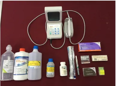
19 MATERIALS USED:
1. 3% Sodium hypochlorite (Vensons India, Bengaluru)
2. Saline (Aculife Healthcare, India)
3. 10% Citric acid (Chenchems, Chennai)
4. 17% EDTA (Chenchems, Chennai)
5. Qmix (Dentsply Tulsa Oklahoma)
6. AH Plus sealer (Dentsply DeTrey GmbH, Konstanz, Germany)
7. Guttapercha (Dentsply Maillefer)
8. Paper points (Dentsply Maillefer)
SOURCE OF SAMPLES:
One hundred freshly extracted human single rooted mandibular premolar teeth that were extracted for periodontal and orthodontic reasons were collected from the Department of Oral and Maxillofacial Surgery, Sri Ramakrishna Dental College and Hospital, Coimbatore.
INFECTION CONTROL PROTOCOL FOR THE TEETH COLLECTED FOR THE STUDY:
Materials and Methods
20
(CDC) recommendations and guidelines as follows:
1. Handling of teeth was always done using gloves, mask and protective eyewear
2. Teeth were cleaned of visible blood and gross debris
3. Teeth were maintained in a hydrated state in a well-constructed closed container containing distilled water during transport
4. Teeth were immersed in 10% formalin for 7 days, following which the liquid was discarded and the teeth were transferred into separate jars containing distilled water
5. The initial collection jars, lids and the gloves employed were discarded into biohazard waste receptacles
6. As and when required, teeth were removed from the jars with cotton pliers
INCLUSION CRITERIA:
1. Intact, single rooted lower premolars with single root canal 2. Teeth with complete root formation
3. Teeth with patent canals
4. Teeth without anatomic variations
5. Teeth free of dental caries and root canal fillings.
EXCLUSION CRITERIA
1. Teeth with open apices 2. Calcified canals
Materials and Methods
21 4. Variations in radicular anatomy 5. Internal or external root resorption 6. Teeth with fractured roots
PROCEDURE:
ASSESSMENT OF DENTIN WETTABILITY OF AH PLUS ROOT CANAL SEALER FOLLOWING DIFFERENT IRRIGATION REGIMENS:
TOOTH SAMPLE PREPARATION:
To reveal root dentine, 20 premolars were cross-sectioned into two separate 1-mm-thick dentine discs per tooth with a diamond disc used under distilled running water. (Figure 2) 40 dentin discs were thus obtained. To create uniform flat surfaces, the dentine discs were subsequently ground with 600-grit silicon carbide paper under distilled water to remove any surface scratches and to provide a smooth surface for the analysis.
SAMPLE TREATMENT:
Dentin discs were then divided randomly into 4 groups (n = 10) depending on the irrigation regimen:
• Group I: 5 ml of 3% NaOCl.
Materials and Methods
22
Irrigation was carried out for one minute. Five milliliters saline was used between the irrigants in groups 2 -4. Following irrigation, all the samples were rinsed with 5 ml of saline and dried with paper points.
CONTACT ANGLE MEASUREMENT
The contact angle was measured using a Dynamic Contact Angle Analyzer (GBX Digidrop, IIT Chennai) under standard conditions of temperature and relative humidity. (Figure 9) This equipment is used for measuring both the static and dynamic contact angles. AH Plus (Dentsply DeTrey, Konstanz, Germany), an epoxy resin-based sealer, was mixed according to the manufacturer’s instructions. (Figure8)
Materials and Methods
23
ASSESSMENT OF PUSH OUT BOND STRENGTH OF AH PLUS ROOT CANAL SEALER FOLLOWING DIFFERENT IRRIGATION REGIMENS:
TOOTH SAMPLE PREPARATION:
The teeth were decoronated at the cementoenamel junction by using a diamond disk under distilled running water. (Figure 1) The root lengths were standardized to 12 mm and radiographed at 2 angulations to confirm the presence of a single canal. A size 10 K file was placed in the canal until it was visible at the apical foramen. The working length was determined by subtracting 1mm from this measurement. Biomechanical preparation was performed with ProTaper Universal NiTi rotary instrument. (Figure 11 &12) Canals were enlarged up to F4 at the working length. The irrigating solution was delivered via a sterile 30-gauge single side vented needle (Ammdent Canal clean) which penetrated 2mm short of the working length.
The samples were divided into 4 groups (n = 20) according to the final irrigation regimen.
Group I: 5 ml of 3% NaOCl
Group II: 5 ml of 3% NaOCl + 5 ml of 17% EDTA.
Group III: 5 ml of 3% NaOCl + 5 ml of 10% citric acid.
Group IV: 5 ml of 3% NaOCl + 5 ml of Q mix.
Materials and Methods
24
irrigants in groups 2 -4. All groups received a final flush with 5 ml of saline and were then dried with absorbent paper points. AH Plus sealer (Dentsply DeTrey GmbH, Konstanz, Germany) was mixed according to the manufacturer’s instructions and was coated on the root canal walls using a letulospiral. Obturation was done with F4 size master cone by cold lateral compaction. The teeth were radiographed at 2 angulations. Samples with voids or bubbles were discarded. Specimens were placed in 100% humidity for 48 hours to ensure complete setting of the sealer. (Figure 13)
After this period, the specimens were embedded in self cure acrylic resin
(Figure 14& 15) and each root was transversely sectioned using a hard tissue
microtome (Leica Germany). One mm slices of mid-root dentin were made to
achieve a main cone diameter approximately 1mm. (Figure 16 - 18)
MEASUREMENT OF DISLOCATION RESISTANCE BY PUSH-OUT BOND STRENGTH TEST
Materials and Methods
25
Bond strength (MPa)= Load in newton /Area of bonded surface .
The area of bonded surface is given by the formula 2πr*h. π is a constant with an approximate value of 3.14, r is the internal diameter of the root canal and h is the height of the specimen. All the data obtained were recorded, tabulated and statistically evaluated.
SCHEMATIC REPRESENTATION OF THE PUSH-OUT TEST
Materials and Methods
[image:37.595.159.476.153.410.2]26
FIGURE 1: DECORONATED TOOTH SPECIMEN FOR PUSH-OUT BOND STRENGTH TEST
[image:37.595.136.502.508.762.2]Materials and Methods
[image:38.595.125.512.124.411.2]27
FIGURE 3: ARMAMENTARIUM USED FOR THE STUDY
[image:38.595.215.418.493.728.2]Materials and Methods
[image:39.595.128.258.125.376.2]28
FIGURE 5: 17% EDTA FIGURE 6: 10% CITRIC ACID
[image:39.595.363.484.127.380.2] [image:39.595.223.412.444.720.2]
Materials and Methods
[image:40.595.153.485.111.341.2]29
FIGURE 8: AH PLUS
[image:40.595.144.496.454.709.2]Materials and Methods
[image:41.595.237.402.125.386.2]30
FIGURE 10: EXPERIMENTAL SET UP WITH DENTIN DISC IN POSITION
FIGURE 11: BIOMECHANICAL PREPARATION OF DECORONATED SAMPLES Materials and Methods
[image:41.595.139.496.506.744.2]33 | P a g e FIG 9: ENDODONTIC PREPARATION OF DECORONATED SAMPLES
Materials and Methods
[image:42.595.148.489.123.356.2]31
[image:42.595.140.499.457.725.2]FIGURE 12: PROTAPER ROTARY SYSTEM AND K FILE
FIGURE 13: TEETH SPECIMEN IN HUMIDITY CHAMBER Materials and Methods
30 | P a g e FIG 3: X-SMART ENDOMOTOR AND HANDPIECE
Materials and Methods
[image:43.595.225.406.142.370.2]32
FIGURE 14: ELASTOMERIC IMPRESSION SAMPLES MOLD WITH MOUNTED ROOT
[image:43.595.152.485.432.680.2]Materials and Methods
[image:44.595.185.447.127.351.2]33
FIGURE 16: HARD TISSUE MICROTOME
[image:44.595.165.467.488.726.2]Materials and Methods
[image:45.595.159.478.125.364.2]34
FIGURE 18: MID ROOT DENTIN SLICES
FIGURE 19: EXPERIMENTAL SET UP WITH UNIVERSAL TESTING MACHINE AND THE SAMPLE IN POSITION
Materials and Methods
35 | P a g e
[image:45.595.183.450.495.736.2]FIG 13: CROSS SECTION AT THE MIDDLE THIRD OF THE SAMPLES
FIG 14: EXPERIMENTAL SET UP WITH THE UNIVERSAL TESTING
Materials and Method
35 STATISTICAL ANALYSIS
Data was analyzed using SPPS VERSION 16 (Statistical Software). The level of significance was set at P < 0.05 and with a confidence interval of 95%. Inter and intra group comparisons were done with one-way ANOVA followed by a pairwise comparison with Turkey’s post hoc test for evaluating the wettability and push out
Results
36
RESULTS
STATISTICAL ANALYSIS OF WETTABILITY OF AH PLUS ON ROOT CANAL DENTIN FOLLOWING VARIOUS IRRIGATION REGIMES
DESCRIPTIVES (TABLE 1)
WETTABILITY
N Mean Std. Deviation Std. Error
95% Confidence Interval for
Mean Minimum Maximum
Lower Bound Upper Bound
Sodium Hypochlorite 10 48.5000 1.65597 .52366 47.3154 49.6846 46.40 51.10
NaOCl + EDTA 10 61.0800 3.45311 1.09197 58.6098 63.5502 54.30 66.20
NaOCl + Citric Acid 10 60.1600 3.66521 1.15904 57.5381 62.7819 53.10 65.20
NaOCl + Q Mix 10 33.9500 2.92204 .92403 31.8597 36.0403 29.30 38.80
Results
37
ONE WAY ANOVA (TABLE 2)
WETTABILITY
Sum of
Squares df Mean Square F Sig.
Between Groups 4824.405 3 1608.135 175.569 .001
Within Groups 329.745 36 9.160
Total 5154.150 39
Results
38
Table 3 shows the intergroup comparison by Post Hoc Tukey test, where high statistical difference was found when group 1 was individually compared with group 2, group 3 and group 4. There was no statistical difference between group 2 and group 3. Group 4 showed high statistical difference when compared individually with group 3 and group 4.
MULTIPLE COMPARISONS (TABLE 3)
WETTABILITY Tukey HSD
(I) Groups (J) Groups
Mean Difference
(I-J) Std. Error Sig.
95% Confidence Interval
Lower Bound Upper Bound Sodium Hypochlorite
NaOCl + EDTA -12.58000* 1.35348 .000 -16.2252 -8.9348
NaOCl + Citric Acid -11.66000* 1.35348 .000 -15.3052 -8.0148
NaOCl + Q Mix 14.55000* 1.35348 .000 10.9048 18.1952
NaOCl +EDTA Sodium Hypochlorite 12.58000* 1.35348 .000 8.9348 16.2252
NaOCl + Citric Acid .92000 1.35348 .904 -2.7252 4.5652
NaOCl + Q Mix 27.13000* 1.35348 .000 23.4848 30.7752
NaOCl + Citric
Acid
Sodium Hypochlorite 11.66000* 1.35348 .000 8.0148 15.3052
NaOCl + EDTA -.92000 1.35348 .904 -4.5652 2.7252
NaOCl + Q Mix 26.21000* 1.35348 .000 22.5648 29.8552
NaOCl + Q Mix Sodium Hypochlorite -14.55000* 1.35348 .000 -18.1952 -10.9048
NaOCl + EDTA -27.13000* 1.35348 .000 -30.7752 -23.4848
NaOCl + Citric Acid -26.21000* 1.35348 .000 -29.8552 -22.5648
[image:50.595.85.551.204.579.2]Results
39
STATISTICAL ANALYSIS OF PUSH OUT BOND STRENGTH OF AH PLUS ON ROOT CANAL DENTIN FOLLOWING VARIOUS IRRIGATION REGIMES
PUSHOUT RESULTS
DESCRIPTIVES (TABLE 4)
Groups Number Mean Std.
Deviation
Minimum Maximum
SodiumHypochlorite 20 4.892105 .8366107 3.7000 6.7500
NaOCl + EDTA 20 7.151000 1.2995906 4.7900 9.9000
NaOCl+ CitricAcid 20 6.814000 1.3374776 4.3700 9.2300
NaOCl + QMix 20 7.194500 1.4243132 4.4600 9.9800
Results
40
ANOVA FOR PUSH OUT TESTS (TABLE 5)
Groups Mean
Std. Deviation
df
Mean Square
F
P value
SodiumHypochlorite 4.892105 .8366107
3 23.043 14.743 .000 NaOCl + EDTA 7.151000 1.2995906
NaOCl + CitricAcid 6.814000 1.3374776
NaOCl + QMix 7.194500 1.4243132
Results
41
MULTIPLE COMPARISONS (TABLE 6) PUSH OUT TESTS
Post hoc analysis (Tukey HSD) to analyse the diffence between the groups
Groups Mean
Differnece P value
SodiumHypochlorite
NaOCl + EDTA -2.2588947* .000
NaOCl + CitricAcid -1.9218947* .000
NaOCl + QMix -2.3023947* .000
NaOCl + EDTA
SodiumHypochlorite 2.2588947* .000
NaOCl + CitricAcid .3370000 .829
NaOCl + QMix -.0435000 1.000
NaOCl + CitricAcid
SodiumHypochlorite 1.9218947* .000
NaOCl + EDTA -.3370000 .829
NaOCl + QMix -.3805000 .771
NaOCl + QMix
SodiumHypochlorite 2.3023947* .000
NaOCl + EDTA .0435000 1.000
NaOCl + CitricAcid .3805000 .771 *. The mean difference is significant at the 0.05 level.
[image:53.595.105.528.166.601.2]Results
[image:54.595.111.530.224.498.2]42
FIGURE 20: DISTRIBUTION OF GROUPS BASED ON MEAN VALUES. (WETTABILITY)
48.5
61.08 60.16
33.95
0 10 20 30 40 50 60 70
Sodium Hypochlorite EDTA Citric Acid Q Mix
Results
43
FIGURE 21: REPRESENTATIVE IMAGES OF SESSILE DROPS OF ROOT
CANAL SEALERS APPLIED TO ROOT CANAL DENTINE TREATED WITH
DIFFERENT IRRIGATING SOLUTIONS.
3 %SODIUM HYPOCHLORITE
17% EDTA
10% CITRIC ACID
Results
[image:56.595.113.537.168.415.2]44
FIGURE 22: DISTRIBUTION OF THE GROUPS BASED ON MEAN VALUES (PUSH OUT BOND STRENGTH)
4.89
7.151
6.814 7.19
0 1 2 3 4 5 6 7 8
SodiumHypochlorite EDTA CitricAcid QMix
Discussion
45
DISCUSSION
Attaining a fluid tight seal in all dimensions, within the entire root canal system, is one of the key factors contributing to the long term success of root canal
treatment.59 This may be achieved by three-dimensional obturation of the root canal
system to prevent oral and apical microleakage. In the context of canal filling, endodontic sealers play an important role as they fill root canal wall irregularities, accessory canals, ramifications and the space between guttapercha and canal walls.60
Optimum adhesion requires intimate contact between the sealer and the substrate to facilitate molecular attraction and allow either chemical adhesion or penetration for micromechanical surface interlocking. The adhesion processes are mainly influenced by the relative surface free energy (wetting ability) of the solid surface.60,61 Wetting is the property of a liquid to sustain contact with a solid surface, depending on the relative surface energies of the solid and the liquid. 63-65Adhesion of a root canal sealer depends on the wetting ability of the sealer.66
Discussion
46
The utilization of sodium hypochlorite (NaOCl) irrigant in endodontics is justified by its undeniable importance as a result of both its wide-spectrum antimicrobial activity and its properties as an organic tissue solvent. However, it is known to be highly irritant to the periapical tissues, mostly at high concentrations and it is unable to remove the smear layer by itself, as it dissolves only organic material .70-72 Chlorhexidine digluconate (CHX) is a potent antiseptic with a broad-spectrum antimicrobial activity and low grade of toxicity; however, it has no tissue-dissolving capability, and consequently, it cannot replace NaOCl.71-73 Ethylenediaminetetraacetic acid (EDTA) is effective at dissolving inorganic material, including hydroxyapatite; on the other hand, it has little or no effect on organic tissue and it does not possess antibacterial activity.72 Thus most of the commonly used irrigants have only few desirable properties.
By virtue of the known drawbacks of all endodontic irrigants, developing improved irrigating solutions for endodontics remains an area of great interest.48 Recently, QMix (Dentsply Tulsa Dental, Tulsa, OK, USA), a novel irrigant for smear layer removal with added antimicrobial agents, has been developed. It contains EDTA, CHX and a detergent. QMix is a clear solution ready to use with no chair- side mixing.15,17 It is effective against bacterial biofilms and removes smear layer equivalent to 17% Ethylenediaminetetraacetic acid (EDTA)15,17
AH plus is a epoxy resin based endodontic sealer which can be used with guttapercha to obtain a three dimensional filling.74 AH Plus has better penetration into
the micro‐irregularities because of its creep capacity and long setting time, which
Discussion
47
properties, such as low solubility, minimal expansion, adhesion to dentin, and very good sealing ability, AH Plus is considered as a benchmark “Gold Standard.” 74
It would be clinically relevant to know whether an irrigation regimen having QMIX as final irrigant would have any effect on dentine wettability and adhesion of root canal sealers when compared to the conventional irrigants namely sodium hypochlorite, EDTA and citric acid.
The tendency of a liquid to spread on a solid surface is expressed in the formation of a contact angle between the surface of the fluid and the surface of the solid.63-65 Thus, the contact angle provides an inverse measure of wettability75.76, meaning that the lower the contact angle, the better the wettability.63,75,76 Therefore, the measurement of contact angles can help indirectly assess the sealing ability of endodontic sealers.
Also adhesion of root canal sealers can be assessed by push-out test as the test
allows an accurate standardization of the specimens,1,2 while the micro-push- out test, for use in smaller areas, yields the development of a more uniform shear strength without the interference of the tensile component, thus producing a stress more
reliably directed at the adhesive interface.22,77
Discussion
48
The contact angle can be measured using a captive bubble or a sessile drop technique. In the present study, the sessile drop method was used. With this approach, the contact angles of a liquid drop on flat surfaces can be maintained in a dry environment. The state of hydration of the dentin surface is also shown to affect the contact angle.78 Although in most studies the solid surfaces were dried with air,78,79 in the present study the samples were dried with paper points to simulate conditions in the root canal system.
The factors affecting the contact angle on the liquid side include the surface tension, and those on the solid side include the hydrophobic effect caused by changes in the porosity as a result of surface roughness, and the surface energy effect based on the changes in the 3-dimensional structure of the surface molecules.80 Polishing the dentine surface was done to reduce the influence of roughness on the surface energy of the root dentine wall and thus to reduce its influence on the contact angle measurement. 81
All static contact angle measurements were performed by using a controlled-volume (0.1 ml) of the sealer using a Digidrop software to measure the controlled-volume. This was done to nullify the effect of volume change on the value of contact angle measurement.82
Discussion
49
reported in previous studies.24,32,81 This occurs because dentine consists predominantly of two different substrates: collagen, which has a low surface free energy, and carbonated apatite, which has a high surface energy.83 Due to the demineralizing ability of EDTA and its absence of a surfactant effect, the use of EDTA as the final canal irrigant results in a thin layer of demineralized collagen fibrils on the dentine surface.84 In the absence of an accompanying surfactant, the presence of this layer of collagen fibrils is responsible for the poor wettability of root canal sealers on the EDTA-irrigated dentine.85
Citric acid produced similar results as EDTA. This may be due to the similar decalcifying effect of EDTA and citric acid21 and also the lack of surfactant.
Contrary to the findings of this study López et al in 2013 discovered that 1% citric acid and 17% EDTA, alone or in combination with sodium hypochlorite, increased the surface free energy of dentine.33 However they measured water contact angles whereas in our study the contact angle of the root canal sealer was measured. Tummala et al in 2012 reported that wettability of AH plus sealer was better when root dentin was treated with EDTA compared to NaOCl alone. This may be explained by the difference in the irrigation time (5 minutes) and the time of contact angle measurement (30 minutes after the droplet was placed).30
Discussion
50
detergent(surface active agent: N‐Cetyl‐N, N, N‐trimethylammonium bromide)
present in QMix. The rationale of adding a surface active agent in QMix is to lower surface tension and increase wettability,86 which also enables better penetration of irrigant into the root canal. Chlorhexidine also has a surfactant molecule which increases the dentin surface free energy.86 Smear layer tends to act as a barrier preventing diffusion of sealer into the dentinal tubule. The presence of EDTA in QMix improves wettability of sealer by removing smear layer and exposing dentinal tubules. 50
Results of the present study show that final rinse with a chelating agent enhanced the bond strength of the AH Plus when compared to the sodium hypochlorite group. This could be attributed to two reasons: 1) Smear layer removal
procedures allow the sealer penetration into the dentinal tubules and thus could increase the dentin bond strength of resin based sealer as well as an enhanced seal.2) The suggested mechanism of bonding of AH Plus to the organic phase of the root dentine, most likely in the collagen network.8
Discussion
51
tension of chelator solutions was lowered with wetting agents which also might account for the similar bond strengths. Similar to our results, Aranda-Garcia et al.16 and Leal et al88 observed similar push-out bond strength values when EDTA and QMix were used as final irrigant, after the use of NaOCl.
Contrary to the results of the present study, Stelzer et al53and Tuncel et al56 reported that EDTA did not enhance the bond strength of AH plus sealer.
LIMITATIONS OF THE STUDY:
Oral cavity temperature, surface roughness of the instrumented root canal
surface, method of sealer placement differ in clinical situations when compared to the experimental scenario of this in vitro study. These factors might affect the contact angle values in vivo.
Studies to evaluate the influence of wettability on sealer penetration are needed.
Improved wettability may enhance the spreading of the sealer circumferentially over the root dentin surface. It may also enhance the penetration of sealers into lateral canals thus contributing to a three dimensional obturation. Future research should focus on the effect of wettability on sealer penetration and three dimensional obturation of root canals.
Summary and Conclusion
52
SUMMARY AND CONCLUSION
The aim of the study was to compare the effect of various final irrigation regimen on the wettability and push out bond strength of a epoxy resin root canal sealer AH plus.
To assess wettability, 20 premolars were cross-sectioned into two separate 1-mm-thick dentine discs thus obtaining 40 dentin discs. Dentin discs were then divided randomly into 4 groups (n = 10) depending on the irrigation regimen:
Group I: 5 ml of 3% NaOCl.
Group II: 5 ml of 3% NaOCl + 5 ml of 17% EDTA.
Group III: 5 ml of 3% NaOCl + 5 ml of 10% citric acid.
Group IV: 5 ml of 3% NaOCl + 5 ml of Q mix.
The contact angle was measured using a Dynamic Contact Angle Analyzer. Images of the droplets were then captured using the Digidrop software to determine the static contact angles made by the sealers on root canal dentine.
Summary and Conclusion
53
specimens were stored at 37 degree Celsius, 100% humidity for 48 hours to ensure complete setting of the sealer.
One mm slices of mid-root dentin were made. The root filling in each section
was subjected to universal testing machine at a crosshead speed of 1mm/minute.
Load was applied in apico-coronal direction until bond failure occurred. The maximum failure load was recorded in Newtons, and push-out bond strength was calculated in megapascals (MPa).
Bibliography
54
BIBLIOGRAPHY
1. Sousa-Neto MD, Coelho FI, Marchesan MA, Alfredo E, Silva- Sousa YTC. In vitro study of the adhesion of an epoxy based sealer to human dentine submitted to irradiation with Er:YAG and Nd:YAG lasers. Int Endod J. 2005;38:866-870.
2. Sousa-Neto M , Passarinho-Neto JG, arvalho-Ju nior JR, ruz- ilho AM, Pe cora JD, Saquy PC. Evaluation of the effect of EDT A, EGT A and CDT A on dentin adhesiveness and microleakage with different root canal sealers. Braz Dent J. 2002;13:123-128.
3. Schwartz RS. Adhesive dentistry and endodontics Part 2: bonding in the root canal system-the promise and the problems: a review. J Endod.
2006;32(12):1125-34.
4. De-Deus G, Di Giorgi K, Fidel S, Fidel RA, Paciornik S. Push-out bond strength of Resilon/Epiphany and Resilon/ Epiphany self-etch to root dentin. J Endod. 2009; 35(7): 1048–1050.
5. Gesi A, Raffaelli O, Goracci C, Pashley DH, Tay FR, Ferrari M. Interfacial strength of Resilon and gutta-percha to intraradicular dentin. J Endod.
2005;31(11):809-13.
Bibliography
55
7. De-Deus G, Namen F, Galan JJ, Zehnder. Soft chelating irrigation protocol optimizes bonding quality of Resilon/ Epiphany root fillings. J Endod.
2008;34(6):703-5.
8. Fisher MA, Berzins DW, Bahcall JK. An in vitro comparison of bond strength of various obturation materials to root canal dentin using a push-out test design. J Endod. 2007;33(7):856-8.
9. Mai S, Kim YK, Arola DD et al. Differential aggressiveness of ethylenediaminetetraacetic acid in causing canal wall erosion in the presence of sodium hypochlorite. J Dent. 2010;38(3):201-6.
10. Zhang K, Kim YK, Cadenaro M et al. Effects of different exposure times and concentrations of sodium hypochlorite/ethylenediaminetetraacetic acid on the structural integrity of mineralized dentin. J Endod. 2010 ;36(1):105-9.
11. LottantiS, Gautschi H, Sener B, Zehnder M. Effects of ethylenediaminetetraacetic, etidronic and peracetic acid irrigation on human root dentine and the smear layer. Int Endod J. 2009;42(4):335-43.
12. Prati C, Biagini G, NucciC,Castaldini C, Zucchini C. Effects of chemical pretreatments on dentin bonding. Am J Dent. 1990;3(5):199-206.
13. Hebling J, Pashley DH, Tjaderhane L, Tay FR. Chlorhexidine arrests subclinical degradation of dentin hybrid layers in vivo. J Dent Res.
Bibliography
56
14. Chandrasekhar V, Amulya V, Rani VS, Prakash TJ, Ranjani AS, Gayathri CH.
Evaluation of biocompatibility of a new root canal irrigant QMixTM 2in1–an in vivo
study. J Conserv Dent. 2013; 16(1): 36–40.
15. Stojicic S, Shen Y, Qian W, Johnson B, Haapasalo M. Antibacterial and smear layer
removal ability of a novel irrigant, QMiX. Int Endod J. 2012; 45(4): 363–71.
16. Aranda-Garcia AJ, Kuga MC, Vitorino KR et al. Effect of the root canal final rinse
protocols on the debris and smear layer removal and on the push-out strength of an
epoxy-based sealer. Microsc Res Tech. 2013;76(5): 533–7.
17. Dai L, Khechen K, Khan S et al. The effect of QMix, an experimental antibacterial
root canal irrigant, on removal of canal wall smear layer and debris. J Endod.
2011;37(1): 80–4.
18. Eliot C, Hatton JF, Stewart GP et al. The effect of the irrigant QMix on removal of
canal wall smear layer: an ex vivo study. Odontology. 2014;102(2): 232–40.
19. Delivanis PD, Mattison GD, Mendel RW. The survivability of F43 strain of Streptococcus sanguis in root canals filled with gutta-percha and Procosol cement. J Endod. 1983;9(10):407-10.
20. Drummond JL, Sakaguchi RL, Racean DC, Wozny J, Steinberg AD. Testing mode and surface treatment effects on dentin bonding. J Biomed Mater Res.
1996;32(4):533-41.
Bibliography
57
22. Goracci C, Tavares AU, Fabianelli A, Monticelli F, Raffaelli O, Cardoso PC, Tay F, Ferrari M. The adhesion between fiber posts and root canal walls: comparison between microtensile and push-out bond strength measurements. Eur J Oral Sci. 2004;112(4):353– 361.
23. Zehnder M, Schicht O, Sener B, Schmidlin P. Reducing surface tension in endodontic chelator solutions has no effect on their ability to remove calcium
from instrumented root canals. J Endod. 2005;31(8):590–592.
24. DoganBuzoglu H, Calt S, Gumusderelioglu M. Evaluation of the surface free energy on root canal dentine walls treated with chelating agents and NaOCl. Int Endod J. 2007;40(1):18-24.
25. Jainaen A, Palamara JEA, Messer HH. Push-out bond strengths of the dentine–sealer interface with and without a main cone. Int Endod J. 2007;40(11):882–890.
26. Teixeira CS, Alfredo E, Thome LH, Gariba-Silva R, Silva-Sousa YT, Sousa-Neto MD. Adhesion of an endodontic sealer to dentin and gutta-percha: shear and push-out bond strength measurements and SEM analysis. J Appl Oral Sci. 2009;17(2):129–35.
27. Balguerie E, van der Sluis L, Vallaeys K, Gurgel-Georgelin M, Diemer F. Sealer penetration and adaptation in the dentinal tubules: a scanning electron microscopic study. J Endod. 2011;37(11):1576-9.
Bibliography
58
29. ilva Guedes akadi e cora ru -Filho AM. Chitosan: a new solution for removal of smear layer after root canal instrumentation. Int Endod J.2013; 46(4):332–338.
30. Tummala M, Chandrasekhar V, Rashmi AS, Kundabala M, Ballal V. Assessment of the wetting behavior of three different root canal sealers on root canal dentin. J Conserv Dent. 2012;15(2):109-12.
31. Vilanova WV, Carvalho-Junior JR, Alfredo E, Sousa- Neto MD, Silva-Sousa YTC. Effect of intracanal irrigants on the bond strength of epoxy resin-based and methacrylate resin- based sealers to root canal walls. Int Endod J. 2012;45(1):42–48.
32. Ballal NV, Tweeny A, Khechen K, Prabhu KN, Satyanarayan, Tay FR. Wettability of root canal sealers on intraradicular dentine treated with different irrigating solutions. J Dent. 2013;41(6):556‐60.
33. López GL, de la Casa ML, Torres PL, del MilagroSáez M, López ME. Effect of combinations of irrigating solutions on dentine wettability. EndodPrac Today. 2013;7(1):65-69.
34. Alkahtani A, Alkahtany SM, Mahmood A, Elsafadi MA, Aldahmash AM, Anil S. Cytotoxicity of QMix™ endodontic irrigating solution on human bone marrow mesenchymal stem cells. BMC Oral Health. 2014;14:27
Bibliography
59
36. Das A, Kottoor J, Mathew J, Kumar S, George S. Dentine microhardness changes following conventional and alternate irrigation regimens: An in vitro study. J Conserv Dent. 2014;17(6):546-9.
37. Elnaghy AM. Effect of QMixirrigant on bond strength of glass fibre posts to root dentine. Int Endod J. 2014;47(3):280–289.
38. Taneja S, Kumari M, Anand S. Effect of QMix, peracetic acid and
ethylenediaminetetraacetic acid on calcium loss and microhardness of root dentine. J Conserv Dent. 2014;17(2):155-8.
39. Arslan H, Uygun AD, Keskin A, Karatas E, Seckin , Yıldırım A. Evaluation of orange-brown precipitate formed in root canals after irrigation with chlorhexidine and QMix and spectroscopic analysis of precipitates produced by a mixture of chlorhexidine/NaOCl and QMix/NaOCl. Int Endod J. 2015; 48(12):1199–1203.
40. Elakanti S, Cherukuri G, Rao VG, Chandrasekhar V, Rao AS, Tummala M.
Comparative evaluation of antimicrobial efficacy of QMixTM 2 in 1, sodium hypochlorite, and chlorhexidine against Enterococcus faecalis and Candida albicans. J Conserv Dent. 2015;18(2):128-31.
41. Jardine AP, Rosa RA, Santini MF, Wagner M, Reis So MV, Kuga MC et al.
The effect of final irrigation on the penetrability of an epoxy resin-based
sealer into dentinal tubules: a confocal microscopy study. Clin Oral Invest.
Bibliography
60
42. Mohan RP, Pai AR. The comparison between two irrigation regimens on the
dentine wettability for an epoxy resin based sealer by measuring its contact angle formed to the irrigated dentine. J Conserv Dent. 2015;18(4):275-8.
43. Neelakantan P, Sharma S, Shemesh H, Wesselink PR. Influence of Irrigation
Sequence on the Adhesion of Root Canal Sealers to Dentin: A Fourier Transform Infrared Spectroscopy and Push-out Bond Strength Analysis. J Endod. 2015;41(7):1108–1111.
44. Uzunoglu E, Turker SA, Karahan S. The Effect of Increased Temperatures of
QMix and EDTA on the Push-out Bond Strength of an Epoxy-resin Based
Sealer. J ClinDiagn Res. 2015; 9(7):ZC98–ZC101.
45. alić M, Lucić R, Mehadžić K, Bago I, Anić I, Jakovljević S, Plečko V. The
efficacy of photon-initiated photoacoustic streaming and sonic-activated irrigation combined with QMiX solution or sodium hypochlorite against intracanal E. faecalis biofilm. Lasers Med Sci. 2016;31(2):335-42.
46. Souza MA, Montagner A, Lana DL, Vidal CM, Farina AP, Cecchin D.
Comparative evaluation of the retaining of QMix and chlorhexidine formulations on human dentin: a chemical analysis. Clin Oral Investig.
2016;30.
47. Azar NG, Heidari M, Bahrami ZS, Shokri F. In vitro cytotoxicity of a new
epoxy resin root canal sealer. J Endod. 2000;26(8):462‐5.
Bibliography
61
49. Khedmat S, Shokouhinejad N. Comparison of the efficacy of three chelating agents in smear layer removal. J Endod. 2008; 34(5): 599–602
50. Hu X, Ling J, Gao Y. Effects of irrigation solutions on dentin wettability and roughness. J Endod. 2010;36(6):1064‐7.
51. Dai L, Khechen K, Khan S, Gillen B, Loushine BA, Wimmer CE, et al. The effect of QMix, an experimental antibacterial root canal irrigant, on removal
of canal wall smear layer and debris. J Endod. 2011;37(1):80-4.
52.Neelakantan P, Subbarao C, Subbarao CV, De-Deus G, Zehnder M. The impact of root dentine conditioning on sealing ability and push-out bond strength of an epoxy resin root canal sealer. Int Endod J.2011; 44(6): 491– 498.
53. Stelzer R, Schaller HG, Gernhardt CR. Push-out bond strength of RealSeal SE and AH Plus after using different irrigation solutions. J Endod.
2014;40(10):1654-7.
54.Gründling GL, de Melo TAF, Montagner F, Scarparo RK, Vier-Pelisser FV. QMix irrigant reduces lipopolysacharide (LPS) levels in an in vitro model. J. Appl. Oral Sci. 2015;23(4): 431-435.
55. Kaushik M, Sheoran K, Reddy P, Roshni, Narwal P. Comparison of the effect
of three different irrigants on the contact angle of an epoxy resin sealer with
intraradicular dentin. Saudi Endod J. 2015;5(3):166-70
56. Tuncel B, Nagas E, Cehreli Z, Uyanik O, Vallittu P, Lassila L. Effect of endodontic chelating solutions on the bond strength of endodontic sealers.
Bibliography
62
57.Centers for Disease Control and Prevention. 2003 CDC infection control
recommendations for dental health-care settings. Compend Contin Educ Dent. 2004;25:43-8,50-3.
58. Kohn WG, Harte JA, Malvitz DM, Collins AS, Cleveland JL, Eklund KJ.
Guidelines for infection control in dental health care set- tings--2003. J Am Dent Assoc. 2004;135:33-47.
59. Sjogren U, Hagglund B, Sundqvist G, Wing K. Factors affecting the long term results of endodontic treatment. J Endod.1990;16(10):498–504.
60. Lee KW, Williams MC, Campus JJ, Pashley DH. Adhesion of endodontic sealers to dentin and gutta-percha. J Endod. 2002;28(10):684–8.
61. Erickson RL. Surface interactions of dentin adhesive materials. Oper Dent 1992;5: 81–94.
62. Eick JD, Gwinnett AJ, Pashley DH, et al. Current concepts on adhesion to dentin. Crit Rev Oral Biol Med. 1997;8(3):306–35.
63. Young T. An Essay on the Cohesion of Fluids. Phil. Trans. R. Soc. Lond. 1805; 95:65-87.
64. Erbil HY. Contact angle of liquid drops on solids. In: Surface Chemistry: Of Solid and Liquid Interfaces. Oxford, UK: John Wiley and Sons; 2009:p. 308– 337.