GALLBLADDER CONTRACTILITY IN CHILDREN WITH CHRONIC
ABDOMINAL PAIN
Dr. C. RAAGHUL
Dissertation submitted to
The Tamil Nadu Dr. M.G.R Medical university, Chennai
In partial fulfillment of the requirements for the degree of
Doctor of Medicine in Paediatrics
Under the guidance of
DR. K. NEELAKANDAN.,
Department of Paediatrics
P.S.G Institute of Medical Sciences &Research, Coimbatore
Tamil Nadu Dr. M.G.R Medical University, Chennai
CERTIFICATE
This is to certify that the thesis entitled
“GALLBLADDER
CONTRACTILITY IN CHILDREN WITH CHRONIC ABDOMINAL
PAIN”
Dr.C.RAAGHUL,
done
under
the
guidance
of
DR. K. NEELAKANDAN
Professor and Head of the Department of Paediatrics
PSG IMS&R, Coimbatore in fulfilment of the regulations laid down by The
Tamilnadu Dr.M.G.R Medical University for the award of MD degree in
Paediatrics.
Dr. K.NEELAKANDAN
Dr.RAMALINGAM
Professor
Dean
Head of the Department
PSGIMS&R
CERTIFICATE
This is to certify that the thesis entitled
“GALLBLADDER
CONTRACTILITY IN CHILDREN WITH CHRONIC ABDOMINAL
PAIN” Dr. C. RAAGHUL, done under my guidance and supervision in the
Department of Paediatrics, PSGIMS&R, Coimbatore in fulfilment of the
regulations laid down by The Tamilnadu Dr.M.G.R Medical University for the
award of MD degree in Paediatrics.
DECLARATION
I, hereby declare that this dissertation entitled
“GALLBLADDER
CONTRACTILITY IN CHILDREN WITH CHRONIC ABDOMINAL
PAIN”
was prepared by me under the guidance and supervision of
Dr. K.NEELAKANDAN
Professor and Head of the Department of Paediatrics,
PSGIMS&R, Coimbatore.
This dissertation is submitted to The Tamilnadu Dr.M.G.R Medical
University, Chennai in fulfilment of the university regulations for the award of MD
degree in Paediatrics. This dissertation has not been submitted elsewhere for the
award of any other Degree or Diploma.
CERTIFICATE-II
This is to certify that this dissertation work titled
“GALLBLADDER
CONTRACTILITY IN CHILDREN WITH CHRONIC ABDOMINAL
PAIN”
of the candidateDr.C. RAAGHUL
with registration Number 201517503 for the award of DOCTOR OF MEDICINE in the branch of PAEDIATRICS. I personallyverified the urkund.com website for the purpose of plagiarism check. I found that the
uploaded thesis file contains from introduction to conclusion pages and result shows 0%
of plagiarism in the dissertation.
ACKNOWLEDGEMENT
I am extremely grateful and indebted to my guide
Dr.K.Neelakandan,
Professor and HOD, Department of Paediatrics, PSG IMS&R, for his invaluable
guidance, concern, supervision and constant encouragement to complete this
dissertation.
I extend my sincere gratitude to
Dr.JOHNMATTHAI,
Professor, Former
Head of the Department of Paediatrics, PSGIMS&R, who gave his unflinching
support and invaluable advice in preparing this dissertation.
I wish to express my gratitude to
Dr.K.JOTHILAKSHMI, and
Dr.JAYAVARDHANA
Professors, Department of Paediatrics, PSG IMS&R, for
their constant support and motivation to complete this work.
I sincerely thank
Dr.N.T. Rajesh Associate Professor Department of
Paediatrics, PSGIMS&R for his valuable suggestions throughout the study period.
I am very thankful to my colleagues
Dr.Bhuvanesh,
Dr.Shyam,
Dr.Lavanya and Dr.Nandhini for their constant support. I also thank my juniors
and all other friends for their support.
I also express my gratitude to the Principal and Dean, faculties of ethical
committee of PSG IMS&R for granting me the permission to conduct the study.
I'm
very
grateful
to
my
Father
Mr.P.Chinnathurai,
Mother
Mrs.Thenmozhi and Brother Mr.Gokul for their love and affection.
CONTENTS
SL.NO
CONTENTS
PAGE NO
1
INTRODUCTION
1
2
OBJECTIVE
3
3
MATERIALS AND METHODS
4
4
STATISTICAL ANALYSIS
8
5
REVIEW OF LITERATURE
9
6
RESULTS
57
7
DISCUSSION
81
8
CONCLUSION
84
9
LIMITATION
86
10
BIBLIOGRAPHY
11
ANNEXURE
I)
CONSENT FORM
II)
PROFORMA
III)
ABBREVIATIONS
LIST OF FIGURES
SL.NO CONTENTS PAGE NO
1 STRUCTURE OF GALLBLADDER 9
2 SPHINCTER OF ODDI 10
3 GALLBLADDER HISTOLOGY 11
4 ALGORITHM OF THE DIAGNOSTIC WORKUP AND
MANAGEMENT OF FUNCTIONAL GB DISORDERS.
21
5 PROPOSED PATHOPHYSIOLOGICAL MODEL FOR
FUNCTIONAL BOWEL DISORDERS.
33
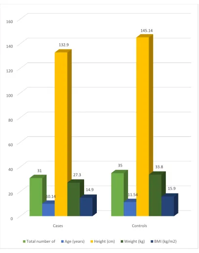
6 BASELINE CHARACTERISTICS OF STUDY
POPULATION
63
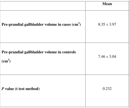
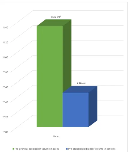
7 PRE-PRANDIAL GALLBLADDER VOLUME IN CASES
AND CONTROLS
66
8 POSTPRANDIAL GALLBLADDER VOLUME IN CASES
AND CONTROLS
68
9 GALLBLADDER EJECTION FRACTION IN CASES
AND CONTROLS
70
10 EJECTION FRACTION IN CASES WITH UPPER
ABDOMINAL PAIN AND LOWER OR PERIUMBILICAL
ABDOMINAL PAIN.
72
11 CORRELATION BETWEEN BMI AND EJECTION
FRACTION IN CASES
74
12 CORRELATION BETWEEN BMI AND EJECTION
FRACTION IN CONTROLS
76
13 CORRELATION BETWEEN EJECTION FRACTION
AND BMI IN CASES
78
14 CORRELATION BETWEEN EJECTION FRACTION
AND BMI IN CONTROLS
LIST OF TABLES
SL.NO CONTENTS PAGE NO
1 BASELINE CHARACTERISTICS OF STUDY
POPULATION
62
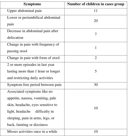
2 CASES PRESENTING WITH VARIOUS SYMPTOMS 64
3 PRE-PRANDIAL GALLBLADDER VOLUME IN CASES
AND CONTROLS
65
4 POSTPRANDIAL GALLBLADDER VOLUME IN CASES
AND CONTROLS
67
5 GALLBLADDER EJECTION FRACTION IN CASES AND
CONTROLS
69
6 EJECTION FRACTION IN CASES WITH UPPER
ABDOMINAL PAIN AND LOWER OR PERIUMBILICAL
ABDOMINAL PAIN.
71
7 CORRELATION BETWEEN BMI AND EJECTION
FRACTION IN CASES
73
8 CORRELATION BETWEEN BMI AND EJECTION
FRACTION IN CONTROLS
75
9 CORRELATION BETWEEN EJECTION FRACTION AND
BMI IN CASES
77
10 CORRELATION BETWEEN EJECTION FRACTION AND
BMI IN CONTROLS
1
INTRODUCTION
Child presenting with chronic abdominal pain is one of the most
commonly encountered symptom. The exact prevalence of chronic abdominal
pain is not exactly known, but the literature reports that 13% of middle school
children and 17% of high school children experience weekly abdominal pain
and this accounts for about 2% to 4% of all pediatric office visits (1). Chronic
abdominal pain and recurrent abdominal pain terms are used interchangeably.
The definition of “chronic” is from definition by Apley of recurrent
paroxysmal abdominal pain in children between the ages of 4 and 16 years that
persists for greater than 3 months duration and affects normal activity (2). At
least as many children experience chronic pain but maintain normal activity
and rarely come to the attention of the physician (3,5). The Pediatric Rome group
has subcategorized the chronic abdominal pain based on clinical presentation:
(1) Isolated paroxysmal abdominal pain,
(2) Abdominal pain associated with symptoms of dyspepsia,
(3) Abdominal pain associated with altered bowel pattern, and
(4) Abdominal migraine (6).
Chronic abdominal pain in children is either organic or inorganic. An organic
cause was found in 70 (82.4%) patients and non-organic cause identified
(NORAP) in 15 (17.6%) cases. Giardiasis was the commonest organic cause in
57 (67.0%) cases, either alone or with other parasitic infestations including
2
Pakistan suggest that RAP is most likely to have an organic cause (up to 82%
of cases), with giardiasis being the most common underlying condition (7, 8).
However, another Indian cohort and a Sri Lankan cohort showed that
non-organic RAP is more prevalent (74% and 76%, respectively) (9, 10). In Malaysia,
both urban and rural population-based cohorts had a similar prevalence of RAP
3
OBJECTIVE
PRIMARY OBJECTIVE:
To know whether gallbladder dysmotility constitutes in children with
chronic abdominal pain
SECONDARY OBJECTIVE:
To find out any associating factors for children with chronic abdominal
4
MATERIALS AND METHODS
Setting:
Place of Study:
The study was done in the department of Pediatrics, PSG Institute of
Medical Sciences and Research, Coimbatore. Cases were recruited from
Pediatric out patient department, PSG Hospitals, Coimbatore. PSG Hospitals is
a nine hundred bedded multispecialty ISO and NABH accredited teaching
hospital that offers comprehensive tertiary level health care for patients with
various ailments and from many nearby districts of western part of Tamilnadu.
Study Design:
The study is a case-control type, where children who fulfilled the
clinical criteria for chronic abdominal pain were compared with age and sex
matched controls. The cases were children between 5 to 15 years of age, who
were brought to the Pediatric OPD with abdominal pain and fulfilled the
inclusion criteria. After enrollment of the cases, healthy age and
gender-matched controls were subjected into the study.
Time Frame:
The study was conducted for one year i.e. from 1stJanuary 2016 to 31st
5
SAMPLE SIZE:
Thirty cases with chronic abdominal pain and an equal number of
healthy controls were included in the study.
INCLUSION CRITERIA:
Children of both gender aged between 5 to 15 years of age with
chronic abdominal pain according to ROME III criteria.
Healthy children, with no clinical evidence of any chronic illness
or on medications were recruited as controls.
EXCLUSION CRITERIA:
Non consent
Children with past abdominal surgical history or known chronic
abdominal pathology
Children who had obvious causes for chronic abdominal pain
following ultrasonographic examination of the gallbladder.
Children who could not tolerate or cannot complete the fatty
meal.
METHODOLOGY:
Children from 5 to 15 years of age attending Pediatrics out patient
department for chronic abdominal pain were requested to respond to a
questionnaire based on the ROME III criteria. Child fulfilling ROME III
criteria for chronic abdominal pain were included in the study after obtaining
6
study. The cases were subjected to ultrasonographic examination of the
gallbladder after a fasting of at least 6 hours. Following the baseline ultrasound
study, the subjects were instructed to eat a fatty meal. The fatty meal is a
special diet prepared by our hospital dietician according to basis of their fat
quantity required to produce gallbladder contraction. The standard meal
contained fat ranging from 30 to 70 g and quantity of fat was decided according
to child's age. Following the fatty meal the subjects were subjected to a second
ultrasonographic examination of the gallbladder to assess for gall bladder
contractility.
During ultrasonographic examination of the gallbladder, the greatest
length (L), greatest transverse width (W), and greatest anteroposterior diameter
(A) of the gallbladder was measured in the fasting state and postprandial period
of 30min. Dodd‟s Formula was used to measure the gall bladder volume.
Volume (V) (cm3) = 0.52 × L × W × A
The gallbladder ejection fraction (EF) for each examination was
obtained from two-volume data using the following equation. Where V0 =
gallbladder volume before the test meal, Vn = gallbladder volume after the test
meal.
EF (%) = ((Vo-Vn) / Vo) X 100%
Children with ejection fraction of less than 40% were considered to have
7
FLOW CHART
Children with chronic abdominal pain and children for routine USG abdomen
USG abdomen after 6 hours of fasting
Statistical analysis between children with chronic abdominal pain and healthy age matched controls
8
Statistical Analysis:
The statistical analysis was done using SPSS software (statistical package
for the social science version-19).
1.The Independent studentst test was applied to compare the means of
quantitative data.
Values were expressed as Mean ± SD. The p values were interpreted
as shown below
a. p> 0.05 was considered not significant.
b. p< 0.05 was considered statistically significant.
2. The ejection fraction of the cases was correlated with their BMI for any
9
REVIEW OF LITERATURE
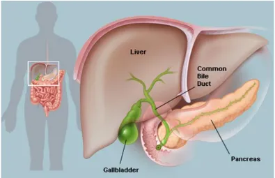
Gallbladder – The gallbladder is a thin-walled sac, placed between both
hepatic lobes. It is divided in to three parts anatomically. It is the fundus,
corpus, and infundibulum (13).Anatomically the human gallbladder and
gallbladder of most mammalian species dogs, cats, guinea pigs, and mice are
fairly similar. The gallbladder ends with cystic duct. Cystic duct has a diameter
of 7 mm with amucosa containing spiral valves (valves of Heister). The cystic
duct then drains into the common bile duct and there is no sphincteric structure.
The course of common bile duct is through the head of thepancreas ending in
[image:20.595.124.520.393.648.2]the sphincter of Oddi.
Figure 1: Structure of gallbladder
Now sphincter of Oddi and common bile duct penetrates theduodenal wall to
form the ampulla of Vater. Cystic duct or the common bile duct has no
10
11
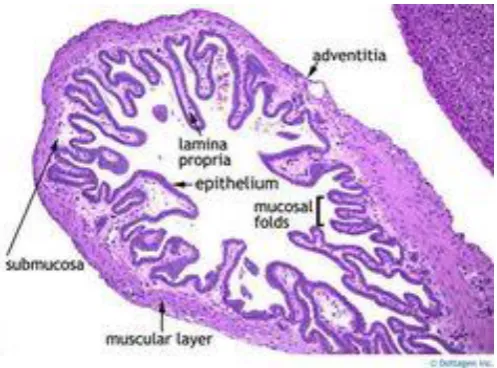
Figure 3: Gallbladder histology
Gallbladder Histology – it consists of mucosa with a single layer of epithelial
cells, a lamina propria, a single layer of muscle that resembles the muscularis
mucosa of the gastrointestinal tract, and a serosal layer (14).
Concentration of hydrophobic bile salts is done by epithelial layer by
absorbing water and electrolytes. Small quantities of bile salts and chloride are
transported by passive diffusion by mucosal layer (15, 16). Hydrophobic bile salts
are aggressive against stomach, esophagus etc. but the epithelial cells tolerates
it by its cytoprotective with help of prostaglandin E2 (PGE2). Epithelial cells
are covered by mucin, which is secreted by prostaglandin and this prevents
damage by inactivating bile salt induced free radicals. Vagus and splanchnic
nerves innervate the muscular layer and this synapses with intramural neurons.
In the fasting and digestive phase of biliary tract, it is integrated with by
neurohormonal mechanism of the digestive tract (14). The bile is continuously
secreted by liver in to intrahepatic and from there it flows in to extra hepatic
12
then stored and concentrated in the fasting state. And during all three phases of
digestive periods the concentrated bile is emptied. Only about 10% of the
hepatic bile drain in to the duodenum during the interdigestive period that is
during intervals between the phasic contractions of the sphincter of Oddi
(diastolic periods). This occurs when sphincter of Oddi basal pressure is
exceeded by the ductal pressure(17). It is the remaining 90% bile that is
redirected towards the cystic duct and gets stored in the gallbladder. By passive
and active mechanisms the gallbladder distends with entry of bile. Active
relaxation or accommodation of the gallbladder is mediated by adrenergic and
noncholinergic nonadrenergic nerves it is gradually induced by the incoming
bile. The vagus and splanchnic nerves and the hormone cholecystokinin (CCK)
are the main neurohormonal mechanisms regulating the motility of the
gallbladder (18–20). The efferent and afferent fibers are from vagus nerve (21). The
efferent fibers form as preganglionic neurons and synapse to intramural
postganglionic cholinergic neurons. The intramural postganglionic neurons are
present within the wall of the gallbladder. Gallbladder contraction is stimulated
by efferent fibers of vagus nerve,hexamethonium and atropine are muscarinic
receptor antagonist that acts by ganglionic blocker. Gallbladder relaxes by
splanchnic nerve stimulation and propranolol blocks this relaxation (22).
Atropine treated cat gallbladder muscle strips demonstrated the inhibitory
innervation (23). The strips blocked by propranolol were stimulated by electrical
field, there was relaxation the strips in response to electrical field stimulation
13
(VIP) suggesting that the stimulus was acting on sympathetic postganglionic
neurons releasing epinephrine as the neurotransmitter that act primarily on
beta-adrenergic receptors. However, different results were obtained when the
atropine-treated cat gallbladder was relaxed by stimulating the vagus nerve.
Under these experimental conditions the relaxation was resistant to propranolol
but was abolished by pretreatment with an antiserum against VIP suggesting
that this peptide is the neurotransmitter responsible for it (24). It is conceivable
that the VIP antiserum is a more effective antagonist against VIP than the
partial VIP antagonists that were used in muscle strips. These discrepancies
between in vitro and in vivo studies cannot be fully explained at this time
although it is possible that both neurotransmitters are involved in the
gallbladder relaxation. Further studies are needed to reconcile these different
observations that were demonstrated using different experimental conditions.
During the fasting period the gallbladder maintains a moderate tonic
contraction that is superimposed with nonpropulsive and propulsive
contractions (25, 26). These tonic and rhythmic contractions were demonstrated in
dogs in vivo. The nonpropulsive contractions are probably designed to keep the
bile insoluble constituents in solution preventing their precipitation,
particularly precipitation of cholesterol that can lead to gallstone formation.
These contractions are even observed in muscle strips from normal human
gallbladders (26). The propulsive contractions empty fractions of gallbladder
bile contributing to the small percentage of bile that enters the duodenum
14
period during the migrating motor complex that is known to stimulate the
gallbladder motility. The increased number of gallbladder contractions
coincides with the phase 3 of the antral migrating motor complex and with the
phase 2 of the duodenum (27). Thesecontractions are responsible for the
additional albeit small discharge of gallbladder bile in the duodenum that takes
place during this period.In the digestive period strong gallbladder contractions
and sphincter of Oddi relaxation lead to the high rates of bile discharge flowing
into the common bile duct and duodenum.During this period, the gallbladder
motor activity like the rest of the gastrointestinal tract is influenced by the three
phases of digestive process: cephalic, antral, and duodenal (28). The cephalic
phase is initiated by stimuli that activate the central nervous system, as
individuals are exposed to olfactory, visual, and the taste of food. This phase is
mediated by preganglionic vagal fibers that synapse with postganglionic
cholinergic neurons. It is estimated that as much as 30–40% of the gallbladder
bile may be emptied during this phase. Once food reaches the stomach it
triggers an antral-gallbladder reflex also mediated by vagal fibers. The
gallbladder emptiesmost of its remaining contents during the intestinal phase
induced by the release of CCK from the duodenum and proximal jejunum (29).
Duodenal CCK contracts the gallbladder mostly by acting directly on
cholinergic neurons (18) and like with the pancreas, and it may also activate long
reflexes through the vagus nerve. Meals containing proteins and fats act on
duodenal CCK containing endocrine cells that stimulate vagal sensory fibers
15
(30). Either mechanism stimulates the final pathway of postganglionic
cholinergic neurons, since atropine pretreatment blocks the gallbladder
contraction induced by physiological concentrations of CCK- 8 (31) or by a
protein-fatty meal. CCK-1 receptors are alsopresent in the gallbladder muscle,
but they only can be stimulated by pharmacological concentrations of CCK,
since they are not blocked by either atropine or tetrodotoxin.
Functional GB Disorder
Definition:
Gallbladder dysfunction is a motility disorder of the GB that manifests
symptomatically with biliary pain as a consequence of either an initial
metabolic disorder (i.e., supersaturated bile with cholesterol)(32) or a primary
motility alteration of the GB in the absence, at least initially, of any
abnormalities of bile composition(33). It is likely that the latter condition, by
causing bile stasis, may alter over a period of time, bile recycling
and bile composition within the GB. Both conditions may eventually lead, over
a period of time, to thedevelopment of organic abnormalities (example
gallstones and acute cholecystitis). The symptoms of these organic and
functional conditions appear to be indistinguishable from one another, and
16
Epidemiology:
The prevalence of GB dysfunction is not known. Large
population-based studies have reported that prevalence of biliary pain in ultrasonography
(USG)-negativesubjects with GB in situ varies from 7.6% in men to 20.7% in
women (34,35).
Clinical Presentation:
The most specific symptom attributed to functional disorders of the GB
appears to be biliary pain, and therefore the crucial steps in the diagnosis are a
thorough history supported by objective evidence of GB dysfunction and
exclusion of structuralabnormalities. These patients will need to be evaluated
with longer follow-ups for at least 1 year after cholecystectomy.
Criteria for this diagnosis:
1. Absence of gallstones, biliary sludge, or microlithiasis
2. An abnormal GB ejection fraction of less than 40% by using a
continuous intravenous cholecystokinin octapeptide infusion over
a 30-minute period
3. A positive therapeutic response with absence of the recurrent pain
for longer than 12 months after cholecystectomy
Laboratory and Instrumental Investigations:
The symptoms of GB dysfunction must be differentiated from organic
disease and other more common functional disorders including functional
17
intervals (few days or weeks). Tests of liver biochemistries and pancreatic
enzymes should be obtained in those patients with the previously mentioned
symptomatic criteria. These tests are normal in the presence of GB motility
dysfunction. The findings of abnormal liver or pancreatic enzyme levels or
both indicate that other diagnoses should be considered.
Ultrasound:
Transabdominal ultrasonographic study of the entire upper abdomen is
mandatory in patients with the previously mentioned symptoms. In the
presence of GB dysfunction, the biliary tract and pancreas appear normal on
USG. In particular, gallstones or sludge cannot be shown. USG usually detect
stones within the GB equal to or greater than 3 to 5 mm in diameter, but it has a
low sensitivity to detect smaller stones (36). USG detection of stones or sludge
within the common bile duct is even more difficult. Endoscopic USGis more
sensitive than traditional transabdominal USG in detecting microlithiasis (tiny
stones 3 mm) and sludge within the biliary tract (37).
Endoscopy:
In the presence of normal laboratory and ultrasonographic findings, an
upper gastrointestinal endoscopy is usually indicated. The diagnosis of GB
dysfunction is suspected in the absence of significant abnormalities in the
18
Microscopic bile examination:
To exclude microlithiasis as a cause for these symptoms, a careful
microscopic examination of GB bile could be performed. The detection of
microlithiasis and cholesterol microcrystals is best accomplished by a careful
examination of GB bile obtained directly at the time of ERCP or by aspiration
from the duodenum during endoscopy after cholecystokinin (CCK) stimulation.
The resultant bile should appear deep golden yellow to dark green-brown. Pale
yellow bile from the common duct is not appropriate. Evenin those patients
with cholesterol gallstones or sludge, this hepatic bile is often free of
cholesterol microcrystals being insufficiently concentrated to nucleate. The
collected bile should be immediately centrifuged and examined.
Two types of deposits may be evident
1. Cholesterol crystals and/or
2. Calcium bilirubinate granules.
Cholesterol microcrystals are birifringent and rhomboid shaped and best
visualized by polarizing microscopy. The presence of cholesterol crystals
provides a reasonably high diagnostic accuracy for microlithiasis (38-40).
Bilirubinate granules are red-brown and can be detected by simple light
19
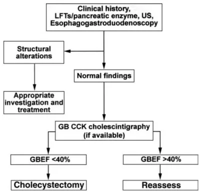
TESTS OF GB MOTOR DYSFUNCTION
Assessment of GB emptying by cholescintigraphy:
Cholescintigraphy is performed after the administration of technetium
99m–labelled iminodiacetic acid analogs. These compounds have a high
affinity for hepatic uptake, are readily excreted into the biliary tract, and
concentrated in the GB. The net activity-time curvefor the GB is then derived
from subsequent serial observations, after either CCK administration or the
ingestion of a meal containing fat. GB emptying is usually expressed as GB
ejection fraction, which is the percentage change of net GB counts after the
cholecystokinetic stimulus. A low GB ejection fraction has been considered
evidence of impaired GB motor function that, in the absence of lithiasis, could
identify patients with primary GB dysfunction. The most widely used and
validated stimulus to contract the GB has been the slow intravenous infusion of
CCK analogs, especially CCK-8 over a 30-minute period (41, 42). Fatty meals
and variable bolus injections of CCK do not yield consistent results. Reduced
emptying can arise from either impaired GB contraction or increased resistance
of the SO because of an elevated basal tone. Furthermore, several other
conditions that do not necessarily present with biliary pain can be associated
with reduced GB emptying such as obesity, diabetes, and several drugs
(example - calcium channel antagonists and oral contraceptives). An accurate
medical history should exclude secondary causes of impaired GB motility. Two
systematic reviews that did not discriminate between slow and rapid
20
evidence to recommend the use of CCK cholescintigraphy to select patients for
cholecystectomy(43, 44).
Assessment of volume changes by transabdominal real-time US:
Unlike cholescintigraphy, this method measures GB volume and obtains
serial measurements during fasting or after a meal or the intravenous infusion
of CCK analogs. In addition, US allows for assessment of residual volume after
emptying and the rate of refilling after GB contraction.
US may be helpful when radiation should be avoided. One deficiency in
the technique is the fact that it is operator dependent, and the results may not be
reproducible between different centers; therefore, the diagnostic role, if any, of
ultrasonographic assessment of GB emptying has not become the standard in
GB dysfunction. Further prospective randomized studies are needed to better
understand the predictive value of CCK cholescintigraphy or CCK USGto
recommend cholecystectomy in patients with suspected GB motility
dysfunction.
Pain provocation test:
A stimulation test with CCK attempting to duplicate biliary pain has
been historically used as a diagnostic investigation. This test has low sensitivity
and specificity in selecting patients with GB dysfunction who respond to
therapy. This may relate to problems in the subjective assessment of pain and
the use of bolus injections of CCK. The latter can induce pain by stimulating
21
22
FUNCTIONAL ABDOMINAL PAIN
Introduction:
Apley and Naish described chronic abdominal pain as children presenting with
intermittent episodes of abdominal pain occurring for at least 3 months without
any identifiable cause and interfering with day to day activities(3). The term
„chronic abdominal pain‟ is often used interchangeably with „recurrent
abdominal pain‟. Abdominal pain that is continuous, persistent, or intermittent
over a period of a few months is called as chronic abdominal pain. The pain
varies in intensity. There may be asymptomatic periods interposed with painful
periods gradually the duration of painful periods prolong, generating a
condition that profoundly causes distresses in the daily life of children. The
term recurrent abdominal pain represents a symptom and not a diagnosis. Many
conditions can cause abdominal pain that is recurrent, but most children and
adolescents presenting with this symptom have a functional disorder without
any evidence of organic disease. They are considered to have functional
abdominal pain.
Epidemiology:
Chronic abdominal pain is a common pediatric complaints (45),
accounting for 2–4% all of pediatric hospital visits (46). Symptoms consistent
with irritable bowel syndrome (IBS),occur in 14% of all high-school students
and 6% of all middle-school students (5). IBS is much more prevalent than other
medical conditions such as hypertension, asthma and diabetes that tend
23
assessing the prevalence of IBS is often difficult. Only one in four patients with
symptoms of IBS seek medical care (48).Reports from Kenya, Poland, Russia,
India and Pakistan reveal that, functional abdominal pain is probably a
universal problem (10, 49-51). A Malaysian study in 1500 schoolchildren found
evidence of chronic abdominal pain in 10% of children, concluding that, in
spite of differences in diet, customs and culture, the overall prevalence of this
entity was similar across regions(52). A Danish study demonstrated that 15% of
children aged 9–12 years had recurrent abdominal pain(53).Quality of life in
individuals with IBS is substantially poorer than in general population (54).
Physiology of the gastrointestinal pain response:
Visceral pain perception passes through a complex pathway of
peripheral and central nervous structures that signal, relay and modify the
afferent stimulus. The enteric nervous system (ENS) is arranged into two main
plexuses providing the intrinsic innervation of the gut. This forms a continuous
network around the circumference of the wall of the intestine along its length.
The mesenteric plexus, alsocalled as Auerbach‟s plexus, is located between the
external longitudinal and internal circular muscle layers, and the submucosal
plexus (Meissner‟s plexus) lies in between the circular muscle layer and the
mucosa. The mesentericplexus is larger and projects fibers mainly to the
smooth muscle of the gut and it controls the motility. Meanwhile, the
submucosal plexus projects into the mucosa and submucosa and control the
glandular secretion. The two intestinal plexuses are connected to each other by
24
nervous system. The characteristics of this mesh of sensory fibers, interneurons
and motor neurons, enables this mini-brain or „gut brain‟ to integrate the
sensory information, and to organize the motor and secretory responses and
influence the luminal absorption, thereby producing a functional state that is
adapted to the well-being of the individual. The ENSthough it receives input
from the central and autonomic nervous systems, it can function independently.
The ENS performs most of its functions in the absence of central nervous
system (CNS) control, locally integrating the information of intrinsic afferent
fibers (for example, luminal distension and chemical stimuli) with efferent
axons, resulting in motor reflexes or secretory or absorptive responses. The
extrinsic nervous system functions to consist connecting the ENS with the
CNS. This communication allows the CNS continuously to integrate the
information from the gastrointestinal tract with incoming information from
other organs and from the environment. Under physiological conditions, most
of these do not reach the level of conscious perception.21 However, sensations
that trigger a particularbehavior, including hunger, satiety and need to defecate,
reach the conscious perception by reaching the cortex. The constant influenceof
the CNS on the ENS through activation of a subset of vagal and sacral
parasympathetic fibers is responsible for psychological stress and
gastrointestinal response, manifested clinically by the occurrence of vomiting
25
The ENS and the brain use multiple neurotransmitters for exchanging of
inhibitory or excitatory information. They include excitatory neurotransmitters
such as acetylcholine and substance P, and gut inhibitory neurotransmitters
such as nitric oxide, ATP, vasoactive intestinal peptide (VIP), cholecystokinin,
enkephalins, calcitonin gene-related peptide (CGRP), norepinephrine
(noradrenaline), epinephrine (adrenaline) and others. Neurotransmitters such as
serotonin (5-hydroxytryptamine) and histamine have more complex effects.
Ninety-five per cent of the total body 5-HT lies within the gut. Of the total gut
5-HT, 90% is found in the granules of the enteroendocrine cells, and 10% in
the neurons of the myenteric plexus (55, 56). 5 HT also plays an important role in
regulating GI motility and intestinal secretion(55). 5-HT receptors appear to
participate in mucosal sensory processing within the gut. Distension and
stroking of mechanosensitive receptors in the enteroendocrine cells triggers the
release of 5-HT (57). There are at least seven main classes of 5-HT receptor and
22 subclasses that can be differentiated on the basis of structure and function.
Four classes have been reported in the human GI tract (5-HT1, 5-HT2, 5-HT3
and 5-HT4)(58). Serotonin action is complex, with mixed effects ranging from
smooth muscle contraction, to relaxationthrough stimulation of inhibitory nitric
oxidereleasing neurons. Higher levels of serotonin are present in
diarrhea-predominant IBS (59). The ENS has the ability to modulate signal transduction
by enhancing or inhibiting the activation of nociceptors through alteration in
smooth muscle tone and contractile activity. Visceral pain may be modulated
26
for the use of centrally acting agents or cognitive behavioral treatments in
functional bowel disorders. Neuroimaging studies have provided information
on differences in brain processing of visceral stimuli between normal
individuals and those suffering from IBS, revealing an increased activity at the
level of the anterior cingulated cortex, prefrontal cortex, insular cortex and
thalamus (the areas associated with emotional responses) in patients with IBS
compared to asymptomatic individuals( 60-62).
Pathophysiology:
Several hypotheses have been put forward to explain the cause of
functional recurrent abdominal pain. We examine them in the following
sections.
Visceral hyperalgesia:
The visceral hyperalgesia hypothesis proposes that greater sensitivity of
visceral afferent pathways or central amplification of visceral input lead to an
enhanced perception of visceral stimuli. There is evidence that the pain and
discomfort of IBS might be due to hyperalgesia and allodynia of the gut. While
in hyperalgesia a painful stimulus is perceived as even more painful, in
allodynia a non-painful stimulus becomes painful (63). Anoxious stimulus
applied to a particular area of thegut may sensitize primary afferent fibers and
nociceptors of adjacent areas, causing painful sensations with a low-intensity
stimulus, resulting in primary hyperalgesia. Most IBS patients experience rectal
discomfort at lower intraluminal volumes or pressures(64, 65) and have
27
presenting with one functional bowel disorder frequently had additional
symptoms referable to other parts of the digestive system, suggesting that
enhanced visceral nociception may be a pan-intestinal phenomenon. For
example, it has been reported that in addition to the features of rectal
hyperalgesia, IBS patients have a decreased sensory threshold to balloon
distension of the esophagus. Children with functional abdominal pain exhibited
generalized visceralhyperalgesia, whereas IBS patients had rectal but not
gastric hyperalgesia(65).Different GI symptoms were reproduced by stimulation
of the predominant site of hyperalgesia, providing a physiological explanation
of symptoms in children who have distinct phenotypic presentations.
Dysmotility:
In addition to a greater intestinal sensitivity, patients with functional
bowel disorders may display abnormal motility. Various types of motor
disturbances have been documented in IBS, apparently reflecting dysfunction
at one or more levels of the brain–gut axis(67). Although the pathophysiology of
IBS is commonly attributed to dysfunction of the large intestine, evidence
exists to incriminate the small bowel as well (68). Postprandial motor
dysfunction in the small bowel appears to be more prevalent among IBS
patients who exhibit underlying visceral hypersensitivity in the fasting state.
Abdominal cramping has been associated with the passage of high-amplitude
contractions through the ileocecalregion(69). Bloating has been explained by an
abnormal transit and pooling of gas in conjunction with guthypersensitivity(66).
28
children and adults with functional dyspepsia(70). However, not all studies have
demonstrated differences between patients and control subjects(71, 72). Motility
changes in IBS are neither specific nor predictable and do not serve as a
diagnostic marker or as an aid to the selection of treatment(73). It has been
suggested than, rather than having a persistent motility abnormality, patients
with functional bowel disorders exhibitan abnormal motor response to a variety
of physiological stimuli(74).
Brain–gut interaction:
The shortcomings of isolated experimental or observational models in
explaining the complex nature of functional bowel disorders have led research
to focus on the alterations in the communications between the CNS and the GI
tract, hence the term „brain–gut‟ interaction(47, 75). There are multiple examples
of brain–gut interaction, the most common being the subjects who, under
emotionally stressful situations, develop diarrhea, nausea or vomiting. Anger
and aggression increase colonic motility, while hopelessness results in
decreased motility(76). The brain–gut model links alterations in peripheral
sensory afferent communication from the gut (e.g. visceral hyperalgesia)
toCNS processing of the sensory stimuli and its efferent signaling to the gut. In
IBS patients multiple studies have shown that both gut and brain show an
exaggerated responsiveness to different stimuli. Patients with IBS have
significantly greater electroencephalogram (EEG) abnormalities than
controls(77). Dynamic brain imaging technologies such as positron emission
29
recently been applied to the study of the gut–brain axis in order to identify the
areas of the brain activated by visceral sensations. Studies with these
techniques have suggested an abnormal cerebral processing of visceral stimuli
in patients with functional bowel disorders (60, 61).
Inflammation:
There is evidence that IBS can occur following a gastrointestinal
infection resulting in transient inflammation. Gwee et al reported that 20–25%
of patients admitted to the hospital for bacterial gastroenteritis developed
symptoms consistent with IBS in the first 3 months (78). Rectal biopsies
prospectively obtained during and after acute gastroenteritis from patients who
developed postinfectious IBS and a control group, showed that the former
group exhibited significantly greater expression of interleukin (IL)-1Beta
mRNA (79). A recent study examining full-thickness biopsies from the
jejunum of patients with severe IBS revealed inflammation and neuronal
degeneration in the mesenteric plexus, suggesting a possible pathogenic role of
inflammation (80).Animal studies also seem to indicate that inflammation may
produce persistent neuromuscular gut dysfunction(81). Mildmucosal
inflammation may perturb neuromuscular function also at remote non-inflamed
sites. The gut dysfunction may persist even after reduction of
the mucosal inflammation. Substances that mediate these changes are not fully
understood, but there is growing recognition of the role of serotonin as a
30
Immunity:
The mucosal immune system mediates the clinical impact of stress and
other psychological factors on the gut. Vagal afferents can be activated by
products of mast cell degranulation, resulting in sensitization of silent
nociceptors. Mast cell mediators may be released in response to luminal
macromolecules, a phenomenon that could explain brain–immune system
interactions within the gut. A descending input by the vagus nerve may also
reciprocally affect mast cell degranulation, resulting in local effects on
secretomotor activity.
Stressors:
Stressful events have long been believed to be important in the
development of symptoms in functional bowel disorders(82). Physiological
reactions to stressors should be considered as attempts by the body to adapt – a
natural coping mechanism, in which, if stressors are not too extreme or long
standing, the subject is usually successful in reaching a homeostatic state.
However, at times the body loses the capacity to adapt and deleterious
behavioral responses may arise. Chronic exposure to threat is associated with
alterations in theautonomic outflow, resulting in activation of
thehypothalamic–hypopituitary–adrenal axis with alteration in pain
modulation. Corticotropinreleasing hormone seems to be the hormonal
mediator of the stress response. Intracerebroventricularinjection of
31
exacerbates nociceptive responses associated with increased release of
histamine(83).
In humans, possibly as a primitive response to danger, stress induces
delayed gastric emptying, slower small bowel activity and accelerated colonic
transit(84-87).These alterations may presumably cause diarrhea, constipation or
abdominal distension depending on the predominant abnormality.
Genetics:
A recent study showed a significant association between subjects with
abdominal pain or bowel disturbances and first-degree relatives with IBS
and dyspepsia(88). Twin studies have shown a 17% concordance for IBS in
monozygotic patients with only 8% concordance in dizygotic twins. Although
these data suggest a specific role for heredity in the development of IBS, the
same study showed ahigher correlation of IBS with parental symptoms,
suggesting that social learning from the patient‟s environment has an equal or
greater influence (89).
Biopsychosocial model:
The biopsychosocial model(90) provides a framework to integrate the
biological and psychosocial processes, in an attempt to understand the
underlying pathophysiological mechanisms determining disease susceptibility,
and to explain the clinical variability and outcome among individuals. The
biopsychosocial model, proposed as an alternative to the traditional biomedical
32
integration of medical and psychosocial factors. To understand this model, one
should differentiate between disease, which is the abnormality of the structure
and/or function of organs and tissues (physical component), and illness, defined
as the patient‟s perception of health and bodily dysfunction (psychological
component). In Engel‟s model (90)
, illness and disease result from interactions at
the cellular, tissue, interpersonal and environmental levels resulting in a clinical
outcome. The biopsychosocial model assumes that genetic influences on
disease susceptibility and behavior result in a biological and psychosocial
predisposition that will influence later psychosocial experiences, physiological
functioning, or susceptibility to a pathological condition. This particular
background is affected by physical and environmental exposures such as
infection, food intolerance and social exposures including friends, family and
community to influence the patient‟s attitude towards illness (91). Stress acting
on a vulnerable GI tract leads to an imbalance in the system, resulting in an
alteration of the brain–gut axis. Multiplicity of stressors reinforces and
up-regulates the response. It is well recognized that some IBSpatients report
initiation or exacerbation of symptoms at times of stress, trauma and major
loss(92). Traumatic early life events such as child abuse may predispose to
functional bowel disorders (93). There have been reports of a greater prevalence
of sexually and physically abusive experiences in individuals with IBS than in
patients with organic gastrointestinal disorders and non-patient populations (94).
The interaction of the previously described subsystems with psychosocial
33
mechanisms) affects the behavior of the individual, the biological nature of the
condition and, ultimately, the clinical outcome. New or uncontrollable
threatening situations may result in emotional and physiological arousal.
Psychosocial factors may affect the end result (clinical presentation and
outcome) acting on gut physiology, modulating symptoms experience, and
influencing health behavior and therapeutic interventions (Figure 5).The
relative contribution of the medical and psychosocial factors varies among
patients. This model should be considered when planning therapy, as failure to
link the disease and illness components will reduce the likelihood of an
[image:44.595.208.427.393.592.2]effective treatment
34
Functional bowel disorders presenting with abdominal pain by ROME III:
1. Functional dyspepsia
2. Irritable bowel syndrome
3. Abdominal migraine
4. Functional abdominal pain
5. Childhood Functional Abdominal Pain Syndrome
Differential diagnosis:
A number of clinical features („red flags‟) are commonly considered as
orienting towards an organic etiology, although definitive proof for their
predictive value is scant. The presence of an isolated symptom (such as isolated
abdominal pain) is usually thought to be consistent with a functional disorder,
while multiple symptoms (such as abdominal pain with weight loss, or
vomiting, or diarrhea) are more likely to be due to an organic condition. There
are almost endless causes of chronic organic abdominal pain in children.
Organic causes of chronic abdominal pain: Gastrointestinal
Esophagitis, gastritis, duodenitis, peptic ulcer, eosinophilic gastroenteritis,
malrotation, cysts (duplication or mesenteric), celiac disease, parasites,
hernias, tumors, foreign body, intussusception, inflammatory bowel disease
Hepatobiliary
Chronic hepatitis, cholelithiasis, cholecystitis, choledochal cyst, sphincter of
35 Pancreatic
Pancreatitis, pseudocyst
Respiratory
Infection, tumor or inflammation vicinity of diaphragm
Genital
Hematocolpos, endometriosis, mittelschmertz tumor
Urinary
Ureteropelvic junction obstruction, recurrent pyelonephritis, recurrent
cystitis
hydronephrosis, nephrolithiasis
Metabolic
Porphyria, diabetes, lead poisoning
Hematological
Angioedema, collagen vascular disease, sickle cell disease
Musculoskeletal
Trauma, inflammation, infection, tumor
Psychiatric
Conversion reaction
Peptic disease:
Pain related to peptic ulcer disease is usually epigastric and
non-radiating, but may also be generalized or periumbilical. It occasionally awakens
36
children experience exacerbation rather than relief with meals. A review of 160
children concluded that epigastric pain, food-related pain, vomiting, bleeding
and family history were important factors in the diagnosis of peptic ulcer in
childhood (95). However, the clinical presentation of ulcer mayhave symptoms
that overlap with some functional bowel disorders such as functional
dyspepsia. A favorable response to antacids or H2 blockers may orient towards
a peptic disease, but there is evidence that anti-secretory therapy may also be
effective in dyspepsia. Therefore, as symptoms and therapeutic response may
be indistinguishable, the definitive diagnosis of peptic disease requires
esophagogastroduodenoscopy (96). While it is accepted that H. pylori infection
causes both gastritis and duodenal ulcer disease, its etiologic role in functional
dyspepsia is still controversial. Dyspepsia associated with epigastric pain was
frequently found in preschool and school children infected by H. pylori(96,
97).These findings are contradicted by other reports. A German study concluded
that H. pylori infection in children was mostly asymptomatic and not associated
with specific gastrointestinal symptoms (98). A review of 2715 children with H.
pylori infection found abdominal pain in 5–17% of cases. However, abdominal
pain was also found in 5–29% ofpatients without H. pylori infection (99).
Universal testing or treatment of H. pylori infection in children with either
recurrent pain referable to the epigastrium or recurrent periumbilical abdominal
37
Pancreatitis:
Pain resulting from pancreatic inflammation is usually epigastric,
radiating to the sides or back, with pain episodes frequently triggered or
aggravated by meals. Nausea and vomiting are often present. Severe abdominal
tenderness in the epigastric area and decreased bowel sounds are characteristic.
Chronic pancreatitis is an inflammatory disease characterized by
recurrentepisodes of abdominal pain that in certain individuals may result in
progressive structural changes and permanent impairment of exocrine and
endocrine functions (100).
Carbohydrate intolerance:
In children with chronic abdominal pain, increased flatulence and
bloating, the diagnosis of carbohydrate intolerance should be considered. The
breath hydrogen test following a lactose or fructose load will confirm the
diagnosis. Exclusion of these offending agents may improve the symptoms. On
the other hand, lactose intolerance has a very high prevalence in the general
population and symptoms after a lactose load develop in only a few of the
self-reported milk-intolerant subjects (101). In a large sample of American patients,
lactose malabsorption was found in 21–25% of IBS patients (102), a prevalence
considered comparable to that in the general American population. Lebenthal et
al (102) found a similar prevalence of lactase deficiency in children with
recurrent abdominal pain and in children of a control group of similar ethnic
background. Moreover, lactose free elimination diet resolved symptoms in a
38
malabsorbers. As a result of the high independent prevalence of both conditions
in the general population, the presence of chronic abdominal pain and lactose
intolerance in one patient may be merely coincidental. Thus, the
recommendation of an exclusion diet should be made with reasonable
expectations, as it may be helpful in the resolution of symptoms in only a
limited number of patients.
Inflammatory bowel disease:
Occult IBD was found in 1% of adult patients(103)and in 3–4% of
pediatric patients(104) evaluated for IBS. Some patients may complain for
months or years of vague abdominal pain and intermittent diarrhea before
being diagnosed as having IBD. The presentation of the abdominal pain is
variable, depending on the site of bowel involvement. Terminal ileum and
cecal disease in the setting of Crohn‟s disease is often associated with right
lower quadrant discomfort and tenderness. Diagnosis is frequently delayed,
with an average lag in diagnosis of approximately 7 months (105). A decrease in
growth preceded the onset of any symptoms by at least 1 year in 24 of 50
patients with Crohn‟s disease (106).
Chronic constipation:
IBS with constipation predominance and chronic constipation present
many descriptive similarities (107). However, constipation in combination with
abdominal pain has a wide differential diagnosis and constipation should not be
39
diagnoses. In chronic constipation physical examination may reveal a fecal
mass in the left lower quadrant and suprapubic region. Examination of the
perineum should include assessment of the lower back, sacrum and site of the
anus. Anal examination may reveal the presence of a fissure or a sentinel skin
tag indicative of a fissure. Rectal examination may reveal the presence of a
dilated rectum containing a hard fecalmass. A flat plate radiograph may help in
the diagnosis in cases where obesityprecludes an appropriate abdominal
examination.
Gallstones:
Cholelithiasis is considered uncommon in infancy, childhood and
adolescence, with a prevalence ranging from 0.2 to 0.5% (108). In older children,
obesity, ileal disease and a family history of childhood gallstones have been
associated with cholelithiasis(109). Children with gallstones may have colicky or
unspecific pain or remain asymptomatic. The pain is mostly located in the right
upper quadrant or in the epigastrium, and may radiate to the back or right
shoulder. Nausea and vomiting are present in more than 50% of cases. Fatty
food intolerance is not commonly reported in children. Other biliary tree
pathologies such as choledochalcyst may present with abdominal pain.
Presentation is age dependent, with jaundice prevailing in children and
abdominal pain in adults (110).
Parasitic infections:
Positive fecal ova and parasite tests were found in only 2% of adult
40
and Blastocystishominismay be found in patients presenting with abdominal
pain, often accompanied by anorexia, abdominal distension and diarrhea.
Blastocystishominisis most likely to be non-pathogenic in the
immunocompetent human host (111).
Small-bowel bacterial overgrowth:
Symptoms of IBS may overlap with those of small-intestinal bacterial
overgrowth (SBBO). Pimentel et al (112), in a prospective study, showed that
78% of IBS patients had SBBO diagnosed by a lactulose hydrogen breathe test,
and that the eradication of the overgrowth improved diarrhea and abdominal
pain. A study of IBS patients with SBBO demonstrated significant motility
abnormalities (113), leading the authors to conclude that dysmotilitywas the
pathogenic mechanism linking SBBO to IBS. Another controlled study looking
at the effects of antibiotics on IBS patients with alterations of the duodenal and
colonic flora has also shown a significant improvement of symptoms in the
group of patients receiving antibiotics (114).
Celiac disease:
Patients with celiac disease may present with symptoms mimicking
other conditions (115). A recent study has shown that celiac disease patients were
initially diagnosed as having IBS in 37% of cases (116). In this study, only 32%
of adults with celiac disease were underweight, and only 50% reported frequent
diarrhea and weight loss. Anemia was present in 67% of the cases. Patients
41
common clinical manifestations of functional bowel disorder. The Dutch
College of General Practitioners states that celiac disease should be added to
the differential diagnosis of IBS (117). A Canadian study revealed that, prior to
being diagnosed as celiac disease, 37% of respondents consulted four or more
family doctors (118). An Israeli study in 270 consecutive patientswhounderwent
endoscopy for abdominal pain demonstratedceliacdisease in one out of 23 of
the patients (119).
Genitourinary disorders:
Pyelonephritis and obstructive conditions such as ureteral or
pelviureteric junction obstruction may present with recurrent cramping
abdominal pain, despite normal physical examination and urinalysis
(120)
.Hematuria can be present in the setting of urinary tract infections, abuse,
trauma, Henoch– Schönleinpurpura, or renal stones. Dysuria associated with
abdominal pain can represent a sign of pyelonephritis, abuse, trauma, or a
sexually transmitted disease(121). In adolescent girls, a history of mid lower
abdominal pain was found to have low sensitivity but high specificity for
gynecological diseases (122).Gynecological pathology such as ovarian cysts,
congenital uterine abnormalities and endometriosis should also be considered
in the differential diagnosis of abdominal pain. Hematocolpos(123) due to
imperforate hymen may present with periodic lower abdominal pain and
urinary retention. Endometriosis may begin 3–4 years after menarche.
Clinically it may manifest as cyclic abdominal pain, nausea, vomiting,
42
Congenital anomalies:
Malrotation presents usually early in life, with 85% of all cases of
midgut volvulus occurring in the first year. In neonates, symptoms are usually
dramatic with sudden onset of bilious vomiting and a visibly seriously ill
patient. Older infantsmay present with episodes of colicky abdominalpain. In
one series, 20% of cases of malrotation in patients over 1 year of age presented
with chronic abdominal pain (125). These patients often have vague,
long-standing abdominal complaints with or without emesis. The pain is often
postprandial and may be accompanied by bilious emesis and diarrhea or
evidence of malabsorption or proteinlosingenteropathy associated with
bacterial overgrowth. Duplications of the alimentary tract are uncommon
congenital abnormalities. The clinical presentations may be vague and diverse,
depending on the location of the duplication (126). Presenting signs and
symptoms include abdominal mass, vomiting, decreased oral intake,
gastrointestinal bleeding, periumbilical tenderness and abdominal distension.
Musculoskeletal pain:
Pain related to trauma is usually well localized and sharp in nature, and
may be exacerbated by movement. Patients usually are able to recall a history
of trauma or strain, but occasionally that history cannot be elicited. The
diagnosis of costochondritis should be considered in adolescent patients
complaining of chest or upper abdominal pain. Costochondritispain originates
43
abdomen. Pain is reproducible by palpating the affected costal cartilage(127).
Diagnostic testing:
In patients with no alarm symptoms, the Rome criteria have a positive
predictive value of approximately 98%, with additional diagnostic tests
providing a yield of 2% or less (128). When needed, the exclusion of an organic
condition can be accomplished by utilizing inexpensive, non-invasive and
easily available diagnostic tests such as complete blood cell count, erythrocyte
sedimentation rate, chemistry panel, liver and thyroid function studies, urine
analysis and stool examination for blood, ova and parasites. Need for other
diagnostic tests should be based on history and physical examination findings.
The physician should avoid the lure of having to „rule out‟ an organic disease at
all cost. Performing multiple tests may provide results that often are unrelated
to the presenting symptom or have no clinical relevance (such as a mildly
elevated sedimentation rate). Repeating tests to confirm the serendipitous
findings may further increase anxiety and undermine the clinical diagnosis of
functional bowel disorder. One could use time as the physician‟s ally, assuring
the patient that no test is necessary at this point but if further symptoms present
or the current symptom worsens the physician will not hesitate to proceed with
further work-up.
Blood and stool studies:
Hamm et al (103) studied 1452 patients with an established diagnosis of
IBS and found that screening tests showed a low incidence of thyroid
44
concluded that limited detection rates, added costs and the inconvenienceof
these tests made the routine use of endoscopy,radiography, thyroid function
tests, fecal ova and parasite determination and the lactulose hydrogen breath
test questionable in the diagnostic evaluation of established IBS patients. In
accordance with these results, Tolliver et al (129) performed fecal ova and
parasite determinations in 196 patients with a possible diagnosis of IBS, and
found no evidence of infection in any of them. In the same study, complete
blood cell count, sedimentation rate, serum chemistries, thyroid profile and
urinalysis were normal or yielded no useful clinical information.
A study designed to investigate the prevalence of elevated
antiendomysial antibody titers in children with recurrent abdominal pain
compared with healthy children found no association between abdominal pain
and celiac disease (130). The study showed that 1% of patients in each group had
positive celiac disease antibodies. An adult investigation studied serum
antibody testing for celiac disease in patients with IBS symptoms and a control
group, followed by upper endoscopy in positive cases. The study revealed that
4.6% of patients in the group with possible IBS had positive antibodies in
comparison with 0.67% in the control group, suggesting that testing for celiac
disease may be one of the few cost-effective evaluations in patients with IBS
45
Endoscopic studies:
A study investigating the presence of gastroesophageal reflux in children
with recurrent abdominal pain concluded that pathological gastroesophageal
reflux is a frequent finding in such children (132). Treatment of gastroesophageal
reflux in this group of patients resulted inresolution or improvement of
abdominal pain in71% of cases.Another study evaluating findings on
endoscopic examinations in 62 Indonesian children with recurrent abdominal
pain revealed pathological abnormalities including esophagitis, erosions and
duodenitis in 50% of the patients(133). In the absence of peptic ulcers, it is
unclear how much these pathological findings contribute to the patients‟
symptoms. Endoscopy and biopsy performed in children evaluated for
dyspepsia demonstrated that most children did not have significant mucosal
disease. Inflammation without evidence of peptic ulceration was found in 38%
of the patients with H. pylori being identified in only five cases (134).
Follow-up at 6 months to 2 years revealed