0022-538X/05/$08.00
⫹
0
doi:10.1128/JVI.79.6.3797–3806.2005
Copyright © 2005, American Society for Microbiology. All Rights Reserved.
Identification of an Essential Domain in the Herpes Simplex Virus 1
U
L
34 Protein That Is Necessary and Sufficient To
Interact with U
L
31 Protein
Li Liang and Joel D. Baines*
Department of Microbiology and Immunology, Cornell University, Ithaca, New York
Received 23 August 2004/Accepted 21 October 2004
Previous results have indicated that the herpes simplex virus 1 U
L31 and U
L34 proteins interact and form
a complex at the inner nuclear membranes of infected cells, where both play important roles in the
envelop-ment of nucleocapsids at the inner nuclear membrane. In the work described here, mapping studies using
glutathione
S
-transferase pull-down assays indicated that amino acids 137 to 181 of the U
L34 protein are
sufficient to mediate an interaction with the U
L31 protein. A recombinant virus (v3480) lacking U
L34 codons
138 to 181 was constructed. Similar to a U
L34 null virus, v3480 failed to replicate on Vero cells and grew to a
limited extent on rabbit skin cells. A U
L34-expressing cell line restored v3480 growth and plaque formation.
Similar to the localization of U
L31 protein in cells infected with a U
L34 null virus, the U
L31 protein was present
in the nuclei of Hep2 cells infected with v3480. Hep2 cells infected with v3480 contained the U
L34 protein in
the cytoplasm, the nucleus, and the nuclear membrane, and this was noted to be similar to the appearance of
cells infected with a U
L31 null virus. In transient expression assays, the interaction between U
L34 amino acids
137 to 181 and the U
L31 protein was sufficiently robust to target green fluorescent protein and emerin to
intranuclear sites that contained the U
L31 protein. These data indicate that amino acids 137 to 181 of the U
L34
protein are (i) sufficient to mediate interactions with the U
L31 protein in vitro and in vivo, (ii) necessary for
the colocalization of U
L31 and U
L34 in infected cells, and (iii) essential for normal viral replication.
Herpes simplex virus (HSV) nucleocapsids bud from the
inner nuclear membrane (INM) to form nascent virions in a
reaction termed primary envelopment. Recent studies have
suggested that HSV type 1 (HSV-1) particles lose this envelope
and acquire a secondary envelope in the cytoplasm (10, 13, 15,
22, 23, 28, 29, 35).
Various HSV gene products are required for primary
envel-opment under different circumstances. For example, the U
L31
and U
L34 proteins are required for envelopment in Vero cells
and Hep2 cells, U
L11 facilitates (but is dispensable for) the
process in most cell types, and gK plays a major role in
envel-opment only in quiescent cells (1, 17, 21, 24, 28, 31). Both the
gK and U
L11 proteins are also required for the efficient
move-ment of viral particles through the cytoplasm (1, 16).
The U
L34 protein is likely a type II integral membrane
protein with a 22-amino-acid transmembrane domain at the C
terminus (27, 31, 33). The U
L34 protein localizes to the nuclear
rim and the cytoplasm in a pattern reminiscent of the
endo-plasmic reticulum when expressed transiently in uninfected
cells, but it is localized exclusively at the nuclear rim in infected
cells (28). The U
L31 protein is necessary and sufficient to
target the U
L34 protein exclusively to the nuclear rim. Both the
U
L31 and U
L34 proteins have been found to associate with
newly enveloped virions in the perinuclear space and at the
INM and outer nuclear membrane, but not in cytoplasmic
virions or extracellular virions, by immunoelectron microscopy
of HSV-infected cells (29). The U
L34 protein (pU
L34) is a
substrate of a viral kinase encoded by the U
S3 protein (26).
The absence of U
S3 delays virion egress but does not preclude
primary envelopment and does not obviate phosphorylation of
the U
L34 protein (26, 27, 29, 32).
The U
L31 protein is a hydrophobic, nucleotidylated nuclear
phosphoprotein (5, 7). The protein is likely associated with the
nuclear matrix, and like many nuclear matrix components such
as lamin A/C, it is resistant to extraction with detergent and
high salt concentrations (7, 8, 14). It is likely that U
L31 plays
multiple roles in viral replication. In restrictive cell types such
as Vero cells, a U
L31 null virus produces 1,000-fold fewer
extranuclear particles than the wild-type virus, reminiscent of
the phenotype of a U
L34 null virus, suggesting that both
pro-teins play important roles in primary envelopment (8, 20). In
the absence of U
L34, the U
L31 protein is more susceptible to
degradation by the proteasome and no longer localizes to the
nuclear rim, but accumulates almost entirely within the
nucle-oplasm (28, 29, 42). Transiently expressed U
L34 is sufficient to
target the U
L31 protein to the nuclear rim in uninfected cells
(28, 40). In addition, noncomplementing cells infected with the
U
L31 null mutant generate decreased levels of viral DNA and
have a three- to fivefold reduction in the ratio of monomeric to
concatemeric viral genomic DNA, suggesting that U
L31 has
minor roles in both viral DNA synthesis and DNA cleavage
and packaging (8).
Its association with the nuclear matrix and presence at the
nuclear rim has led to the deduction that the pU
L31/pU
L34
protein complex is associated with the nuclear lamina of
in-fected cells (7, 28). The observations that both pU
L31 and
pU
L34 interact in vitro with lamin A/C (a major lamina
com-ponent) and that both are required for the HSV-mediated
modification of lamin A/C and chromatin (30, 34) support this
hypothesis. pU
L31 binds to lamin A domains that normally
* Corresponding author. Mailing address: Department of
Microbi-ology and ImmunMicrobi-ology, Cornell University, Ithaca, NY 14853. Phone:
(607) 253-3391. Fax: (607) 253-3384. E-mail: [email protected].
3797
on November 8, 2019 by guest
http://jvi.asm.org/
interact with chromatin (30, 36, 38), suggesting a mechanism
for the U
L31-mediated chromatin alteration. The
overexpres-sion of U
L31 is also sufficient to displace lamin A/C from the
nuclear rim into the nucleoplasm (30). Disruption of the
lam-ina and associated chromatin may serve to facilitate the
pas-sage of nucleocapsids through the lamina barrier to budding
sites within the INM. It is also possible that the pU
L31/pU
L34
complex mediates nucleocapsid budding by interactions with
nucleocapsids destined for envelopment at the nuclear
mem-brane. This hypothesis is supported by the observations that (i)
pU
L34 can interact with ICP5, the major capsid protein, and
(ii) both pU
L31 and pU
L34 become incorporated into nascent
virions located within the perinuclear space (29, 41).
To understand the mechanisms by which the U
L31 and U
L34
proteins mediate nucleocapsid envelopment, we have begun a
series of studies to define the structure and function(s) of the
pU
L31/pU
L34 complex. As a first step in this process, we
sought to identify domains in the U
L34 protein that mediate
the interaction with pU
L31.
MATERIALS AND METHODS
Cells and viruses. Hep2, Vero, and rabbit skin cells were maintained in Dulbecco’s modified Eagle’s medium supplemented with 10% newborn calf serum (growth medium). Wild-type herpes simplex virus type 1 (strain F), a UL31
deletion virus, and UL34 deletion and repair viruses were described previously
(9, 20, 28, 31).
To construct a UL34-expressing cell line, we transfected rabbit skin cells with
the plasmid pRR1099 containing full-length UL34 as described previously (31).
Cells were selected for the ability to propagate in growth medium containing 200 g of Geneticin/ml. Several clonal cell lines were derived from the stably trans-fected population by limiting dilution and were cloned twice before being tested for the ability to support the replication of the UL34 deletion virus. One of the
cloned cell lines was chosen for further studies and was designated clone R1310.
Plasmid constructions.The plasmids used for mapping assays were con-structed as described below. All of the plasmids were sequenced to verify the expected genotypes.
Full-length UL31 was cloned into the pCDNA3 vector such that UL31
expres-sion was driven by the human cytomegalovirus immediate early promoter-en-hancer. The construct was designated pJB261.
The full-length UL34 sequence (encoding amino acids [aa] 1 to 275) was PCR
amplified from HSV-1(F) viral DNA by use of the primers 5⬘GTA GTC GAC ATA TGG CGG GAC TGG GCA AG 3⬘and 5⬘GCG GTC GAC AGG GCT GTG TGG GGC GAA GGC GTC 3⬘. The PCR product was then removed with SalI and ligated into the pGEX4T-1 SalI restriction site. This plasmid was designated pJB253. The UL34 fragment was then cut from pJB253 with the SalI
enzyme and ligated into the XhoI site of pCDNA3, and the construct was designated pJB280a. To include sequences sufficient to promote translation of the gene, we PCR amplified a full-length UL34 fragment from pJB253 by using
the primers 5⬘GTA CTC GAG ATA TGG CGG GAC TGG GCA AG 3⬘and 5⬘GTG CTC GAG AGG GCT GTG TGG GGC GAA GGC GTC 3⬘. The PCR product was digested with XhoI and ligated into pCDNA3, and the construct was named pJB280b.
Codons 1 to 245 of UL34 were PCR amplified from pJB253 by use of the
primers 5⬘GTA GTC GAC ATA TGG CGG GAC TGG GCA AG 3⬘and 5⬘ GAA GTC GAC GTG CTT AAG ACC CCG CAG 3⬘. The PCR product was cut with SalI and ligated into the SalI restriction site of pGEX4T-1. The resultant plasmid was designated pJB273. Using the SalI sites, we transferred the UL34
sequences (encoding aa 1 to 245) from pJB273 into the XhoI restriction site of pCDNA3 and named the resultant plasmid pJB274.
Codons 1 to 203 of UL34 were PCR amplified from pJB253 by use of the
primers 5⬘GTA GTC GAC ATA TGG CGG GAC TGG GCA AG 3⬘and 5⬘ CTA CCC GTA CGC CTC CCG 3⬘(UL34-609B-TAG). The PCR product was
then ligated into the pCR2.1 vector (Invitrogen). This clone was named pJB336. Codons 205 to 275 of UL34 were PCR amplified from pJB253 by use of the
primers 5⬘CGA GGC CGG GCT GGG GGT G 3⬘and 5⬘GCG GTC GAC AGG GCT GTG TGG GGC GAA GGC GTC 3⬘. The PCR product was ligated into the pCR2.1 vector, and the construct was named pJB338. The UL34
se-quences were then removed by digestion with EcoRI and ligated into the EcoRI
site of pGEX4T-1; the resultant plasmid was named pJB339. This EcoRI frag-ment was also subcloned into the EcoRI site of pEGFPC2 (Clontech) in frame with the gene for green fluorescent protein (GFP), and the resulting plasmid was named pJB354.
UL34 codons 81 to 203 were PCR amplified from pJB253 by use of the primers
5⬘CTC GAG ATG GCG TGC AAC CCT TAC CTG 3⬘and 5⬘CTA CCC GTA CGC CTC CCG 3⬘, and the resulting amplicon was ligated into pCR2.1. The resulting clone was named pJB337.
UL34 codons 79 to 275 were PCR amplified by use of the primers 5⬘CTC
GAG ATG GTC CCG TGC AAC CCT TA 3⬘and 5⬘GTG CTC GAG AGG GCT GTG TGG GGC GAA GGC GTC 3⬘. The PCR product was ligated into pCR2.1, and the clone was named pJB340. The UL34 sequences were then
removed with EcoRI and subcloned into the EcoRI site of pEGFPC3 in frame with the GFP gene. The resulting plasmid was designated pJB341.
UL34 codons 79 to 141 were PCR amplified by use of the primers 5⬘TGT CCC
GTG CAA CCC TTA CCT G 3⬘and 5⬘GCC GAG CCG CCC CTT GAT G 3⬘. The PCR product was ligated into pCR2.1 and subcloned into the pGEX4T-1 EcoRI site. This clone was designated pJB342.
A UL34 gene lacking codons 1 to 80 and 181 to 207 was generated as follows.
A UL34 fragment (encoding aa 209 to 275) was PCR amplified by use of the
primers 5⬘GGA TCC TGG GGG TGG CCG GAA CG 3⬘and 5⬘GCG GTC GAC AGG GCT GTG TGG GGC GAA GGC GTC 3⬘, and the amplicon was cloned into pCR2.1. The 210-bp double digestion products created with EcoRI and BamHI were ligated with the EcoRI and BamHI digests from pJB337 into pCR2.1. The clone was named pJB343.
A UL34 gene lacking codons 138 to 181 was generated by a three-step PCR
cloning strategy. First, a fragment bearing UL34 codons 182 to 275 was generated
by the use of pJB280a as a template with the primers UL34-F-XhoI (5⬘CTG
GAC ACC ATC AAG TGC CGC GCC GCC GAG CAG 3⬘) and 5⬘GTG CTC GAG AGG GCT GTG TGG GGC GAA GGC GTC 3⬘. Second, a DNA amplicon bearing UL34 codons 1 to 137 was generated by the use of pJB280a as
a template with the PCR primers 5⬘GTA CTC GAG ATA TGG CGG GAC TGG GCA AG 3⬘and UL34-B-XhoI (5⬘CTC GGC GGC GCG GCA CTT GAT
GGT GTC CAG GTC 3⬘). Third, a UL34 DNA fragment lacking the region
encoding aa 138 to 181 was generated by the use of gel-purified PCR products from the first two steps as templates with UL34-F-XhoI and UL34-B-XhoI as
primers. The final PCR product was cloned into pCR3.1, and the construct was named pJB347. The same fragment was then cut out of pJB347 with XhoI and subcloned into the XhoI site of pGEX4T-2 in frame with the gene encoding glutathioneS-transferase (GST). The resultant plasmid was named pJB348.
UL34 codons 137 to 181 were PCR amplified by the use of pJB253 as a
template with the primers 5⬘CAT GAA GGG GCG GCT CGG C 3⬘and 5⬘CTA CAG GAT CCG TCT CGT CCG TC 3⬘, and the product was ligated to the pCR2.1 vector. The UL34 sequences were then subcloned into the EcoRI site of
pEGFPC2 in frame with the gene for GFP, and the total fragment was desig-nated pJB345. This fragment was then subcloned into the EcoRI site of pGEX4T-1 and designated pJB346.
Emerin was PCR amplified from the plasmid Emerin-pOTB7 (MGC-2126; American Type Culture Collection [ATCC]) by use of the primers e1F-XhoI (5⬘ CTA CTC GAG ATG GAC AAC TAC GCA G 3⬘) and e256B-XhoI (5⬘GTT CTC GAG CTA GAA GGG GTT GCC 3⬘). The PCR product was cloned into pCR2.1, cut with XhoI, and ligated into the XhoI site of pCDNA3. The resulting plasmid was named pJB351.
A plasmid bearing codons 61 to 100 of emerin replaced with UL34 codons 137
to 181 was constructed by a three-step PCR cloning strategy. First, codons 1 to 60 of emerin were PCR amplified from Emerin-pOTB7 (ATCC) by use of the primers e1F-XhoI (5⬘CTA CTC GAG ATG GAC AAC TAC GCA G 3⬘) and e60-B-new (5⬘CGA GCC GCC CGC TAT AAG AGG AGG 3⬘). UL34 codons
137 to 181 were PCR amplified from pJB253 by use of the primers UL
34-138F-ov-e60 (5⬘CCT CTT ATA GCG GGC GGCTCG GCC TG 3⬘) and UL
34-181B-ov-e101 (5⬘CTG GTG GTC AGG ATCCGT CTC GTC 3⬘). Emerin codons 101 to 256 were PCR amplified from Emerin-pOTB7 by use of the primers e101F-ov-34-181 (5⬘ GAC GGA TCC TGA CCA CCA GGA CTT ATG 3⬘) and e256B-XhoI (5⬘GTT CTC GAG CTA GAA GGG GTT GCC 3⬘). Second, a DNA fragment was generated by a PCR using amplicons containing UL34
codons 137 to 181 and emerin codons 101 to 256 as templates along with the primers UL34-138F-ov-e60 and e256B-XhoI. Third, the final chimeric DNA
fragment was generated by using the PCR product for emerin (codons 1 to 60) from step 1 and the PCR product from step 2 as templates. This reaction used the primers e1F-XhoI and e256B-XhoI. The final PCR product was ligated into pCR2.1, removed with XhoI, and ligated into the XhoI site of pCDNA3. The final plasmid was named pJB352.
on November 8, 2019 by guest
http://jvi.asm.org/
Construction of a UL34 mutant virus deficient in interaction with UL31.A
DNA fragment containing a UL34 sequence lacking codons 138 to 181
neigh-boring a kanamycin resistance (Kanr) gene was generated by PCR. These genes
were flanked by Flp recombination sites. The UL34-Flp Kanr-Flp construct was,
in turn, flanked by UL34 flanking sequences and BamHI restriction sites. This
construct was generated as follows. First, a PCR amplicon was generated that contained the UL34 sequence lacking codons 138 to 181. The UL34 sequences
were flanked by 50 bp upstream of UL34 and by 12 bp of sequences overlapping
Flp-Kanr
-Flp. This amplicon was generated by the use of plasmid UL34
(del138-181aa)-pCR3.1 as a template with the primers 50UL34F-Bam (5⬘CCT TTG
GTG GGT TTA CGC GGG CAC GCA CGC TCC CAT CGC GGG CGC CGG ATC CAT GGC GGG ACT GGG CAA GCC CTA CAC CG 3⬘) and UL34Bovflip (5⬘CGA ACT GCA GGT CGA CTT ATA GGC G 3⬘). Second, a
fragment containing Flp-Kanr
-Flp was generated by using Flp-Kanr
-Flp as a template (a kind gift of Klaus Osterrieder and Bernd Karsten Tischer, Cornell University) with the primers flpKanF-ov34 (5⬘GCG CGC GCC TAT AAG TCG ACC TGC AGT TCG 3⬘) and flpkanB-Bam-ov34down (5⬘GGC GTC CGG AAC GCA CTG GCG ATT AGG GCG GCG GTG CGT CCT TTT GGA TCC GCT TCG AAG TTC CTA TAC TTT CTA GAG 3⬘). This fragment was flanked by sequences overlapping the 3⬘end of UL34 at one end and 50 bp of sequences
downstream of UL34 at the other end. Third, the final 2.2-kb fragment was
generated by a PCR using the gel-purified PCR products from the amplicons generated in steps 1 and 2 (above) as templates with the primers 50UL34F-Bam
and flpkanB-Bam-ov34down.
About 500 ng of the 2.2-kb PCR product was electroporated into a strain of
Escherichia coliEL250 harboring (i) the full-length HSV-1(F) genome main-tained as a bacterial artificial chromosome (BAC) called Yebac102 (37), (ii) a prophage system encoding the Gam, Exo, and Beta recombinases, and (iii) an arabinose-inducible Flp recombinase to facilitate BAC modification at Flp re-combinase recognition target (FRT) sites (19). Electroporation was performed in an ice-cold cuvette at 2.3 kV and 25F by use of a Bio-Rad Gene Pulser controller set to 200⍀. Electroporated bacteria were resuspended in 1 ml of Luria-Bertani medium at room temperature, and the material was transferred to a 1.5-ml microcentrifuge tube and incubated at 30°C for 1 h with shaking. Bacteria were then plated on Luria-Bertani agar plates containing 30g of chloramphenicol/ml and 50 g of kanamycin/ml. Kanr
Cmpr
colonies were picked, and BAC DNA was extracted by alkaline lysis. The Kanrfragment was
deleted by the induction of Flp recombinase with 0.1%L-arabinose overnight at 30°C. BAC DNA was then cotransfected into R1310 cells with a Cre expression plasmid to delete the BAC vector sequences (pCAGGS-nlsCre, a gift from Michael Kotlikoff, Cornell University), and the resulting viral plaques were purified twice. The resulting virus was named v3480.
Purification of GST fusion proteins and GST pull-down assays.Full-length UL31 fused to GST (encoded by pJB187) and different UL34-GST mutant fusion
proteins were purified as described previously (11). The full-length UL34 protein
(from plasmid 280b) or UL31 protein (from pJB261) was radiolabeled with
[35S]methionine in vitro by use of a rabbit reticulocyte lysate-based transcription
and translation expression system (TNT System; Promega). Five microliters of the UL31 radiolabeling reaction was preclarified overnight with Sepharose beads
and subsequently incubated for 2 to 5 h at 4°C with 5g of GST or a UL34-GST
fusion protein conjugated to glutathione-Sepharose beads in cold phosphate-buffered saline supplemented with 1% (vol/vol) Triton X-100. In reciprocal reactions, an [35
S]methionine-labeled UL34 mutant protein expressed in rabbit
reticulocyte lysates was incubated with 5g of a UL31-GST fusion protein or
GST. After the incubation periods, the beads and bound proteins were washed six times with cold 1% Triton X-100. The bound proteins were eluted by boiling in 3⫻sodium dodecyl sulfate (SDS) loading buffer and then electrophoretically separated in an SDS–12% polyacrylamide gel. The gel was soaked for 30 min in 20% sodium salicylate, dried, and either exposed in a PhosphorImager cassette (Molecular Dynamics) followed by quantification with Imagequant software or subjected to fluorography with X-Omat (Kodak) film exposed overnight at ⫺80°C.
Southern blot analysis. Vero cells in 25-cm2 dishes were infected with a
wild-type virus derived from Yebac102 or with the v3480, Kan, or v3480-Kan-BAC virus until a cytopathic effect was visible in 80% of the cells. Viral DNAs were then purified as described previously (20). The viral DNAs were digested with BamHI. The viral DNA digests were then separated in 1% agarose gels, transferred to nitrocellulose membranes, probed with [␣-32
P]dCTP (Am-ersham)-labeled DNA, and exposed to radiographic film as described previously (20). The DNA probes used for the experiments were derived from the purified full-length UL34 PCR product, a fragment bearing the Kanrgene generated by
PCR, and the True Blue BAC II plasmid (Genomics One Corporation) (BAC probe).
Confocal microscopy.Hep2 cells were seeded on glass coverslips in a six-well plate to reach 80% confluence. The cells were transfected with the plasmid pJB345, bearing UL34 codons 137 to 181 fused to the GFP gene or UL34 codons
205 to 275 fused to the GFP gene, either alone or together with pJB261 con-taining full-length UL31. The cells were fixed in 3% paraformaldehyde for 15 min
at room temperature and then permeabilized with 0.1% Triton X-100 for 3 min. The cells were blocked in 10% human serum and stained with a pUL31-specific
rabbit antibody diluted 1:50 (28), followed by extensive washing and incubation with a Texas Red-conjugated donkey anti-rabbit antibody (Jackson Immunore-search). Fluorescence values attributable to GFP and Texas Red were recorded as described previously (28).
In a separate experiment, Hep2 cells were transfected with emerin (pJB351) or emerin-UL34 (pJB352) alone or together with full-length UL31. The cells were
fixed and permeabilized with ice-cold methanol for 20 min at⫺20°C. Nonspecific immunoreactivity was blocked with 10% human serum for 1 h, and the cells were stained with the pUL31-specific rabbit antibody diluted 1:50 and with a mouse
monoclonal anti-emerin antibody (NCL-Emerin; Novocastra) diluted 1:40. The cells were then incubated with fluorescein isothiocyanate (FITC)-conjugated donkey anti-rabbit and Texas Red-conjugated donkey anti-mouse antibodies.
In a final experiment, Hep2 cells were infected with the UL31 deletion virus,
the UL34 deletion virus, the UL34 mutant virus v3480, or the UL34 repair virus.
Sixteen hours after infection, the cells were fixed with ice-cold methanol for 20 min at⫺20°C. The cells were then stained with pUL31- and pUL34-specific
antibodies as described above.
In all cases, glass coverslips were examined with an Olympus confocal laser scanning microscope using 10⫻ocular and 40⫻stage objectives and excitation wavelengths of 488 and 568 nm. The cells were also examined by Nomarski differential interference contrast imaging.
RESULTS
Mapping of U
L34 protein domains that interact with U
L31
protein.
Different portions of U
L34 were cloned into plasmid
vectors that placed fragments of U
L34 in frame with the gene
encoding GST, as described in Materials and Methods. The
resultant GST fusion proteins were affinity purified as detailed
in Materials and Methods. Equal amounts of the fusion
pro-teins bound to glutathione-Sepharose beads were incubated
with
35S-labeled U
L
31 protein, followed by extensive washing,
elution in an SDS-containing buffer, separation by
electro-phoresis, fluorography, and analysis on a Molecular Dynamics
PhosphorImager. Some of the results are shown in Fig. 1A and
B. The results of all experiments are summarized in Fig. 1C.
We determined that full-length pU
L34 pulled down 43 times
more radiolabeled pU
L31 than did GST alone, confirming
previous results indicating that the U
L31 and U
L34 proteins
interact in this assay (28). In contrast, codons 1 to 137 (codon
1 encodes methionine) and 182 to 275 of the 275-codon U
L34
open reading frame were dispensable for the interaction
inas-much as fusion proteins bearing these sequences did not pull
down the radiolabeled U
L31 protein significantly more than
did GST alone. Importantly, a GST fusion protein containing
only U
L34 amino acids 137 to 181 was sufficient to pull down 20
times more radiolabeled U
L31 protein than was GST alone.
The deletion of this region from full-length U
L34 produced a
protein that pulled down only five times more pU
L31 than did
GST alone. This was significantly less than the amount that was
pulled down with full-length pU
L34-GST. We therefore
con-cluded that amino acids 137 to 181 were necessary and
suffi-cient to mediate an interaction with pU
L31 in vitro.
Amino acids 137 to 181 of U
L34 can interact with full-length
U
L31 in eukaryotic cells.
Because GST pull-down assays do not
necessarily indicate that a given region mediates the
interac-tion in more relevant contexts, a series of experiments were
undertaken to investigate the role of U
L34 amino acids 137 to
on November 8, 2019 by guest
http://jvi.asm.org/
181 in mediating the interaction in both HSV-infected and
uninfected eukaryotic cells. Specifically, the putative
interact-ing region (U
L34 codons 137 to 181) and pU
L34 codons 205 to
275 (a region shown not to interact with pU
L34 in the GST
pull-down assays) were fused to the gene encoding GFP, and
the fluorescence levels in cells that transiently expressed the
construct in the presence or absence of pU
L34 were compared.
The cells were also immunostained with antibodies specific for
the pU
L31 protein.
When expressed alone, pU
L34 137-181/GFP localized in a
smooth pattern either within the cytoplasm or in the nucleus
and cytoplasm (Fig. 2, top two rows). When coexpressed with
the U
L31 protein, however, the majority of pU
L34 137-181/
GFP localized to the nucleus, where it partially colocalized
with the U
L31 protein (Fig. 2). In contrast, the construct
en-FIG. 1. Mapping of regions of U
L34 that interact with pU
L31.
(A) GST fusion proteins bearing portions of pU
L34 were purified by
the use of Sepharose beads, electrophoretically separated, and stained
with Coomassie blue. Lane 1, protein standard; lane 2, full-length
pU
L34-GST. The other lanes contain portions of pU
L34 fused to GST
as follows: lane 3, amino acids 79 to 141; lane 4, amino acids 137 to 181;
lane 5, full-length pU
L34 lacking amino acids 138 to 181; lane 6, amino
acids 205 to 275; lane 7, GST alone. (B) Equal amounts of the bound
proteins were mixed with [
35S]methionine-labeled pU
L
31 produced in
a rabbit reticulocyte lysate. After extensive washing, the proteins bound to
the GST fusion protein were eluted and separated in a denaturing
polyacrylamide gel, followed by fluorography and quantification by a
Molecular Dynamics PhosphorImager. Lane 1, in vitro-translated
pU
L31; lane 2, GST pull-down reaction containing full-length pU
L34-GST. The other lanes show the results of similar pull-down reactions
containing the same segments of pU
L34 fused to GST as those shown
in panel A. (C) Summary of mapping studies. The schematic diagram
shows the pU
L34 sequences that were fused to GST and tested in GST
pull-down assays. Dashed lines in the diagram indicate portions of
pU
L34 that are missing from some of the constructs.
⫹
, the fusion
protein pulled down levels of pU
L31 above the background levels
[image:4.585.313.530.68.458.2]obtained with GST alone.
FIG. 2. Confocal microscopic images of chimeric pU
L34-contain-ing proteins in the presence and absence of wild-type pU
L31. Hep2
cells were transfected with a plasmid encoding pU
L34 (137–181) or
pU
L34 (205–275) fused to GFP alone or together with the wild-type
U
L31 protein. The cells were fixed, permeabilized, and stained with a
pU
L31-specific rabbit polyclonal antibody, and bound IgG was
de-tected with a Texas Red-conjugated donkey anti-rabbit secondary
an-tibody.
on November 8, 2019 by guest
http://jvi.asm.org/
[image:4.585.57.271.82.513.2]coding pU
L34 (aa 205 to 275) fused to GFP did not colocalize
with coexpressed U
L31.
To further show that pU
L34 aa 137 to 181 were sufficient to
interact with pU
L31 in eukaryotic cells, U
L34 codons 137 to
181 were used to replace codons 61 to 100 of emerin, a type II
integral membrane protein that localizes within the inner
nu-clear membrane (2, 3, 25, 39). The construct bearing the
emerin chimera was designated pJB352. The localization of the
pJB352-encoded protein was similar to the localization of
emerin cloned into the same vector, pJB351 (Fig. 3B),
inas-much as both proteins localized to the nuclear rim. However,
the appearance of the emerin chimera-specific
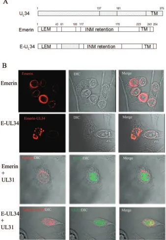
immunofluo-FIG. 3. (A) Schematic diagram of emerin, pU
L34, and chimeric pU
L34. Most pU
L34 sequences are indicated by diagonally hatched rectangles,
whereas the putative pU
L31 interaction region is indicated by a vertically hatched rectangle. Emerin sequences are indicated by open rectangles.
The final chimera (E-UL34) contains the putative pU
L31 interaction region of pU
L34 in place of codons 61 to 100 of emerin. Numbers above the
diagrams indicate the amino acids delimiting the domains. TM, transmembrane domain; INM, region shown to be necessary for targeting emerin
to the inner nuclear membrane; LEM, domain common to many INM-targeted proteins. (B) Hep2 cells were transfected with a plasmid encoding
emerin or the emerin-U
L34 chimeric construct (E-UL34) alone or together with a plasmid encoding the U
L31 protein. The cells were stained with
an emerin-specific mouse monoclonal antibody and a pU
L31-specific rabbit polyclonal antibody. Bound antibodies were detected with Texas
Red-conjugated donkey anti-mouse and FITC-conjugated donkey anti-rabbit antibodies.
on November 8, 2019 by guest
http://jvi.asm.org/
[image:5.585.122.460.74.562.2]rescence at the nuclear rim was slightly altered from that of
native emerin and often exhibited a punctate distribution,
es-pecially in cells containing large amounts of chimeric protein.
It should also be noted that under these conditions, the
stain-ing of native emerin was readily diststain-inguished from that of the
expressed transgenes, presumably as a consequence of the
relatively low expression level of the former. In control
exper-iments, the localization of transiently expressed emerin did
not change, regardless of whether the U
L31 protein was
coexpressed (Fig. 3B, third row). In contrast, the
localiza-tion of the emerin chimera bearing U
L34 amino acids 137 to
181 changed significantly upon coexpression of the U
L31
protein such that the chimera largely colocalized with
pU
L31 in the nucleoplasm, with some emerin-specific
stain-ing remainstain-ing at the nuclear rim. We suspect but cannot
prove that the residual fluorescence at the nuclear rim
rep-resents native emerin as opposed to that derived from the
transiently expressed chimera. Irrespective of this possibility,
these experiments indicate that U
L34 amino acids 137 to 181
are sufficient to cause a partial colocalization of two different
proteins (GFP and emerin) with the U
L31 protein in the
con-text of a eukaryotic cell.
Construction and characterization of U
L34 (del 138-181)
virus.
The next set of experiments was designed to determine
if amino acids 138 to 181 of U
L34 were essential for
colocal-ization of the U
L31 and U
L34 proteins in infected cells and to
determine if this region was also essential for the contribution
of U
L34 to viral replication.
A recombinant virus bearing a mutant U
L34 protein lacking
codons 138 to 181 was used to replace native U
L34 within a
BAC containing the HSV-1(F) genome as detailed in
Materi-als and Methods. Briefly, the recombinant virus was
con-structed as follows. (i) A DNA fragment that contained a
mutant U
L34 gene lacking codons 137 to 181 and a kanamycin
resistance gene that was flanked by Flp recombination sites
and by 50 bp of U
L34 sequence and BamHI restriction sites
was generated by PCR. (ii) The PCR amplicon was
electropo-rated into bacteria harboring the HSV-1(F) genome within the
BAC pYEC102 (37), and kanamycin-resistant colonies were
selected at 30°C. (iii) One colony was picked, genotypically
verified (see below), and designated v3480-Kan-BAC. (iv) Flp
recombinase was induced in
E. coli
cells harboring
v3480-BAC-Kn to remove the kanamycin resistance gene. The
result-ing recombinant BAC was designated v3480-BAC. (v) The
BAC DNA was purified from bacteria and cotransfected with
a Cre expression plasmid into R1310 cells (a U
L34-expressing
cell line derived from rabbit skin cells), viral plaques were
purified, and the virus from one of the plaques was designated
v3480. The Cre recombinase-mediated removal of the BAC
vector sequences inserted between U
L3 and U
L4 was expected
inasmuch as the BAC sequences were flanked by Lox
recom-bination sites (37).
FIG. 4. (A) Schematic diagram of steps involved in the
construc-tion of a virus bearing a U
L34 gene lacking codons 137 to 181. Line 1,
sequences within a 2.2-kb PCR product containing the U
L34 gene
lacking codons 137 to 181 and a gene encoding kanamycin resistance
(Kan) flanked by Frt recombination sites (not shown), with 50 bp of
sequence homologous to regions flanking U
L34 in wild-type viral
ge-nomes and BamHI restriction sites. Line 2, wild-type HSV-1 sequences
containing U
L34 and schematic diagram of the U
L34 probe (UL34-P,
gray rectangle) used to hybridize to DNA sequences, as shown in panel
B. The lengths of the expected DNA fragments are indicated in
kilo-base pairs and are delimited by BamHI (B) sites. Line 3, genome of the
U
L34 mutant virus v3480-Kan after homologous recombination within
the 50-bp sequences flanking the Kan
rgene and U
L
34. The sizes of
the expected BamHI fragments are indicated in kilobase pairs
be-low the rectangles. Line 4, genome of the U
L34 mutant virus v3480
after deletion of the kanamycin resistance gene at the Frt sites by the
induction of Flp recombinase. (B) Digitally scanned image of a
flu-orograph of viral DNAs probed with radiolabeled DNAs. Viral DNAs
were purified, digested with BamHI, and electrophoretically separated
in a 1% agarose gel. The DNAs were transferred to nitrocellulose and
probed with [
␣
-
32P]dCTP-labeled U
L
34 (schematically diagramed as
UL34-P in panel A), the gene encoding kanamycin resistance, or the
BAC vector sequence. Lanes 1, HSV-BAC; lanes 2, v3480 DNA; lanes
3, v3480-Kan DNA; lanes 4, v3480-Kan-BAC DNA. The numbers
indicate the sizes of the DNA fragments in kilobase pairs.
on November 8, 2019 by guest
http://jvi.asm.org/
For verification of the expected genotypes of the
recombi-nant BACs, HSV-1(F) BAC, v3480, Kan, and
v3480-Kan-BAC DNAs were purified, digested with BamHI, and
electrophoretically separated in a 1% agarose gel. The
digest-ed DNAs were transferrdigest-ed to nitrocellulose and probdigest-ed with
[
␣
-
32P]dCTP-labeled DNA from either U
L
34, the gene
encod-ing kanamycin resistance, or the BAC vector sequence. As
shown in Fig. 4B, the U
L34 probe recognized 9.3- and 1.3-kb
bands from the HSV-1(F) BAC (lane 1, left panel). However,
both bands were missing from the v3480, v3480-Kan, and
v3480-Kan-BAC DNAs; instead, a 0.8- or 2.2-kb band was
recognized due to the presence or absence of the kanamycin
resistance gene, respectively (lanes 2 to 4). A band of
approx-imately 9.0 kb was also recognized for all three recombinant
viruses due to sequences within the probe that hybridized to
U
L34 (Fig. 4A shows a diagrammatic representation of these
sequences). As expected, the kanamycin probe only recognized
bands from the v3480-Kan and v3480-Kan-BAC DNAs (lanes
3 and 4, middle panel), and the BAC probe only recognized
bands from the HSV-BAC and v3480-Kan-BAC DNAs (lanes
1 and 4, right panel). In addition, the U
L34 gene of v3480 was
amplified by PCR and sequenced to ensure that the viral
ge-notype was as designed (not shown). As confirmation of the
truncation of pU
L34, as shown in Fig. 5A, the pU
L34-specific
antibody recognized a protein with an apparent relative
mo-lecular weight of 34,000 in lysates of cells infected with the
wild-type virus, whereas cells infected with v3480 contained
a protein with an apparent relative molecular weight of
28,000. As expected, no U
L34 protein was detected in lysates of
mock-infected cells or cells infected with the U
L34 deletion virus.
FIG. 5. (A) Scanned digital image of immunoblot of cell lysates probed with an antibody to pU
L34. Hep2 cells were either mock infected or
infected with wild-type HSV-1(F), the U
L34 deletion virus, or the mutant virus v3480. The cells were lysed, and associated proteins were denatured,
electrophoretically separated, transferred to nitrocellulose, and probed with a pU
L34-specific antibody. (B) Confocal images of the localization of
pU
L31 and pU
L34 in infected cells. Hep2 cells were infected with the U
L31 deletion virus (UL31
⫺
), the U
L34 deletion virus (UL34
⫺
), the mutant
virus v3480, or the U
L34 repair virus (UL34R) for 16 h and then fixed with ice-cold methanol at
⫺
20°C. The cells were then stained with pU
L31-and pU
L34-specific rabbit and pU
L34-specific chicken antibodies, and bound antibodies were detected with a FITC-conjugated donkey anti-rabbit
antibody and a Texas Red-conjugated donkey anti-chicken antibody. Differential interference contrast (DIC) images were collected separately. The
separately collected channels of fluorescent and DIC images were coprojected in the rightmost column. The intensity of the red and green channels
was increased in the merged image to allow comparison with light-refracting features.
on November 8, 2019 by guest
http://jvi.asm.org/
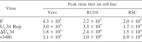
[image:7.585.125.463.71.452.2]To determine whether precluding an interaction with U
L31
played a role in viral replication, we infected Vero cells, rabbit
skin cells, and R1310 cells with 1.0 PFU of virus per cell.
Twenty-four hours after infection, the cells were lysed by
freez-ing and thawfreez-ing, and infectious titers were determined by
ti-tration and a plaque assay on R1310 cells; the data are shown
in Table 1. Like the U
L34 null virus (31), v3480 failed to
replicate on Vero cells to an appreciable extent and replicated
up to 6.9
⫻
10
4PFU/ml on rabbit skin cells. Titers produced in
rabbit skin cells containing U
L34 were increased
⬎
10- to
100-fold compared to titers obtained from normal rabbit skin cells.
Experiments to characterize the ability of v3480 to form viral
plaques were also performed (data not shown). Neither the
U
L34 deletion mutant nor v3480 formed plaques on Vero cells.
However, both the U
L34 deletion mutant and v3480 formed
small plaques on the more permissive rabbit skin cells. In
summary, these data indicate that v3480 replication in Vero
and rabbit skin cells resembles that of the U
L34 deletion virus,
suggesting that amino acids 137 to 181 are essential for normal
pU
L34 function.
To determine if the inability of mutant U
L34 to support viral
replication was reflected in an inability to properly localize
U
L31 to the nuclear rim of infected cells, we infected Hep2
cells with the U
L31 deletion virus, the U
L34 deletion virus, the
U
L34 restored virus, or v3480. The cells were then fixed 16 h
after infection and stained with antibodies directed against the
U
L31 and U
L34 proteins. The results are shown in Fig. 5. As
previously described (28), cells infected with the virus bearing
a restored U
L34 gene contained both U
L31 and U
L34 proteins
almost exclusively at the nuclear rim. In contrast, cells infected
with the U
L34 deletion mutant contained U
L31 exclusively
within the nucleoplasm, and cells infected with the U
L31
de-letion virus contained the U
L34 protein at the nuclear rim and
within the nucleus and the cytoplasm. The latter was present in
a pattern emanating from the nuclear membrane that strongly
resembled the distribution of the endoplasmic reticulum.
In cells infected with v3480, the U
L31 protein localized
ex-clusively in the nucleoplasm in a pattern that closely resembled
that of the distribution of the U
L31 protein in cells infected
with the U
L34 null mutant. We also noted that some of the
nucleoplasmic aggregates containing the U
L31 protein also
contained lamin A/C (not shown). The colocalization of the
U
L31 and U
L34 proteins at the nuclear membrane was
re-stored in the complementing cell line R1310 infected with
v3480 (not shown). We therefore concluded that the U
L34
protein of v3480 lacks the capacity to correctly target the U
L31
protein to the nuclear membrane. Although we cannot
ex-clude the possibility that amino acids 137 to 181 mediate
other functions that contribute to viral replication, these
data suggest that these amino acids constitute a domain that
is necessary for interacting with the U
L31 protein in vitro and
in vivo.
DISCUSSION
Previous studies have shown that the U
L31 and U
L34
pro-teins form a complex at the nuclear membrane and act
to-gether to play important roles in the primary envelopment of
both HSV-1 and pseudorabies virus (8, 12, 18, 28, 31). The
deletion of either protein from HSV-1 abolishes its replication
on some cell lines and causes mislocalization of the other
protein (28, 29, 31).
The goal of this study was to identify the domain in pU
L34
that mediates the interaction with pU
L31 and to test the
sig-nificance of the pU
L31-pU
L34 interaction for viral replication.
Previous studies showed that charged cluster mutations did not
disrupt the interaction with pU
L31, but some of these
muta-tions were lethal for virus replication and mislocalized the
pU
L31/pU
L34 complex elsewhere in the cell (4). For the
present study, it was determined that amino acids 137 to 181 of
pU
L34 were sufficient to mediate the interaction with pU
L31 in
three different contexts, including GST pull-down assays,
tar-geting of GFP to sites containing pU
L31, and targeting of
chimeric emerin to intracellular sites that contained pU
L31.
Moreover, pU
L34 amino acids 137 to 181 were necessary for
viral replication and were shown to be essential for
colocaliza-tion of the pU
L31 and pU
L34 proteins in infected cells.
The peptide comprising amino acids 137 to 181 is less polar
(9.7% by weight) and more basic (26.4% by weight) than
pU
L34 as a whole (18.3 and 17.0% by weight, respectively [not
shown]). A comparison of the sequences of the U
L34 homologs
of HSV-1, pseudorabies virus, varicella zoster virus, and equine
herpesvirus 1 has shown that the identified interaction region
is highly conserved (not shown). In particular, the consensus
for residues 155 to 173 is
155CF(V/T)RMP(X)VQL(A/S)FRF
MGP(E/D)D
173, with 79% identity among the various
ho-mologs. Two HSV-1 mutations that were assessed for their
effects on the pU
L31-pU
L34 interaction are particular
interest-ing inasmuch as they are located near or in the pU
L31
inter-action region detected in the present study. Thus, while
chang-ing
136IKGR
139(a mutant designated CL10) or
158RMPR
161(designated CL13) to alanines did not disrupt the interaction
with pU
L31 or the localization of the complex at the nuclear
membrane, it precluded complementation of a U
L34 viral
de-letion mutant when the proteins were expressed transiently (4).
These data indicate that the alanine clusters, while insufficient
to disrupt the pU
L31-pU
L34 interaction, may have functionally
altered the complex such that it was unable to perform its
role(s) in viral egress.
A comprehensive genetic analysis of the more distantly
re-lated murine cytomegalovirus homolog of U
L34 (M50) has also
been conducted in the context of viral infection (6). In previous
studies, residues
54YPVEY
57of M50 were shown to be
essen-tial for coimmunoprecipitation with M53, the HSV-1 U
L31
homolog. Although this region is conserved in the
alphaher-pesviruses equine herpesvirus 1, varicella zoster virus, and
HSV [consensus sequence, (F/Y)P(I/L/V)EY], the N-terminal
80 amino acids of pU
L34 were dispensable for the interaction
with pU
L31 in GST pull-downs (Fig. 1C), and a U
L34 protein
[image:8.585.43.283.81.153.2]lacking the first 78 residues maintained the ability to colocalize
TABLE 1. Peak titers of viruses on different cell lines
Virus
Peak virus titer on cell line
Vero R1310 RSC
F
4.3
⫻
10
72.2
⫻
10
72.0
⫻
10
7U
L34 Rep
3.0
⫻
10
7
3.3
⫻
10
71.7
⫻
10
7⌬
U
L34
1.8
⫻
10
32.4
⫻
10
61.5
⫻
10
4v3480
3.1
⫻
10
31.0
⫻
10
66.9
⫻
10
4on November 8, 2019 by guest
http://jvi.asm.org/
with U
L31 at the nuclear membrane (not shown). Similarly, a
cluster mutation near this region (
67EYVLR
71) did not prevent
colocalization of this U
L34 mutant protein with the U
L31
pro-tein at the nuclear membrane in cells infected with a U
L34 null
virus (4). HSV-1 pU
L34 amino acids 137 to 181 are mostly
conserved in murine cytomegalovirus M50, but this region of
M50 also contains insertions of 12, 12, 8, and 9 amino acids
that are not conserved in alphaherpesviruses (not shown). The
insertion of 5 amino acids into the 12-amino-acid
noncon-served regions precluded rescue of the murine cytomegalovirus
M50 null mutant (6). Other lethal mutations in M50 mapped
within regions that are conserved in HSV-1 pU
L34 137-181.
The data therefore suggest that the interactions of the HSV
and cytomegalovirus homologs and the structures of the
re-sulting complexes may differ significantly. It is also possible
that mutations at the N terminus of M50 cause it to aberrantly
interact with other regions of M50 (or other proteins) and thus
preclude an interaction with M53. In our studies with pU
L34,
such effects would not be expected because the N-terminal
region was deleted from the constructs tested in vitro.
In cells infected with the U
L34 null virus or v3480, the U
L31
protein localized to the nucleus and within intranuclear
aggre-gates (Fig. 5). Recent analyses indicated that these aggreaggre-gates
also contained lamin A/C (not shown). This is consistent with
the observation that overexpression of the U
L31 protein is
sufficient to relocalize lamin A/C to the nucleoplasm and may
reflect the possibility that the interaction between U
L31 and
lamin A/C is sufficient to mediate a partial dissolution of the
lamina (30). Such relocalization may not be readily apparent in
infected cells expressing wild-type pU
L34 because pU
L31 is
retained at the nuclear rim, as is the lamin A/C to which the
pU
L31/pU
L34 complex binds.
ACKNOWLEDGMENTS
This study was supported by National Institutes of Health grant R01
AI52341.
We thank Jarek Okulicz Kozaryn for technical support and Yasushi
Kawaguchi, Richard Roller, Michael Kotlikoff, Klaus Osterrieder,
Bernd Karsten Tischer, and Carol Duffy for reagents and helpful
discussions.
REFERENCES
1.Baines, J. D., and B. Roizman.1992. The UL11 gene of herpes simplex virus
1 encodes a function that facilitates nucleocapsid envelopment and egress from cells. J. Virol.66:5168–5174.
2.Bengtsson, L., and K. L. Wilson.2004. Multiple and surprising new functions for emerin, a nuclear membrane protein. Curr. Opin. Cell Biol.16:73–79. 3.Bione, S., E. Maestrini, S. Rivella, M. Mancini, S. Regis, G. Romeo, and D.
Toniolo.1994. Identification of a novel X-linked gene responsible for Emery-Dreifuss muscular dystrophy. Nat. Genet.8:323–327.
4.Bjerke, S. L., J. M. Cowan, J. K. Kerr, A. E. Reynolds, J. D. Baines, and R. J. Roller.2003. Effects of charged cluster mutations on the function of herpes simplex virus type 1 UL34 protein. J. Virol.77:7601–7610.
5.Blaho, J. A., C. Mitchell, and B. Roizman.1994. An amino acid sequence shared by the herpes simplex virus 1 alpha regulatory proteins 0, 4, 22, and 27 predicts the nucleotidylylation of the UL21, UL31, UL47, and UL49 gene
products. J. Biol. Chem.269:17401–17410.
6.Bubeck, A., M. Wagner, Z. Ruzsics, M. Lo¨tzerich, M. Iglesias, I. R. Singh, and U. H. Koszinowski.2004. Comprehensive mutational analysis of a her-pesvirus gene in the viral genome context reveals a region essential for virus replication. J. Virol.78:8026–8035.
7.Chang, Y. E., and B. Roizman.1993. The product of the UL31 gene of herpes
simplex virus 1 is a nuclear phosphoprotein which partitions with the nuclear matrix. J. Virol.67:6348–6356.
8.Chang, Y. E., C. Van Sant, P. W. Krug, A. E. Sears, and B. Roizman.1997. The null mutant of the UL31 gene of herpes simplex virus 1: construction and
phenotype of infected cells. J. Virol.71:8307–8315.
9.Ejercito, P. M., E. D. Kieff, and B. Roizman.1968. Characterization of
herpes simplex virus strains differing in their effects on social behavior of infected cells. J. Gen. Virol.2:357–364.
10.Elliott, G., and P. O’Hare.1999. Live-cell analysis of a green fluorescent protein-tagged herpes simplex virus infection. J. Virol.73:4110–4119. 11.Frangioni, J. V., and B. J. Neel.1993. Solubilization and purification of
enzymatically active glutathioneS-transferase (pGEX) fusion proteins. Anal. Biochem.210:179–187.
12.Fuchs, W., B. G. Klupp, H. Granzow, N. Osterrieder, and T. C. Mettenleiter.
2002. The interacting UL31 and UL34 gene products of pseudorabies virus are involved in egress from the host cell nucleus and represent components of primary enveloped but not mature virions. J. Virol.76:364–378. 13.Granzow, H., B. G. Klupp, W. Fuchs, J. Veits, N. Osterrieder, and T. C.
Mettenleiter.2001. Egress of alphaherpesviruses: comparative ultrastruc-tural study. J. Virol.75:3673–3684.
14.He, D. C., J. A. Nickerson, and S. Penman.1990. Core filaments of the nuclear matrix. J. Cell Biol.110:569–580.
15.Holland, D. J., M. Miranda-Saksena, R. A. Boadle, P. Armati, and A. L. Cunningham.1999. Anterograde transport of herpes simplex virus proteins in axons of peripheral human fetal neurons: an immunoelectron microscopy study. J. Virol.73:8503–8511.
16.Hutchinson, L., and D. C. Johnson.1995. Herpes simplex virus glycoprotein K promotes egress of virus particles. J. Virol.69:5401–5413.
17.Jayachandra, S., A. Baghian, and K. G. Kousoulas.1997. Herpes simplex virus type 1 glycoprotein K is not essential for infectious virus production in actively replicating cells but is required for efficient envelopment and trans-location of infectious virions from the cytoplasm to the extracellular space. J. Virol.71:5012–5024.
18.Klupp, B. G., H. Granzow, and T. C. Mettenleiter.2000. Primary envelop-ment of pseudorabies virus at the nuclear membrane requires the UL34 gene product. J. Virol.74:10063–10073.
19.Lee, E.-C., D. Yu, J. M. de Velasco, L. Tessarollo, D. A. Swing, D. L. Court, N. A. Jenkins, and N. G. Copeland.2001. A highly efficient Escherichia coli-based chromosome engineering system adapted for recombinogenic tar-geting and subcloning of BAC DNA. Genomics73:56–65.
20.Liang, L., M. Tanaka, M. Kawaguchi, and J. D. Baines.2004. Cell lines that support replication of a novel herpes simplex virus 1 UL31 deletion mutant
can properly target UL34 protein to the nuclear rim in the absence of UL31.
Virology329:68–76.
21.Loomis, J. S., J. B. Bowzard, R. J. Courtney, and J. W. Wills.2001. Intra-cellular trafficking of the UL11 tegument protein of herpes simplex virus type 1. J. Virol.75:12209–12219.
22.Mettenleiter, T. C. 2002. Herpesvirus assembly and egress. J. Virol.76:
1537–1547.
23.Miranda-Saksena, M., P. Armati, R. A. Boadle, D. J. Holland, and A. L. Cunningham.2000. Anterograde transport of herpes simplex virus type 1 in cultured, dissociated human and rat dorsal root ganglion neurons. J. Virol.
74:1827–1839.
24.Neubauer, A., J. Rudolph, C. Brandmu¨ller, F. Just, and N. Osterrieder.
2002. The equine herpesvirus 1 UL34 gene product is involved in an early step in virus egress and can be efficiently replaced by a UL34-GFP fusion protein. Virology300:189–204.
25.Ostlund, C., J. Ellenberg, E. Hallberg, J. Lippincott-Schwartz, and H. J. Worman.1999. Intracellular trafficking of emerin, the Emery-Dreifuss mus-cular dystrophy protein. J. Cell Sci.112:1709–1719.
26.Purves, F. C., D. Spector, and B. Roizman.1991. The herpes simplex virus 1 protein kinase encoded by the US3 gene mediates posttranslational modifi-cation of the phosphoprotein encoded by the UL34 gene. J. Virol.65:5757–
5764.
27.Purves, F. C., D. Spector, and B. Roizman.1992. UL34, the target gene of the
herpes simplex virus US3 protein kinase, is a membrane protein which in its
unphosphorylated state associates with novel phosphoproteins. J. Virol.66:
4295–4303.
28.Reynolds, A. E., B. J. Ryckman, J. D. Baines, Y. Zhou, L. Liang, and R. J. Roller.2001. UL31 and UL34 proteins of herpes simplex virus type 1 form a
complex that accumulates at the nuclear rim and is required for envelopment of nucleocapsids. J. Virol.75:8803–8817.
29.Reynolds, A. E., E. G. Wills, R. J. Roller, B. J. Ryckman, and J. D. Baines.
2002. Ultrastructural localization of the herpes simplex virus type 1 UL31,
UL34, and US3 proteins suggests specific roles in primary envelopment and
egress of nucleocapsids. J. Virol.76:8939–8952.
30.Reynolds, A. E., L. Liang, and J. D. Baines.2004. Conformational changes in the nuclear lamina induced by herpes simplex virus type 1 require genes UL31 and UL34. J. Virol.78:5564–5575.
31.Roller, R. J., Y. Zhou, R. Schnetzer, J. Ferguson, and D. DeSalvo.2000. Herpes simplex virus type 1 UL34 gene product is required for viral envel-opment. J. Virol.74:117–129.
32.Ryckman, B. J., and R. J. Roller.2004. Herpes simplex virus type 1 primary envelopment: UL34 protein modification and the US3-UL34 catalytic rela-tionship. J. Virol.78:399–412.
33.Shiba, C., T. Daikoku, F. Goshima, H. Takakuwa, Y. Yamauchi, O. Koiwai, and Y. Nishiyama.2000. The UL34 gene product of herpes simplex virus
on November 8, 2019 by guest
http://jvi.asm.org/
type 2 is a tail-anchored type II membrane protein that is significant for virus envelopment. J. Gen. Virol.81:2397–2405.
34.Simpson-Holley, M., J. D. Baines, R. Roller, and D. M. Knipe.2004. Herpes simplex virus 1 UL31 and UL34 gene products promote the late maturation of viral replication compartments to the nuclear periphery. J. Virol. 78:
5591–5600.
35.Skepper, J. N., A. Whiteley, H. Browne, and A. Minson.2001. Herpes sim-plex virus nucleocapsids mature to progeny virions by an envelopment3
deenvelopment3reenvelopment pathway. J. Virol.75:5697–5702. 36.Stierle, V., J. Couprie, C. Ostlund, I. Krimm, S. Zinn-Justin, P. Hossenlopp,
H. J. Worman, J. C. Courvalin, and I. Duband-Goulet.2003. The carboxyl-terminal region common to lamins A and C contains a DNA binding domain. Biochemistry42:4819–4829.
37.Tanaka, M., H. Kagawa, Y. Yamanashi, T. Sata, and Y. Kawaguchi.2003. Construction of an excisable bacterial artificial chromosome containing a full-length infectious clone of herpes simplex virus type 1: viruses
reconsti-tuted from the clone exhibit wild-type properties in vitro and in vivo. J. Virol.
77:1382–1391.
38.Taniura, H., C. Glass, and L. Gerace.1995. A chromatin binding site in the tail domain of nuclear lamins that interacts with core histones. J. Cell Biol.
131:33–44.
39.Tews, D. S.1999. Emerin. Int. J. Biochem. Cell Biol.31:891–894. 40.Yamauchi, Y., C. Shiba, F. Goshima, A. Nawa, T. Murata, and Y. Nishiyama.
2001. Herpes simplex virus type 2 UL34 protein requires UL31 protein for its relocation to the internal nuclear membrane in transfected cells. J. Gen. Virol.82:1428.
41.Ye, G.-J., K. T. Vaughan, R. B. Vallee, and B. Roizman.2000. The herpes simplex virus 1 UL34 protein interacts with a cytoplasmic dynein
intermedi-ate chain and targets nuclear membrane. J. Virol.74:1355–1363. 42.Ye, G.-J., and B. Roizman.2000. The essential protein encoded by the UL31
gene of herpes simplex virus 1 depends for its stability on the presence of UL34 protein. Proc. Natl. Acad. Sci. USA97:11002–11007.
on November 8, 2019 by guest
http://jvi.asm.org/