Int. J. Electrochem. Sci., 13 (2018) 4071 – 4084, doi: 10.20964/2018.05.24
International Journal of
ELECTROCHEMICAL
SCIENCE
www.electrochemsci.orgMini review
Recent Progress in Electrochemical Biosensors for Detection of
Prostate-Specific Antigen
Yong Chang, Mengmeng Wang, Luyan Wang and Ning Xia*
Henan Province of Key Laboratory of New Optoelectronic Functional Materials, College of Chemistry and Chemical Engineering, Anyang Normal University, Anyang, Henan 455000, People's Republic of China
*
E-mail: [email protected]
Received: 1 January 2018 / Accepted: 29 January 2018 / Published: 10 April 2018
Prostate-specific antigen (PSA) is a biomarker for preoperative diagnosis and screening of prostate cancer and monitoring of its post-treatment. Because of their sensitivity, simple in operation, rapid response, and compatibility with miniaturization, electrochemical biosensors have attracted considerable attention for PSA detection. In this work, we focused on the recent progress in the electrochemical methods for PSA detection, including immunosensors, aptasensors and peptide-based biosensors.
Keywords: Prostate cancer; electrochemical biosensors; prostate-specific antigen
1. INTRODUCTION
blood [10]. Once the concentration of PSA is within the danger zone of 4 ng/mL to 10 ng/mL, PCa is often suspected [7]. As a result, the development of ultrasensitive methods for the detection of low-level PSA is of vital importance for the early diagnosis, therapeutic treatments and post-treatment monitoring of PCa.
Currently, many efforts have provoked the appearance of various kinds of PSA detection methods, such as fluorescence [11, 12], enzyme-linked immunosorbent assay (ELISA) [13] and bioluminescent enzyme immunoassay [14]. In spite of their good sensitivity, most of these methods are complicated, time-consuming, and expensive. Because of their sensitivity, simple in operation, rapid response, and compatibility with miniaturization, electrochemical biosensors have attracted considerable attention for PSA sensing. Given the exuberant advancement, this review aims to summarize the recent advancement in electrochemical biosensors for the detection of PSA biomarker, including immunosensors, aptasensors and peptide-based biosensors.
2. ELECTROCHEMICAL IMMUNOSENSORS
Antibodies-based electrochemical PSA biosensors include label-free immunosensors and sandwich-like immunosensors. The label-free immunosensor is based on antibody-PSA interaction. Sandwich-like immunosensor is mainly composed of a capture antibody, PSA, and secondary antibody. Preparation procedure and electrode materials of these two types of electrochemical immunosensors are reviewed.
2.1 Label-free immunosensors
For the label-free detection, the capture of PSA by antibodies immobilized onto the electrode surface can induce the change of faradaic current and electrochemical impedance, depending upon the modified electrodes. If the electrode is modified with non-electroactive materials, the antibody-PSA interaction can induce the impedance or current change, which is usually monitored in [Fe(CN)6]
3-/4-solution. If the electrode is modified with electroactive substrate, the interaction can cause the current change of the redox probes modified on electrode surface.
2.1.1 Non-electroactive electrode
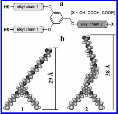
are attached onto the electrode surface through mechanical entrapment or avidin-biotin affinity for PSA binding. The linear ranges of these two immobilization methods are 1 ~ 100 pg/mL and 1 ~ 200 ng/mL, and the detection limits are found to be 1 pg/mL and 1 ng/mL, respectively. Thus, the sensor prepared with an affinity immobilization strategy is more sensitive than that of the entrapment method.
Figure 1. (a) Structural features of dithiols derived from 3,5-dihydroxybenzyl alcohol. (b) Energy minimized geometries and molecular dimensions of 1 and 2 (calculated using the semiempirical PM3 algorithm supported on HyperChem software, Version 7.0). Reprinted with permission from reference [15]. Copyright 2008 American Chemical Society.
Electrode materials with excellent conductivity are especially welcomed in the construction of label-free electrochemical immunosensors [17]. The interfacial electron transfer of [Fe(CN)6]3-/4- probe
[image:3.596.104.490.158.531.2]
composite modified electrode [19]. The 3D immunosensor operates in a linear range of 0 ~ 10 ng/mL with a detection limit of 0.59 ng/mL. Moreover, polypyrrole electropolymerized on a three-dimensional Au nanowire array and high molecular-weight silk peptide (SP)-functionalized reduced graphene oxide (rGO) nanosheets have also been used for the immobilization of PSA antibodies, which allowed for the determination of PSA with a low detection limit [20, 21]. Recently, Li and Wei utilized binary reductants to synthesize highly conductive composites containing reduced graphene oxide with silver nanoparticles (rGO/Ag NPs) for PSA assay [22]. Through one-pot reaction under mild conditions, the strong reduction capacity of hydrazine hydrate and sodium citrate induced the formation of small Ag NPs on graphene sheets, which effectively inhibited rGO stacking. Furthermore, the decoration with Ag NPs repaired the structural defects in rGO, which in turn contributed to the formation of conducting path between rGO and Ag NPs. The modification of the obtained rGO/Ag NPs composites on the screen-printed electrode was beneficial to the further binding of anti-PSA antibody. The proposed electrochemical immunosensor showed a wide linear response range (1.0 ~ 1000 ng/mL) and a low detection limit (0.01 ng/mL).
2.1.2 Electroactive electrode
When the antibodies were modified onto the electroactive electrode, capture of PSA by the modified electrode may impede the electronic transfer. For this view, many groups have developed electrochemical PSA biosensors. For example, Ma’s group demonstrated that the conductive redox hydrogel composed of aniline and viny-ferrocene can be used for the immobilization of PSA antibodies. The capture of PSA decreases the catalytic oxidation of ascorbic acid by ferrocene. As a result, PSA can be detected with a linear range of 0.001 ~ 200 ng/mL and a detection limit of 0.54 pg/mL [23]. Furthermore, they used a glassy carbon electrode modified with cadmium-organic coordination nanoparticle (CdOP) film, Nafion and graphene oxide to detect PSA. The CdOP film exhibited an electrochemical redox activity at –0.71 V. Binding of PSA at the concentration of 0.01 ~ 100 ng/mL on the electrode surface led to the decrease in the electrochemical signal [24]. Cao and co-workers demonstrated that the nanocomposite of halloysite nanotubes with polypyrrole shell and palladium nanoparticles (HNTs@PPy-Pd) shows high electrocatalytic activity toward H2O2 reduction
[25]. The PSA-antibody interaction on the HNTs@PPy-Pd covered electrode surface decreases the electrocatalytic activity, thus facilitating the PSA detection with a detection limit of 0.03 pg/mL and a linear range of 0.0001 ~ 25 ng/mL. Metal organic frame-works (MOFs) have showed great applications as new materials because of their hierarchical structures and high surface areas. However, the poor electrical conductivity limits their applications in developing electrochemical sensing devices. Recently, Deep’s group demonstrated that the tetracyanoquinodimethane (TCNQ)-doped copper-MOF can be used in the electrochemical sensing platform for PSA detection in a linear range of 0.1 ~ 100 ng/mL [26]. Li and co-workers prepared an amino functionalized cuprousoxide@ceric dioxide (Cu2O@CeO2-NH2) core-shell nanocomposites loaded with AuNPs and found that they exhibit high
electrocatalytic activity towared H2O2 reduction. With nanocomposites as electrode materials, a PSA
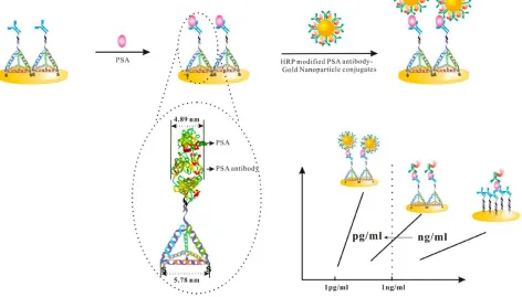
utilizing the target-induced proximity hybridization for the release of electroactive methylene blue (MB) from the inner pores of mesoporous silica nanoprobe (MSN) (Fig. 2) [28]. Briefly, the flexible DNA could be wrapped on the surface of the positively charged amine-functionalized MSN due to the electrostatic adsorption, which impeded the release of MB molecules entrapped in the MSN. In the presence of target protein PSA, two DNA-labeled antibodies formed into proximate complex, leading to the hybridization with the DNA strand into a rigid double-stranded structure. As a result, the bioconjugate was opened and the sealed MB molecules came out, which was monitored with a screen-printed carbon electrode. Moreover, the addition of nicking endonuclease Nt.BbvCI could initiate in situ enzymatic recycling binding of the proximate complex and opened more DNA biogates for the detachment of more MB. A wide detection range from 0.002 to 100 ng/mL of PSA with a detection limit of 1.3 pg/mL was achieved for the proposed assay.
Figure 2. Schematic illustration of homogeneous electrochemical immunoassay using proximity hybridization-responsive mesoporous silica nanoprobe. Reprinted with permission from reference [28]. Copyright 2014 American Chemical Society.
2.2 Sandwich-like immunosensors
[image:5.596.62.539.278.452.2]
Figure 3. Formats suitable for electrochemical immunosensing include competitive (A, B) and sandwich (C) immunoassays. For competition, the immobilized recognition molecule can be either an antibody (A) or an antigen (B, antigen, hapten, or their derivatives); the signalgenerating labeled molecule, or the tracer, includes analyte−NP (A) or antibody−NP (B1) conjugates. The latter variant can also employ an unlabeled primary antibody and a secondary labeled antibody (B2). For sandwich assays, the capture (primary) antibody is immobilized (C), and a signal is generated using the labeled secondary antibody (C1) or even the third labeled antibody (C2). The general calibration graphs for both competitive and sandwich assays are shown in the right-hand section of the image. Reprinted with permission from reference [35]. Copyright 2017 American Chemical Society.
[image:6.596.174.423.71.321.2]
the stepwise enlargement of AuNP tag was described by Laocharoensuk and teammates to improve the electrochemical detection sensitivity of the sandwich immunoassay for the PSA assay [37]. For the fabrication of the sandwich electrochemical sensor, the capture moiety of anti-PSA antibody was firstly assembled on the carbon nanotube powder-modified screen-printed carbon electrodes (SPCE). After binding of PSA, the detector of anti-PSA antibody-AuNP bioconjugates was successively captured onto the electrode based on the antigen-antibody interaction. Then, a triple signal process was performed. Firstly, a nucleation reaction was done to obtain large AuNP by reducing gold chloride ion over small nucleation seeds AuNP tag. Secondly, further increase in the size and surface area of AuNP was achieved by initiating the growth of spiky AuNP. Thirdly, an electroactive layer on the obtained AuNP was accomplished by the silver enhancement step. As a result, the coupling use of small AuNP and triple enhancement process offered a great sensitivity improvement for the detection of PSA with over 260-fold of signal increase relative to conventional silver enhancement process.
[image:7.596.64.536.295.564.2]
3. ELECTROCHEMICAL APTASENSORS
In contrast to antibody, DNA aptamer can be inexpensively prepared and modified with high purity and reproducibility. With aptamer as the biorecognition element, many electrochemical biosensors have been developed in the formats of direct detection, competitive assay and sandwich-like assay. The development of the electrochemical PSA aptasensors is therefore introduced in this part.
3.1. Direct detection
For direct detection of PSA by electrochemical impedance spectroscopy (EIS), aptamer SAMs are still welcomed. Usually, thiol or amine terminated DNA aptamers are immobilized on a bare gold electrode surface through Au-S interaction or attached onto a carboxyl SAMs electrode surface through amino coupling reaction. The optimized experimental conditions for the formation of SAMs on gold electrode surface have been investigated by Estrela and co-workers, including DNA density, spacer molecule and buffer solution [38, 39]. Electrode materials exhibit a key factor in the sensitivity and binding ability of bioreceptors. With the development of nanotechnology, many nanomaterials have been modified onto electrode surface to improve the analytical performances [40-42]. For example, with AuNPs-covered graphitized mesoporous carbon nanoparticles (AuNPs@GMCs) as the electrode materials for aptamer immobilization, PSA can be quantitatively determined with a detection limit down to 0.25 ng/mL [42]. The rGO-MWCNT/AuNPs nanocomposite modified electrode can be used to detect PSA within a detection range of 0.005 ~ 100 ng/mL [41]. By virtue of the synergistic recognition of molecular imprinting and DNA aptamer, a novel bioreceptor has been constructed for selective PSA sensing [43]. DNA aptamer complexed with PSA was immobilized on the surface of a gold electrode, which served as the template of controlled electropolymerisation of dopamine. After removal of PSA, a selective molecularly imprinted polymer (MIP) cavity with the embedded aptamer was fabricated. The formed hybrid bioreceptor exhibited superior recognition properties relative to that of aptamer alone. According to the EIS, the fabricated bioreceptor shows excellent response to the PSA with the concentration ranging from 100 pg/mL to 100 ng/mL. A low limit of detection of 1 pg/mL was obtained for PSA, and three-fold increase in sensitivity was achieved than that of conventional aptasensors. Additionally, the sensor displayed good selectivity in the presence of homologous protein hK2 and serum protein HSA.
3.2 Competitive assay
inducing a strong decrease in redox current intensity. In order to demonstrate the binding of PSA to the aptamer instead of non-specific adsorption, another DNA strand with a complementary sequence to the aptamer was added in solution for aptamer hybridization, which detached the PSA from the surface of conducting substrate and gave rise to the current increase. Such a crossing of current intensity can effectively avoid false positives and be utilized to estimate the dissociation constant KD. A good
calibration curve from 1 ng/mL up to 10 μg/mL of PSA was established and the KD value was estimated at 2.670.8 nM.
Methylene blue (MB) displayed strong binding ability to DNA and RNA sequences via electrostatic attractions and intercalation. However, in the presence of the working solution of Tris, MB preferred to bind to the free guanine bases through hydrophobic forces of guanine with the fused aromatic rings in MB structure rather than electrostatic interaction. Heli and co-workers utilized the interaction of MB with guanine-rich aptamers to construct a label-free electrochemical aptasensing of PSA [45]. For the fabrication of the gold nanospears modified electrode, arginine as a shape-directing agent combined with high negative potential (compared to the formal potential of gold) was employed for the electrodeposition of gold. The thiolated PSA aptamer molecules were then immobilized on the modified electrode. Based on the strong interaction between PSA aptamer and MB, a large number of electroactive MB were adsorbed on the PSA aptamer and a strong electrochemical reduction peak of bonded MB was recorded by cyclic voltammograms (CVs) and differential pulse voltammograms (DPVs). Upon the capture of PSA, the aptamer structure changed into a double stranded, leading to the release of the bonded MB. Thus, the decreased current of MB was obtained. An excellent linear interaction between the plot and the concentration of PSA in the range of 0.125 ~ 200 ng/mLwith a detection limit of 0.05 ng/mL was attained. Furthermore, the proposed aptasensor displayed good reproducibility, regeneration and stability, which could be applied in the detection of PSA in real samples.
3.3 Sandwich structure
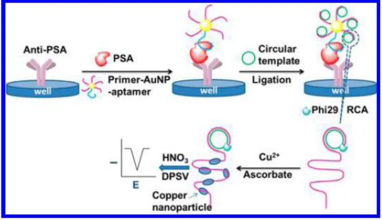
Figure 5. Schematic representation of cascade signal amplification for the detection of a prostate cancer biomarker. Reprinted with permission from reference [48]. Copyright 2016 American Chemical Society.
An abnormal expression of vascular endothelial growth factor (VEGF) is associated with numerous types of human cancers, including brain tumors, lung cancer, breast cancer, urinary tract tumors, and gastrointestinal cancer, which may serve as a contrast for enhancing the accuracy of PCa diagnosis with PSA. Yang’s group developed a dual-modality biosensor that can simultaneously detect VEGF and PSA for early diagnosis of Pca [49]. Highly soluble amine-terminated graphene oxide GO resulting from the carboxylated GO (GO-COOH) and branched polyethylenimine (bPEI) was fabricated for the conjugation with 5′-thiol-modified ssDNA by using sulfo-SMCC as the linker. The obtained GO-ssDNA bioconjugate was then modified on a glass chip with an Au-electrode for the capture of VEGF. The dual-antibody modified poly-L-lactide (PLLA) nanoparticles were recognized by VEGF and PSA, which enabled the successive modification of PLLA nanoparticles and PSA on the electrode. The current response induced by the modification of VEGF and PSA was measured using a 0.05-V bias voltage. The biosensor showed wide linear detection ranges with the concentration of 0.05 ~ 100 ng/mL for VEGF and 1 ~ 100 ng/mL for PSA. Additionally, high levels of sensitivity and selectivity were achieved.
4. PSA PROTEASE DETECTION
[image:10.596.105.492.74.297.2]
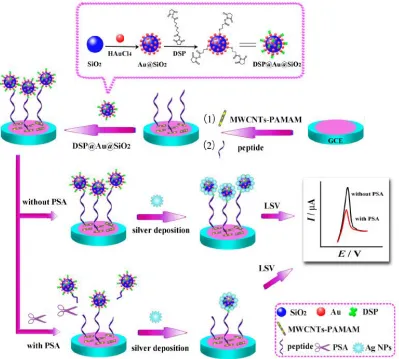
multiwalled carbon nanotubes/poly(amidoamine) dendrimers (MWCNTs-PAMAM) nanohybrids were firstly assembled on the electrode for the further binding of the substrate peptide. The dithiobis(succinimidylpropionate) (DSP) @Au@SiO2 composites were then attached on the peptide
via the inherent interaction between DSP and the amino of peptide. The DSP@Au@SiO2 nanohybrids
served as a tracing tag for the subsequent silver deposition process. Upon the addition of PSA, the specific substrate peptide was recognized and cleaved, which caused the separation of the tracing tag from the electrode surface. Accordingly, the amount of the deposited silver in the electrode decreased, resulting in decrement of the electrochemical stripping signal of the deposited silver. The concentration of PSA could be monitored by the change of the electrochemical stripping signal. Because the formed MWCNTs-PAMAM nanohybrids as the sensor platform provided a large surface area for the immobilization of a large amount of peptide and facilitated electron transfer, a wide line from 0.001 to 30 ng/mLwith a detection limit of 0.7 pg/mL was achieved.
Figure 6. Schematic diagram of the electrochemical-peptide biosensor for detection of PSA. Reprinted with permission from reference [52]. Copyright 2015 American Chemical Society.
[image:11.596.101.500.292.651.2]
bromide (CTAB). The binding of positively charged surfactant CTAB caused AuNPs with a positive charge, which was beneficial for the further adsorption of the negatively charged redox probe [Fe(CN)6]3−/4−. A low value of electro-transfer resistance was thus obtained. However, the addition of
PSA initiated the cleavage of the molecular recognition peptide modified with positively charged AuNPs. The release of positively charged AuNPs from the electrode surface contributed to a high electro-transfer resistance. Hence, a “signal-on” approach for PSA activity assay was achieved. Due to the signal amplification of AuNPs, low level of PSA with a concentration of 0.2 pg/mL was readily measured.
Ma’s group firstly utilized BSA as an effective sensitivity enhancer for the construction of a peptide-based amperometric biosensor for the ultrasensitive detection of PSA [54]. For the proposed biosensor, a porous and conductive substrate of chitosan-lead ferrocyanide-(poly(diallyldimethylammonium chloride)-graphene oxide) was assembled on a glassy carbon electrode (GCE) through drop coating method. Among the porous substrate, (poly(diallyldimethylammonium chloride)-graphene oxide could effectively enlarge the specific area and conductivity of the sensing interface, while chitosan was employed to adsorb conductive redox species lead ferrocyanide and link with peptide through coordination effect. In order to improve the sensitivity, the co-immobilization of 1-aminopropyl-3-methylimidazolium and peptide on the substrate was performed for the space of peptide. Additionally, further modification of BSA on the immobilized peptide contributed to the increase of resistance of the sensing interface, leading to signal amplification. The target analysts PSA could effectively recognize and cleave the substrate peptide, which caused a dramatic increase of current signal. As a result, a wide linear range (100 ng/mL ~ fg/mL) and a low detection limit (0.01 fg/mL) was obtained with the use of multiple amplification strategies.
5. CONCLUSION
Nowadays, exuberant progresses have been made in the field of PSA sensing, including excellent linear detection ranges and low detection limits (down to a femto-value). However, most of these advancements are still confined to laboratories and cannot be applied into the commercialization field. The major challenge may be the interference from the complicated real sample of blood or serum. In addition, the relative long analysis process is difficult in clinical trials. As a result, continuous studies on the biosensor with rapid response, improved sensitivity and long-term stability are equally critical, which would enable the shift from in-lab PCa sensing devices into a portable detection system with commercial values.
ACKOWLEDGMENTS
References
1. S. R. Denmeade and J. T. Isaacs, Nat. Rev. Cancer, 2 (2002) 389.
2. R. L. Siegel, K. D. Miller and A. Jemal, Ca-Cancer J. Clin., 66 (2016) 7.
3. W. J. Catalona, D. S. Smith, T. L. Ratliff, K. M. Dodds, D.E. Coplen, J. J. Yuan, J. A. Petros and G. L. Andriole, New Engl. J. Med., 324 (1991) 1156.
4. H. Grönberg, J. Adolfsson, M. Aly, T. Nordström, P. Wiklund, Y. Brandberg, J. Thompson, F. Wiklund, J. Lindberg, M. Clements, L. Egevad and M. Eklund, Lancet Oncol., 16 (2015) 1667. 5. F. Zhao, C. Cheng and N. Xia, Int. J. Electrochem. Sci., 12 (2017) 7580.
6. H. Yu, M. Giai, E. P. Diamandis, D. Katsaros, D. J. Sutherland, M. A. Levesque, R. Roagna, R. Ponzone and P. Sismondi, Cancer Res., 55 (1995) 2104.
7. D. A. Healy, C. J. Hayes, P. Leonard, L. McKenna and R. O'Kennedy, Trends Biotechnol., 25 (2007) 125.
8. L. Yu, Z. Fan, W. Li, S. Li, P. Wang and H. Wang, Int. J. Electrochem. Sci., 12 (2017) 8188. 9. N. Chen, M. Rong, X. G. Shao, H. Zhang, S. P. Liu, B. J. Dong, W. Xue, T. Y. Wang, T. H. Li
and J. H. Pan, Int. J. Nanomed., 12 (2017) 5399.
10. H. Lilja, A. Christensson, U. Dahlén, M.-T. Matikainen, O. Nilsson, K. Pettersson and T. Lövgren, Clin. Chem., 37 (1991) 1618.
11. H. Härmä, T. Soukka and T. Lövgren, Clin. Chem., 47 (2001) 561.
12. Q. W. Zhang, L. Wu, T. I. Wong, J. L. Zhang, X. H. Liu, X. D. Zhou, P. Bai, B. Liedberg and Y. Wang, Int. J. Nanomed., 12 (2017) 2307.
13. L. I. Stowell, L. E. Sharman and K. Hamel, Forensic Sci. Int., 50 (1991) 125. 14. Y. Seto, T. Iba and K. Abe, Luminescence, 16 (2001) 285.
15. A. Fragoso, N. Laboria, D. Latta and C. K. O’Sullivan, Anal. Chem., 80 (2008) 2556. 16. A. Chem., Anal. Chem., 80 (2008) 6198.
17. Z. Gu, M. Zhao, W. Zhang, T. Jiang and M. Sun, Int. J. Electrochem. Sci., 12 (2017) 10726. 18. H. Wang, Y. Zhang, H. Yu, D. Wu, H. Ma, H. Li, B. Du and Q. Wei, Ana. Biochem., 434 (2013)
123.
19. H. D. Jang, S. K. Kim, H. Chang and J.-W. Choi, Biosens. Bioelectron., 63 (2015) 546. 20. J.-M. Moon, Y. H. Kim and Y. Cho, Biosens. Bioelectron., 57 (2014) 157.
21. Y. Wang, Y. Qu, G. Liu, X. Hou, Y. Huang, W. Wu, K. Wu and C. Li, Microchim Acta, 182 (2015) 2061.
22. L. Han, C. M. Liu, S. L. Dong, C. X. Du, X. Y. Zhang, L. H. Li and Y. Wei, Biosens. Bioelectron., 87 (2017) 466.
23. W. Li and Z. Ma, Sensor. Actuat. B: Chem., 248 (2017) 545. 24. W. Li, Q. Rong and Z. Ma, New J. Chem., 41 (2017) 1124.
25. Y. Li, M. S. Khan, L. Tian, L. Liu, L. Hu, D. Fan, W. Cao and Q. Wei, Anal. Bioanal. Chem., 409 (2017) 3245.
26. S. K. Bhardwaj, A. L. Sharm, N. Bhardwaj, M. Kukkar, A. A. S. Gill, K.-H. Kim and A. Deep, Sensor. Actuat. B: Chem., 240 (2017) 10.
27. F. Li, Y. Li, J. Feng, Y. Dong, P. Wang, L. Chen, Z. Chen, H. Liu and Q. Wei, Biosens. Bioelectron., 87 (2017) 630.
28. K. Ren, J. Wu, Y. Zhang, F. Yan and H. Ju, Anal. Chem., 86 (2014) 7494.
29. B. Qu, L. Guo, X. Chu, D.-H. Wu, G.-L. Shen and R.-Q. Yu, Anal. Chim. Acta, 663 (2010) 147. 30. Z. Biniaz, A. Mostafavi, T. Shamspur, M. Torkzadeh-Mahani and M. Mohamadi, Microchim.
Acta, 184 (2017) 2731.
31. G. G. Gutiérrez-Zúñiga and J. L. Hernández-López, Anal. Chim. Acta, 902 (2016) 97. 32. L. Liu, Y. Xing, H. Zhang, R. L. Liu, H. J. Liu and N. Xia, Int. J. Nanomed., 9 (2014) 2619. 33. W. Wen, X. Yan, C. Zhu, D. Du and Y. Lin, Anal. Chem., 89 (2017) 138.
35. Z. Farka, T. Juřík, D. Kovář, L. Trnková and P. Skládal, Chem. Rev., 117 (2017) 9973.
36. X. Chen, G. Zhou, P. Song, J. Wang, J. Gao, J. Lu, C. Fan and X. Zuo, Anal. Chem., 86 (2014) 7337.
37. P. Duangkaew, T. Wutikhun and R. Laocharoensuk, Sens. Actuators, B, 239 (2017) 430. 38. N. Formisano, P. Jolly, N. Bhalla, M. Cromhout, S. P. Flanagan, R. Fogel, J. L. Limson and P.
Estrela, Sensor. Actuat. B: Chem., 220 (2015) 369.
39. P. Jolly, N. Formisano, J. Tkáčb, P. Kasák, C. G. Frost and P. Estrela, Sensor. Actuat. B: Chem., 209 (2015) 306.
40. P. Jolly, P. Zhurauski, J. L. Hammond, A. Miodek, S. Liébana, T. Bertok, J. Tkáč and P. Estrela, Sensor. Actuat. B: Chem., 251 (2017) 637.
41. E. Heydari-Bafrooei and N. S. Shamszadeh, Biosens. Bioelectron., 91 (2017) 284. 42. B. Liu, L. Lu, E. Hua, S. Jiang and G. Xie, Microchim. Acta, 178 (2012) 163.
43. P. Jolly, V. Tamboli, R. L. Harniman, P. Estrela, C. J. Allender and J. L. Bowen, Biosens. Bioelectron., 75 (2016) 188.
44. M. Souada, B. Piro, S. Reisberg, G. Anquetin, V. Noel and M. C. Pham, Biosens. Bioelectron., 68 (2015) 49.
45. A. Rahi, N. Sattarahmady and H. Heli, Talanta, 156-157 (2016) 218.
46. N. Xia, C. Cheng, L. Liu, P. Peng, C. Liu and J. Chen, Microchim. Acta, 184 (2017) 1.
47. N. Xia, D. Deng, L. Zhang, B. Yuan, M. Jing, J. Du and L. Liu, Biosens. Bioelectron., 43 (2013) 155.
48. Y. Zhu, H. Wang, L. Wang, J. Zhu and W. Jiang, ACS Appl. Mater. Interfaces, 8 (2016) 2573. 49. L. H. Pan, S. H. Kuo, T. Y. Lin, C. W. Lin, P. Y. Fang and H. W. Yang, Biosens. Bioelectron., 89
(2017) 598.
50. D. Deng, Y. Shi, H. Feng, Q. Chen, D. Li and L. Liu, Int. J. Electrochem. Sci., 8 (2013) 6933 51. N. Xia, D. Deng, Y. Wang, C. Fang and S.-J. Li, Int. J. Nanomed., 13 (2018) 154046.
52. Y. He, S. B. Xie, X. Yang, R. Yuan and Y. Q. Chai, ACS Appl. Mater. Interfaces, 7 (2015) 13360. 53. D. Wang, Y. Zheng, Y. Chai, Y. Yuan and R. Yuan, Chem. Commun., 51 (2015) 10521.
54. Z. Tang, Y. Fu and Z. Ma, Biosens. Bioelectron., 94 (2017) 394.