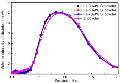
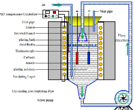
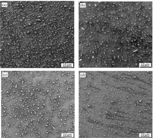
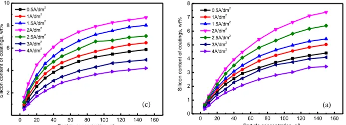
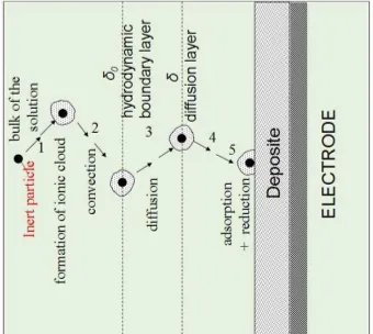
Effects of Particle Conductivity on the Fe-Si co-deposition Process
Full text
Figure
Related documents
The cell esds are taken into account individually in the estimation of esds in distances, angles and torsion angles; correlations between esds in cell parameters are only used
These determine the curriculum content and its organization, the teaching methods and strategies, the courses offered, the assessment process, the educational
From this representation, it seems that the law of this equation is bounded which is rational for which, there may be a fixed number of contexts to define either ‘ ’ or ‘ ’
Using two different migration assays, Western blotting, conventional and super-resolution (dSTORM) fluorescence microscopy we examine the effects of the dual PI3K/mTOR-
Histologic analysis revealed that overexpression of NICD1 drastically increased the amount of trabecular bone with significantly fewer undifferentiated cells left in the masses
Earth Planets Space, 51, 1215?1222, 1999 MD simulation for H2 formation on amorphous ice Junko Takahashi1,2,3? 1National Astronomical Observatory, 2 21 1, Osawa, Mitaka, Tokyo 181 8588,
Survival outcomes and multivariate analysis The multivariable Cox proportion hazards model adjusted for the mex3a expression group, and the group that included age, gender,
The authors reported gray matter volume loss in the cerebellar hemispheres, vermis, and whole brain stem and white matter loss in the whole brain stem, midbrain, pons, middle
