Sequencing a piece of history: complete genome sequence of
the original
Escherichia coli
strain
Karl A. Dunne,1Roy R. Chaudhuri,2Amanda E. Rossiter,1Irene Beriotto,1Douglas F. Browning,1Derrick Squire,1 Adam F. Cunningham,1Jeffrey A. Cole,1Nicholas Loman1and Ian R. Henderson1,
*
Abstract
In 1885, Theodor Escherich first described theBacillus coli commune, which was subsequently renamedEscherichia coli. We report the complete genome sequence of this original strain (NCTC 86). The 5 144 392 bp circular chromosome encodes the genes for 4805 proteins, which include antigens, virulence factors, antimicrobial-resistance factors and secretion systems, of a commensal organism from the pre-antibiotic era. It is located in theE. coliA subgroup and is closely related toE. coliK-12 MG1655.E. colistrain NCTC 86 and the non-pathogenic K-12, C, B and HS strains share a common backbone that is largely co-linear. The exception is a large 2 803 932 bp inversion that spans the replication terminus fromgmhBtoclpB. Comparison withE. coliK-12 reveals 41 regions of difference (577 351 bp) distributed across the chromosome. For example, and contrary to current dogma,E. coliNCTC 86 includes a nine genesillocus that encodes a silver-resistance efflux pump acquired before the current widespread use of silver nanoparticles as an antibacterial agent, possibly resulting from the widespread use of silver utensils and currency in Germany in the 1800s. In summary, phylogenetic comparisons with other E. coli strains confirmed that the original strain isolated by Escherich is most closely related to the non-pathogenic commensal strains. It is more distant from the root than the pathogenic organismsE. coli042 and O157 : H7; therefore, it is not an ancestral state for the species.
DATA SUMMARY
Supplementary figures have been deposited in Figshare (DOI:10.6084/m9.figshare.4235762) (https://figshare.com/s/ 91b570d54d27b0ecf9ea). Supplementary tables are available with the online Supplementary Material. The genome
sequence ofEscherichia coliNCTC 86 has been deposited in
GenBank with project accession number PRJEB14041 (www.ebi.ac.uk/ena/data/view/PRJEB14041) and in EMBL with accession number LT601384 (https://www.ncbi.nlm. nih.gov/nuccore/LT601384).
INTRODUCTION
Escherichia coliis unsurpassed as a model organism in the field of biology. Its leading role is predicated on its ability to replicate rapidly, to adjust easily to nutritional and environ-mental changes, and the relative simplicity with which it
can be genetically manipulated. The origin of E. coli as a
model organism began in the early part of the twentieth century with Charles Clifton’s study of oxidation–reduction reactions inE. coliK-12 and Felix d’Herelle’s studies on bac-teriophage interactions withE. coliB [1–3]. However,E. coli
became widely studied after the work of Tatum and Leder-berg on amino acid biosynthesis and exchange of genetic material that lead to the award of a Nobel Prize [3]. Subse-quently, many Nobel Prizes have been awarded for various studies using this versatile organism.
The depth of knowledge about the biochemistry of E. coli
and the non-pathogenic character of laboratory strains has madeE. colithe workhorse of molecular biology. No other organism is exploited more widely in research laboratories to manipulate DNA and to produce native and mutant pro-teins for studies in a variety of settings. Indeed, Neidhardt’s
Received 18 November 2016; Accepted 24 January 2017
Author affiliations: 1Institute of Microbiology and Infection, University of Birmingham, Birmingham, UK; 2Department of Molecular Biology and Biotechnology, University of Sheffield, Sheffield, UK.
*Correspondence:Ian R. Henderson, i.r.henderson@bham.ac.uk
Keywords:Escherichia coli; Theodor Escherich; genome sequence.
Abbreviations:CDS, coding DNA sequence; CPS, capsular polysaccharide; CU, chaperone–usher; ETEC, enterotoxigenicEscherichia coli; LPS, lipopoly-saccharide; NCTC, National Collection of Type Cultures; PTS, phosphotransferase system; ROD, region of difference; T1SS, type 1 secretion system; T2SS, type 2 secretion system; T3SS, type 3 secretion system; T5SS, type 5 secretion system; T6SS, type 6 secretion system; UPEC, uropathogenic
Escherichia coli.
The GenBank/EMBL/DDBJ accession number for the genome sequence ofEscherichia coliNCTC 86 is LT601384.
Data statement:All supporting data, code and protocols have been provided within the article or through supplementary data files. Nine supplementary figures and two supplementary tables are available with the online Supplementary Material.
eloquent statement exemplifies the diverse role ofE. coliin research‘Although not everyone is mindful of it, all cell biolo-gists have two cells of interest, the one they are studying and E. coli’ [4]. Therefore, as the biotechnology industry arose out of the molecular and cell biology research laboratories, it was logical forE. colito become the cornerstone of these revolutionary endeavours. Today, many biopharmaceuticals
are produced in E. coli, and these products impact on the
lives of millions of individuals worldwide on a daily basis [5].
WhilstE. colirose to prominence in the twentieth century,
the origin of this organism dates back to the late nineteenth
century. In about 1885, Theodor Escherich (1857–1911)
first describedBacterium coli commune, later named
Bacil-lus coli communisand eventuallyE. coliin Escherich’s hon-our after his death [6]. In his initial description, Escherich made several important observations that are widely reported in the modern literature, often without supporting citations. Escherich noted: (i) that the intestinal tracts of infants were sterile at birth, but were colonized byE. coli
within hours of birth; (ii) that bacterial colonization of the intestine was attributable to the infant’s environment; (iii) thatE. coliwas a Gram-negative, rod-shaped motile
organ-ism; (iv) thatE. coliwas a dominant member of the
micro-biota; (v) thatE. coliproduced acid and fermented glucose;
and (vi) that E. coli adopted a commensal lifestyle in the
normal host [7]. Soon after, Escherich and others noted the pyogenic and pathogenic properties of certainE. colistrains, although these were predominantly thought to be associated with individuals whose health was compromised in some manner. The association ofE. coliwith urinary tract disease was observed relatively quickly [8], although it would be
over 50 years before specific diarrhoeal strains of E. coli
were identified [9].
When Theodor Escherich first described his bacillus some 125 years ago, he could not possibly have imagined the major impact his discovery would have on subsequent generations of scientists. Even at the time of his death, he could not have known howE. coliwould influence the study of biological sci-ence. Despite the significance of his discovery, the historically important strain he isolated has remained largely unstudied. Here, we present the whole-genome sequence of Escherich’s original isolate and compare this genome with the genomes of other pathogenic and commensalE. coli.
METHODS
Bacterial strain and sequencing
TheE. colistrain NCTC 86 was isolated in 1885 from a child with no overt signs of diarrhoeal disease [7]. The strain was deposited in the National Collection of Type Cultures (NCTC) in 1919 by the Lister Institute and is officially recog-nized as Escherich’s originalBacillus coli commune[7]. The isolate sequenced here was obtained from the UK Health
Pro-tection Agency’s NCTC and is derived from batch 1. DNA
was prepared using Qiagen genomic DNA preparation kits
according to the manufacturer’s instructions. DNA was
fragmented using a Hydroshear (Digilab) using the recom-mended protocol for 20 kb fragments and further size-selected on a BluePippin instrument (Sage Science) with a 7 kb mini-mum size cut-off. The library was sequenced on two SMRT Cells using the Pacific Biosciences RS II instrument at the Norwegian Sequencing Centre, Oslo, Norway, using C4-P2
chemistry. The E. coli genome was generated by de novo
assembly using the Pacific Biosciences sequencing data. Reads
were assembled using the ‘RS_HGAP_Assembly.3’ pipeline
within SMRT Portal V2.2.0. Illumina reads from the same sample were mapped to this draft genome assembly in order to correct remaining indel errors in the assembly using Pilon (www.broadinstitute.org/software/pilon).
Gene prediction, annotation and comparative analysis
Automated annotation was performed using Prokka v1.8 (PMID: 24642063), which uses Prodigal (PMID: 20211023) for coding sequence prediction, Barrnap (www.vicbioinfor-matics.com/software.barrnap.shtml) to identify rRNA genes, Aragorn to identify tRNA genes (PMID: 14704338) and Infer-nal (PMID: 24008419) to identify non-coding RNA genes. To assess the size of the core genome and pan-genome ofE. coli, a set of 35 completeE. coliandShigellagenomes was selected. A pan-genome analysis was performed using Roary v3.7.0 (PMID: 26198102) to determine the sizes of the core and pan-genomes. A whole-genome phylogeny was reconstructed from the Roary core genome alignment using RAxML (PMID: 24451623).
Nucleotide sequence accession number
The annotated genome sequence of E. coli NCTC 86 has
been deposited in the EMBL database with the accession number LT601384.
RESULTS AND DISCUSSION
Structure and general features of theE. coliNCTC 86 chromosome
TheE. coliNCTC 86 genome consists of a circular
chromo-some of 5 144 392 bp. The general features of the E. coli
NCTC 86 chromosome are presented in Table 1. We identi-fied 4805 protein-encoding genes (coding DNA sequences,
IMPACT STATEMENT
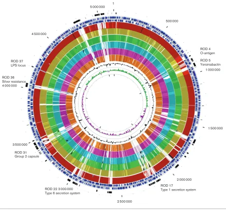
CDSs) in the chromosome, which included 414 (8.62%)that encoded conserved hypothetical proteins with no known function and 544 (11.32 %) genes associated with mobile ele-ments such as integrases or transposases, or that were phage related. We have identified 41 regions of difference (RODs) inE. coliNCTC 86 compared with other sequenced E. coli
chromosomes (Fig. 1, Table S1, available in the online Sup-plementary Material). The combined size of these RODs was 577 351 bp (11.22 % of the chromosome). They included 10 prophages distributed across the chromosome (Fig. 1).
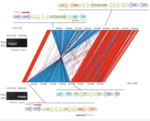
Comparison of E. coli NCTC 86 with the non-pathogenic
E. coli K-12, C, B and HS strains revealed that these
genomes shared a common backbone that is largely collin-ear, with the exception of a 2 803 932 bp inversion (Fig. 2).
The inversion spans the replication terminus fromgmhBto
clpB. The inversion was apparent from long Pacific
Bio-sciences (PacBio) reads and was generated by
recombina-tion between the equivalent DNA of theE. coliK-12rrfH
andrrsG RNA genes. PCR was used to confirm this
intra-chromosomal recombination event. Amplification ofE. coli
K-12 genomic DNA with primers corresponding to regions
within gmhBand dkgB resulted in a product of 6.8 kb. No
amplification product could be detected from similar
reac-tions withE. coliNCTC 86 genomic DNA (Fig. 2). In
con-trast, PCR amplification ofE. coliNCTC 86 genomic DNA
with primers corresponding to regions within gmhB and
kgtPresulted in a product of 7.2 kb, while no PCR product
was obtained from reactions with the same primers and
E. coli K-12 DNA (Fig. 2). The size of the PCR products observed in these experiments is consistent with the dis-tance between these genes predicted from the genome sequence data. The absence of an amplification product indicates that the target genes are located distally on the chromosome. Whilst these results confirm the inversion, the functional significance of this recombination event is unknown. This phenomenon has been noted before to occur in laboratory strains, but it was suggested that strains with such inversions rarely survive in the environment [10]. The
inversion in E. coli NCTC 86 may have arisen due to
prolonged culture and storage under laboratory conditions. However, such inversions have been noted in strains more recently isolated from humans [11].
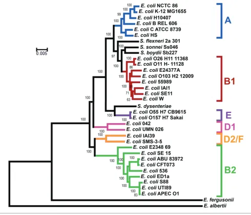
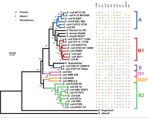
Phylogeny ofE. coliNCTC 86
The phylogeny of E. coli NCTC86 was resolved through
comparisons with the genome sequences of otherE. coliand
Shigellastrains. The core genome was used to reconstruct a
phylogenetic tree (Fig. 3), with Escherichia albertii and
Escherichia fergusoniiincluded as outgroups. As previously
noted, the E. coli subgroups A, B1, B2, D and E are all
monophyletic, with the exception of group D; group D is divided at the root [12].E. coliNCTC 86 is located in the A subgroup and clusters closely with the non-pathogenic labo-ratory strains of E. coli K-12. E. coli K-12 is considered a commensal derivative on the basis that it was isolated from a patient with diphtheria and without diarrhoeal disease or urinary tract infections [13]. Thus, the location of E. coli
NCTC 86 close toE. coliK-12 is consistent with Escherich’s observations of a non-pathogenic organism that was part of the normal microbiota [7].
A recurrent issue in the field ofE. colibiology has been the
assumption thatE. coliK-12 represents an ancestral
evolu-tionary lineage of the species. This misplaced belief probably arose from the factE. coliK-12 was an early isolate of the species. Further confusion arises when the genomes of path-ogenicE. coliare considered:E. coliK-12 is often used as a baseline genome with the simplistic view that pathogens are
E. coli K-12 derivatives that have acquired extra DNA
encoding disease-causing functions. These observations have been challenged before [14], and are again challenged here. Despite their early isolation, it is clear from their phy-logenetic distribution that neither E. coliK-12 nor E. coli
[image:3.595.44.558.565.706.2]NCTC 86 represents an ancestral state for the species. Indeed, the observation that these strains are distant from the root, and the smaller size of their genetic complement, suggest these strains are undergoing reductive evolution as they transition to a state of commensalism. Similar reductive evolution has been noted for many intracellular obligate
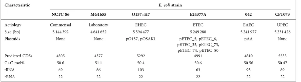
Table 1.Major features of the genome ofE. coliNCTC 86 and several archetypalE. colistrains
Characteristic E. colistrain
NCTC 86 MG1655 O157 : H7 E24377A 042 CFT073
Aetiology Commensal Laboratory EHEC ETEC EAEC UPEC
Size (bp) 5 144 392 4 641 652 5 594 477 5 249 288 5 241 977 5 231 428
Plasmids None None pO157, pOSAK1 pETEC_5, pETEC_6,
pETEC_35, pETEC_73, pETEC_74, pETEC_80
pAA None
Predicted CDSs 4805 4377 5292 4991 4810 5533
G+C mol% 50.6 51.1 50.4 50.6 50.56 50.47
tRNA 69 86 103 63 93 89
rRNA 22 22 22 22 22 22
organisms as they shed their free-living lifestyles in favour of a less competitive existence in a host-restricted environ-ment [15, 16]. Genetic attrition can also be observed for
E. coli NCTC 86. For example, the locus encoding ETT2
(ROD 24), a cryptic type 3 secretion system (T3SS), appears to be intact in E. coli strains O157 : H7 and 042, but like otherE. coli strains, the locus is severely eroded in E. coli
NCTC 86 (Fig. S1). In a similar manner, the majority of the locus encoding Flag-2, a cryptic lateral flagellum found in
E. coli042 and UMN026, is absent inE. coliNCTC 86 (Fig.
S2). Such observations support the hypothesis that E. coli
NCTC 86 is a modern rather than an ancestral lineage of
E. coli.
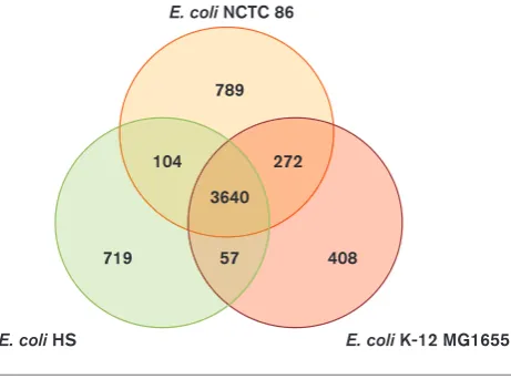
Comparative genomics ofE. coliNCTC 86
As previously stated, phylogenetic mapping revealedE. coli
NCTC 86 is located in the A subgroup along with the
non-pathogenic laboratory strainE. coliK-12 and the
com-mensal isolateE. coliHS. Comparison ofE. coliNCTC 86
with the closely related E. coli K-12 and HS strains
revealed that these chromosomes are largely similar, with 3640 genes conserved in all three strains (Fig. 4). Analyses of the gene contents of all three strains revealed thatE. coli
NCTC 86 contains 789 CDSs that are not present in the other two non-pathogenic strains. The majority (54 %) of these genes are predicted to encode transposons, integra-ses, prophage genes and other mobility factors or
1 000 000
1 500 000
2 000 000 500 000
3 500 000
4 500 000
5 000 000 1
2 500 000
ROD 5 Yersiniabactin ROD 4 O-antigen
ROD 17
Type 1 secretion system ROD 22 3 000 000
Type 6 secretion system ROD 31
Group 2 capsule ROD 36
Silver resistance 4 000 000
[image:4.595.66.531.64.496.2]ROD 37 LPS locus
hypothetical proteins that are located throughout the chro-mosome. These 601 CDSs are contained in RODs distrib-uted across the genome: they are discussed in the sections
below. E. coliHS and NCTC 86 share 104 common genes
not present in E. coli K-12, the majority of which encode
hypothetical proteins. The rest contain a variety of genes,
such as a type 6 secretion system (T6SS) NCTC86_02960–
76. In contrast, there are 272 genes present in E. coliK-12
and NCTC 86 not found in E. coli HS. Around half of
these are genes of unknown function; the rest are an
assortment of genes such as thefecABCDEIRgenes, which
encode a ferric citrate uptake system, and the
wcaABC-DEFGHIJKLMN genes, which encode proteins involved in
colanic acid synthesis. Additionally, E. coli HS and K-12
contain 57 genes not found inE. coli NCTC 86, over half
of which consist of genes of unknown function or mobile
elements. The rest are an assortment of genes such as the cold shock protein genes,cspBSHI, and the
antibiotic-resis-tance genes, such asacrR and blr (Table S2).E. coli K-12
contains 408 genes not present in either E. coli NCTC 86
or HS. Again, most of these are mobile elements.
Serotype antigens
Three major structures associated with the cell envelope of Gram-negative bacteria are flagella, lipopolysaccharide (LPS) and capsular polysaccharide (CPS), which mediate motility and protect the organism by providing a barrier to noxious substances and components of the immune system, respectively. The flagella, LPS and CPS are the three major
antigenic determinants ofE. coli: named the H-, O- and
K-antigens, respectively. These structures can demonstrate high degrees of polymorphism that give rise to differences
yfiH clpB dkgB
yfaC
yfiH clpB rrsG rrlG
rrfG kgtP yfiM
yfiM kgtP NCTC86_00220
metN
dkgB rrfH
metN rrsH
NCTC86_02992
yfaC gmhB
gmhB
Primer 3
Primer 1
Primer 2
Primer 1
NCTC 86 MG1655
8000 6000
Primers 1 and 2
NCTC 86 MG1655
8000 6000
Primers 1 and 3 NCTC 86
[image:5.595.50.546.61.459.2]MG 1655
in antigenicity. This polymorphism has been exploited for many years for the serological detection and typing ofE. coli
isolates [17].
Flagellin (FliC) is the major protein subunit of the flagellum. Amino acid variation in flagellin is responsible for changes to the antigenic profile of the flagellum. Examination of the
E. coliNCTC 86 genome sequence revealed the that major flagellar locus encodes an H10-serotype flagellin subunit that is identical to the flagellar locus ofE. coliBi 623–42, an O11 : H10 strain isolated from a patient with peritonitis [18]. This is consistent with the detection of the H10 flagel-lin reported forE. coliNCTC 86 [19].
Smooth LPS is a repeating structure termed the O-antigen side chain polysaccharide, which is chemically linked to the core oligosaccharide. The core oligosaccharide is fur-ther divided into the inner and outer core. In E. coli, the genes encoding the lipid A-core oligosaccharide and the O-antigen polysaccharide portions of LPS are encoded separately on the chromosome and are synthesized by independent pathways before linkage at the inner mem-brane [20]. The inner core and lipid A portions of the molecule are highly conserved. The outer core exhibits variation giving rise to five different core oligosaccharide structures designated K-12, R1, R2, R3 and R4. Inspection
[image:6.595.51.542.62.481.2]of theE. coliNCTC 86 genome revealed a locus (ROD 37)
encoding an R2 oligosaccharide core. This locus is 99 % identical over 8 kb to the R2 prototype strainE. coliF632,
which is an O-antigen-deficient derivative of E. coli
100 : K?(B) : H2 [21]. The biological significance of the R2 core is unknown: it is the least frequently occurring core
type amongst commensal and pathogenic E. coli [22].
However, the presence of an R2 oligosaccharide core in a commensal organism suggests that efforts to target vac-cines to this core region might be unwise, as it might neg-atively impact on the ability of a commensal organism to colonize the gut [23].
In striking contrast to the lipid A-core components, the O-antigens of LPS are chemically and structurally diverse. Over 180 serologically distinct O-antigens have been identi-fied inE. coli[24]. Scrutiny of theE. coliNCTC 86 genome revealed a locus encoding the proteins to produce an O-antigen of serotype O15 (ROD 4). This locus is 99 % identi-cal to the nucleotide sequence of the O-antigen locus from
E. coli G1201 (O15 : K14 : H4) over 9.2 kb. Analysis of the
O-antigen-encoding locus revealed that theE. coliNCTC 86
locus is 2.3 kb smaller than the E. coli G1201 locus. This
deletion appears to have occurred through recombination of
the ISE3C element with thewzygene, which encodes the
O-antigen polymerase, resulting in truncation ofwzyand loss
of thewzxandwbuSgenes encoding the O-antigen flippase
and a glycosyltransferase, respectively [25–27]. The loss
of O-antigen expression inE. coli is a common feature of
strains cultured in the laboratory for extended periods of time [28, 29].
Capsules are a series of high-molecular-mass polysacchar-ides that coat the bacterial surface. They are widely distrib-uted and are found in many diverse pathogens such as
Neisseria meningitidis, Staphylococcus aureus, Streptococcus pneumoniaeandE. coli. At least 80 different polysaccharide capsules have been identified inE. coli. They have been sep-arated into four different types (groups 1–4) based on their genetic organization, as well as their physical and biochemi-cal properties [30].E. coliNCTC 86 possesses thekpslocus
on ROD 31 (Fig. S3). The arrangement of thekpsFEDUCS
capsular biosynthesis genes in region I and the ABC
trans-porter genes kpsMT in region III are consistent with the
production of group 2 CPS. The variable region II, which is the determinant of antigenicity, is 98 % identical to the genes in region II ofE. coliCFT073 (Fig. S3). This suggests
that theE. coliNCTC 86 kpslocus encodes a K-2 serotype
capsule. Such capsules are often associated with strains that cause extra-intestinal infection, where they provide protec-tion against complement-mediated killing. However, as commensals do not need to evade the immune system, it might play a role in protection in the environment, such as desiccation.
Metal binding and resistance
Iron plays an essential role in metabolic and cellular pro-cesses, in particular acting as a cofactor for several critical enzymes [31]. However, free iron is not readily available. In an aqueous environment at physiological pH it forms
insol-uble Fe(OH)3. Pathogens and commensals encounter
addi-tional problems in vivo as iron is extensively chelated by
host proteins, such as ferritin and lactoferrin [32]. Thus, bacteria require efficient iron-acquisition mechanisms in order to survive. Some bacteria secrete molecules termed siderophores that have high affinity for iron. The iron-bound forms of the siderophore are recognized by specific proteinaceous machines that facilitate their uptake into the bacterial cell [33].E. coliNCTC 86 possesses a locus, located on ROD 5, which encodes the siderophore yersiniabactin (Ybt) (Fig. S4). Ybt was first described inYersinia, but it is
widespread amongst the Enterobacteriaceae [34]. TheE. coli
NCTC 86 locus is most similar to theE. coli042 locus (97 % identity over 24 kb). Interestingly, in addition to the reported iron-acquisition functions of Ybt, some studies suggest that Ybt contributes to bacterial resistance to copper [35]. Copper ions have been shown to be toxic toE. coli[36, 37]. Therefore, Ybt might act as a countermeasure to copper toxicity by sequestering host-derived copper and preventing its catechol-mediated reduction to copper (I), this enables
bacteria to proliferate in vivo and can confer protection
from redox-based phagocytic defences [38].
Like copper, silver ions show antibacterial activity against
E. coli. Interestingly,E. coliNCTC 86 harbours a 36 kb locus
(sil) comprised of nine ORFs (ROD36) that are associated
with silver resistance (Fig. S5). Thesil locus confers
resis-tance though a sliver ion efflux system. Although the sil
locus is frequently found on plasmids harbouring antibi-otic-resistance genes, such as the pMG101 plasmid [39], in
272 104
3640
408 57
719
789 E. coli NCTC 86
[image:7.595.49.280.66.237.2]E. coli K-12 MG1655 E. coli HS
E. coli NCTC 86 it was found on the chromosome. The
locus had a similar genetic arrangement to E. coli C
ATCC8739, H10407 and inE. coli55989,Enterobacter
cloa-caeATCC 13047 andCronobacter sakazakiiCMCC 45402
(Fig. S5), where it is also found on the chromosome. It was suggested that this locus was only recently acquired by
E. coli after the introduction of silver nanoparticles as an antibacterial agent. This hypothesis was based on the
obser-vation that E. coli strains recently isolated from humans
possessed thesillocus, but that noE. colistrain of an avian origin was found to contain the locus [40]. However,E. coli
NCTC 86 clearly acquired this locus before the current widespread use of silver nanoparticles, suggesting there is an alternative explanation for the appearance of this locus in human isolates. It is tempting to speculate that the reason
E. coliNCTC 86 possesses thesillocus was the widespread use of silver utensils and currency in Germany in the 1800s.
Metabolism
The metabolic properties encoded in a bacterium are one of the primary factors in determining whether it is successful in an ecological niche.E. coliNCTC 86 contains a variety of RODs encoding loci associated with metabolic functions, such as ethanolamine utilization, a set of acyl-CoA synthe-tase and permease genes, and putative C4-dicarboxylate and sugar-uptake systems.
The phosphotransferase system (PTS) is a translocation sys-tem that transports a variety of different sugars into the cell. The PTS consists of two proteins, an enzyme I and HPr, and a number of carbohydrate-specific enzymes, the enzymes II. In addition to the PTSs encoded in the conserved coreE. coli
genes, E. coliNCTC 86 possesses two sugar-uptake systems
encoded on RODs. Thevpelocus is located on ROD 40 and is
99.77 % identical to thevpelocus ofE. coliO104 : H4
2009EL-2050 strain. The vpe operon was first described in the
uropathogenicE. coli(UPEC) strain AL511 in which mutants
were shown to produce much smaller amounts of group two capsule [41]. As noted earlier,E. coliNCTC 86 encodes a
sim-ilar group two capsule. ROD 40 also contains the deoK
operon. This region conferring the ability to utilize deoxyri-bose as a carbon source is thought to have been transferred fromSalmonella entericatoE. coli[42]. ThedeoKoperon is commonly connected with strains isolated from infected blood and urine, in which it is usually found in extensive islands carrying genes contributing to the adaptive properties and/or virulence of UPEC strains, such as the sepsis strain
E. coli AL862 [43]. The second putative PTS is located on ROD 34 ofE. coliNCTC 86 and is 98 % identical to that inE. coliO157 : H7 Sakai. However, the function of this system is unknown.
E. coliNCTC 86 contains a putative C4-dicarboxylate uptake (Duc) transporter system located on ROD 30 that is com-posed of three genes clustered together: a tripartite ATP-independent periplasmic (TRAP) transporter; an ABC
transporter permease; and a substrate-binding protein. E.
coliNCTC 86 system is 100 % identical to the C4 -dicarboxy-late system found inE. coliIAI39. However, inE. coliIAI39
there is a transposon inserted in the centre of the periplas-mic (TRAP) transporter gene, indicating that it might not be functional in this isolate. While many bacteria utilize molecules such as aspartate, malate, fumarate and succinate for anaerobic respiration (aspartate is metabolized under both aerobic and anaerobic conditions), the function and
substrate of this specific C4-dicarboxylate uptake system
remains unknown.
Protein secretion systems
Protein secretion involves translocation across the cyto-plasmic membrane, and in Gram-negative bacteria the addi-tional barriers of a peptidoglycan-filled periplasm and an outer membrane. To combat these latter obstacles, Gram-negative bacteria have evolved a number of highly special-ized protein secretion systems [44]. Inspection of theE. coli
NCTC 86 genome revealed loci encoding a type 1 secretion system (T1SS), type 2 secretion system (T2SS), T3SS, type 5 secretion system (T5SS) and a T6SS. The ETT2 and Flag-2 T3SSs were discussed earlier.
Conventional T1SSs of Gram-negative bacteria are com-posed of a secreted substrate protein and three envelope-associated subunits: a TolC-like outer membrane pore-forming protein (OMP), and two inner-membrane-associ-ated proteins, termed the ATP-binding cassette (ABC) pro-tein, which provide energy to the system, and the membrane-fusion protein (MFP), which contacts with the TolC-like protein to form the secretion channel across the cell envelope on ROD 17 [45]. Like the conventional
sys-tems, the E. coli NCTC 86 system possesses an OMP
(NCTC86_02172), MFP (NCTC86_02173) and ABC protein (NCTC86_02170). In contrast to the conventional T1SSs, the locus also encodes an additional ABC protein (NCTC86_02173) and an
additional component whose function is unknown
(NCTC86_02170) (Fig. S6). Unlike the conventional T1SS, in which the C-terminal sequence is uncleaved, the putative substrate molecules for the unconventional systems possess an N-terminal Sec-dependent signal sequence that is cleaved. Located downstream of the genes encoding the translocation machinery is a pseudogene that would have encoded a substrate molecule (NCTC86_02180) but is dis-rupted by the insertion of genes encoding transposases. However, located 11.2 kb upstream is NCTC86_02122, which appears to encode a complete putative substrate mol-ecule. This secretion system is homologous to the
Aat/dis-persin previously described for enteroaggregative E. coli
042. InE. coli042, the substrate molecule is localized to the bacterial cell surface where it promotes fimbrial-mediated adherence by altering the surface charge of the bacterium [46]. The presence of this gene cluster in a commensal bac-terium suggests its primary role is in colonization rather than in pathogenesis as previously suggested [47].
Several pathogenic strains ofE. colipossess a
chromosom-ally encoded T2SS. For enterotoxigenicE. coliit is essential for secretion of heat-labile enterotoxin (LT) [48]. InE. coli
yghJ–gspO (pppA) –gspC(yghF) and the distal gspL–gspM
genes, but not the remainder of the T2SS (Fig S7). E. coli
NCTC 86 also possesses the distalgspL–gspMgenes.
How-ever, the T2SS locus appears to be in an even later stage of
genetic attrition. TheyghJGFandpppAgenes have also been
lost. The erosion of this locus is further evidence for the adaptation ofE. coliNCTC 86 to a commensal lifestyle.
T5SSs are a large and diverse superfamily of proteins. Based on structural differences and variations in the mode of bio-genesis, T5SS has been divided into five subclasses: classical autotransporters (T5aSS); the two-partner secretion systems (T5bSS); the trimeric autotransporters (T5cSS); the chimeric autotransporters (T5dSS); and the inverted autotransporters
(T5eSS).E. coliNCTC 86 does not appear to possess
mem-bers of the T5bSS, T5cSS or T5dSS subfamilies. However,
E. coli NCTC 86 possesses 15 loci encoding polypeptides with homology to the T5aSS subfamily and 3 polypeptides
with homology to the T5eSS subfamily (Fig S8). Of the 15
T5aSS-encoding loci, 9 are found in E. coli K-12 and are
scattered throughout the chromosome; 4 of these are
pseu-dogenes. Remarkably, E. coliNCTC 86 possesses five loci
(NCTC86_00823, NCTC86_02105, NCTC86_02886, NCTC 86_03415 and NCTC86_04998) encoding polypeptides with homology to the autotransporter antigen 43. These loci are found on RODs 5, 17, 21, 31 and 41, although one (ROD 31; NCTC86_03415) is a pseudogene. These classical autotrans-porters have been implicated in adherence and biofilm for-mation. The three loci encoding members of the T5eSS
subfamily can also be found inE. coliK-12. This family of
[image:9.595.49.551.287.686.2]proteins has been implicated in mediating attachment to host cells [49]. Scrutiny of the distribution of the T5SS genes across the evolutionary spectrum ofE. colireveals no strong association of these genes with pathogenic or non-patho-genic isolates. This suggests that they do not play a specific
role in pathogenesis but rather promote colonization, a function that is consistent with their presence in a commen-sal strain.
E. coli NCTC 86 contains a T6SS. This locus encodes the Hcp and Vgr proteins that form a needle-like injection device and are essential for the T6SS to function. The locus also contains the TssA, E, F, G and K core component pro-teins. It is this type of T6SS that is most widespread amongst
E. coli[50–52]. This 30 kb locus is located on ROD 22 and is >97 % identical to the T6SS ofE. coli55989, and is highly
homologous to loci in E. coli 042, urinary tract isolates
UMN026, 536 and UTI89, and avian isolates APEC O1 (Fig. S9). T6SSs have been shown to be widespread among the proteobacteria and have roles in inhibiting eukaryote cell division as well as possessing antibacterial properties, by acting as a mechanism to attack other bacterial cells [53].
Therefore, it is likely the locus provides E. coli NCTC 86
with a colonization advantage within the host.
Proteobacteria use chaperone–usher (CU) pathways to
assemble fimbriae on the bacterial surface. The CU system is divided into six subfamilies, designateda-,b-,g-,k-,p
-ands-fimbriae [54].E. coliNCTC 86 contains 12 CU
sys-tems, 9 of which are also present inE. coliK-12 (Fig. 5). It lacks members of thek-,b- ors- subfamilies. As inE. coli
K-12, these CU operons appear to be scattered throughout
the chromosome. The other three CU systems in E. coli
NCTC 86 (NCTC86_00020–6, NCTC86_03382–8 and
NCTC86_02125–9) are found on RODs 1, 17 and 31,
respectively. The CU system encoded on ROD 1 is homolo-gous to thematfimbriae ofE. coliO157, but the first gene
in this locus is truncated inE. coliNCTC 86 rendering the
system non-functional. The CU locus on ROD 31 is dis-rupted by the insertion of two transposable elements and is unlikely to be functional. The locus on ROD 17 also encodes a non-functional CU system with homology to the
aggrega-tive adherence fimbriae ofE. coli042. There is no obvious
phylogenetic signature for the presence or absence of the CU systems. Furthermore, there is no obvious association with pathogenic or non-pathogenic lineages, with the excep-tion of shigellae, which appear to have lost many of the loci encoding these CU systems. As with the T5SS, these data suggest that the CU systems do not play a specific role in pathogenesis, but rather promote colonization. The diver-sity of systems may allow colonization of different hosts or colonization of different niches within the same host.
Conclusions
The complete genetic content ofE. coliNCTC 86 described
in this article provides a modern interpretation for observa-tions made by Escherich over 125 years ago. During the preparation of this manuscript, a draft sequence of the same strain and its historical provenance were reported [55].
These studies demonstrate thatE. coliNCTC 86 is
phyloge-netically closely related to other commensalE. colistrains. The anthropocentric view of bacteriology has largely driven the study of pathogenicE. coliat the expense of understand-ing commensalism and, perhaps, of truly understandunderstand-ing the
repertoire of genes that are required for pathogenesis and those which are simply required for colonization. The cur-rent study adds to the body of literature underpinning the
study of commensalE. coliand hints that these organisms
arose by reductive evolution as they adapted to an intimate life with their mammalian hosts. However, given the plastic-ity of theE. coligenome, further genomic studies are essen-tial to determine those factors that are widely conserved for
commensalE. colifrom geographically diverse locations and
distinctive human populations. With such studies, E. coli
will undoubtedly remain as a model organism.
Funding information
This study was funded by the University of Birmingham and by the Darwin Trust.
Acknowledgements
The other authors would like to thank Professor Jeff Cole for his advice in writing the manuscript.
Conflicts of interest
The authors declare that there are no conflicts of interest.
Ethical statement
No human/animal experiments were involved in this study.
Data bibliography
1. Aslett MA. GenBank accession number FN554766 (2009). 2. Aslett MA. GenBank accession number FN554767 (2009).
3. Brzuszkiewicz E, Brueggemann H, Liesegang H, Emmerth M, Oelschlaeger Tet al.GenBank accession number CP000247 (2006). 4. Genoscope CEA. GenBank accession number CU928145 (2008). 5. Brzuszkiewicz EB, Liesegang H, Zdziarski J, Svanborg C, Hacker Jet al. GenBank accession number CP001671 (2009). 6. Brzuszkiewicz EB, Liesegang H, Zdziarski J, Svanborg C, Hacker Jet al. GenBank accession number CP001833 (2009). 7. Johnson TJ, Nolan LK. GenBank accession number CP000468 (2009). 8. Johnson TJ, Nolan LK. GenBank accession number DQ381420 (2006). 9. Johnson TJ, Nolan LK. GenBank accession number DQ517526 (2006). 10. Kim JF, Jeong H, Choi S-H, Yu D-S, Hur C-Get al. GenBank accession number CP000819 (2007).
11. Copeland A, Lucas S, Lapidus A, Glavina del Rio T, Dalin Eet al.
GenBank accession number CP000946 (2008).
12. Welch RA, Burland V, Plunkett GD III, Redford P, Roesch Pet al. GenBank accession number AE014075 (2002).
13. Thomson NR. GenBank accession number FM180568 (2008). 14. Thomson NR. GenBank accession number FM180569 (2008). 15. Thomson NR. GenBank accession number FM180570 (2008). 16. Rasko DA, Rosovitz MJ, Brinkley C, Myers GSA, Seshadri Ret al. GenBank accession number CP000795 (2007).
17. Rasko DA, Rosovitz MJ, Brinkley C, Myers GSA, Seshadri Ret al. GenBank accession number CP000796 (2007).
18. Rasko DA, Rosovitz MJ, Brinkley C, Myers GSA, Seshadri Ret al. GenBank accession number CP000797 (2007).
19. Rasko DA, Rosovitz MJ, Brinkley C, Myers GSA, Seshadri Ret al. GenBank accession number CP000798 (2007).
20. Rasko DA, Rosovitz MJ, Brinkley C, Myers GSA, Seshadri Ret al. GenBank accession number CP000799 (2007).
21. Rasko DA, Rosovitz MJ, Brinkley C, Myers GSA, Seshadri Ret al. GenBank accession number CP000800 (2007).
23. Fiedoruk K, Daniluk T, Swiecicka I, Murawska E, Sciepuk Met al. GenBank accession number CP007025 (2013).
24. Genoscope CEA. GenBank accession number CU928162 (2008). 25. Genoscope CEA. GenBank accession number CU928144 (2008). 26. Genoscope CEA. GenBank accession number CU928158 (2008). 27. Archer CT, Kim JF, Jeong H, Park J, Vickers CEet al. GenBank accession number CP002185 (2010).
28. Archer CT, Kim JF, Jeong H, Park J, Vickers CEet al. GenBank accession number CP002186 (2010).
29. Archer CT, Kim JF, Jeong H, Park J, Vickers CE, Lee S et al. GenBank accession number CP002187 (2010).
30. Aslett MA. GenBank accession number FN649414 (2009). 31. Aslett MA. GenBank accession number FN649415 (2009). 32. Aslett MA. GenBank accession number FN649416 (2009). 33. Aslett MA. GenBank accession number FN649417 (2009). 34. Aslett,M.A. GenBank accession number FN649418 (2009). 35. Rasko DA, Rosovitz M, Kaper JB, Nataro JP, Myers GS et al. GenBank accession number CP000802 (2007).
36. Genoscope CEA. GenBank accession number CU928160 (2008). 37. Genoscope CEA. GenBank accession number CU928164 (2008). 38. Blattner FR, Plunkett G III. GenBank accession number U00096 (2013). 39. Hattori M, Toh H, Oshima K, Yamashita A, Hayashi Tet al. GenBank accession number AP010958 (2008).
40. Hattori M, Toh H, Oshima K, Yamashita A, Hayashi Tet al. GenBank accession number AP010959 (2008).
41. Hattori M, Toh H, Oshima K, Yamashita A, Hayashi Tet al. GenBank accession number AP010960 (2008).
42. Hattori M, Toh H, Oshima K, Yamashita A, Hayashi Tet al. GenBank accession number AP010961 (2008).
43. Hattori M, Toh H, Oshima K, Yamashita A, Hayashi Tet al. GenBank accession number AP010962 (2008).
44. Hattori M, Toh H, Oshima K, Yamashita A, Hayashi Tet al. GenBank accession number AP010963 (2008).
45. Hattori M, Toh H, Oshima K, Yamashita A, Hayashi Tet al.GenBank accession number AP010964 (2008).
46. Hattori M, Toh H, Oshima K, Yamashita A, Hayashi Tet al. GenBank accession number AP010965 (2008).
47. Makino K. GenBank accession number AB011548 (1998). 48. Makino K. GenBank accession number AB011549 (1998).
49. Hattori M, Ishii K, Shiba T. GenBank accession number BA000007 (2000).
50. Hattori M, Toh H, Oshima K, Yamashita A, Hayashi Tet al. GenBank accession number AP010953 (2008).
51. Hattori M, Toh H, Oshima K, Yamashita A, Hayashi Tet al. GenBank accession number AP010954 (2008).
52. Hattori M, Toh H, Oshima K, Yamashita A, Hayashi Tet al. GenBank accession number AP010955 (2008).
53. Hattori M, Toh H, Oshima K, Yamashita A, Hayashi Tet al. GenBank accession number AP010956 (2008).
54. Hattori M, Toh H, Oshima K, Yamashita A, Hayashi Tet al. GenBank accession number AP010957 (2008).
55. Wang L, Zhou Z, Li X, Liu B, Beutin Let al. GenBank accession number CP001846 (2009).
56. Wang L, Zhou Z, Li X, Liu B, Beutin Let al. GenBank accession number CP001847 (2009).
57. Genoscope CEA. GenBank accession number CU928146 (2008). 58. Genoscope CEA. GenBank accession number CU928161 (2008). 59. Jin Q, Yang F, Yang J, Jiang Y, Yan Yet al. GenBank accession number CP000036 (2004).
60. Yang J, Chen L, Jin Q. GenBank accession number CP000037 (2007).
61. Jin Q, Yang F, Yang J, Jiang Y, Yan Yet al. GenBank accession number CP000034 (2004).
62. Yang J, Chen L, Jin Q. GenBank accession number CP000035 (2007). 63. Yang J, Yang F, Chen L, Jin Q. GenBank accession number CP000640 (2007).
64. Hattori M, Oshima K, Toh H, Sasamoto H, Shiba T. GenBank accession number AP009240 (2006).
65. Hattori M, Oshima K, Toh H, Sasamoto H, Shiba T. GenBank accession number AP009241 (2006).
66. Hattori M, Oshima K, Toh H, Sasamoto H, Shiba T. GenBank accession number AP009242 (2006).
67. Hattori M, Oshima K, Toh H, Sasamoto H, Shiba T. GenBank accession number AP009243 (2006).
68. Hattori M, Oshima K, Toh H, Sasamoto H, Shiba T. GenBank accession number AP009244 (2006).
69. Hattori M, Oshima K, Toh H, Sasamoto H, Shiba T. GenBank accession number AP009245 (2006).
70. Hattori M, Oshima K, Toh H, Sasamoto H, Shiba T. GenBank accession number AP009246 (2006).
71. Hattori M, Yamashita A, Toh H, Oshima K, Shiba T. GenBank accession number AP009378 (2007).
72. Hattori M, Yamashita A, Toh H, Oshima K, Shiba T. GenBank accession number AP009379 (2007).
73. Jin Q, Shen Y, Wang JH, Liu H, Yang Jet al. GenBank accession number AE005674 (2011).
74. Jin Q, Zhang JY, Liu H, Yang J, Yang Fet al. GenBank accession number AF386526 (2007).
75. Fricke WF, Wright MS, Lindell AH, Harkins DM, Baker-Austin Cet al. GenBank accession number CP000970 (2008).
76. Fricke WF, Wright MS, Lindell AH, Harkins DM, Baker-Austin Cet al. GenBank accession number CP000971 (2008).
77. Fricke WF, Wright MS, Lindell AH, Harkins DM, Baker-Austin Cet al. GenBank accession number CP000972 (2008).
78. Fricke WF, Wright MS, Lindell AH, Harkins DM, Baker-Austin Cet al. GenBank accession number CP000973 (2008).
79. Fricke WF, Wright MS, Lindell AH, Harkins DM, Baker-Austin Cet al. GenBank accession number CP000974 (2008).
80. Jin Q, Yang F, Yang J, Jiang Y, Yan Yet al. GenBank accession number CP000038 (2004).
81. Yang J, Chen L, Jin Q. GenBank accession number CP000039 (2007). 82. Yang J, Yang F, Chen L, Jin Q. GenBank accession number CP000641 (2007).
83. Yang J, Yang F, Chen L, Jin Q. GenBank accession number CP000642 (2007).
84. Yang J, Yang F, Chen L, Jin Q. GenBank accession number CP000643 (2007).
85. Genoscope CEA. GenBank accession number CU928148 (2008). 86. Genoscope CEA. GenBank accession number CU928149 (2008). 87. Genoscope CEA. GenBank accession number CU928163 (2008). 88. Chen SL, Hung C-S, Xu J, Reigstad CS, Magrini Vet al. GenBank accession number CP000243 (2006).
89. Chen SL, Hung C-S, Xu J, Reigstad CS, Magrini Vet al. GenBank accession number CP000244 (2006).
References
1. Clifton CE, Cleary JP.Oxidation-reduction potentials and ferricya-nide reducing activities in glucose-peptone cultures and suspen-sions ofEscherichia coli.J Bacteriol1934;28:561–569.
2. D’Herelle FH.Sur une microbe invisible antagoniste des bacilles dysenteriques.C R Acad Sci1917;165:373–375.
3. Lederberg J, Tatum EL.Gene recombination in Escherichia coli.
4. Neidhardt FC, Curtiss R, Ingraham JL, Lin ECC, Low KB et al.
Escherichia coliandSalmonella: Cellular and Molecular Biology. Washington, DC: ASM Press; 1996. pp. 123–145.
5. Ferrer-Miralles N, Domingo-Espín J, Corchero JL, Vazquez E, Villaverde A.Microbial factories for recombinant pharmaceuticals.
Microb Cell Fact2009;8:17.
6. Migula W. Bacteriaceae (St€abchenbacterien). . Die Natürlichen Pflanzenfamilien. Leipzig: W. Engelmann; 1895. pp. 20–30. 7. Escherich T.Die darmbakterien des neugeborenen und sauglings.€
Fortsch Der Med1885;3:547–554.
8. Hyman M, Mann LT.The nitrite reaction as an indicator of urinary infection.J Urol1929;521.
9. Bray J.Isolation of antigenically homogeneous strains ofBact. coli neapolitanumfrom summer diarrhœa of infants.J Pathol Bacteriol
1945;57:239–247.
10. Hill CW, Gray JA.Effects of chromosomal inversion on cell fitness inEscherichia coliK-12.Genetics1988;119:771–778.
11. Bergholz TM, Wick LM, Qi W, Riordan JT, Ouellette LM et al. Global transcriptional response of Escherichia coli O157:H7 to growth transitions in glucose minimal medium. BMC Microbiol
2007;7:97.
12. Chaudhuri RR, Henderson IR.The evolution of theEscherichia coli
phylogeny.Infect Genet Evol2012;12:214–226.
13. Lederberg J.Genetic studies with bacteria. In: Dunn LC (editor).
Genetics in the 20th Century. New York: Macmillan; 1951. pp. 263–289.
14. Ren CP, Chaudhuri RR, Fivian A, Bailey CM, Antonio Met al.The ETT2 gene cluster, encoding a second type III secretion system fromEscherichia coli, is present in the majority of strains but has undergone widespread mutational attrition.J Bacteriol2004;186: 3547–3560.
15. Andersson SG, Zomorodipour A, Andersson JO, Sicheritz-Ponten T, Alsmark UCet al.The genome sequence ofRickettsia prowaze-kiiand the origin of mitochondria.Nature1998;396:133–140. 16. Stephens RS, Kalman S, Lammel C, Fan J, Marathe R et al.
Genome sequence of an obligate intracellular pathogen of humans:Chlamydia trachomatis.Science1998;282:754–759. 17. Garrity G, Brenner DJ, Krieg NR, Staley JR.Bergey’s Manual of
Systematic Bacteriology. New York: Springer; 2005.
18. Orskov I, Orskov F, Jann B, Jann K.Serology, chemistry, and genetics of O and K antigens of Escherichia coli. Bacteriol Rev
1977;41:667–710.
19. Kauffmann F.Zur serologie der coli-gruppe.Acta Pathol Microbiol Scand1944;21:20–45.
20. Raetz CR, Whitfield C.Lipopolysaccharide endotoxins. Annu Rev Biochem2002;71:635–700.
21. H€ammerling G, Lüderitz O, Westphal O, M€akel€a PH. Structural investigations on the core polysaccharide ofEscherichia coli0100.
Eur J Biochem1971;22:331–344.
22. Amor K, Heinrichs DE, Frirdich E, Ziebell K, Johnson RPet al. Dis-tribution of core oligosaccharide types in lipopolysaccharides fromEscherichia coli.Infect Immun2000;68:1116–1124.
23. Santos MF, New RR, Andrade GR, Ozaki CY, Sant’anna OAet al. Lipopolysaccharide as an antigen target for the formulation of a universal vaccine against Escherichia coli O111 strains. Clin Vaccine Immunol2010;17:1772–1780.
24. Iguchi A, Iyoda S, Kikuchi T, Ogura Y, Katsura Ket al.A complete view of the genetic diversity of theEscherichia coliO-antigen bio-synthesis gene cluster.DNA Res2015;22:101–107.
25. Feldman MF, Marolda CL, Monteiro MA, Perry MB, Parodi AJ et al.The activity of a putative polyisoprenol-linked sugar translo-case (Wzx) involved inEscherichia coliO antigen assembly is inde-pendent of the chemical structure of the O repeat.J Biol Chem
1999;274:35129–35138.
26. Grozdanov L, Z€ahringer U, Blum-Oehler G, Brade L, Henne A et al.A single nucleotide exchange in the wzy gene is responsible
for the semirough O6 lipopolysaccharide phenotype and serum sensitivity ofEscherichia colistrain Nissle 1917.J Bacteriol2002; 184:5912–5925.
27. Marolda CL, Feldman MF, Valvano MA.Genetic organization of the O7-specific lipopolysaccharide biosynthesis cluster ofEscherichia coliVW187 (O7:K1).Microbiology1999;145:2485–2495.
28. Browning DF, Wells TJ, França FL, Morris FC, Sevastsyanovich YR et al. Laboratory adapted Escherichia coli K-12 becomes a pathogen ofCaenorhabditis elegansupon restoration of O antigen biosynthesis.Mol Microbiol2013;87:939–950.
29. Stevenson G, Neal B, Liu D, Hobbs M, Packer NHet al.Structure of the O antigen ofEscherichia coliK-12 and the sequence of its rfb gene cluster.J Bacteriol1994;176:4144–4156.
30. Whitfield C.Biosynthesis and assembly of capsular polysacchar-ides inEscherichia coli.Annu Rev Biochem2006;75:39–68. 31. Andreini C, Bertini I, Cavallaro G, Holliday GL, Thornton JM.Metal
ions in biological catalysis: from enzyme databases to general principles.J Biol Inorg Chem2008;13:1205–1218.
32. Weinberg ED.Iron availability and infection.Biochim Biophys Acta
2009;1790:600–605.
33. Andrews SC, Robinson AK, Rodríguez-Quiñones F.Bacterial iron homeostasis.FEMS Microbiol Rev2003;27:215–237.
34. Schubert S, Rakin A, Heesemann J.TheYersinia high-pathogenic-ity island (HPI): evolutionary and functional aspects. Int J Med Microbiol2004;294:83–94.
35. Koh EI, Henderson JP. Microbial copper-binding siderophores at the host-pathogen interface. J Biol Chem 2015;290:18967– 18974.
36. Clark HW, Gage SD.On the bactericidal action of copper.Public Health Pap Rep1905;31:175–204.
37. Macomber L, Imlay JA.The iron-sulfur clusters of dehydratases are primary intracellular targets of copper toxicity.Proc Natl Acad Sci USA2009;106:8344–8349.
38. Chaturvedi KS, Hung CS, Crowley JR, Stapleton AE, Henderson JP.The siderophore yersiniabactin binds copper to protect patho-gens during infection.Nat Chem Biol2012;8:731–736.
39. Gupta A, Matsui K, Lo JF, Silver S.Molecular basis for resistance to silver cations inSalmonella.Nat Med1999;5:183–188.
40. Sütterlin S, Edquist P, Sandegren L, Adler M, T€angden Tet al. Sil-ver resistance genes are oSil-verrepresented amongEscherichia coli
isolates with CTX-M production. Appl Environ Microbiol 2014;80: 6863–6869.
41. Martinez-Jehanne V, Pichon C, du Merle L, Poupel O, Cayet N et al.Role of the vpe carbohydrate permease inEscherichia coli
urovirulence and fitness in vivo. Infect Immun 2012;80:2655– 2666.
42. Bernier-Febreau C, du Merle L, Turlin E, Labas V, Ordonez Jet al. Use of deoxyribose by intestinal and extraintestinal pathogenic
Escherichia colistrains: a metabolic adaptation involved in com-petitiveness.Infect Immun2004;72:6151–6156.
43. Lalioui L, Le Bouguenec C.afa-8 gene cluster is carried by a path-ogenicity island inserted into the tRNA(Phe) of human and bovine pathogenic Escherichia coli isolates. Infect Immun 2001;69:937– 948.
44. Abby SS, Cury J, Guglielmini J, Neron B, Touchon M et al. Identifi-cation of protein secretion systems in bacterial genomes.Sci Rep
2016;6:23080.
45. Henderson IR, Navarro-Garcia F, Desvaux M, Fernandez RC, Ala’aldeen D. Type V protein secretion pathway: the autotrans-porter story.Microbiol Mol Biol Rev2004;68:692–744.
46. Sheikh J, Czeczulin JR, Harrington S, Hicks S, Henderson IRet al. A novel dispersin protein in enteroaggregativeEscherichia coli.J Clin Invest2002;110:1329–1337.
48. Tauschek M, Gorrell RJ, Strugnell RA, Robins-Browne RM. Identi-fication of a protein secretory pathway for the secretion of heat-labile enterotoxin by an enterotoxigenic strain ofEscherichia coli.
Proc Natl Acad Sci USA2002;99:7066–7071.
49. Leo JC, Oberhettinger P, Schütz M, Linke D.The inverse auto-transporter family: intimin, invasin and related proteins.Int J Med Microbiol2015;305:276–282.
50. Brzuszkiewicz E, Brüggemann H, Liesegang H, Emmerth M, Olschl€ager Tet al. How to become a uropathogen: comparative genomic analysis of extraintestinal pathogenic Escherichia coli
strains.Proc Natl Acad Sci USA2006;103:12879–12884.
51. Johnson TJ, Kariyawasam S, Wannemuehler Y, Mangiamele P, Johnson SJ et al. The genome sequence of avian pathogenic
Escherichia coli strain O1:K1:H7 shares strong similarities with
human extraintestinal pathogenic E. coli genomes. J Bacteriol
2007;189:3228–3236.
52. Touchon M, Hoede C, Tenaillon O, Barbe V, Baeriswyl Set al. Organised genome dynamics in the Escherichia coli species results in highly diverse adaptive paths. PLoS Genet 2009;5: e1000344.
53. Hood RD, Singh P, Hsu F, Güvener T, Carl MAet al. A type VI secretion system of Pseudomonas aeruginosatargets a toxin to bacteria.Cell Host Microbe2010;7:25–37.
54. Nuccio SP, B€aumler AJ.Evolution of the chaperone/usher assem-bly pathway: fimbrial classification goes Greek.Microbiol Mol Biol Rev2007;71:551–575.
55. Meric G, Hitchings MD, Pascoe B, Sheppard SK.From Escherich to theEscherichia coligenome.Lancet Infect Dis2016;16:634–636.
Five reasons to publish your next article with a Microbiology Society journal 1. The Microbiology Society is a not-for-profit organization.
2. We offer fast and rigorous peer review–average time to first decision is 4–6 weeks. 3. Our journals have a global readership with subscriptions held in research institutions around
the world.
4. 80% of our authors rate our submission process as‘excellent’or‘very good’.
5. Your article will be published on an interactive journal platform with advanced metrics.